ELECTRON PARAMAGNETIC RESONANCE STUDIES OF POLYMER CHAIN DYNAMICS IN SOLUTION
Sooyeon Sim
A dissertation submitted to the faculty of the University of North Carolina at Chapel Hill in partial fulfillment of the requirements for the degree of Doctor of Philosophy in the Department
of Material Science.
Chapel Hill 2014
Approved by:
Malcolm D. E. Forbes
Andrew M. Moran
Edward T. Samulski
Sergei S. Sheiko
Yue Wu
ABSTRACT
SOOYEON SIM: Electron Paramagnetic Resonance Studies of Polymer Chain Dynamics in Solution
(Under the direction of Malcolm D. E. Fobes)
The temperature and side-chain structure dependencies of the time-resolved electron
paramagnetic resonance (TREPR) spectra of acrylic polyhedral oligosilsesquioxane (POSS)-based
copolymers and acrylic block copolymers are presented. The carbon–centered radicals within the
main chain of an acrylic polyhedral oligosilsesquioxane (POSS)-based copolymers and acrylic
block copolymers are produced in dilute solution at temperatures ranging from 25 °C to over
120 °C by direct excitation (248 nm) of the ester group in the polymers leading to Norrish I α–
cleavage of the side chain ester moiety and provides a means to investigate the conformational
energy landscape using a highly localized, minimally perturbative spin probe.
The observed photochemical degradation mechanism of those copolymers is quite
similar to that previously observed in photolysis of homo poly (methacrylate)s. As expected,
chromophores with POSS group side chains do not participate in the photodegration process,
which suggests that POSS utilize a beneficial effect against UV light photodegradation in these
copolymers. The resulting experimental spectra suggest since all of the investigated
methacrylate-POSS copolymers show the same photodegradation phenomena, that methacrylate-POSS could play an
important roles in preventing UV degradation and improving photoresistance in such materials.
photophysical and photochemical behavior of acrylic blocks in dilute solution, especially the site
selective photochemistry was studies here by TREPR.
It is interesting note that alternating line broadening effects due to the modulated hyperfine
constant cause by a bulky ester side chain are observed in high temperature TREPR spectra of both
t-butyl methacrylate-POSS block copolymer and poly t-butyl methacrylate rich block copolymers. The excellent fit of the simulations allow for identification of the signal carriers. The spectra show
a strong signal from the tert-butyl radical after decarboxylation of the oxo-acyl radical. This
process is rarely observed in high temperature fast limit TREPR spectra.
The temperature dependence of TREPR spectra of all block copolymers presented here shows
similar conformational dynamics to those reported in our previous papers.
Radical-triplet pair interactions are used to investigate the dynamics of acrylic polymers in
dilute solution. Methyl methacrylate was randomly copolymerized with a small amount of an
amine–containing monomer to create the polymers. The amine subunits were then oxidized to
nitroxide moieties (stable free radicals). Using TREPR spectroscopy on the sub-microsecond time
scale, competition is observed between two deactivation processes of the ester side chain triplet
state: 1) Norrish I –cleavage, leading to a main chain free radical studied previously in our
laboratory, and 2) spin polarization transfer or quenching by a nearby stable nitroxide radical. The
two processes have TREPR spectral signatures that are easily distinguished by their widths,
number of transitions, and chemically induced electron spin polarization (CIDEP) mechanisms.
processes clearly demonstrates the influence of polymer chain dynamics on the observed
ACKNOWLEDGMENTS
I take this opportunity to express my profound gratitude and deep regards to my guide
Professor Malcolm D. E. Forbes for his exemplary guidance, monitoring and constant
encouragement throughout the course of this thesis. The blessing, help and guidance given by him
time to time shall carry me a long way in the journey of life on which I am about to embark.
My great mentor, Prof. Chongseung Yoon in Hanyang University, encouraged me to study in US.
He introduced me joy of studying science and has been a continued source of friendship and
wisdom.
I also take this opportunity to express a deep sense of gratitude to all our group members,
for their kindly support, valuable information and encouragement, which helped me in completing
this task through various stages and particularly to Lauren Jarocha, for being able to explain or fix
anything; Renat , for making me happy and encouraging me all the time. Prof. Sang Wook Wu
who was a research associate in Pederson group, we had nice discussion about quantum mechanics,
and Spin Hamiltonian. It was the best discussion ever in my life.
For their support of my graduate school experience, I must acknowledge the members of my
family, mom, dad, my brother and my Korean fiends in NC and in Korea, especially, Alley Choo,
TABLE OF CONTENTS
LIST OF TABLES ... xi
LIST OF FIGURES ... xii
LIST OF ABBREVIATIONS AND SYMBOLS ... xvii
CHAPTER 1. Introduction ... 1
1.1 Study of Polymer Chain Dynamics in Solution ... 1
1.2 Characterization of Main-Chain Radicals from the Photodegradation of Acrylic Polymers ... 6
1.3 Experimental overview of TREPR ... 15
1.3.1 Steady-state EPR (SSEPR) vs. TREPR ... 15
1.3.2 Experimental considerations for TREPR ... 21
1.4 Chemically Induced Electron Spin Polarization (CIDEP) Mechanisms ... 25
1.4.1 The Triplet Mechanism (TM) ... 28
1.4.2 The Radical-Triplet Pair Mechanism (RTPM) ... 30
2. Photochemistry of acrylate-POSS block copolymers and dynamic effects in copolymer radicals ... 37
2.3 Summary and Implications ...
2.4 Experimental ... 58
3. Photodegradation of Acrylic Block Copolymers ... 60
3.1 Introduction ... 60
3.1.1 Structure of Block Copolymers ... 61
3.1.2 Photochemistry of Acrylic Polymers ... 64
3.2 Result and Discussion ... 69
3.2.1 TREPR spectra of Block Copolymers ... 77
3.2.1.1 Poly (methyl methacrylate)–b–Poly (tert-butyl methacrylate) Copolymers ... 77
3.2.1.2 Poly (methyl methacrylate)–b–Poly (n-butyl methacrylate) Copolymers ... 84
3.2.1.3 Poly (methyl methacrylate)–b–Poly (ethyl acrylate) Copolymers.... 87
3.3 Summary and Outlook ... 92
3.4 Experimental ... 93
3.4.1 Materials ... 93
3.4.2 Synthesis ... 93
3.4.2.1 Anionic Polymerization ... 93
3.4.2.2 Free radical Polymerization ... 94
3.4.3 Analysis of polymer samples ... 94
3.4.3.1 Gel Permeation Chromatography (GPC) ... 94
3.4.3.2 High temperature TREPR set-up ... 95
4. Radical-Triplet Pair Interactions as Probes of Long-Range Polymer Motion in Solution ... 97
4.2 The Microscopic Order–Macroscopic Disorder Model (MOMD) ... 99
4.3 Studies of Segmental Rotational Dynamics of Spin-labeled PMMA in Dilute Solution ... 103
4.4 TREPR Studies of Chain Dynamics of Spin-labeled Polymers in Dilute Solutions ... 111
4.5 Conclusions ... 127
4.6 Experimental ... 128
4.6.1 Analysis of polymer samples ... 129
LIST OF TABLES
Table 1.1 Parameters used to simulate TREPR spectra (Fig. 1.3) of Polymer Main-Chain
radicals and Acyl Radicals...11
Table 3.1 The structure and molecular weight of block copolymers...70
Table 4.1 Parameters used in the simulation temperature dependence of the SSEPR spectra of 1 mol % nitroxide copolymer sample in PC...107
LIST OF FIGURES
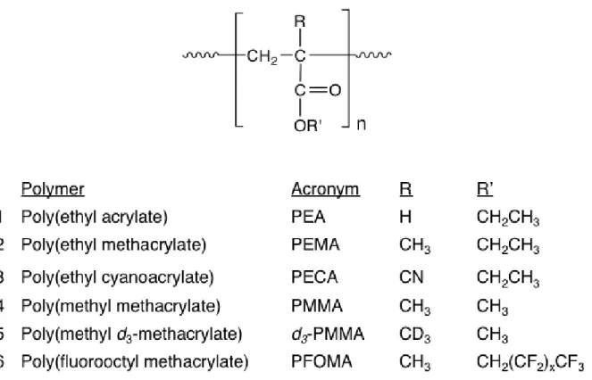
Figure 1.1 Photochemistry and free radicals resulting from 248 nm excimer laser excitation of acrylic polymers in solution ··· 7 Figure 1.2 Structures of polymers characterized by TREPR ··· 8 Figure 1.3 (A–E) Experimental TREPR spectra (left) and simulated spectra (right) for
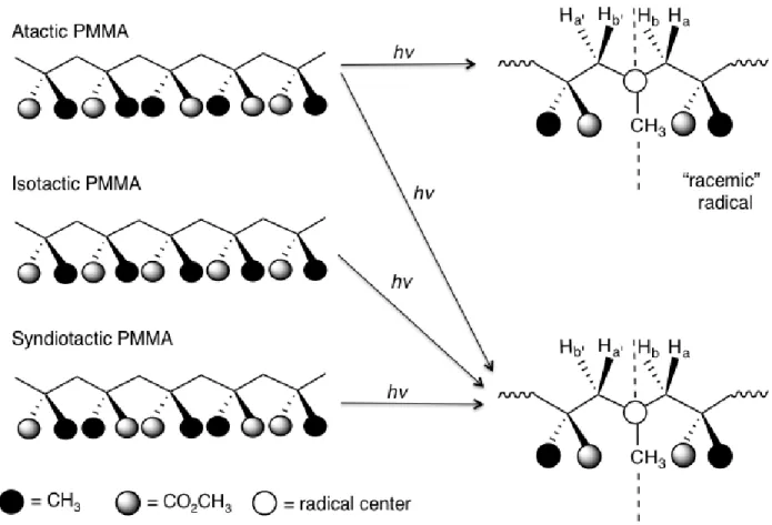
main-chain acrylic radicals observed at 0.8 µs after 248 nm laser flash photolysis for the polymers indicated (see Fig. 1.2 for polymer structures and Table 1.1 for radical structures). (A) PEA, (B) PEMA, (C) PECA, (D) PMMA, (E) PMMA-d3, all obtained in propylene carbonate. (F) TREPR spectrum (left, experimental; right, simulated) of the oxo-acyl radical from PFOMA, obtained in the high boiling fluorinated solvent mixture FC-70. For PMMA, the material is isotactic (91% by NMR), but all other polymer samples are atactic material. Simulation parameters (ref. [10]) are listed in Table 1.1. ··· 10 Figure 1.4 Symmetry relationships between the nuclear hyperfine coupling constants in acrylic
polymers as a function of polymer tacticity ··· 13 Figure 1.5 Energy level diagram depicting a single spin system in a external magnetic field.
When the resonance condition is met, a single observable transition can occur
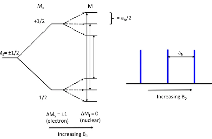
between the two spin states, as shown by the double arrow in the figure ··· 17 Figure 1.6 Energy level diagram demonstrating the hyperfine interaction and observable
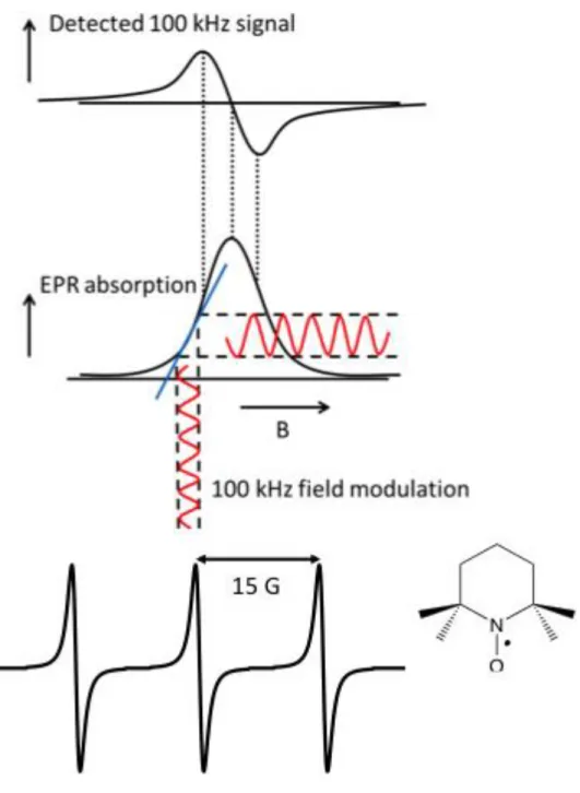
transitions of a radical coupled to a neighboring nucleus with I=1 ··· 18 Figure 1.7 Steady-state EPR detection using 100 kHz field modulation and the resulting first
derivative line shape ··· 20 Figure 1.8 Block diagram of the TREPR experiment using a boxcar signal averager ··· 22 Figure 1.9 Timing sequence for the TREPR experiment using a boxcar signal averager ··· 24 Figure 1.10 (A) Energy level diagram of a single electron in the magnetic field with population
Figure 1.13 Energy levels of a radical–triplet pair as a function of their separation. Schematic explanation of net absorption CIDEP created by excited singlet quenching by stable free radical. ··· 33 Figure 2.1 Chemical structures of silsesquioxanes. non-caged (left) vs. caged (right)··· 40 Figure 2.2 Schematic drawing of chemical structures of POSS-based polymeric
nanocomposites: (left) norbornyl-POSS copolymers; (middle) methacrylate-POSS copolymers and (right) styryl-POSS copolymers. ··· 42 Figure 2.3 Structures of polymers characterized by TREPR: (1)
[(propylmethacryl-heptaisobutyl-POSS)-co-(t-butyl methacrylate)]; (2) [(propylmethacryl-heptaisobutyl-POSS)-co-(n-butyl methacrylate)];
(3)[(propylmethacryl-heptaisobutyl-POSS)-co-(methyl methacrylate)]; (4) Poly (t-butyl methacrylate); (5) Poly (methyl methacrylate) and (6) Poly (n-butyl methacrylate)]··· 44 Figure 2.4 Two solvents for TREPR expetiments at high Temperature··· 46 Figure 2.5 Experimental High-temperature TREPR spectra for the main-chain radical from 248
nm irradiation of the following polymers: (A) PSS 25 wt % of Poly
[(propylmethacryl-heptaisobutyl-POSS)-co-(t-butyl methacrylate)], (B) PSS 45 wt % of Poly [(propylmethacryl-heptaisobutyl-POSS)-co-(t-butyl methacrylate)], (C) P(t-butyl methacrylate) in diethyl glycol dimethyl ether at 125 °C, shown for comparison ··· 47 Figure 2.6 The 10-line t-butyl radical spectrum from decarboxylation of poly (t-butyl
methacrylate) oxo–acyl radical in diethylene glycol dimethyl ether at 125 °C with following total sweep width: (top) 300 G, 100 G, 50 G, and (bottom) 30 G and delay time: 500 ns ··· 49 Figure 2.7 Photochemistry and free radicals resulting from 248nm excimer laser excitation of
Poly (t-butyl methacrylate) in solution ··· 50 Figure 2.8 (Top) The photochemistry of PFOMA. (Bottom left) experimental and (Bottom
right) simulated high temperature TREPR spectra of the oxo-acyl radical from PFOMA in perfluorinated solvent FC-70 at 110 °C and delay time 0.5 s. ··· 52 Figure 2.9 Two proposed simulated high temperature TREPR spectra of poly (t-butyl
methacrylate) after photolysis at 120°C and delay time of 500 ns ··· 53 Figure 2.10 The temperature dependence of TREPR spectra for the main-chain radical from 248
nm irradiation of Poly [(propylmethacryl-heptaisobutyl-POSS)-co-(n-butyl
Figure 2.11 The temperature dependence of TREPR spectra for the main-chain radical from 248 nm irradiation of Poly [(propylmethacryl-heptaisobutyl-POSS)-co-(nethyl
methacrylate)] in diglyme ··· 57 Figure 3.1 Phase diagram for polymer solution ··· 62 Figure 3.2 (Top) The photochemistry of PFOMA. (Bottom left) experimental and (Bottom
right) simulated high temperature TREPR spectra of the oxo-acyl radical from PFOMA in perfluorinated solvent FC-70 at 110 °C and delay time 0.5 μs ··· 68 Figure 3.3 Two solvents for TREPR expetiments at high Temperature··· 73 Figure 3.4 Experimental high temperature TREPR spectra for radicals observed after 248 nm
laser flash photolysis of the following polymers and their structures ··· 74 Figure 3.5 Simulated high temperature TREPR spectra for radicals observed after 248 nm laser
flash photolysis of the following polymers and their structures ··· 75 Figure 3.6 Block chain length ratio dependence of Poly (methyl methacrylate)–b–poly (t-butyl
methacrylate) copolymer TREPR spectra in diglyme at 500 ns at 125 °C ··· 78 Figure 3.7 The 10-line t-butyl radical spectrum from decarboxylation of poly (t-butyl
methacrylate) oxo–acyl radical in diethylene glycol dimethyl ether at 125 °C with following total sweep width: (top) 300 G, 100 G, 50 G, and (bottom) 30 G and delay time: 500 ns ··· 80 Figure 3.8 Structure of propagating radical of poly (t-butyl methacrylate) and simulated EPR
spectra of the propagating radical of poly (t-butyl methacrylate) (left) direct
detection and (right) 1’st derivative at 150 °C ··· 82 Figure 3.9 The temperature dependence of TREPR spectra for the main-chain radical from 248
nm irradiation of all three Poly (methyl methacrylate)-b–poly (t-butyl methacrylate) copolymer in either diglyme or propylene carbonate ··· 83 Figure 3.10 Block chain length ratio dependence of Poly (methyl methacrylate)-b–poly (n-butyl
methacrylate) copolymer TREPR spectra in diglyme at 500 ns at 115 °C ··· 85 Figure 3.11 The temperature dependence of TREPR spectra for the main-chain radical from 248
Figure 3.13 The temperature dependence of TREPR spectra for the main-chain radical from 248 nm irradiation of all two Poly (methyl methacrylate)-b–poly (ethyl acrylate)
copolymer in propylene carbonate (1g of polymer in 25 ml of solvent) at delay time of 500 ns ··· 91 Figure 4.1 Norrish I α–cleavage reaction of poly(methyl methacrylate) (PMMA) and graphical
representation of the fast rotation of the side chain along one axis and the much slower conformational motion along the polymer chain. ··· 98 Figure 4.2 (a) Different coordinate systems (laboratory: L, director: D, and magnetic: m) used
to define motion parameters for a nitroxide spin label. (b) Diffusion rotation angles used to define the magnetic axes relative to the diffusion axes. Note that the
reference system for these angles is the is diffusion frame. (c) Structure of spin-labeled PMMA with 2,2,6,6,-tetra-methyl-4-hydroxypiperidinyl-1-oxy and the assumed conformation of spin label bounded at the end of the side chain (ref. 104, 105) ··· 101 Figure 4.3 The structure of nitroxide containing random PMMA copolymer studied in this
chapter ··· 104 Figure 4.4 Top: X-band SSEPR (dark) spectra of the nitroxide-doped copolymer from Figure
4.3, acquired at room temperature in the solvents indicated. Bottom: Computer simulations using the Freed model REF with rotational correlation times of (left to right) 0.18 ns, 0.45 ns, and 0.72ns respectively ··· 105 Figure 4.5 (Left )X-band SSEPR spectra of the nitroxide-doped copolymer and (Right) the
same mol % nitroxide (TEMPO) and PMMA in PC at the given temperature ··· 107 Figure 4.6 Arrhenius plot for rotational correlation time data from nitroxide-doped polymer
(Figure 4.5). Squares are the simulated data, and the solid line is the linear fit with Ea = 23 KJ/mol ··· 110 Figure 4.7 Graphical representation of spin polarization created by TM, RTPM and ESPT ·· 113 Figure 4.8 TREPR spectra of the nitroxide–containing copolymer shown in figure 4.3, acquired
in propylene carbonate solution at 120°C, as a function of nitroxide incorporation (in mol %) the indicated to the left. Asterisks in the 1.0 mole % spectrum indicate transitions from the main chain polymeric radical of PMMA that is dominant in the 0.5 and 0.1 mol % spectra ··· 117 Figure 4.9 TREPR spectra of the nitroxide–containing polymer shown in Figure 4.3 (1 mol %
nitroxide) in propylene carbonate solution at the temperatures indicated. Asterisks in the 120°C spectrum indicate transitions from the main chain polymeric radical of PMMA, which is very broad but still observable in the 25 °C spectrum (top). The
Figure 4.10 Inter– vs. intramolecular competition between TM and RTPM. (Top) TREPR spectra of unattached TEMPO (1.5mM) and PMMA (5 wt %) in propylene
carbonate after 248 nm laser excitation at 120°C. (Bottom) TREPR spectrum of the nitroxide containing PMMA copolymer (loading level is 1 mol %) under the same conditions. Asterisks in the bottom spectrum indicate transitions due to the main chain polymeric radical of PMMA. The delay time is 500 ns and the sweep width is 150 G in both spectra. ··· 123 Figure 4.11 TREPR spectra of the 1 mol% of nitroxide–containing PMMA polymer as a function
of concentration. The spectra were acquired in propylene carbonate solution at 120 °C. Asterisks in the bottom spectrum indicate transitions due to the main chain polymeric radical of PMMA. The delay time is 500 ns and the sweep width is 150 G in both spectra ··· 124 Figure 4.12 TREPR spectra of nitroxide–containing poly(n-butyl methacrylate) at 70°C (top) and poly(tert-butyl methacrylate) at 130°C (bottom) in diglyme. The sweep width is 200 G and the delay time is 500 ns in both spectra. The degree of nitroxide
LIST OF ABBREVIATIONS AND SYMBOLS A. ABBREVIATIONS AIBN a-PMMA CH3 CH2 CO CO2 CW CIDEP DPE DLS
d3-PMMA
diglyme EA EPR ESPT GPC ISC i-PMMA LW MeOH 2,2’-azobisisobutyronitrile
atactic poly(methyl methacrylate)
methyl group
methylene group
carbon monoxide
carbon dioxide
continuous wave
chemically induced electron polarization
1,1-diphenylethylene
dynamic light scattering
poly(methyl d3-methacrylate)
diethylene glycol dimethyl ether
ethyl acrylate
electron paramagnetic resonance
electron spin polarization transfer
gel permeation chromatography
intersystem crossing
isotactic poly (methyl methacrylate)
linewidth
MMA NMR n-BMA n-BuLi PC PDI PEA PEMA PFOMA P(isoBMA) POSS PMMA P(n-BMA) PS P(t-BMA) RPM RTPM S/N s-PMMA SSEPR methyl methacrylate
nuclear magnetic resonance
n-butyl methacrylate
n-Butyllithium
propylene carbonate
poly disperse index
poly(ethyl acrylate)
poly(ethyl methacrylate)
poly(fluorooctyl methacrylate)
poly(iso-butyl methacrylate)
polyhedral oligomeric silsesquioxane
poly(methyl methacrylate)
poly(n-butyl methacrylate)
poly(styrene)
poly(t-butyl methacrylate)
radical pair mechanism
radical triplet pair mechanism
signal to noise ratio
syndiotactic poly(methyl methacrylate)
TREPR
UV
UV/VIS
time resolved electron paramagnetic resonance
ultraviolet
ultraviolet/visible absorption spectroscopy
B. ROMAN SYMBOLS
A a aH B C C 13C C* d E E G g g mg GHz H 1H Absorption
polymer segment length
hyperfine coupling constant
applied magnetic field
h h Hz I I J K k k KJ KrF M Mn Ms Mw mL μL mM mm nm Plank constant hour Hertz
nuclear spin quantum number
Intensity exchange interaction Kelvin Boltzmann constant rate constant kilojoules krypton-fluoride molarity
number average molecular weight
quantum number
weight average molecular weight
milliliters
microliters
millimolar
millimeter
RF Rg μs ns S S0 S1 s T T Tg Tm T1 T1 t t v
end-to-end distance of the chain
radius of gyration
microseconds
nanoseconds
excited singlet state
ground state
first excited singlet state
singlet
temperature
excited triplet energy level
glass transition temperature
melting temperature
first excited triplet state
spin-lattice relaxation
triplet
time
volumn
C. GREEK SYMBOLS
α
β
βe
γe
upper energy level of electron
lower energy level of electron
Bohr magneton
δ ε Φ η λ ν ν ν θ ρ τ χ ω ϕ ϕ* chemical shift extinction coefficient quantum yield viscosity wavelength
excluded volume index
frequency
Flory exponent
dihedral angle
density
rotational correlation time
Flory-Huggins segmental interaction parameter
frequency
concentration
Chapter 1. Introduction
1.1 Study of Polymer Chain Dynamics in Solution
Polymer chain dynamics in melts and highly concentrated solutions is a well–studied area
of science [1, 2]. In such systems, the chain dynamics are not affected by the solvent and instead
reflect bulk properties, where entanglement plays a major role, e.g., in the rheology of melts.
It can be difficult to understand their macroscopic behavior of polymer solutions due to
the complexities of entanglement. In this regard, physical measurements of polymer
conformational dynamics in dilute solution can be very informative as they can provide more
information about the intrinsic polymer properties.
The conformational dynamics of macromolecules is a topic of great interest and has been
well-studied in polymer physics [3, 4], however most theoretical models in the field are based on
so-called “coarse-grain” methods, i.e., balls and springs with standard potentials such as
Lennard-Jones or modifications thereof. There are very few models in the literature that use information at
the molecular level [5a,b]; indeed there is only one published report of a molecular dynamics
simulation of a polymer chain with solvent molecules included explicitly [5c]. Unlike a protein
simulation, most polymer solution simulations cannot use water as their solvent, and therefore
need to add one more term related to the interaction energies between polymer and solvent.
potentials (well depths and activation barriers), are sought in order to assess the intrinsic properties of polymer chains in the absence of entanglements. By careful analysis of intrinsic polymer
properties the role of entanglement in melt properties might be better understood (by eliminating
them).
In order to understand polymer behaviors at the molecular level, a polymer dilute solution
can be used without entanglement, and with no overlapping chains. The polymer chain in solution
can be isolated at special conditions where it is below critical concentration, and at theta (θ)
temperature where the effective monomer and monomer interaction are weak thus polymer coil
can consider as ideal chain.
Nuclear magnetic resonance (NMR) has been the most widely used to study of polymer
motions and conformation so far. 13C can be used in a NMR spin probe of polymer chain dynamic
because of the sensitivity of 13C resonance frequencies or chemical shifts, δ13C, to polymer
configuration and conformation [6]. Also, the measurement of segment diffusion and NMR
spin-lattice relaxation is a suitable method for studying polymer chain dynamics [7].
Like NMR, Electron paramagnetic resonance (EPR) spectroscopy has been used to obtain
information about polymer motions and local chain behavior by using a spin probes, commonly
stable nitroxide radicals. Depending on their rotational and translational motion, with correlation
times (τc) along the polymer chain, the appearance of the EPR spectrum can be quite different [8].
Thus, studying their line shapes and widths as a function of the correlation times (τc) provides
polymer chains in dilute solutions as a function of polymer main chain and side chain structure,
temperature, solvent and concentration using a EPR study of localized vs. non-localized spin
probes.
The basic strategy for these studies is summarized in scheme 1. On the left side of the
scheme 1 is the creation of carbon–centered radicals within the main chain of an acrylic polymer
by photolysis (248nm) creates a highly localized, minimally perturbative spin probe, thus an EPR
study of this localized spin probe is suitable to investigate the conformational energy landscape of
polymer chains [9,10]. The main chain radical can only be observed in solution using the
time-resolved EPR (TREPR) technique: under steady-state conditions its rearrangement to the
propagating radical via beta-scission is facile at room temperature and above. Because the
electron-nuclear hyperfine interactions in the main chain radical depend on the dihedral angle
between the p orbital containing the unpaired electron and the neighboring C-H sigma bond, the
TREPR spectra of these main chain radicals show a strong temperature dependence for both line
widths and line positions [11].Changes in line width are related to the “jump time” between
conformations, while changes in line position (at constant width) reflect changes in the populations
of certain rotational conformers (“rotamers”). For this reason, the observed TREPR spectra are
rich in information regarding the conformational landscape of the polymers, and this data can be
obtained with only a minimal perturbation of the polymer chain structure. Thus, the degradation
of acrylic polymers has been a subject of intense study in the Forbes laboratory for the past decade,
and this thesis seeks to extend this work to create a workable “atom-specific” model for
macromolecular chain dynamics that has so far proven elusive for these complex structures. In the
process of creating the model, we will be using TREPR to study acrylic polymers that have not
show novel degradation pathways and different chain dynamics.
The right side of Scheme 1 shows a longer-range conformational change that can also be
probed using TREPR. In this case, the polymer is synthesized with a small percentage of stable
nitroxide free radicals as side chains. When a photoexcited triplet states is created, it can be
quenched by the nitroxide, or it can engage in Heisenberg spin exchange with the stable radical.
In both cases the nitroxide radical receives additional population of its electron spin states that are
non-Boltzmann. The magnitude of this polarization is directly proportional to the number of
encounters made between the stable radical and the excited triplet state, and is therefore an indirect
measure of the rate of intra-chain contact between the two sites.
The photodegradation mechanism of several acrylate polymers and copolymers (block,
random and hybrid) and TREPR characterization of their free radical reactive intermediates
created after UV laser flash photolysis in dilute solution are described in Chapters 2 and 3. In
these chapters, the dynamic effects and conformational s of several blockcopolymers will be
presented and discussed. Their dynamics behavior depends on polymer chain stiffness,
temperature, block chain length and chain configuration will be discussed. Fast motion TREPR
spectra obtained at high temperature will be used to allow for accurate simulations of their
main-chain polymer radicals. Chapter 4 describes radical-triplet pair interactions, which are used to
detect long–range chain motion in acrylic polymers in dilute solution by incorporating a nitroxide
into the polymer chain by means of covalent bonds, we can generate RTPM polarization via
intramolecular encounters that depend on several structural and physical features of the polymer/solvent system. Additionally, Chapter 4 will present the dependence of the RTPM
polarization in the covalently linked system as a function of nitroxide incorporation (mol%),
This chapter provides background information about the previous work in the Forbes
laboratory on acrylic polymer degradation, the TREPR experiment itself and the relevant CIDEP
mechanisms to the new work presented in this thesis.
1.2 Characterization of Main-Chain Radicals from the Photodegradation of Acrylic Polymers
The photodegradation mechanisms of polymers have been a subject of intense interest in
for many years [12], and many of these reactions involve free-radical intermediates [13]. In our
laboratory, we have used TREPR spectroscopy to investigate the degradation of acrylic polymers
as a function of polymer structure, solvent, pH, and temperature [9, 10, 14, 15].The generic
main-chain acrylic polymer radical (Figure 1.A, top center), obtained after 248 nm laser flash photolysis
of the ester side chain, rapidly rearranges to the so-called propagating radical (Figure 1.B, bottom
left) and an alkene via β-scission. For this reason, the main-chain radical is not generally
observable with high spectral resolution in SSEPR experiments involving acrylic polymers [16].
However, the TREPR experiment is fast enough to observe both main-chain acrylic radicals and,
in some cases, the corresponding oxo-acyl counter radicals (Figure 1.C, top right) on the
microsecond timescale. Here is the brief review of our recent findings regarding the structure and
dynamics of acrylic main-chain radicals created by 248 nm laser flash photolysis of the polymers
shown in Figure 1.2.
Poly(ethyl acrylate) (PEA) was the starting point for our investigation as it has the simplest
the appearance of TREPR spectra, especially the line widths. The next polymer, poly
(methyl methacrylate) (PMMA), has been extensively studied in our previous papers because of
the interesting nuclear spin symmetry properties of their main-chain radicals as a function of
polymer tacticity. In addition, PMMA-d3 was also investigated to confirm the spectral assignments
by isotopic labeling with TREPR. The last polymer structure listed in Figure 1.2 is
Poly(fluorooctyl methacrylate) (PFOMA), where, the β-substituent is the –CH2(CF2)6CF3 bulky
ester side chain, which has very different physical properties from other acrylate polymers [17].
The stiffness of the main chain due to the bulky side chain produces a huge change in the TREPR
spectral appearance of the resulting radicals.
The TREPR spectra obtained 0.8 μs after 248 nm laser flash photolysis of six acrylic
polymers are shown in Figure 1.2, along with simulations for some of the spectra computed using
magnetic parameters listed in Table 1.1. All TREPR spectra shown in Figure 1.3were recorded at
high temperature (above 100 °C) where the “fast-motion” regime of the polymer dominated. The
TREPR spectra of the main-chain polymeric radicals mentioned in our previous work exhibit
alternating line widths at room temperature, a phenomenon that is highly indicative of
conformationally modulated hyperfine interactions (slow motion). However, upon heating, most
acrylic polymer main-chain radicals show motional narrowing with sharp line widths, allowing us
to simulate the spectra using an average set of hyperfine coupling constants.
Figure 1.3A shows the experimental high-temperature TREPR spectrum obtained after photolysis
of PEA in propylene carbonate solution. This radical shows six major transitions with the doublets
of the innermost lines attributed to two β-methylene hyperfine couplings of 23.0 and 24.7 G and
one α-hyperfine coupling of 21.5 G, which were comparable to the reported literature values for
observed at high temperature because the signal from this radical decays rapidly due to fast spin
relaxation [18].Compared to PEA, the α-substituent methyl group in PEMA has a large effect on
the appearance of the TREPR spectrum. For example, the spectrum resulting from photolysis of
PEMA (Figure 1.3B) consists of a 27 line spectrum associated with three separate isotropic
hyperfine coupling constants. There is coupling to the methyl group to form a quartet (22.9 G),
which is split further into a triplet from one set of diastereotopic β-methylene protons (15.8 G) and
another triplet from the other set (11.2 G). Technically, the maximum expected number of
observable transitions is 36 (a quartet of triplets of triplets) for the PEMA radical spectrum, but
there are accidental degeneracies, leading to a smaller number of observed transitions.
Similar to the α-substituent dependence described earlier, the spectrum from the PECA
radical is quite different from the TREPR spectrum of the PEA radical. At a glance, there are five
transitions (Figure 1.3C) from coupling to the β-methylene protons, but each peak is further split
into three additional lines from the γ-nitrogen (I=1) in the nitrile group. From the simulation
parameters in Table 1.1, a hyperfine coupling of 3.3 G is assigned to the γ-nitrogen and couplings
of 16.3 and 14.8 G are assigned to the β-methylene protons. Figure 1.3D shows the TREPR
spectrum of the main-chain polymeric radical from photolysis of i-PMMA along with
computer-simulated TREPR spectrum.
We have previously studied the temperature dependence of all three tacticities (isotactic,
syndiotactic, or atactic) of main-chain radicals from PMMA and noticed that β-methylene
radicals can exist as either meso radicals or racemic radicals. The fast-motion spectrum of
the PMMA radical from i-PMMA, which has a more flexible chain due to its slightly lower Tg
than the other two PMMA tacticities, consists of 21 lines attributed to three separate isotropic
hyperfine coupling constants. As noted in Fig. 1.3D, the splitting pattern for PMMA is expected
to show 36 lines, but a smaller number of transitions are observed, while syndiotactic and atactic
PMMA show 27-lines, due to a lifting of an accidental degeneracy. It is also possible that we have
not completely reached true “fast motion” at these temperatures for these stiffer polymers. These
accidental degeneracies arise because one of the β-methylene coupling constant (11.7 G)
is almost exactly half of the value of the methyl proton coupling constant (22.9 G).
Figure 1.3E shows the experimental and simulated TREPR spectra obtained during the photolysis
of PMMA-d3, which was used to confirm the assignment of the experimental spectrum from
PMMA in Figure 1.3D.
All of the spectra from polymeric radicals shown here exhibit strong net emissive spin
polarization from the TM, and none from the Radical pair Mechanism (RPM) or the Spin correlated
radical pair (SCRP) mechanism. As shown in Fig. 1, both main-chain radicals and oxo-acyl
radicals are created after photolysis, and these radicals have drastically different diffusional
properties in solution. The oxo-acyl radical is small and will diffuse faster than the main-chain
radical. This will lead to weak RPM polarization and will completely quench the SCRP mechanism.
The radical-producing reaction here is predominantly from the triplet state and quite efficient,
It is also of interest to consider why the oxo-acyl radicals are not observed at high
temperatures. All of the spectra from the polymeric radicals previously presented and discussed
(Figure 1.3A–E) show very intense main-chain radical signals, whereas the oxo-acyl radical signal
is weak or absent from the TREPR spectrum at high temperature due to fast spin relaxation. The
opposite relative intensities are observed for PFOMA (Figure 1.3F): In this case, the polymeric
main-chain radical did not show a fast-motion spectrum even at very high temperatures, and only
the oxo-acyl radical from the side chain was observed. This is understandable because of the steric
bulk and conformational rigidity of the perfluoroalkyl ester side chains. Also, the fluorinated
oxo-acyl radical has a much longer rotational correlation time than its alkyl analog.
1.3 Experimental overview of TREPR
1.3.1 Steady-state EPR (SSEPR) vs. TREPR
Paramagnetic species such as radicals and molecules in the excited triplet state are
important intermediate species in photochemical reactions [19]. EPR is one of the desirable tools
that give us direct information about their structure and dynamics, and, typically, there are two
different techniques which are used: SSEPR and TREPR.
EPR deals with the interaction between electromagnetic radiation and the magnetic
moment of one or more electrons with an external magnetic field. In the absence of an external
magnetic field, the two spin states of an unpaired electron, with symbols α and β (or with the
numbers Ms = ± 1
2), are degenerate. In the presence of an applied field, the magnetic moment of
the electron aligns itself either parallel (-1
2) or antiparallel (+ 1
2) to the field, with the parallel
alignment being lower in energy. The separation in energy levels of the two states of the electron
∆𝐸 = 𝑔𝑒 𝛽𝑒𝐵0 = ℎ𝜈 (1.1)
where 𝛽𝑒is the Bohr magneton, 𝐵0 is the applied magnetic field, and g is the chemical shift
of the electron. These energies are called Zeeman energies, and Figure 1.5 shows the energy level
diagram for one unpaired electron in a magnetic field. Transitions can occur between two energy
levels (labeled α and β in figure 1.5) via absorption of electromagnetic radiation from a specific
magnetic field when the frequency of the microwave (𝜈) matches the separation energy (∆𝐸).
The EPR spectrum of a radical is sensitive to the spin state of neighboring nuclei and other
nearby electrons [19]. The interaction between an unpaired electron and a neighboring,
nuclear-dipole moment causes splitting of the resonance (referred to as the hyperfine interaction) and the
resulting splitting in the EPR spectrum is known as the hyperfine structure.
Figure 1.6 shows the energy level diagram of a radical hyperfine interaction with neighboring
nuclei for I=1. All observable EPR transitions must follow quantum mechanical selection rules,
𝛥𝑀𝑠 = 1 and 𝛥𝑀𝐼 = 0, which results in possible transitions for three transitions when I=1. The
EPR spectrum of a radical is sensitive to the spin state of neighboring nuclei and other nearby
electrons [19]. The interaction between an unpaired electron and a neighboring, nuclear-dipole
moment causes splitting of the resonance (referred to as the hyperfine interaction) and the resulting
splitting in the EPR spectrum is known as the hyperfine structure.
TREPR is a direct detection technique, and the time response of TREPR (50 ns) is about 3
orders of magnitude faster than is that of the SSEPR (40 μs). For all experiments described in this
Figure 1.6 Energy level diagram demonstrating the hyperfine interaction and observable transitions of a radical coupled to a neighboring nucleus with I=1
the strength of the magnetic field was gradually increased. TREPR uses continuous wave
excitation in the same way as a SSEPR spectrometer. A standard “out of the box” commercial
instrument operating at X-band will typically use a cylindrical microwave resonator tuned to its
resonant frequency with a capillary sample in the center. The EPR transitions are detected by
sweeping an external magnetic field B0 (provided by an electromagnetic with a range of 0 to ~ 6
kG) through each resonance at a constant microwave frequency ω0 [19].
A key feature that separates the SSEPR experiment from any others is that the external
field is modulated as it is swept, usually at a frequency of 100 kHz, so that phase-sensitive
detection can be used to increase the signal-to-noise (S/N) ratio. The resulting spectra have
first-derivative line shapes, which often help to improve spectral resolution (Figure 1.7). Care is taken
to keep the amplitude of the field modulation smaller than the line widths of the signals to avoid
line shape distortions. A consequence of the use of 100-kHz field modulation is that the time
response of the spectrometer becomes limited to, at best, the inverse of the modulation frequency.
Practically, for good S/N, three or four cycles of modulation are necessary, which means that
species with chemical lifetimes less than about 40μs become difficult to detect. Since most organic
radicals have lifetimes in solution on the order of 10–100μs, their detection can be problematic
when using SSEPR at room temperature.
The “direct detection EPR” or Time-resolved (CW) EPR is distinguished from SSEPR
methods. The term “direct detection” arises from the fact that the 100-KHz field modulation
employed in SSEPR is bypassed and instead the EPR signal coming directly from the
spectrometer’s microwave bridge preamplifier circuit is sampled electronically on short timescale
after its creation. The method finds a balance between sensitivity (about the same as SSEPR in
and 10 ns at Q-band, three orders of magnitude faster than SSEPR). The time response at X-band
is generally limited by the resonator quality factor (more on this later). The TREPR method is most
useful for obtaining high-resolution EPR spectra of organic free radicals in the sub-microsecond
time domain without the restriction on spectral width associated with pulsed methods.
In addition, a major advantage of the direct detection TREPR technique is the observation,
on the timescale of typical free-radical electron spin relaxation times (0.1–10 μs), of several
chemically induced spin polarization processes that give insight into the mechanism of the reaction
that generated the radicals, their motional dynamics (both intra- and intermolecular), and other
characteristics regarding the photochemical precursors such as their spin multiplicity.
1.3.2 Experimental considerations for TREPR
Figure 1.8 shows how the components for TREPR experiments are arranged and connected to the
commercial instrument [20].
In addition to an operational EPR spectrometer (commercial or home-built), the TREPR
experiment requires the following instrumentation: a pulsed laser to initiate radical reactions along
with any required optics to guide the beam into the resonator, a boxcar signal averager or transient
digitizer for trapping the EPR signal on the sub-microsecond timescale, a computer interface to
the spectrometer to collect field-swept EPR data, a fast photodiode for observation of the laser
pulse, an oscilloscope to monitor the timing of the experiment, and a pulse delay generator to
control the timing of the laser pulses and fast signal detection. Other useful additions include a
micro pump for flowing samples, a source of dry nitrogen gas for purging the samples as they are
beam (the flat cell also allows for the experiment to be conducted with high dielectric solvents
such as water). A mechanism for heating or cooling the sample as it enters or exits the EPR
resonator can be desirable.
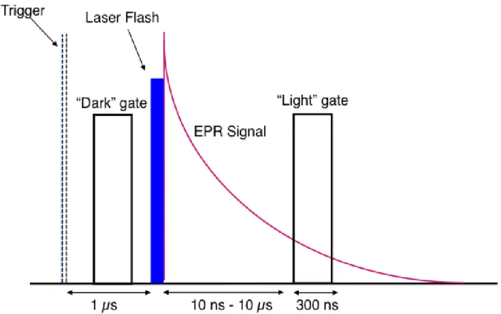
It is instructive to outline the timing sequence for the various electronic components and
discuss how they affect the overall performance of a TREPR experiment. Such a sequence is shown
in Figure 1.9. For the TREPR experiment, the external magnetic field sweep, which is controlled
either internally by clock pulses in the spectrometer or externally by a computer-generated ramp.
In either case, the field sweep is divided up into a certain number of data points (in our instrument
1000 points) that can be saved, along with the TREPR signal intensity at that point, as an x–γ array in the computer.
The timing sequence originates from a pulse generator (in our case, a Stanford DG535
digital delay generator providing TTL output pulses), from which the laser and boxcar (or digitizer)
are triggered. The repetition rate of the experiment is 60–100 Hz for an excimer laser and 10–30
Hz for a Nd:YAG laser, with the actual rate depending on the laser model in each case. Typical
sweep times are 2 or 4 min with the laser repetition rate set at 60 Hz. This means that in a 4-min
scan, there are 60 s-1 ~ 240 s/1000 ~14 laser flashes per magnetic field data point. The sampling
gates are 100–300 ns wide, with one positioned in front of the laser flash to collect the dark EPR
signal and one after the flash to collect the light+dark signal. Subtraction of the two gate voltages
gives the light induced TREPR signal. The fast photodiode allows the laser flash to be observed
on the oscilloscope so that the boxcar gates can be properly positioned.
It is important to note here two disadvantages of the TREPR technique. It is not generally
possible to observe a Boltzmann (equilibrium) population of spin states using the boxcar method,
gate is placed close in time to the laser flash. This is a consequence of having the microwave
excitation running continuously in TREPR. Near the laser flash, and during the photochemical
events that produce radicals, the apparatus is attempting to excite spin states that are still in the
process of forming. As stated earlier, small interactions such as hyperfine couplings take time to
evolve and may not be visible in the TREPR spectrum for several hundreds of nanoseconds after
the laser flash. When structural information about an unknown radical is desired, it is important to
collect spectra at multiple delay times to make sure maximum spectral resolution has been
achieved.
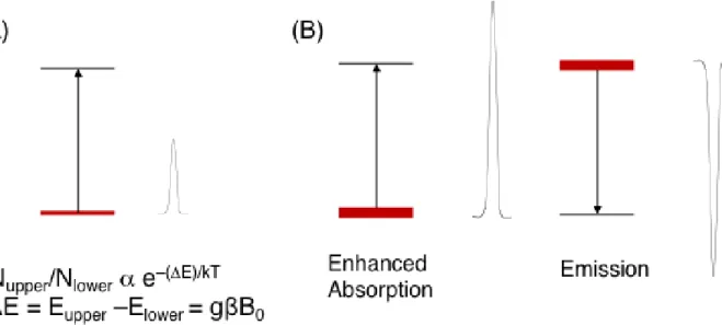
1.4 Chemically Induced Electron Spin Polarization (CIDEP) Mechanisms
In spectroscopy, it is common for transitions to be observed as absorptive lines because the
Boltzmann distribution, at equilibrium, ensures a higher population of the lower state than the
upper state (Figure 1.10 A). Examples where emission is observed, which are by definition non-
equilibrium situations, are usually cases where an excess population is created in a higher energy
level by adding energy to the system from an external source such as photolysis. CIDEP
phenomena in EPR spectroscopy can show both enhanced absorption (greater absorptive signal
intensity than predicted by the Boltzmann factors) and emission in the observed spectra. (Figure
1.10 B) What makes the non-Boltzmann populations observed in TREPR experiments so unusual
is that, in some cases, electron- and/or nuclear spin dependent chemical reactions (homolytic bond
breaking or forming) are responsible for the process. While it requires energy to break a chemical
bond, once it is broken, the mixing of spin wave functions in the resulting radical pair (RP) is all
that is necessary to make some NMR and EPR transitions appear with the so-called spin
The idea that the nuclear spin state energy-level differences (which are much smaller than
kT at room temperature) could be responsible for different chemical reaction rates was
revolutionary and somewhat controversial, especially with the observation of the nuclear spin
polarization phenomenon (CIDNP) in NMR spectroscopy. As more and more experiments were
performed to investigate this phenomenon, the idea rapidly gained acceptance and in fact helped
connect solution dynamics of small molecules to spin quantum mechanics in a very natural and
informative way. Later on in the development of CIDEP theories, the requirement for chemical
reaction to take place was lifted - many of these cases will be presented in this work.
CIDEP phenomena are observed in most TREPR-detected photochemical reactions that
produce radicals, radical ions, or biradicals. Indeed, it is from CIDEP that some of the sensitivity
is regained that was lost in bypassing the phase-sensitive detection unit (100 kHz field modulation)
is regained.
An additional improvement in TREPR sensitivity comes from the use of the boxcar to
signal average. In all of the TREPR spectra shown in this dissertation, transitions below the
baseline are in emission (E), while those above the baseline are in enhanced absorption (A). This
is different from conventional SSEPR spectra, which, as noted in Figure 1.7, are displayed as
first-derivative curves representing the change in detected intensity with the external field.
There are four well-known CIDEP mechanisms: the Triplet Mechanism (TM), the Radical
Triplet Pair Mechanism (RTPM), Radical Pair Mechanism (RPM), Spin Correlated Radical Pair
Mechanism (SCRP). The spectra presented in this dissertation exhibit polarization from the TM
and RTPM, and the following two sections of this chapter will briefly outline the basic
phenomenon of these two types of CIDEP mechanisms and provide an example of each
1.4.1 The Triplet Mechanism (TM)
The TM is often considered to be the simplest CIDEP mechanism [21].Qualitatively, the
mechanism is outlined in Figure 1.11, which suggests that the intersystem crossing (ISC) process,
usually by spin-orbit coupling, from excited singlet state (S1) to excited triplet state (T) in the
organic molecules will populate the triplet spin states TX, TY, and TZ differently even in the absence of a magnetic field.
The labels TX, TY, and TZrepresent the triplet state energy levels in the molecular coordinate system (frame of reference); these are known as the zero-field basis functions. Because spin orbit coupling
is anisotropic in this frame, the TZ spin state is overpopulated in Figure 1.11 (thicker bars represent higher populations) during ISC process, and this excess population is then transferred to the spin
state T+ (=|𝛼𝛼⟩) of the laboratory frame of reference (in which the Z-axis is directed along the applied magnetic field of the spectrometer). The net polarization of the electron spins in the triplet,
created during the spin–orbit coupling (SOC) process from S1 to T, is transferred to the radicals
resulting from photochemical reactions of the triplet. Over time, the
populations of the spin states of the triplet relax to their Boltzmann equilibrium values.
Experimentally, it is observed that all of the resonance lines of both radicals in the TREPR
spectrum of such systems are polarized equally and either are polarized positively (enhanced
absorption, A) or polarized negatively (emission, E). Several examples of TM will be discussed in
1.4.2 The Radical-Triplet Pair Mechanism (RTPM)
The quenching of excited states by molecules has been extensively investigated on
photochemical and physical processes, such as energy transfer, triplet-triplet annihilation, and
quenching by radicals. In particular, the system which includes excited triplet state quenching by
free radicals, shows CIDEP effects which are observable by TREPR.
Two mechanisms have been proposed to explain the polarization associated with
triplet-doublet (T-D) systems; one is a radical triplet pair mechanism (RTPM), and the other is an electron
spin polarization transfer (ESPT) mechanism.
The radical-triplet pair mechanism (RTPM) of CIDEP, as its name implies, for its
polarization requires an interaction between an excited molecular triplet state and a free radical,
usually a stable nitroxide structure. The participation of a stable radical is not a requirement for
the appearance of the RTPM, nor is the phenomenon restricted to liquid solution. In some
photochemical reactions, a large concentration of excited triplet states are produced, leading to
spin–spin interactions between the excess triplets and the unstable free radicals produced from
previous reactions. The polarization mechanism itself is explained as follows: In free solution,
encounters between molecules (M) in their electronically excited singlet (1M*) or triplet (3M*)
states with radicals (R) in their ground doublet state (2R) result in either enhanced intersystem
crossing (EISC) or deactivation of the electronic excitation. The quenching reaction of an
3M* + 2R → 4[3M* ⋅⋅⋅2R] →1M + 4R* (1.3)
2[3M* ⋅⋅⋅2R] →1M + 2R* (1.4)
2[3M* ⋅⋅⋅2R] →1M + 2R (1.5)
In the non-adiabatic potential surface, where free radicals have no electron spin
polarization because four quartet spin states are equally populated after dissociation, but Reaction
(1.2) is possible has never been conclusively observed. Relying on numerous transient optical
absorption spectroscopy experiments [22–27],it was concluded that quenching reactions (1.3) and
(1.4) occur at diffusion-controlled rates if the electronic energy of the excited molecular triplet
state is higher than the energy of the doublet excited state of the radical. In other words, quenching
is effective when it is followed by energy transfer (reaction 1.4) and is ineffective when energy
transfer is impossible (reaction 1.5). Reactions (1.3)–(1.5) are collectively called the EISC
processes shown in figure 1.12 and 1.13.
Imamura et al. [28] discovered that stable nitroxide radicals that encounter excited
molecular triplet states can develop ESP (electron spin polarization). This phenomenon has been
confirmed in numerous other experimental studies [29–33] and is now included in the CIDEP
family with two separate abbreviations: RTPM and ESPT (electron spin polarization transfer).
These are used to differentiate between two possible mechanisms for the generation of ESP.
The RTPM polarization is determined by the multiplicity of an excited state (S1 or T1) and
the sign of an exchange parameter, J, between the triplet and the radical. On the other hand, in the case of ESPT, the spin polarization of the excited T1 state via a spin-selective ISC is transferred to
a stable radical via an energy transfer or a spin exchange interaction, and the overpopulation of the
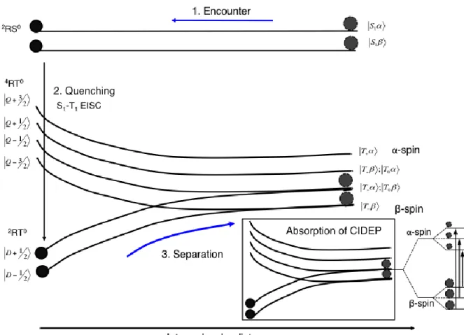
Figure 1.13 Energy levels of a radical–triplet pair as a function of their separation. Schematic explanation of net absorption CIDEP created by excited singlet quenching by stable free radical. The black dots represent the population.1. Encounter, 2. Quenching and 3. During the separation of the
2RT0 -spin is enriched on stable
and the overpopulation of the this level results in the observation of emission in the observed
TREPR spectrum.
As shown in the figure 1.12, neglecting the Boltzmann spin polarization, we can assume that when
a molecular triplet collides with a stable nitroxide radical, the resulting Radical-Triplet (R-T) (or
collision complex) pair creates six equally populated electron spin states a molecular triplet
collides with a stable nitroxide radical, the resulting Radical-Triplet (R-T) (or collision complex)
pair creates six equally populated electron spin states {|𝑇+𝛼⟩, |𝑇+𝛽⟩, |𝑇0𝛼⟩, |𝑇0𝛽⟩, |𝑇−𝛼⟩, |𝑇−𝛽⟩. At
the distance of closest approach (R) between the radical and the triplet state, the exchange
interaction dominates all of the magnetic interactions in the R-T pair. The electronic spin levels
of the system can be labeled according to the total spin F=T+ D, resulting in four quartet spin
states: |𝑄+3 2⁄ ⟩, |𝑄+1 2⁄ ⟩, |𝑄−1 2⁄ ⟩, |𝑄−3 2⁄ ⟩ and two doublet spin states |𝐷+1 2⁄ ⟩ and |𝐷−1 2⁄ ⟩, the
latter two being lower in energy than the former four if the J is negative (Fig. 1.12). The doublet states are depopulated because of quenching reaction (1.4)earlier. If the exchange interaction is
the only interparticle interaction, the |𝐷+1 2⁄ ⟩ spin state of the R-T complex correlates with the
|𝑇+𝛽⟩ state of the separated R-T pair, and the |𝐷−1 2⁄ ⟩ correlates with the |𝑇−𝛼⟩ state. For this
reason, spin-selective quenching does not lead to ESP. However, the spin–spin dipolar interaction
between the electron spins located in the triplet (ZFS) does not commute with the Hamiltonian of
the R-T pair, the elements of which are coupled by the exchange interaction. This creates mixing
of the spin states in the vicinity of the avoided crossings of the spin states, which are the circled
remain passive.
This increases the population of the|𝑇+𝛼⟩ and |𝑇0𝛼⟩ spin states of the separate R-T pair,
creating an excess of the |𝛼⟩ spins located in the radicals. In the TREPR experiment, this
depopulation process manifests itself as emissive polarization in the three-line nitroxide radical
signal. It should be noted that a corresponding correlation diagram for a positive exchange
interaction (quartets with lower energy than the doublet spin states) could be constructed in a
similar fashion and leads to radicals that have escaped from the R-T encounter with positive
polarization (enhanced absorption) as shown in figure 1.13.
Evans[34] was the first to discover that strongly forbidden singlet–triplet UV optical
absorptions in aromatic molecules can be induced by paramagnetic substances, particularly oxygen.
Later, Hoijtink[35] and Murrell[36] proved that the intermolecular exchange interaction was a
driving force for this 1M* → 3M* EISC in electronically excited molecules.
There are five general spin-allowed reactions of 2R with electronically excited singlet
molecules 1M*:
i. EISC: 1M* + 2R ⟶ 2[1M*⋅⋅⋅ 2R] ⟶ 3M* + 2R (1.6)
ii. Quenching: 1M* + 2R ⟶ 1M + 2R (1.7)
iii. Energy transfer: 1M* + 2R ⟶2[1M*⋅⋅⋅ 2R] ⟶1M + 2R* (1.8)
iv. Charge transfer: 1M* + 2R ⟶2[2M±⋅⋅⋅2R∓] ⟶ 3M* + 2R (1.9)
1M* + 2R ⟶2[2M±⋅⋅⋅2R∓] ⟶ 1M + 2R (1.10)
Among these possible processes, EISC 1M* → 3M* (reactions 1.7 and 1.9) are the most
effective [37,38].Reaction (1.7)is followed by the generation of ESP [39,40].The ESP generated
exchange interaction, QP RTPM always results in emissive net polarization of radicals, and the
DP RTPM mechanism results in positive polarization. The notable examples of RTPM will be
discussed in Chapter 4. The radical-triplet pair interactions can be used to investigate the dynamics
of acrylic polymers in dilute solution. By incorporating a nitroxide into the polymer chain via
covalent bonds, we can generate RTPM polarization via intramolecular encounters that depend on several structural and physical features of the polymer/solvent system. The magnitude of this
polarization is directly proportional to the number of encounters made between the stable radical
and the excited triplet state, and is therefore an indirect measure of the rate of intra-chain contact
Chapter 2. Photochemistry of acrylate-POSS block copolymers and dynamic effects in copolymer radicals
2.1 Introduction
As discussed in Chapter 1.4.1, the degradation of acrylic polymers has been a subject of
intense study in our laboratory for the past decade, and our TREPR techniques are powerful enough
to investigate the photochemical and photophysical properties of acrylic homopolymers. The
creation of carbon–centered radicals within the main chain of an acrylic homopolymer provides a
means to investigate the conformational energy landscape using a highly localized, minimally
perturbative spin probe.
Because the electron-nuclear hyperfine interactions in the main chain radical depend on
the dihedral angle between the p orbital containing the unpaired electron and the neighboring C-H
sigma bond, the TREPR spectra of these main chain radicals show a strong temperature
dependence on both line widths and line positions. Changes in line width are related to the “jump
time” between conformations, while changes in line position (at constant width) reflect changes in
the populations of certain rotational conformers (“rotamers”). Additionally, alternating hyperfine
line width effect is shown in lower temperature spectra of acrylate polymers because system
involves dynamic effects due to conformational motion. As mentioned in chapter 1.4.1, polymer
tacticity plays a strong role in both the overall spectral appearance and in the
temperature-dependent line broadening processes.
conformational landscape of the polymers. Furthermore, because the photochemistry involved in
these studies is destructive, characterization of the observed free radicals can offer insight into the
degradation mechanism of acrylates in solution with high spectral resolution and unambiguous
assignment of the reactive intermediates involved. However, the photodegradation mechanisms of
block copolymers are not yet fully understood because the dynamics of block copolymer chains
are expected to be much more complicated than that of the homopolymer.
The polyhedral oligosilsesquioxane (POSS)-based polymers, one of the most studied
inorganic-ogranic hybrid copolymers, are of significant interest, with diverse potential applications
such as biomaterial [41], molecular optics [42], electronics [43], and coatings and films [44]. This
is because they show unique physical properties such as a high modulus, a high resistance to
oxidation and an increased stability to UV light compared to other acrylic structures.
In this chapter, we will discuss the photochemistry of several acrylate-POSS block
copolymers that have not been investigated before and we will show novel degradation pathways,
the relationship between degradation stability and microstructure, and interesting chain dynamics.
Before going into detail regarding the TREPR results of these POSS-based copolymers, it is useful
to present some background on previous studies on acrylate-POSS polymer degradation, including
the structure of both POSS and POSS based polymers.
2.1.1 POSS and POSS based Polymers
1) By modifying and incorporating inorganic filters into organic groups via covalent
bonds; or,
2) By using melt-mixing or in-situ polymerization to improve compatibility between
the inorganic and the polymer matrix
By using the former method, POSS can be produced and the inorganic filters naturally
disperse into the matrix.
POSS is one of many kinds of silsesquioxane molecules. The term silsesquioxane refers to
the chemical structures following the basic composition of (RSiO1.5)n, n is typically 8, 10, 12, 16,
17. Here, the R-group, also called the vertex group for polyhedral molecules, may be hydrogen,
alkyl, alkylene, and aryl arylene among others6.
The molecular architecture of silsesquioxanes can be classified into two categories: (a)
non-caged structure and (b) caged structure (the most prevalent structure shown in Figure 2.1).
Only caged silsesquioxanes are usually called POSS and these are well-defined, highly symmetric
molecules. POSS molecules with an inorganic core composed of silicon-oxygen (R8 Si8 O12) are
the most common structures used in studies of physical and photochemical properties.
The overall performance of POSS base polymers relies on how POSS moieties are
incorporated into the polymer chains, and this in turn affects the local molecular interaction and
chain mobility. Highly symmetric POSS molecules are tethered to eight organic groups including
methyl, isobutyl and phenyl, which surround and are bonded to the silicon vertices [46].
One or more corner groups can be substituted by functional groups, such as methacrylate,
In 1946, Scott successfully synthesized well-defined POSS structures, after which the Air
Force Research Lab developed a series of linear random and block POSS structures containing
copolymers [49]. So far, research on POSS-based polymer and copolymers has continued in
several research labs, but especially at the Air Force Research Lab due to an interest in their
development as aerospace coatings [50-52].
2.1.2 Degradation studies of POSS-based Polymer
Common polymers such as acrylate polymers can be easily degraded by interactions with
UV-light [53] and under high temperature [54], so it can be undesirable to use these polymers in
outdoor applications such as architectural coatings or packaging materials that may be exposed to
light. Several studies have been undertaken to understand the mechanism of degradation so that
the degradation of such polymers can be controlled by means of additives and/or modifications to
the structure of polymer. One attempt incorporated inorganic nanofillers in a polymer chain similar
to the POSS-based polymer. This approach makes the polymer thermally and chemically robust,
to such an extent that one of the promising applications of POSS-based polymers is for use in the
highly oxidizing environment of orbiting space vehicles [55,56]. Accordingly, the photo-stability
of polyolefin-based nanocomposites including POSS polymers is a very important and desirable
asset that may positively contribute to the resolution of a wide range of industrial challenges.
The study of the degradation mechanism in POSS based polymers has been carried out