EXAMINING THE BIOLOGICAL FUNCTIONS OF THE H3K36 METHYLATION STATES IN TRANSCRIPTION REGULATION AND THE POST-TRANSLATIONAL MODIFICATION
LANDSCAPE OF SET2
Julia Veronica DiFiore
A dissertation submitted to the faculty at the University of North Carolina at Chapel Hill in partial fulfillment of the requirements for the degree of Doctor of Philosophy in the Curriculum in
Genetics and Molecular Biology in the School of Medicine.
Chapel Hill 2020
© 2020
ABSTRACT
Julia Veronica DiFiore: Examining the Biological Functions of the H3K36 Methylation States in Transcription Regulation and the Post-Translational Modification Landscape of Set2
(Under the direction of Brian D. Strahl)
To package DNA, eukaryotes fold and compact DNA into chromatin. The fundamental building block of chromatin is the nucleosome, which plays a crucial role in DNA accessibility. The unstructured N-terminal tails of histones can be post-translationally modified with chemical moieties such as acetylation and methylation, amongst others. These post-translational
modifications (PTMs) can serve as binding sites for proteins that alter chromatin structure or affect the interactions between histones and DNA, thus making the DNA more or less accessible to other cellular machinery. Additionally, other proteins in the cell can be post-translationally modified with similar chemical moieties, which can alter their enzymatic activity, create binding sites for other proteins, or other outcomes. The role of PTMs in regulating cellular processes underscores their importance and the need to understand their function and
ACKNOWLEDGEMENTS
First, I would like to thank my advisor, Dr. Brian Strahl, for his guidance and support throughout the years. No matter the circumstances, Brian was always a source of enthusiasm. Whether it was troubleshooting experiments, analyzing new data, or discussing slightly crazy hypotheses, he could always be counted on for spirited discussion, new ideas, and a fresh outlook on my research. I would also like to thank my thesis committee members, Dr. Bob Duronio, Dr. Kerry Bloom, Dr. Ian Davis, and Dr. Mauro Calabrese. Initially, the thought of regularly presenting my work to a group of accomplished scientists was intimidating, but I quickly realized that they were all there with the intent to help my research be the best that it could be. I am grateful that our meetings were always full of thoughtful questions, ideas, and encouragement.
Andrew Lerner have been in the lab with me since day one. We started off having no idea what we were doing and now, we sometimes know what we are doing. We did it, guys. We made it.
I have also had the opportunity to work with a lot of fantastic collaborators, both at UNC and otherwise. Catherine Fahey headed up a great group for a collaboration between the Strahl, Davis, and Rathmell labs. Working on that collaboration gave me confidence that I could
meaningfully contribute to projects. Bing Li at Shanghai Jiao Tong University graciously provided some of the foundational data for my thesis work and has been helpful in providing additional data. Brenda Temple at the Structural Bioinformatics Core helped with protein modeling, which has proven to be instrumental in explaining the molecular basis underlying my thesis. Jeremy Simon and Travis Ptacek provided a thorough analysis of my RNA-seq data, which is another linchpin of my work. While normally rivals, collaborating with Dave MacAlpine, Heather MacAlpine, and Bonnie Chen at Duke has been great. Their expertise in MNase-seq has provided answers to long-standing questions and their enthusiasm makes for a fun
collaboration. Finally, a recent project with Shu Zhang in the Dohlman lab has given new insight to some of the processes I have studied in graduate school. Shu’s curiosity, hard work, and dedication make it easy to work with her and I look forward to hearing about her incredible discoveries in the years to come.
As Hermione Granger said in Harry Potter and the Sorcerer’s Stone, “Books and
best way possible), and introduced me to Game of Thrones, The Bachelor, How to Train Your Dragon, and at least a dozen other things I now love. Michelle Engle has also been part of my graduate school tribe since the beginning and was always down for new adventures – whether locally or across the country. Her kindness and encouragement have gotten me through the many challenges of graduate school. The Yinzers – Casey Schmidt, Talia Hatkevich, Aaron Crain, and Chris Abdullah – watched many Steelers and Penguins games together and brought a small taste of home to North Carolina. Debbie and Ken Tunnel, Adam and Melanie Long, Holly Anderson, and many others at Chatham Community Church reminded me that I am so much more than what happens in lab. Most recently, Kevin Smith has been there to support, listen, and love me through all of this. He kept me fed, caffeinated, and laughing at his ridiculous dancing, puns, and unending episodes of Brooklyn Nine-Nine, and for that, I am forever grateful. Finally, I would of course, not be here without my family. From my very first science fair in first grade when I was too scared to talk to anyone, to now, my parents have always
TABLE OF CONTENTS
LIST OF FIGURES ... xiv
LIST OF TABLES ... xvi
LIST OF ABBREVIATIONS ... xvii
CHAPTER 1 – INTRODUCTION ... 1
Histone Code Hypothesis ... 2
Histone Lysine Methylation ... 3
Set2 and H3K36 Methylation in Transcription ... 4
Regulation of Set2 and H3K36 Methylation ... 4
Set2 Protein Structure ... 5
H3K36 Methylation Interacting Proteins ... 6
Cryptic Transcription ... 8
Additional Functions in Budding Yeast and Set2 Homologs ... 10
Additional Functions in Budding Yeast ... 10
Schizosaccharomyces pombe ... 12
Caenorhabditis elegans ... 13
Drosophila melanogaster ... 13
Humans and Cancer Relevance ... 14
Non-histone PTMs and their Biological Functions ... 18
Concluding Remarks and Contributions of This Work ... 19
Figures ... 20
Introduction ... 23
Results ... 26
Phe/Tyr Switch in Set2 separates H3K36 Methylation States in vitro ... 26
Phe/Tyr Switch Mutations in Set2 are Tools to Separate H3K36 Methylation States in vivo ... 27
Distinct Methylated Forms of H3K36 are Deposited within or near Transcribed Regions of Genes ... 29
H3K36me1/2 and H3K36me3 Function Redundantly in Some Cellular Contexts ... 30
Genetic Interactions of SET2 with BUR1 and SPT16 Reveal Unique Functions for H3K36me1/2 and H3K36me3 ... 31
H3K36me1/2 and H3K36me3 Have Unique and Shared Functions in Repressing Cryptic Transcription at Reporter Loci ... 31
During Nutrient Deprivation, H3K36me1/2 or H3K36me3 Prevent Antisense Transcription ... 32
Alteration of Global H3K27ac and H3K56ac in set2 Methylation Mutants ... 35
H3K36me1/2 and H3K36me3 are Important for Proper H3K27ac and H3K56ac Localization ... 36
Discussion ... 38
Materials and Methods ... 41
Yeast Strains and Plasmids ... 41
Alignment and Molecular Modeling ... 41
Western Blotting ... 42
Recombinant Protein Purification from Baculovirus Expression System ... 42
Histone Methyltransferase Assay ... 43
Co-immunoprecipitation ... 43
Chromatin Immunoprecipiration and Real-Time PCR ... 44
Spotting Assays ... 44
RNA-seq Library Preparation and Sequencing ... 44
Figures ... 53
CHAPTER 3 – MODIFYING A MODIFIER: DISSECTING THE POST-TRANSLATIONAL MODIFICATION LANDSCAPE OF SET2 ... 69
Summary ... 69
Introduction ... 69
Results ... 72
Lysine Residues on Set2 are Methylated and Acetylated ... 72
set2 Lysine Point Mutants Do Not Affect Set2 Protein Abundance or H3K36 Methylation . 73 set2 Lysine Point Mutants Do Not Affect Set2 Function in Certain Cellular Contexts ... 73
Predicted Phosphorylation Sites are Found on Set2 ... 74
set2 Serine Point Mutants Affect Set2 Protein Abundance, but not H3K36 Methylation ... 74
set2 Serine Point Mutants Repress Cryptic Transcription at Reporter Loci but Have Transcriptional Elongation and DNA Double Strand Break Repair Defects ... 75
Discussion ... 76
Materials and Methods ... 78
Yeast Strains and Plasmids ... 78
Mass Spectrometry ... 78
Database Searching ... 78
Western Blotting ... 78
Spotting Assays ... 79
Tables ... 80
Figures ... 84
CHAPTER 4 – CONCLUSIONS AND FUTURE DIRECTIONS ... 89
H3K36me States Work Redundantly to Repress Cryptic Transcription During Nutrient Stress ... 89
H3K36me3 Has a Unique Function Related to Bur1 and Spt16 ... 92
Final Thoughts ... 96
APPENDIX A – STRUCTURE/FUNCTION ANALYSIS OF RECURRENT MUTATIONS IN SETD2 REVEALS A CRITICAL AND CONSERVED ROLE FOR A SET DOMAIN RESIDUE IN MAINTAINING PROTEIN STABILITY AND H3K36 TRIMETHYLATION ... 97
Summary ... 97
Introduction ... 98
Results ... 100
SETD2 and Set2 Share a High Degree of Structural and Sequence Homology at their SET and SRI Domains ... 100
SET domain mutation destabilizes SETD2 in Cells ... 101
Histone H3 Lysine 36 Trimethylation is Linked to SETD2 Mutational Status ... 102
The SETD2 R1625C Variant is Enzymatically Inactive in vitro and has Diminished Substrate Binding ... 103
Domain-Specific Mutations in Yeast Set2 Separate Roles of H3K36 Methylation States . 105 Human Kidney Cells Display an H3K36 Trimethylation-Dependent DNA Damage Response ... 107
Discussion ... 108
Materials and Methods ... 110
Modeling SETD2 and Set2 ... 110
Mammalian Cell Lines Transfections and Phenotypic Assays ... 111
Sequencing and Allelic Analysis ... 112
Immunoblot Analysis ... 112
Antibodies ... 112
Immunocytochemistry ... 113
Chromatin Immunoprecipitation ... 113
Quantitative RT-PCR ... 114
Expression and Purification of Human SETD2 ... 114
Histone Methyltransferase Assays ... 115
Yeast Growth Assays ... 116
Immunofluorescence Staining for γH2A.X ... 116
Figures ... 118
LIST OF FIGURES
Figure 1.1: Set2 Domain Map ... 20
Figure 1.2: H3K36 Methylation Interacting Proteins ... 21
Figure 1.3: Schematic of Cryptic Transcription in wild-type and set2∆ ... 21
Figure 2.1:4Phe/Tyr Switch in Set2 Separates H3K36 Methylation States in vitro ... 53
Figure 2.2:5Phe/Tyr Switch Mutations in Set2 are Tools to Separate H3K36 Methylation States in vivo ... 54
Figure 2.3:6set2 Mutatns Differentially Methylate H3K36 and Interact with RNAPII ... 55
Figure 2.4:7Distinct Methylated Forms of H3K36 are Deposited within or near Transcribed Regions of Genes ... 56
Figure 2.5:8H3K36me1 is Deposited within or near Transcribed Regions of Genes ... 57
Figure 2.6:9H3K36me1/2 and H3K36me3 have Unique Phenotypes in Some Cellular Contexts ... 58
Figure 2.7:10Function of H3K36 Methylation States in Cryptic Transcription Regulation ... 58
Figure 2.8:11Activity of H3K36 Methylation States in Cryptic Transcription Regulation ... 60
Figure 2.9:12Alteration of Global H3K27ac and H3K56ac in set2 Methylation Mutants ... 63
Figure 2.10:13Set2 and H3K36 Methylation Levels are Similar at 0 and 60 Minutes after Nutrient Deprivation ... 64
Figure 2.11:14H3K36me1/2 and H3K36me3 are Important for Proper H3K27ac Localization ... 66
Figure 2.12:15H3K26me1/2 and H3K36me3 are Important for Proper H3K56ac Localization ... 66
Figure 3.1:16set2 Lysine Point Mutants Do Not Affect Set2 Protein Abundance or H3K36 Methylation ... 84
Figure 3.2:17set2 Lysine Point Mutatns Do Not Affect Set2 Function in Certain Cellular Contexts ... 85
Figure 3.3:18Predicted Phosphorylation Sites are Found on Set2 and set2 Serine Point Mutants Affect Set2 Protein Abundance, but not H3K36 Methylation ... 87
Figure 3.4:19set2 Serine Point Mutants Repress Cryptic Transcription at Reporter Loci and Have Transcriptional Elongation and DNA Double Strand Repair Defects ... 88
Figure A.3:24The SETD2 R1625C variant is catalytically inactive and has reduced
substrate-binding capacity ... 121 Figure A.4:24Modeling of ccRCC specific mutations in Set2 results in separate effects
based on H3K36me status ... 122 Figure A.5:24H3K36me3 loss delays γH2A.X foci resolution after DNA damage but
LIST OF TABLES
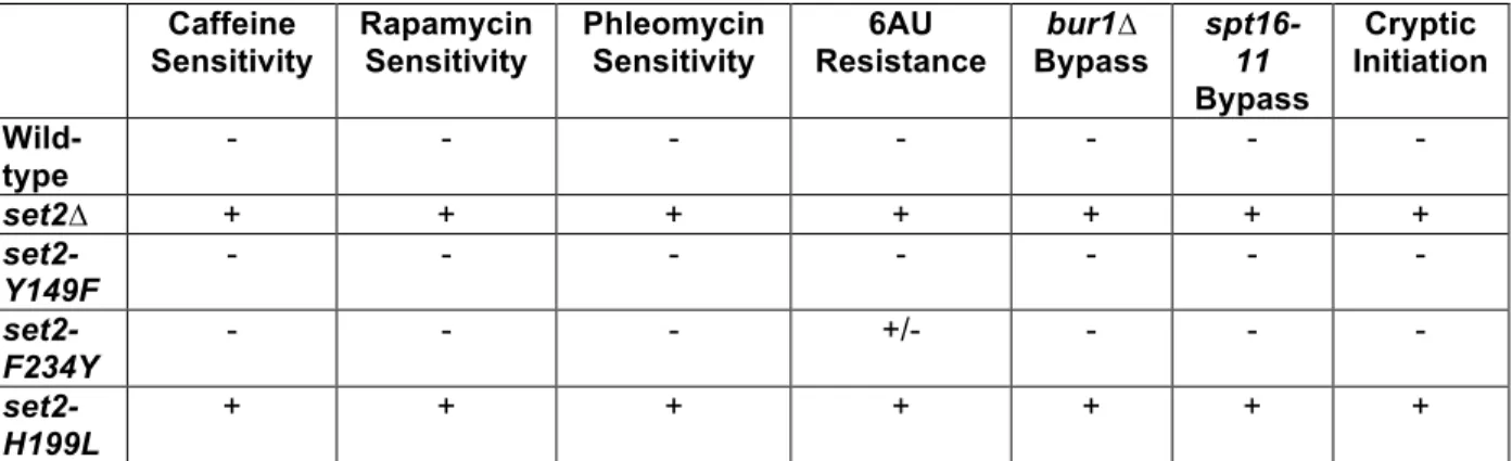
Table 2.1: Summary of set2 Mutant Phenotypes ... 46
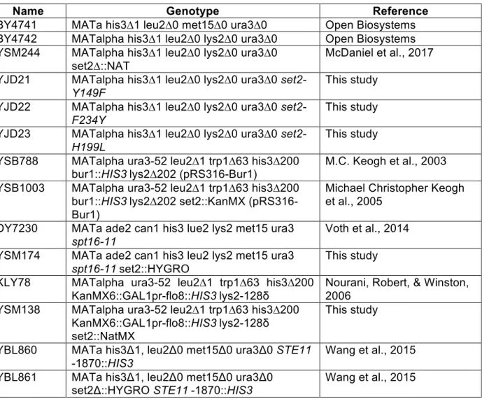
Table 2.2: Yeast Strains and Genotypes ... 47
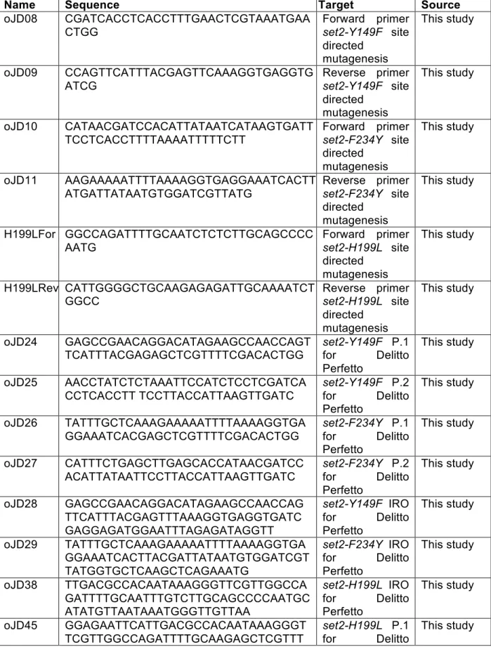
Table 2.3: List of Primers ... 48
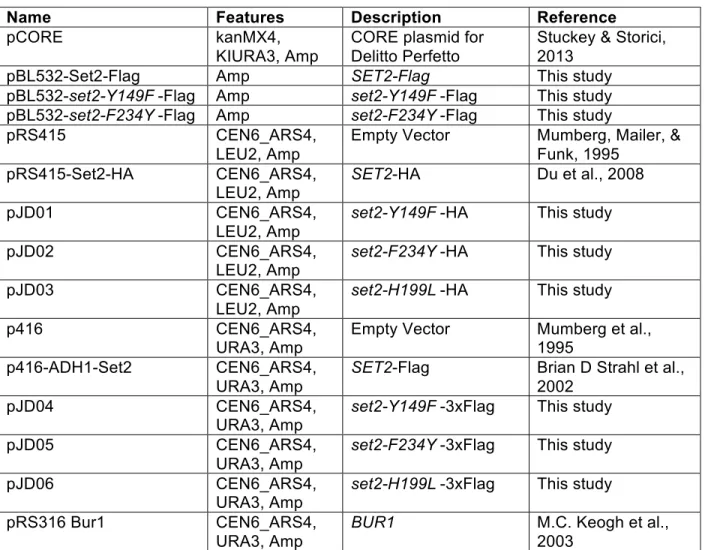
Table 2.4: List of Plasmids ... 52
Table 3.1:5Lysine Residues on Set2 are Methylated and Acetylated ... 80
Table 3.2:6List of Yeast Strains and Genotypes ... 80
Table 3.3:7List of Primers ... 81
LIST OF ABBREVIATIONS
° degree
3D three dimensional
5-FOA 5-fluoroorotic acid
6-AU 6-azauracil
Å angstrom
ac acetyl
ALL acute lymphocytic leukemia AID autoinhibitory domain
APC/C anaphase promoter cyclosome complex AWS associated with SET
BLAST basic local alignment search tool BSA Bovine serum albumin
C celcius
CC coiled coil
ccRCC clear cell renal cell carcinoma CD circular dichroism
ChIP chromatin immunoprecipitation
ChIP-seq chromatin immunoprecipitation sequencing CIS cryptic initiation site
COSMIC catalogue of somatic mutations in cancer cryo-EM cryogenic electron microscopy
CTD C-terminal domain
DRB 5,6-dichlorobenzimidazole 1-β-D-ribofuranoside DSB double strand break
DTT dithiothreitol
ECL enhanced chemiluminescence EDTA ethylenediaminetetraacetic acid ELM eukaryotic linear motif
FACT facilitates chromatin transcription complex
FAIRE-seq formaldehyde assisted isolation of regulatory elements sequencing FBS fetal bovine serum
G1 Gap 1
G2 Gap 2
GST glutathione S-transferase
Gy gray
H2BK120 histone H2B lysine 120 H3K4 histone H3 lysine 4 H3K9 histone H3 lysine 9 H3K14 histone H3 lysine 14 H3K18 histone H3 lysine 18 H3K27 histone H3 lysine 27 H3K36 histone H3 lysine 36 H3K44 histone H3 lysine 44 H3K56 histone H3 lysine 56 H3K79 histone H3 lysine 79 H3S10 histone H3 serine 10
HAT histone acetyltransferase HDAC histone deacetylase complex
HEPES 4-(2-hydroxyethyl)-1-piperazineethanesulfonic acid HMT histone methyltransferase
hMSC human mesenchymal stem cells HR homologous recombination HRP horseradish peroxidase
ICC immunocytochemistry
IPTG isopropyl β-d-1-thiogalactopyranoside
kDA kilodalton
LANS light-activated nuclear shuttle
LC-MS liquid chromatography mass spectrometry LEDGF lens epithelium-derived growth factor LON long oligonucleotides
M mitosis
MALDI-TOF matrix assisted laser desorption/ionization-time of flight MAP mitogen-activated protein
MBF MluI cell-cycle box binding factor
me methylation
me1 mono-methylation
me2 di-methylation
me3 tri-methylation
mg miligram
min minute
mM milimolar
MMR mismatch repair
MNase-seq microccocal nuclease sequencing mRNA messenger ribonucleic acid
MS mass spectrometry
MRX Mre11, Rad50, Xrs2
MSL male-specific lethal
MUD-PIT multidimensional protein identification technology NHEJ Non-homologous end joining
NFR Nucleosome free region
nM nanomolar
OD optical density
ORF open reading frame
PCR polymerase chain reaction PBS phosphate-buffered saline
PBS-T phosphate-buffered saline-tween 20 PDB protein data bank
PHD plant homeodomain
Phe/Tyr phenylalanine/tyrosine
PS post-SET
PTM post-translational modification PVDF polyvinylidene fluoride
PWWP proline-tryptophan-tryptophan-proline domain
rep replicate
RNAPII RNA polymerase II RNA-seq RNA sequencing rpm rotations per minute
rRNA ribosomal RNA
RT-qPCR quantitative reverse transcription polymerase chain reaction
S DNA synthesis
SAH S-adenosyl-L-homocycteine SAM S-adenosyl-L-methionine
SC synthetic complete
SD synthetic depleted
SDS-PAGE sodium dodecyl sulface-polyacrylamide gel electrophoresis SEM standard error of the mean
SET Su(var)3-9, Enhancer of zeste and Trithorax SRAT Set2-repressed antisense transcript
SRI Set2 Rpb1 interacting domain
STAR spliced transcripts alignment to a reference SUMEB SDS urea MOPS EDTA bromophenol blue SUT stable unannotated transcript
TALEN TAL effector nucleases TAP tandem affinity purification TFA rribluroacetic acid
TOR1C target of rapamycin 1 complex tSETD2 truncated SETD2
ub ubiquitination
µl microliter
µM micromolar
UV ultraviolet
V(D)J variable diversity joining
qPCR quantitative polymerase chain reaction TCA trichloroacetic acid
TEV tobacco etch virus
Tm melting temperature
WT wild-type
CHAPTER 1 – INTRODUCTION
The enormous diversity observed in the natural world underscores the complexity of life and has long pushed us to better understand its origins. Toward that end, in 1865, Gregor Mendel completed his studies on plant hybridization and decades later, the results would be recognized as foundational genetic principles (Mendel, 1901). Mendel’s work and those who rediscovered it at the beginning of the 20th century founded the basis for understanding life, its diversity, and who we are at a molecular level (De Castro, 2016). Seminal work throughout the first half of the 20th century established a molecule called deoxyribonucleic acid (DNA) as the blueprint for all life on earth and the material that was responsible for passing traits from one generation to the next. Subsequent research established the “Central Dogma” of molecular biology, stating that DNA is transcribed into another molecule, ribonucleic acid (RNA), which is then translated into functional units called protein. However, recent discoveries have challenged this dogma and given us a more nuanced view of molecular biology and life itself (Gayon, 2016). Continued research will no doubt answer many of today’s questions and raise more for future generations to grapple with.
access is regulated through post-translational modifications (PTMs) on histone and non-histone proteins.
Histone Code Hypothesis
In order to package DNA, cells fold it into a higher order structure called chromatin. The basic unit of chromatin is the nucleosome, made up of two copies each of histones H2A, H2B, H3, and H4 with 147 base pairs of DNA wrapped around it (Luger et al., 1997). Histones can be post-translationally modified with chemical groups such as acetylation, methylation,
phosphorylation, and ubiquitination, amongst others (Rothbart and Strahl, 2014). Many of these PTMs occur on the flexible histone tails, but some are found on the histone fold or globular domains (Cosgrove et al., 2004). These PTMs are functionally important in allowing or preventing cellular machinery to access DNA during cellular processes like transcription, replication, DNA damage repair, amongst others.
For years, it was thought that chromatin simply acted as a physical barrier to DNA-templated processes and organized DNA to fit into the nucleus. Beginning in the 1960s with the discovery of histone acetylation by Vincent Allfrey, evidence began to emerge that chemical modifications can affect DNA-histone contacts (Allfrey et al., 1964). For example, lysine
acetylation changes histone tail from positively charged to neutral, therefore disrupted the binding with negatively charged DNA. Later, phosphorylation on serine 10 of histone H3 (H3S10) was shown to be important for two opposing functions: chromosome condensation during mitosis and chromatin decompaction in transcription (Bradbury, 1992; Mahadevan et al., 1991). This
Histone Lysine Methylation
One of the most well studied histone PTMs is lysine methylation. Since the detection of methylation on lysine 4 of histone H3 and the subsequent identification of SUV39H1 as the first lysine specific histone methyltransferase, methods have rapidly improved to detect histone lysine methylation, identify histone methyltransferases (HMT), and study their biological functions (Rea et al., 2000; Strahl et al., 1999). Histone lysine methylation is found on histone H3 at lysines 4, 9, 14, 18, 23, 27, 36, and 79, along with histone H4 at lysine 20 (Wozniak and Strahl, 2014). HMTs catalyze methylation at these residues by transferring a methyl group from the cofactor S-adenosyl-L-methionine (SAM) to the amino group of the lysine residue (Dillon et al., 2005). Each HMT has a specific lysine residue they target for methylation as well as specificity for how many methyl groups they add to the target lysine (mono-, di-, or
trimethylation) (Black et al., 2012). Unlike acetylation, methylation does not change the charge of the lysine residue and is thought to primarily function as a way to recruit chromatin-modifying proteins (Beaver and Waters, 2016). By serving as a binding site for other proteins, histone lysine methylation has a diverse set of functions, particularly in gene transcription.
Set2 and H3K36 Methylation in Transcription
Regulation of Set2 and H3K36 Methylation
In Saccharomyces cerevisiae (hereafter, budding yeast), Set2 is the sole H3K36 methyltransferase and catalyzes mono-, di-, and trimethylation (H3K36me1/2/3) in actively transcribed genes (Strahl et al., 2002). In multicellular organisms, such as Caenorhabditis elegans, Drosophila melanogaster, and humans, there are multiple enzymes that catalyze H3K36me. In D. melanogaster and humans, certain enzymes are specific for H3K36me1 and H3K36me2, while the Set2 homologs in those organisms catalyze only H3K36me3 in vivo
(Venkatesh and Workman, 2013). Nevertheless, budding yeast serves as an excellent model system for Set2 and H3K36me since the loss (set2∆) or mutation of one enzyme can alter H3K36me globally and the consequent changes can be studied.
Set2 catalyzes H3K36me in actively transcribed genes and is targeted there in part through its interaction with the phosphorylated serine 2 and serine 5 on the C-terminal domain (CTD) of RNA polymerase II (RNAPII) (Li et al., 2003; Xiao et al., 2003; Youdell et al., 2008). While serine 5 is phosphorylated at the promoter by Kin28, serine 2 phosphorylation depends on Ctk1 and Bur1 (Zaborowska et al., 2016). Loss of Ctk1 results in reduced Set2 protein levels and loss of H3K36me (Chu et al., 2006; Fuchs et al., 2012; Youdell et al., 2008). Similarly, bur1 mutants have reduced H3K36me3 (Chu et al., 2006). Another protein that is important for Set2 regulation is the histone chaperone Spt6, which also associates with the phosphorylated CTD of RNAPII (Bortvin and Winston, 1996; Diebold et al., 2010; Sun et al., 2010). Critically, the
association of Spt6 with the CTD stabilizes Ctk1 protein levels and Ctk1 and serine 2
phosphorylation also maintain Spt6 protein levels (Dronamraju and Strahl, 2014). Moreover, in
spt6-1004 mutants, there is no detectable Set2 or H3K36me (Youdell et al., 2008).
over H3K36. Furthermore, the associated with SET (AWS) domain (discussed below) interacts with H2BK120ub, which increases Set2 activity on nucleosomes (Bilokapic and Halic, 2019). Overall, a network of transcriptional machinery and key nucleosomal contacts are important for targeting Set2 and H3K36me to the correct genomic locations, maintaining normal protein levels, and positioning the enzyme over its substrate.
Set2 Protein Structure
Set2 contains several, well-defined protein domains (Figure 1.1). Two of the most crucial domains to Set2 function are the catalytic SET domain and the Set2 Rpb1 Interacting (SRI) domain. The catalytic SET domain is responsible for methylating H3K36. It is located at the N-terminus and is flanked by the associated with SET (AWS) domain and the post-SET (PS) domains (Strahl et al., 2002). Both the AWS and PS domains contain three conserved cysteines that coordinate zinc near the active site and are necessary for catalysis (Cheng et al., 2005). The SRI domain is located at the C-terminus of Set2. It is necessary for binding to the phosphorylated serine 2 and serine 5 residues on the CTD of RNAPII during transcriptional elongation (Kizer et al., 2005). Without the SRI domain, Set2 cannot catalyze H3K36me3 and only low levels of H3K36me2 are present (McDaniel et al., 2017). Interestingly, in the absence of the SRI domain, Set2 is still recruited to gene bodies, indicating that other mechanisms work along with the SRI domain to recruit Set2 to genes (Youdell et al., 2008).
domains, the WW domain and the coiled-coil (CC) domain. Both are located between the AID and the SRI domain, however, there are no known functions for either of these domains in budding yeast. The WW domain is named for two conserved tryptophans (W) and tends to bind to proline-rich regions in other proteins (Sudol et al., 1995). Thus, the WW domain could
mediate non-histone interactions of Set2. CC domains are often involved with subunit oligomerization and phosphorylation within the domain can modulate its binding ability
(Burkhard et al., 2001). Consequently, the CC domain in Set2 could be functionally relevant for oligomerizing Set2 and dynamically regulated by phosphorylation.
H3K36 Methylation Interacting Proteins
H3K36 methylation acts as a binding site for several chromatin-modifying enzymes and prevents the binding of a histone chaperone (Figure 1.2). The known proteins that bind to H3K36me bind through either a PWWP or chromo domain, which are common reader domains of lysine methylation (Rona et al., 2016; Yap and Zhou, 2011; Yun et al., 2011). Overall, the combined function of the H3K36 effector proteins is to create a hypoacetylated environment after RNAPII-mediated transcription and prevent aberrant transcriptional events (Venkatesh and Workman, 2013).
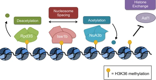
One protein that cannot bind in the presence of H3K36me is Asf1, a histone chaperone that contributes to the deposition of H3K56ac by Rtt109 (Tsubota et al., 2007). H3K56ac is associated with histone exchange, particularly over promoters (Williams et al., 2008). However, Asf1 cannot bind to peptides that are di- or trimethylated at H3K36, which likely contributes to suppressing histone exchange and maintaining a hypoacetylated chromatin environment in the wake of RNAPII (Venkatesh et al., 2012).
allosterically activates Rpd3 to remove H3 and H4 acetylation (Carrozza et al., 2005; Joshi and Struhl, 2005; Keogh et al., 2005; Merker et al., 2008; Ruan et al., 2015). Additionally, binding by the PHD of Rco1 to unmodified H3 helps target Rpd3S to gene bodies (Li et al., 2007; McDaniel et al., 2016). Overall, the Rpd3S complex creates a hypoacetylated environment to prevent aberrant transcription from initiating within gene bodies.
Another important protein that can bind H3K36me is Ioc4, which is part of the Isw1b ATP-dependent chromatin-remodeling complex (Vary et al., 2003). Through its PWWP domain, Ioc4 binds to H3K36me3 in vivo and H3K36me3 and H3K36me2 in vitro. By associating with H3K36me, Ioc4 localizes Isw1b to sites of active transcription to reposition nucleosomes (Maltby et al., 2012; Smolle et al., 2012). Isw1b nucleosome remodeling activity helps position
nucleosomes to be an appropriate substrate for Rpd3S HDAC activity and create a hypoacetylated chromatin environment (Lee et al., 2013).
Cryptic Transcription
A well-documented function for Set2 and H3K36 methylation is to prevent cryptic transcription. When normal chromatin architecture is disrupted, transcription can initiate from intragenic regions instead of canonical promoter regions (Kaplan et al., 2003). Cryptic
transcripts typically arise from nucleosome free regions (NFRs) and can be transcribed in the sense or antisense direction (Neil et al., 2009; Xu et al., 2009). Several classes of cryptic
transcripts have been defined, including cryptic unstable transcripts (CUTs), stable unannotated transcripts (SUTs), and Xrn1-sensitive unstable transcripts (XUTs). CUTs are rapidly degraded by the exosome or cytoplasmic decay pathways. CUTs were first detected in exosome mutants (rrp6∆) and later detected in 5’-to-3’ exonuclease mutants (xrn1∆) and cytoplasmic decapping mutants (dcp1∆ or dcp2∆) (Thompson and Parker, 2007; Wyers et al., 2005). Later, a specific class of transcripts degraded by Xrn1 was defined as XUTs (van Dijk et al., 2011). The rapid detection and degradation of these transcripts suggest they may have a negative effect on cellular processes. In contrast, SUTs are more resistant to decay mechanisms and are
detectable in wild-type cells (Xu et al., 2009). Overall, cryptic transcripts are widespread in the budding yeast genome and differ in their sensitivity to degradation pathways.
In addition to the rapid degradation of cryptic transcripts, there are several mechanisms to suppress their transcription. In particular, Set2 and H3K36me recruit the Rpd3S HDAC to create a hypoacetylated environment in the wake of RNAPII and prevent additional polymerases and transcription factors from binding and initiating transcription from intragenic regions
mechanisms have been mostly studied in set2∆ cells and the contributions of individual H3K36me states is still not well understood (Figure 1.3).
While Set2 and H3K36me play a critical role in preventing cryptic transcription, there are other important factors that repress cryptic transcription. Spt6 is a histone chaperone that binds H3/H4 dimers and requires H2A/H2B-binding FACT to reassemble nucleosomes after
transcription by RNAPII (Bortvin and Winston, 1996; Mccullough et al., 2015). Cryptic transcripts were first identified in an spt6-1004 mutant and subsequently observed in other mutant strains, including SPT16, a member of the FACT complex. spt6-1004 strains showed increased MNase sensitivity compared to wild-type, indicating a depletion of histones, altered chromatin structure, or both (Kaplan et al., 2003). Future work revealed that spt6 mutants had decreased
nucleosome occupancy at the 5’ ends of genes and increased cryptic transcription from these regions compared to wild-type (Dronamraju et al., 2018). FACT and Spt6 also work together to prevent the promoter-specific histone variant H2A.Z from being incorporated into gene bodies and promoting cryptic transcription (Jeronimo et al., 2015). In addition to Spt6 and FACT complex members, a genome-wide screen identified 50 genes that were important for repressing cryptic transcription. Most of these genes included histones, chromatin modifying proteins, and transcription elongation factors (Cheung et al., 2008). Robust evidence
demonstrates that chromatin structure is a key regulatory step in repressing cryptic transcription and its misregulation contributes to aberrant transcription.
important for repressing transcription at certain genes. Interestingly, most genes regulated by the Set2 and Rpd3S pathway have an overlapping lncRNA over their promoter. The
transcription of the lncRNA brings Set2, H3K36me, and Rpd3S to the gene and contributes to its repression (Kim et al., 2016). Similarly, work from our lab found that under nutrient
deprivation conditions, set2∆ cells produced cryptic transcripts whose presence was correlated with decreased sense transcription at corresponding genes (McDaniel et al., 2017). In
accordance with a decrease in sense transcription, others detected decreased protein
abundance for genes whose promoters overlap with a SUT (Huber et al., 2016). Such evidence suggests that cryptic transcription may function in the cellular response to stress and regulate transcript and protein abundance, but more work is needed to better understand the relationship between cryptic transcription and stress response. Interestingly, some cryptic transcripts appear to be translated, however the function of these truncated polypeptides is still not understood (Cheung et al., 2008; Wei et al., 2019). Overall, cryptic transcripts can expand the yeast transcriptome and proteome and future work focusing on their functionality will be key in understanding the significance of this phenomenon.
Additional Functions in Budding Yeast and Set2 Homologs
Additional Functions in Budding Yeast
In addition to transcription elongation, Set2 and H3K36me have functions in DNA damage repair, the cell cycle, aging, and mRNA splicing. Set2 and H3K36me function in the early stages of checkpoint activation and require the interaction with RNAPII to function at sites of DNA damage (Jha and Strahl, 2014; Winsor et al., 2013). In the absence of Set2 and
2014). Future work examining the possibility that H3K36me recruits specific repair machinery will further our understanding of Set2 and H3K36me in DNA damage response.
During the cell cycle, Set2 and H3K36me function to repress cryptic transcription at cell cycle genes. Repressing cryptic transcription and thereby preventing transcriptional interference ensures that the cell cycle genes are properly expressed. However, in set2∆ and K36A strains, cells show delayed progression through G1 and rapidly progress through S phase. Interestingly, Set2 and H3K36me3 are most abundant during G2/M and targeted for destruction during M phase, with evidence suggesting that the APC/CCDC20 complex degrades Set2 (Dronamraju et al., 2017). Similar to its function in repressing cryptic transcription during carbon source shifting and nutrient stress, Set2 and H3K36me also contribute to maintaining the precise transcriptional programming of the cell cycle (Dronamraju et al., 2017; Kim et al., 2016; McDaniel et al., 2017).
Global chromatin alterations have been linked to aging, including changes to H3K36me. An overarching model is that reduced nucleosome occupancy contributes to expression of genes that are normally repressed (Hu et al., 2014). Interestingly, cells with H3K36 mutants (H3K36R or H3K36E) or set2∆ displayed a decreased life span, while cells lacking the H3K36 demthylase, Rph1, had an increased life span. The decreased longevity in H3K36me deficient and set2∆ cells was attributed to the increased acetylation across gene bodies, thus more open chromatin environment, and increased cryptic transcription found in those cells (Sen et al., 2015). Overall, the role of Set2 and H3K36me in maintaining a proper chromatin environment and repressing cryptic transcription serves as an important mechanism in aging.
and better mechanistic insight into how it interacts with and recruits the spliceosome will improve our understanding of the combined roles of chromatin, transcription, and splicing in gene expression.
Schizosaccharomyces pombe
Schizosaccharomyces pombe, or fission yeast, is a distant cousin to budding yeast. It is considered a more ancient yeast species since fewer evolutionary changes occurred in fission yeast than budding yeast since diverging from their common ancestor. Overall, more genes are conserved between fission yeast and mammals than between budding yeast and mammals. The similarities and differences between budding yeast and fission yeast provide valuable evolutionary insight. Conserved mechanisms between the two species indicate a mechanism that is likely conserved across eukaryotes, while differences between the two suggest more diversity in other eukarytoes (Hoffman et al., 2015). In fission yeast, as in budding yeast, Set2 is the sole enzyme responsible for H3K36me1/2/3 and binds to phosphorylated serine 2 on the RNAPII CTD through its SRI domain (Kizer et al., 2005; Strahl et al., 2002; Suzuki et al., 2016). Likewise, Set2 and H3K36me influence DSB repair choice in fission yeast, tipping the balance in favor of NHEJ. However, data in fission yeast suggests that H3K36me peaks in S/G2 of the cell cycle and Set2 protein levels are constant throughout (Pai et al., 2014). The difference in cell cycle regulation is intriguing, especially considering the human homolog, SETD2, is regulated similarly to budding yeast Set2 (discussed below) (Dronamraju et al., 2017).
between fission and budding yeast continue to better our understanding of the molecular mechanisms regulated by Set2 and H3K36me.
Caenorhabditis elegans
In Caenorhabditis elegans, there is no longer a single enzyme that catalyzes H3K36me, but two: MET-1 and MES-4. MET-1 catalyzes H3K36me co-transcriptionally, while MES-4 does not require an association with RNAPII and is important for germline development (Andersen and Horvitz, 2007; Bender et al., 2006; Furuhashi et al., 2010; Rechtsteiner et al., 2010). Previously, it was thought that only MET-1 was capable of generating H3K36me3, but recent evidence demonstrates that MES-4 can also catalyze H3K36me3. MES-4 is the major
H3K36me3 methyltransferase in early embryos and requires the H3K36me3 transmitted to the progeny by the sperm and oocyte for its recruitment, thus establishing a mechanism for the epigenetic memory of H3K36me3 across generations (Kreher et al., 2018). Genome-wide analysis shows that H3K36me3 is enriched over exons, a pattern that is similar to humans (discussed below) (Kolasinska-Zwierz et al., 2009). Additionally, C. elegans is one of the premiere model systems for studying development and aging because of their short life span and ability to produce many progeny (Corsi et al., 2015). Multiple studies observed shorter lifespans in C. elegans that have decreased H3K36me3 levels (Pu et al., 2015; Sen et al., 2015). Mechanistically, it appears that H3K36me3 acts to prevent cryptic transcription and maintain proper gene expression, similar to budding yeast (Sen et al., 2015). Continued work in
C. elegans will help elucidate the function of H3K36me in multicellular organisms, particularly in
splicing, aging, and epigenetic memory.
Drosophila melanogaster
for development and cell biology (Hales et al., 2015). In Drosophila, like in C. elegans, there are two enzymes that catalyze H3K36me. Mes-4 catalyzes H3K36me1 and H3K36me2, while dSet2, the homolog of budding yeast Set2, catalyzes H3K36me3 (Venkatesh and Workman, 2013). dSet2 and H3K36me3 is required for proper development and like in budding yeast, it associates with the hyperphosphorylated CTD of RNAPII (Bell et al., 2007; Stabell et al., 2007). In H3K36R mutants, there is an increase in H4 acetylation, but no cryptic transcripts detected, indicating differences in how cryptic transcription in metazoans is regulated compared to budding yeast. Interestingly, evidence suggests that H3K36me is important
post-transcriptionally and functions in proper mRNA maturation (Meers et al., 2017). Another function of H3K36me in Drosophila that is distinct from its function in budding yeast is in recruiting the MSL complex to the male X chromosome for dosage compensation (Larschan et al., 2007). A recently developed histone gene replacement platform in Drosophila is an excellent system to study the function of specific histone residues and will help continue to increase our knowledge of H3K36me in metazoans (McKay et al., 2015).
Humans and Cancer Relevance
Given the well-established role for H3K36me3 in transcription, it is not surprising that H3K36me3 is also important for splicing. The first evidence of its role in splicing came from examining alternative splicing of the FGFR2 gene in human cells. Exon IIIb of FGFR2 is
included in PNT2 cells and exon IIIc is excluded, while the opposite occurs in hMSCs; exon IIIb is excluded and exon IIIc is included. The PTB protein regulates the alternative splicing of FGFR2 by binding to silencing elements around exon IIIb and ensuring its exclusion.
Interestingly, H3K36me3 was enriched at the FGFR2 gene in hMSCs, cells where exon IIIb is excluded. When SETD2 was overexpressed and H3K36me3 levels increased, there was increased exon IIIb exclusion, establishing a causal relationship between H3K36me3 and alternative splicing (Luco et al., 2010). Additionally, in tumor cells with mutated SETD2 and reduced H3K36me3, there was increased intron retention (Simon et al., 2014). The exact mechanism for H3K36me3 in splicing is not known. H3K36me3 may recruit MRG15, the human homolog of Eaf3, to splice sites which may in turn recruit other proteins for splicing (Luco et al., 2010). Tumor cells with mutated SETD2 had changes in chromatin accessibility in addition to splicing defects, suggesting the ability of H3K36me3 to recruit chromatin remodelers may also contribute to its role in splicing (Simon et al., 2014).
transcriptional regulation of cell cycle genes and targeted by APC/C for destruction (Dronamraju et al., 2017).
In addition to its functions in splicing, DNA repair, and the cell cycle, SETD2 has been implicated in a variety of other cellular processes. Both de novo DNA methyltransferases, DNMT3a and DNMT3b, bind H3K36me3 through their PWWP domains (Dhayalan et al., 2010; Rondelet et al., 2016). In vitro work and studies in mouse stem cells demonstrate that DNMT3a and DMNT3b activity is increased by binding to H3K36me3 and targets the enzymes to gene bodies (Baubec et al., 2015; Dhayalan et al., 2010; Morselli et al., 2015). Interestingly, methylation by DNMT3b represses cryptic transcription and depends upon recruitment and activation by H3K36me3 (Neri et al., 2017). Another silencing mechanism that SETD2 is
functionally related to is PRC2-mediated gene repression. The Tudor domain of PCL1 can bind H3K36me3 and recruit PRC2, the enzyme responsible for H3K27me3 and transcriptional repression at many developmental loci (Cai et al., 2013). Recent work in mice has also shown that SETD2 and H3K36me3 are necessary for lymphocyte development and V(D)J
recombination (Ji et al., 2019). Finally, in addition to its role in methylating H3K36, SETD2 has been connected to tubulin methylation and cytoskeleton reorganization (Park et al., 2016). All of these functions underscore the importance of SETD2 and H3K36me3 in maintaining genomic integrity.
With SETD2 and H3K36me3 playing an integral role in many cellular functions, it is no surprise that SETD2 is loss or mutated in many forms of cancer. SETD2 mutations in cancer were first observed in clear cell renal cell carcinoma (ccRCC) (Dalgliesh et al., 2010). SETD2 is located on chromosome 3p and one allele of chromosome 3p is frequently loss in ccRCC. Along with SETD2, VHL, PBRM1, and BAP1 are located on chromosome 3p and the combined
myriad of cancers, including lung adenocarcinoma, endometriod carcinoma, and hematopoietic cancers, amongst others (Jiang et al., 2018; Li et al., 2016; Yuan et al., 2017; Zhu et al., 2014). In addition to the loss or mutation of SETD2, mutations to H3K36 have recently emerged in several types of cancer. Initially, a lysine to methionine (H3K36M) mutation was identified and characterized in chondroblastomas, a type of bone cancer. Tumors with H3K36M showed a global decrease in H3K36me2 and H3K36me3 along with a redistribution of H3K27me3 and PRC1/PRC2-mediated silencing. While only 1 allele out of the 32 encoding for H3K36 was mutated to H3K36M, the mutation acted as a dominant-negative by trapping SETD2 and inhibiting its methyltransferase activity (Fang et al., 2016; Lu et al., 2016). Based on the crystal structure of the SETD2 catalytic domain bound to H3K36M peptide, conformational changes in the PS domain contribute to the increased association with H3K36M and decreased enzymatic activity (Yang et al., 2016; Zhang et al., 2017). In addition to chondroblastoma, H3K36M mutations are observed in pediatric soft tissue sarcoma and head and neck squamous cell carcinoma (Lu et al., 2016; Papillon-Cavanagh et al., 2017). Continued efforts to understand how H3K36M mutations contribute to cancer development will likely reveal similar mechanisms as cancers with SETD2 mutations.
Despite the lack of mechanistic understanding of how SETD2 and H3K36m3 contribute to tumorigenesis, their functions in transcription, DNA repair, and the cell cycle have already been implicated. Approximately 25% of genes expressed in one cohort of ccRCC tumors had RNA processing defects (Simon et al., 2014). Transcriptional data from The Cancer Genome Atlas demonstrates that transcriptional read-through (transcription occurring past the 3’
important checkpoint activation signaling (Carvalho et al., 2014; Kanu et al., 2015; Mar et al., 2017). Additionally, cell cycle genes are improperly expressed when SETD2 and H3K36me3 are absent in a variety of tumors (Dominguez et al., 2016). Examining the interplay of transcription, DNA repair, and cell cycle regulation by SETD2 and H3K36me3 will be critical in understanding their contributions to cancer development.
Non-histone PTMs and their Biological Functions
PTMs on non-histone proteins have important biological functions. Cellular processes such as transcription, the cell cycle, and DNA repair rely on non-histone PTMs for proper activation, recruitment, and destruction of key proteins involved (Biggar et al., 2017; Duan and Walther, 2015; Narita et al., 2019). As discussed above, phosphorylation on the CTD of RNAPII regulates the recruitment of transcriptional machinery. Without proper CTD phosphorylation, key proteins are not recruited and normal gene expression cannot occur (Zaborowska et al., 2016). Interestingly, advances in mass spectrometry have revealed that chromatin-modifying enzymes are also post-translationally modified (Biggar et al., 2017). Furthermore, changes to the PTMs on chromatin-modifying enzymes have already been associated with disease (Habibian and Ferguson, 2019).
interplay between non-histone PTMs, transcription, and other cellular processes is a key regulatory feature across organisms.
Concluding Remarks and Contributions of This Work
Since the proposal of the Histone Code Hypothesis, significant progress has been made toward understanding how histone PTMs function in a variety of cellular processes.
Furthermore, studies focused on non-histone PTMs have also increased our understanding of cell signaling and gene expression regulation. A well-studied methyltransferase, Set2, and its catalytic product, H3K36me, are conserved from yeast to humans, thus underscoring their importance in transcriptional regulation and other cellular processes. While substantial progress has been made in understanding the functions of Set2 and H3K36me, various questions remain unanswered. Many studies of Set2 and H3K36me take place in the context of wild-type or set2∆ cells; however there has not been a comprehensive examination of the different H3K36me states and their functions. In Chapter 2, I describe biochemical, genetic, and genomic assays that elucidate the unique and shared functions of the H3K36me states. Furthermore, no studies have examined the PTMs that occur on Set2 and how those affect Set2 function. In Chapter 3, I present biochemical and phenotypic data that provides evidence for PTMs on Set2 and their functions. As discussed earlier in this Chapter, the human homolog of Set2, SETD2, is
Figures
Figure 1.1: Set2 Domain Map
The H4 interacting domain is at the N-terminus (green). The catalytic domain is comprised of the Associated with SET domain (AWS, yellow), the SET domain (tan), and the Post-SET domain (PS, orange). In the middle of the protein is the autoinhibitory domain (blue). In the C-terminus, there are three protein-protein interacting domains, including the WW domain (red), the coiled-coiled domain (teal), and the Set2 Rpb1 Interacting domain (SRI, purple).
Figure 1.2: H3K36 Methylation Interacting Proteins
Rpd3S (green), Isw1b (pink), and NuA3b (blue) complexes bind to H3K36me (yellow) and carry out their respective functions on nucleosomes. Asf1 (purple) cannot bind to nucleosomes marked with H3K36me.
Isw1b Rpd3S
Deacetylation Asf1
Histone Exchange Nucleosome
Spacing
Acetylation
NuA3b
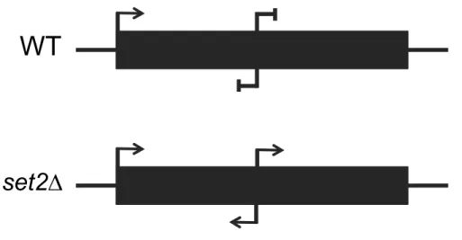
Figure 1.3: Schematic of Cryptic Transcription in wild-type and set2∆
In wild-type cells, transcription starts from the canonical 5’ promoter and is repressed from within gene bodies. In set2∆ cells, cryptic transcription is observed and can occur bidirectionally from within the gene body.
WT
CHAPTER 2 – UNIQUE AND SHARED ROLES FOR HISTONE H3K36
METHYLATION IN TRANSCRIPTION REGULATION FUNCTIONS
Summary
Set2 co-transcriptionally methylates lysine 36 of histone H3 (H3K36), producing mono-, di-, and trimethylation (H3K36me1/2/3). These modifications recruit or repel chromatin effector proteins important for transcriptional fidelity, mRNA splicing, and DNA repair. However, it was not known whether the different methylation states of H3K36 have distinct biological functions. We used engineered forms of Set2 that produced different methyl states to identify unique and shared functions for H3K36 modifications. Although H3K36me1/2 and H3K36me3 were
functionally redundant in many SET2 deletion phenotypes, H3K36me3 had a unique function related to Bur1 kinase activity and FACT complex function. Further, during nutrient stress, either H3K36me1/2 or H3K36me3 repressed high levels of histone acetylation and cryptic transcription that arises from within genes. Our findings uncover the potential for regulation of diverse
chromatin functions by different H3K36 methylation states.
Introduction
Histone post-translational modifications affect a great variety of DNA-templated processes. Methylation, acetylation, and other modifications are added to histones by chromatin-modifying enzymes (Rothbart and Strahl, 2014; Soshnev et al., 2016). These
replication, and DNA repair (Papamichos-Chronakis and Peterson, 2013; Smolle and Workman, 2013). A similar coordinated effort is employed to make the genome unreachable by RNAPII at appropriate times to prevent aberrant transcription.
Set2 is a chromatin-modifying enzyme that contributes to prevention of inappropriate transcription. Set2 methylates histone H3 at lysine 36 (H3K36) (McDaniel and Strahl, 2017; Strahl et al., 2002; Venkatesh and Workman, 2013). In Saccharomyces cerevisiae (hereafter, budding yeast), Set2 is responsible for all forms of H3K36 methylation, mono-, di-, and
trimethylation. Set2 binds to the C-terminal domain (CTD) of transcribing RNAPII and catalyzes H3K36 methylation (H3K36me) in actively transcribed genes (Kizer et al., 2005; Xiao et al., 2003). H3K36me provides docking sites for several proteins, such as Rpd3S, a histone
deacetylase complex, and Isw1b, a nucleosome remodeler. Rpd3S is recruited to chromatin by the PHD fingers in Rco1 and the activity of Rpd3S is stimulated by binding of the Eaf3
chromodomain to H3K36me2 (Carrozza et al., 2005; Joshi and Struhl, 2005; Keogh et al., 2005; Li et al., 2007; McDaniel et al., 2016; Ruan et al., 2015). Additionally, Isw1b associates with chromatin by way of the Ioc4 PWWP domain binding to H3K36me3 (Maltby et al., 2012; Smolle et al., 2012). Collectively, these processes ensure transcriptional fidelity by preventing
transcription initiation from within gene bodies, a process known as cryptic transcription. In the absence of Set2 and H3K36me, both sense and antisense cryptic transcription occur across the genome. Cryptic transcription tends to be a consequence of bi-directional transcriptional events at cryptic promoters within gene bodies (Carrozza et al., 2005;
Churchman and Weissman, 2011; Joshi and Struhl, 2005; Lickwar et al., 2009; Neil et al., 2009; Xu et al., 2009). Precisely how cryptic sites become accessible in SET2 deletion mutants
al., 2012). From a functional standpoint, cryptic transcription in the absence of Set2 does not result in significant growth defects when examined in nutrient-rich medium, although cell cycle progression is partially disrupted (Dronamraju et al., 2018; Kim et al., 2016; Lenstra et al., 2011; McDaniel et al., 2017; Venkatesh et al., 2016). In contrast, during nutrient deprivation or carbon source shifting, cryptic antisense transcription can occur at a level sufficient to impair normal transcription and lead to improper gene expression and poor cell growth (Kim et al., 2016; McDaniel et al., 2017).
Although the functions of Set2 and H3K36 methylation have been studied intensely, there are no studies that have thoroughly interrogated which functions of Set2 are directed by the different (mono- di-, and tri-) H3K36 methylation states. Previous reports suggest that H3K36me3 is dispensable for suppression of cryptic transcription (Hacker et al., 2016; Li et al., 2009; Youdell et al., 2008). In contrast, set2 mutants that harbor only H3K36me1 exhibit cryptic transcription, suggesting that the main functions of H3K36 methylation occur through
H3K36me2 (Hacker et al., 2016). However, it is not known whether H3K36me3 function is unique or overlapping with the other H3K36me states.
In this study, we engineered the SET domain of Set2 so that it performed only
function to ensure proper levels of H3K27ac and H3K56ac in genes, pointing to a potential mechanism for preventing inappropriate transcriptional initiation by control of nucleosome remodeling and histone exchange. In sum, our data provide key evidence for the independent and overlapping functions of H3K36me1/2 and H3K36me3 in chromatin biology and in
transcriptional regulation.
Results
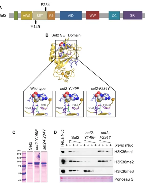
Phe/Tyr Switch in Set2 separates H3K36 Methylation States in vitro
Although previous studies have assessed the effects of losing or limiting H3K36 methylation, no study has comprehensively examined the individual functions of each H3K36 methylation state produced by Set2. To create a system in which we could control production of H3K36 methylation states, we employed a structural mutagenesis approach for Set2 based on the Phe/Tyr switch phenomenon that alters the SET domain lysine-binding pocket (Cheng et al., 2005; Collins et al., 2005). Briefly, two well-positioned aromatic residues within the SET domain (a phenylalanine [Phe, F] and a tyrosine [Tyr, Y]) can be switched to the opposite aromatic residue to control the size of the catalytic domain. An F to Y mutation creates steric hindrance in the active site with lysine trimethylation, thereby limiting the enzyme to only mono- and
dimethylation. However, a Y to F change creates more space and promotes the catalysis of trimethylation. In Set2, Y149 and F234 were predicted to comprise the Phe/Tyr switch (Cheng et al., 2005; Collins et al., 2005) (Figure 2.1A).
configured to enable Set2 to mono-, di-, and trimethylate H3K36. We then investigated this model for the effect of deploying the Phe/Tyr switch. Modeling showed that the Y149F mutation opened the SET domain binding pocket and eliminated hydrogen bonding between the hydroxyl group of tyrosine and H3K36me (Figure 2.1B, middle panel). These changes would permit rapid rotation of the target lysine in the presence of S-adenosylmethionine (SAM) and likely increase the potential for higher methylation states on H3K36. Conversely, the F234Y substitution created steric hindrance between the tyrosine hydroxyl group and the target lysine, thus
suggesting that this mutant would limit H3K36me3 (Figure 2.1B, right panel). These findings are consistent with these residues encompassing a working Phe/Tyr switch in Set2.
Lastly, we measured the effect of the Phe/Tyr switch on Set2 enzymatic activity. We performed histone methyltransferase (HMT) activity assays with recombinant Set2 (wild-type,
set2-Y149F, and set2-F234Y ), SAM, and recombinant Xenopus nucleosomes; methylation states were detected by Western blot analysis with antibodies specific to the three modifications (Figures 2.1C and 2.1D). We found that set2-Y149F catalyzed predominately H3K36me3, whereas set2-F234Y catalyzed H3K36me1 and H3K36me2 (Figure 2.1D). Overall, these results demonstrated that the SET domain of Set2 contains a functional Phe/Tyr switch that can be used to produce specific H3K36 methylation states.
Phe/Tyr Switch Mutations in Set2 are Tools to Separate H3K36 Methylation States in vivo
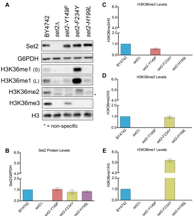
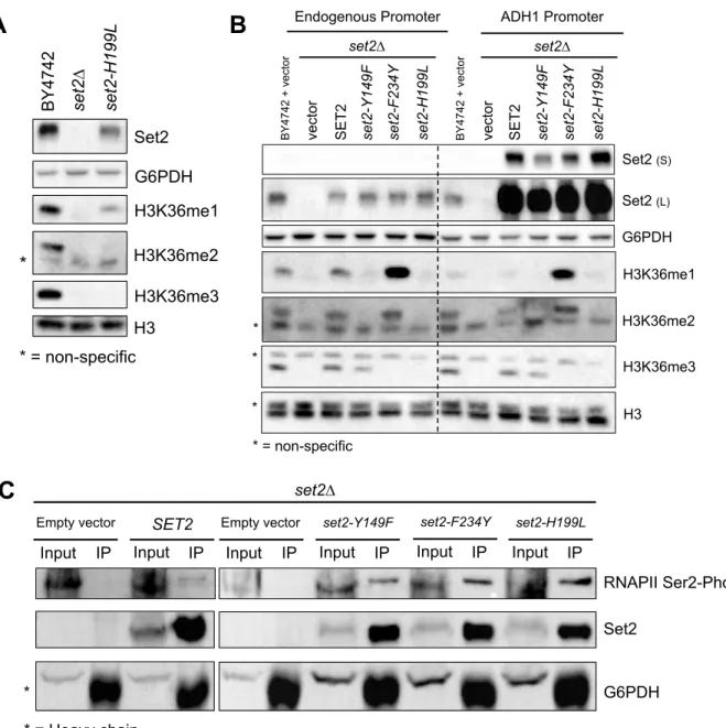
To test the effect of the Set2 Phe/Tyr switch on H3K36 methylation in vivo, we generated yeast strains harboring endogenous set2-Y149F or set2-F234Y mutations. For controls, we included a set2∆ strain and a catalytic domain mutant strain (set2-H199L) that was capable of producing only low levels of H3K36me1 in vivo (Hacker et al., 2016; Jha and Strahl, 2014). The
set2-Y149F mutation predominately resulted in H3K36me3 and a low level of H3K36me1
(Figure 2.2A). As previously described, the absence of Set2 resulted in complete loss of H3K36 methylation and the catalytic domain mutant, set2-H199L, showed only a low level of
H3K36me1 (Figure 2.2A and Figure 2.3A). The SET domain mutations did not alter the amount of Set2 protein (Figure 2.2B), however, global H3K36 methylation differed from wild-type. The
set2-Y149F mutant had an average of 56% of the wild-type H3K36me3 (Figure 2.2C).
set2-F234Y showed an H3K36me2 signal similar to wild-type (93%), but a 5-fold increase in
H3K36me1 (Figures 2.2D and 2.2E). set2-H199L had a reduced H3K36me1 signal compared with wild-type. Overall, these results confirmed that the Phe/Tyr switch in Set2 functioned in vivo
and, although global H3K36me levels varied from wild-type, we could largely control different methylation states.
Because some of our downstream genetic analyses would require expression of SET2
from plasmids, we also examined the effect of the set2 mutants in two exogenous expression systems: expression of SET2 and the set2 mutants from their native promoter and
overexpression of the same genes from the ADH1 promoter. Western blot analyses for H3K36 methylation in these two systems revealed two important finings. First, the set2-Y149F and set2-F234Y mutants, regardless of expression system, recapitulated the initial in vivo findings that the Phe/Tyr switch could control the methylation states of H3K36 (Figure 2.3B). Second, and surprisingly, overexpression of SET2 via the ADH1 promoter resulted in robust overexpression of Set2 protein compared with expression from its endogenous promoter, yet global H3K36 methylation levels in the wild-type or set2 mutants were similar to or lower than normal. Thus, although it was possible to greatly overexpress Set2, the amount of methylation that Set2 can produce on chromatin must have a limit, perhaps due to a finite number of Set2 binding sites on the CTD of RNAPII, restricted histone accessibility, limited SAM availability, or a combination of these factors.
H3K36me1/2. These provided tools to examine in vivo the functions of different methylation states of H3K36.
Distinct Methylated Forms of H3K36 are Deposited within or near Transcribed Regions of Genes
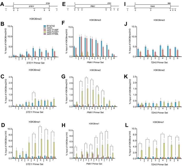
We next asked whether the set2 mutants still deposited H3K36me within or near the coding regions of genes in a manner consistent with its known localization and function (Pokholok et al., 2005; Rao et al., 2005; Weiner et al., 2015). We first confirmed by
co-immunoprecipitation that the set2 mutants still associated normally with phosphorylated RNAPII (Figure 2.3C). Consistent with RNAPII association, ChIP-qPCR of all H3K36me forms in the
set2 mutants confirmed that they were deposited within or near the coding regions of several loci tested (STE11, PMA1, and TDH3, see Figures 2.4A, 2.4E, and 2.4I). Additionally, the H3K36me states detected by ChIP-qPCR were consistent with the aforesaid in vitro HMT and Western blot assays (Figures 2.1C and 2.2A).
Closer comparison of the differently methylated H3K36 forms with wild-type revealed several key observations. As shown in Figures 2.4B, 2.4F, and 2.4J, there were no significant differences in the H3K36me3 levels between wild-type and set2-Y149F at all loci tested.
However, for H3K36me2, we observed differences between wild-type and set2-F234Y at STE11
and PMA1. A significantly higher H3K36me2 level was detected in the middle of STE11 in set2-F234Y (Figure 2.4C), whereas, at PMA1, a significantly higher H3K36me2 level was detected across the entire gene body (Figure 2.4G). In contrast, we did not detect any significant difference in H3K36me2 levels between set2-F234Y and wild-type at TDH3 (Figure 2.4K). As expected from the high H3K36me1 Western blot signal, set2-F234Y had significantly higher levels of H3K36me1 in the 3’ end of STE11 and across the entirety of PMA1 and TDH3
set2-H199L at the loci examined. At STE1, we did not find significant differences in H3K36me1 between set2-H199L and wild-type (Figure 2.4D and Figure 2.5). However, at PMA1, there was significantly more H3K36me1 deposited by set2-H199L in the gene body (Figure 2.4H Figure 2.5D). In contrast, we detected less H3K36me1 at the 5’ end of TDH3 in set2-H199L compared with wild-type, although the rest of that locus did not show a significant difference between the mutant and wild-type. Overall, these results established that the levels of H3K36me in the set2
mutants differed from wild-type at certain loci, but the results further confirmed the specificity of the set2 mutants and indicated that differentially methylated forms of H3K36 were localized within or near the bodies of transcribed genes.
H3K36me1/2 and H3K36me3 Function Redundantly in Some Cellular Contexts
To determine whether the different forms of H3K36me are responsible for distinct biological outcomes, we examined the sensitivity of set2 methylation mutants to drugs that affect set2∆ cell growth. We tested caffeine (a TOR1C and MAP kinase inhibitor), rapamycin (a TOR1C inhibitor), phleomycin (a double-strand break-inducing agent), and 6-azauracil (6-AU, a transcriptional elongation inhibitor) (Jha and Strahl, 2014; Kizer et al., 2005; McDaniel et al., 2017). Like previous work, we found that set2∆ and set2-H199L mutant cells were sensitive to caffeine, rapamycin, and phleomycin, and they were resistant to 6-AU (Figures 2.6A and 2.6B and Table 2.1). Intriguingly, similar to wild-type, the set2-Y149F and set2-F234Y mutants rescued the drug-induced growth defects from caffeine, rapamycin, and phleomycin (Figure 2.6A and Table 2.1). However, only set2-Y149F was as sensitive to 6AU as wild-type, with set2-F234Y having an intermediary phenotype between wild-type and set2∆ (Figure 2.6B). Thus, in
Genetic Interactions of SET2 with BUR1and SPT16 Reveal Unique Functions for H3K36me1/2 and H3K36me3
In addition to phenotypes of set2∆ cells challenged with drugs, deletion of SET2
bypasses the lethal or slow growth phenotypes associated with the inactivation or deletion of members of the BUR kinase and FACT complexes (Biswas et al., 2006; Chu et al., 2006; Keogh et al., 2003). Both complexes regulate aspects of transcription; the BUR kinase complex
promotes RNAPII elongation and the FACT complex is responsible for nucleosome disruption and reassembly. Cells lacking BUR1 (bur1∆) are not viable, but they survive when SET2 is also deleted (bur1∆set2∆). Likewise, cells harboring the FACT mutant allele spt16-11 grow
extremely slow at 34°C, and deletion of Set2 bypasses this phenotype. Therefore, we examined our set2 methylation mutants to determine whether the different H3K36me states would bypass the bur1∆ and spt16-11 phenotypes. We confirmed that the presence of set2∆ in the bur1∆ or
spt16-11 cells rescued the lethal or slow growth phenotypes, whereas exogenously expressed wild-type SET2 did not rescue growth (Figures 2.6C and 2.6D and Table 2.1). Surprisingly, cells containing the set2-Y149F allele that catalyzed only H3K36me3 phenocopied wild-type Set2, whereas the set2-F234Y and set2-H199L alleles that catalyzed H3K36me1/2 or low levels of H3K36me1, respectively, phenocopied the set2∆ cells (Figures 2.6C and 2.6D and Table 2.1). Thus, these findings indicated that H3K36me3 has a function distinct from H3K36me2 and H3K36me1 in relation to Bur1 kinase activity and FACT complex function.
H3K36me1/2 and H3K36me3 Have Unique and Shared Functions in Repressing Cryptic Transcription at Reporter Loci
H3K36me1/2. To answer this question, we used two reporter genes, FLO8 and STE11, that each have internal cryptic initiation sites (CIS) and the HIS3 gene integrated out-of-frame and downstream of the CIS. When the FLO8 and STE11 loci are transcribed from canonical promoters, HIS3 is not transcribed in-frame, and the cells cannot survive on media lacking histidine (Silva et al., 2012; Wang et al., 2015). However, when cryptic transcription occurs at the CIS, HIS3 is transcribed in-frame, and cells survive without histidine (Figure 2.6E and 2.6F). Thus, we first confirmed that cells with wild-type SET2 suppressed cryptic transcription and did not grow on media lacking histidine, whereas set2∆ cells did not suppress cryptic transcription at either locus (Figures 2.6G and 2.6H and Table 1). At the FLO8 locus, which was under the GAL promoter and constitutively expressed in cells growing on galactose-containing media, both H3K36me3 and H3K36me1/2 were sufficient to suppress cryptic transcription similar to wild-type SET2 (Figure 2.6G and Table 2.1). Surprisingly, at the STE11 locus, expressed from its native promoter and at a lower level than FLO8, only H3K36me3 prevented cryptic transcription, whereas cells with H3K36me1/2 or a low level of H3K36me1 did not suppress (Figure 2.6H and Table 2.1). Thus, at STE11, H3K36me3 alone is sufficient to suppress cryptic transcription. Additionally, these results suggested that H3K36me3 and H3K36me1/2 have different, but sometimes overlapping, functions in preventing cryptic transcription. Particularly, highly and lowly expressed genes may have different requirements for H3K36me.
During Nutrient Deprivation, H3K36me1/2 or H3K36me3 Prevent Antisense Transcription
performed stranded RNA-seq at 0 and 60 minutes following nutrient deprivation in wild-type,
set2∆, set2-Y149F, set2-F234Y, and set2-H199L cells. We examined sense and antisense transcriptional signals in the genomic regions that surround the high- and intermediate-confidence cryptic initiation sites of the aforesaid 439 genes. After 60 minutes of nutrient
deprivation, we observed a global increase in sense and antisense transcription surrounding the CIS in set2∆ and set2-H199L cells (Figure 2.7A). Interestingly, cryptic transcription was not completely repressed in set2-Y149F or set2-F234Y; both mutants showed a slight increase in antisense transcription upstream of the CIS compared with wild-type (Figure 2.7A). When we separated the CIS-containing genes based on gene expression levels, we observed that set2-Y149F or set2-F234Y repressed sense and antisense cryptic transcription similar to wild-type in
highly and lowly expressed genes (Figure 2.8A). In contrast, set2∆ and set2-H199L did not repress any form of cryptic transcription in highly or lowly expressed genes (Figure 2.8A).
Importantly, we verified that nutrient deprivation did not alter existence of the H3K36 methylation states catalyzed by the set2 mutants (Figure 2.7A). These results revealed that H3K36me1/2 or H3K36me3 can function to prevent cryptic transcription genome-wide and low levels of
H3K36me1 do not have a large part in repressing cryptic transcription.
Next, we wanted to further characterize the antisense transcription that occurred in the
the regions between the CIS and transcription start sites. Any signal in these regions should reflect normal sense transcription and cryptic antisense transcription, but not cryptic sense transcription because cryptic sense transcription would only occur downstream of the CIS. Indeed, we frequently observed decreases in sense transcription and concomitant increases in antisense transcription, with the strongest effects on both strands occurring in set2∆ and set2-H199L cells (Figure 2.7C). The effects observed here were more pronounced than the effects
we observed between two wild-type replicates compared as a control (Figure 2.8C). Moreover, many of the same genes exhibited similar effects for all the set2 mutants, particularly, set2∆,
set2-H199L, and set2-F234Y cells (Figure 2.7D). Together, these data suggested that, during nutrient deprivation, H3K36me1/2 or H3K36me3 suppresses cryptic antisense transcription, which appears to drive the downregulation of sense transcription from certain CIS-containing genes.
Lastly, we wondered whether the sequences surrounding the CIS harbored any particular sequence motifs that may explain why cryptic transcription initiates there and not elsewhere. We searched the sequences ±100 bp of the 439 high- and intermediate-confidence CIS and found a significant enrichment of a (T/C)AAT motif, which may represent a degenerate TATA box element (Figure 2.7E) (Basehoar et al., 2004; Lubliner et al., 2013). We did not observe this motif for a random sampling of 439 low-confidence CIS (Figure 2.8D). Therefore, these data suggested that cryptic transcription may initiate from TATA-like elements, but their suppression requires H3K36me1/2 or H3K36me3. Mechanistically, H3K36me1/2 and