The Neural Cell Adhesion Molecule (NCAM) Promotes
Clustering and Activation of EphA3 Receptors in GABAergic
Interneurons to Induce Ras Homolog Gene Family, Member A
(RhoA)/Rho-associated protein kinase (ROCK)-mediated
Growth Cone Collapse
*
Received for publication, September 22, 2016, and in revised form, October 24, 2016 Published, JBC Papers in Press, November 1, 2016, DOI 10.1074/jbc.M116.760017
Chelsea S. Sullivan, Maike Kümper, Brenda S. Temple, and Patricia F. Maness1
From the Department of Biochemistry and Biophysics, R. L. Juliano Structural Bioinformatics Core, School of Medicine, University of North Carolina, Chapel Hill, North Carolina 27599-7264
Edited by F. Anne Stephenson
Establishment of a proper balance of excitatory and inhibitory connectivity is achieved during development of cortical networks and adjusted through synaptic plasticity. The neural cell adhesion molecule (NCAM) and the receptor tyrosine kinase EphA3 regu-late the perisomatic synapse density of inhibitory GABAergic interneurons in the mouse frontal cortex through ephrin-A5-in-duced growth cone collapse. In this study, it was demonstrated that binding of NCAM and EphA3 occurred between the NCAM Ig2 domain and EphA3 cysteine-rich domain (CRD). The binding interface was further refined through molecular modeling and mutagenesis and shown to be comprised of complementary charged residues in the NCAM Ig2 domain (Arg-156 and Lys-162) and the EphA3 CRD (Glu-248 and Glu-264). Ephrin-A5 induced co-clustering of surface-bound NCAM and EphA3 in GABAergic cortical interneurons in culture. Receptor clustering was impaired by a charge reversal mutation that disrupted NCAM/EphA3 association, emphasizing the importance of the NCAM/EphA3 binding interface for cluster formation. NCAM enhanced ephrin-A5-induced EphA3 autophosphorylation and activation of RhoA GTPase, indicating a role for NCAM in activating EphA3 signaling through clustering. NCAM-mediated clustering of EphA3 was essential for ephrin-A5-induced growth cone collapse in cortical GABAergic interneurons, and RhoA and a principal effector, Rho-associated protein kinase, mediated the collapse response. This study delineates a mechanism in which NCAM promotes ephrin-A5-dependent clustering of EphA3 through interaction of the NCAM Ig2 domain and the EphA3 CRD, stimulating EphA3 auto-phosphorylation and RhoA signaling necessary for growth cone repulsion in GABAergic interneuronsin vitro, which may extend to remodeling of axonal terminals of interneuronsin vivo.
Neural cell adhesion molecule (NCAM)2is a key regulator of cell adhesion, axon growth, and fasciculation (1). NCAM also plays a critical role in the regulation of perisomatic synapses in the developing prefrontal cortex (2). For information to be transmitted effectively within the brain, an intricate balance of excitation and inhibition in the cortical circuitry must be estab-lished. Because inhibitory synapses perform essential roles in network function (3–7), the elucidation of how they are pro-duced and refined is of vital importance. Inhibitory GABAergic synapses make and break connections in response to activity-dependent and -inactivity-dependent stimuli throughout development and into adulthood in a dynamic manner (6 –9). Alterations of inhibition disrupt the excitation and inhibition balance and are associated with cognitive deficits in schizophrenia (10 – 12) and autism (13–16). Although the mechanisms regulat-ing formation and fine-tunregulat-ing of excitatory synapses have been extensively studied (17, 18), the molecular pathways underlying inhibitory synapse formation and refinement are poorly understood.
Previous work identified a novel role for the neural adhesion molecule NCAM as an obligate component of a receptor com-plex that acts in the early postnatal mouse medial frontal cortex to constrain basket interneuron connectivity with pyramidal neuron soma (2). Deletion of NCAM, EphA3, or ephrin-A2/ A3/A5 in mice increases the number of perisomatic basket cell synaptic terminals in the mPFC (2). NCAM associates with EphA3 tyrosine kinase, an ephrin-A5 receptor, and mediates growth cone collapse of GABAergic interneuron terminals in cortical neuron cultures (2). Furthermore, ephrin-A5 treat-ment of cortical slices promotes loss of perisomatic basket cell synaptic puncta, and this loss is NCAM-dependent (2). Although NCAM and EphA3 co-localizein vivoat perisomatic synapses (2), the significance and mechanism of NCAM/EphA3 binding and clustering are not understood. Highlighting the importance of NCAM and ephrinA/EphA signaling in human *This work was supported, in whole or in part, by National Institute of Health
Grants R01 MH101605, F32 MH111189, and P30 CA016086. The authors declare that they have no conflicts of interest with the contents of this article. The content is solely the responsibility of the authors and does not necessarily represent the official views of the National Institutes of Health.
1To whom correspondence should be addressed: Dept. of Biochemistry and
Biophysics, University of North Carolina Chapel Hill School of Medicine, 3020 Genetic Medicine Research Bldg., Campus Box 7260, 120 Mason Farm, Chapel Hill, NC 27599-7264. Tel.: 919-966-3532; Fax: 919-966-2852; E-mail: [email protected].
2The abbreviations used are: NCAM, neural cell adhesion molecule; LBD,
ligand-binding domain; CRD, cysteine-rich domain; FN, fibronectin III domain; PSA, polysialylation; ROCK, Rho-associated protein kinase; PBD, Pak binding domain; RBD, Rho binding domain; ANOVA, analysis of vari-ance; DIV, day(s)in vitro; E, embryonic day; EC, extracellular.
cortical circuitry, genetic polymorphisms and dysregulation of NCAM (19 –25) or EphA/ephrinA (26 –28) are associated with schizophrenia, autism, and bipolar disorder.
EphA3 kinase activation and signaling depend on receptor clustering and are important for growth cone collapse, axon repulsion, and synaptic plasticity (29). The extracellular do-main of EphA receptors consists of a ligand-binding dodo-main (LBD), a cysteine-rich domain (CRD), and two fibronectin III (FN) domains (30). Crystallographic studies of ephrin-A5 or ephrin-A2 bound EphA2 demonstrated that ephrinA-EphA interactions are mediated by the LBD and an additional surface in the CRD, enabling formation of large multimeric receptor/ ligand clusters. Studies of EphA4 clustering suggested that only the CRD interface is important for clustering of EphA4, rather than the additional LBD interface used by EphA2 (31). Together, these studies indicate that even Eph receptors in the same family can have varying clustering properties based on subtle differences in their ectodomains. The EphA3 structure has not been solved, but its overall sequence is more similar to EphA4 than EphA2.
How NCAM engages EphA3 to promote repellent responses is unclear. NCAM has five Ig-class and two FN domains in its extracellular region and a cytoplasmic domain that differs in size among three principal isoforms (32). The NCAM extracel-lular region can be modified by polysialylation (PSA-NCAM), which is implicated in mediating synaptic plasticity (33). PSA-NCAM is highly expressed in the embryonic and early postnatal stages and in interneuron populations in the mature cortex (33–36). The 140-kDa isoform NCAM140 interacts physically and functionally with EphA3 to a greater extent than other iso-forms (2) and is most prominent in axonal growth cones of developing neuronal cells (37). NCAM forms trans and cis homodimers in the plasma membrane, and these interactions are mediated by the Ig1, Ig2, and Ig3 domains as well as the first FN domain (32, 38). Based on the knowledge that NCAM and EphA3 colocalize at perisomatic synapsesin vivo, interact bio-chemically, and promote remodeling of GABAergic interneu-ron terminals (2), we hypothesized that NCAM may promote EphA3 receptor clustering and signaling to bring about growth cone collapse and axon terminal retraction.
In this study, we utilized an approach combining molecular modeling based on crystal structures of rat NCAM Ig1–3 (PDB code 1QZ1) and the LBD/CRD regions of human EphA4 (PDB code 4M4P), analysis of receptor clustering, and functional assays to define the molecular and cellular mechanism by which NCAM promotes EphA3 signaling required for growth cone collapse of GABAergic interneurons. It was demonstrated that NCAM binding to EphA3 promotes ephrin-A5-dependent receptor clustering to activate EphA3 kinase signaling, nec-essary for EphA3 autophosphorylation, RhoA-GTPase acti-vation, and growth cone collapse of cortical GABAergic interneurons.
Results
The Ig2 Domain of NCAM Binds the Cysteine-rich Domain of EphA3 through a Charged Interface—Previous studies identi-fied an interaction between NCAM and EphA3 in the mouse forebrain, and the interaction site on NCAM was mapped to the
extracellular region (2). We hypothesized that NCAM may pro-mote receptor clustering and signaling by binding EphA3 through association with specific extracellular domains. To identify the binding determinants on EphA3 and NCAM, pull-down experiments were performed using domain deletions of each molecule. HEK293T cells were transfected with WT EphA3, EphA3 lacking the LBD (EphA3⌬LBD), and EphA3 lacking both the LBD and CRD (EphA3⌬LBD/CRD) (39). Transfected lysates were incubated with purified fusion pro-teins consisting of the entire extracellular region of NCAM, Ig1–5/FN1–2 with a C-terminal Fc tag (NCAM-EC). Protein A/G-Sepharose pulldown of NCAM-EC showed that the extracellular domain of NCAM bound WT EphA3 and EphA3⌬LBD, indicating that the ligand-binding domain of EphA3 was not required for interaction with NCAM. How-ever, EphA3⌬LBD/CRD was unable to bind NCAM-EC (Fig. 1B), identifying the CRD as the critical binding domain. To delineate the EphA3 binding domain on NCAM, full-length NCAM-EC or NCAM truncations containing Ig1–3, Ig1–2, Ig2, or Ig1 as Fc fusion proteins were incubated with lysates of HEK293T cells transfected with WT EphA3. Fc pulldown assays showed that NCAM Ig1–3, Ig1–2, and the Ig2 domain alone bound EphA3, whereas NCAM Ig1 was not sufficient for binding (Fig. 1,CandD). These experiments demonstrated that the Ig2 domain of NCAM contains the required binding site for EphA3.
experiments demonstrate that the Ig2 domain of NCAM and the CRD domain of EphA3 comprise a charged interface for interaction.
NCAM Binding EphA3 Promotes Ephrin-A5-dependent Clus-tering of Receptors in Cortical GABAergic Interneurons—EphA3 signaling depends on ligand-induced receptor clustering and is important for growth cone collapse, axon repulsion, and synap-tic plassynap-ticity (45, 46). Studies of EphA family members demon-strated that EphA2 forms extended arrays through two distinct interfaces (47) in the LBD and CRD, whereas EphA4 clusters are typically smaller and oligomeric because of their reliance on only one of these two corresponding interfaces, the CRD inter-face, suggesting that EphA receptors can have different cluster-ing tendencies based on their ectodomains (31). However, the extent to which these clusters form in the absence of NCAM is not known.
The importance of NCAM/EphA3 binding for receptor clus-tering was assessed in cortical neuron cultures from NCAM KO mice co-transfected with WT or mutant NCAM140 together with pCMV-IRES-eGFP (Fig. 2A). We focused on GABAergic (GABA-immunopositive) interneurons in these cultures based on the identified role of NCAM at interneuron perisomatic puncta. After treatment with preclustered Fc or ephrin-A5-Fc for 30 min, cells were immunostained for GABA, NCAM, and EphA3 and imaged confocally. GFP/GABA double-positive cells were selected in single optical sections, and colocalization of NCAM and EphA3 was assessed. GABA staining allowed the identification of individual axons to ensure that clustering was observed in single neurites and not bundles.
The proportion of colocalized signal for each channel was evaluated by colocalization analysis yielding thresholded Manders coefficients (Fig, 2,BandC; tM1, proportion of red
pixels colocalizing with green pixels; tM2, proportion of green pixels colocalizing with red pixels). As a further measure of correlation of the two fluorescent signals, Pearson correlation coefficients (R⬎0 indicates positive correlation, R⬍0 indi-cates negative correlation) were generated (Fig. 2D). Ephrin-A5-Fc strongly promoted colocalization/clustering of WT NCAM and EphA3 compared with control Fc, as shown by merged fluorescent images, heat maps of colocalization, and colocalization coefficients (Fig. 2). Ephrin-A5-Fc also pro-moted EphA3 colocalization with NCAM R192E, a mutant that did not disrupt association of NCAM and EphA3. On the other hand, ephrin-A5-Fc did not promote colocalization of EphA3 with NCAM R156E. These colocalization analyses were consis-tent with co-immunoprecipitation studies that showed interac-tion of EphA3 with WT NCAM and NCAM R192E but not NCAM R156E (Fig. 1,EandF). The results demonstrate that binding of NCAM to EphA3 promotes ephrin-A5-dependent clustering of receptors in cortical GABAergic interneurons.
previously (2), and only GFP-positive (transfected) cells were included in the analysis.
Neurons expressing WT-NCAM exhibited robust growth cone collapse in response to ephrin-A5-Fc, consistent with a requirement for NCAM in ephrin-A5-mediated growth cone collapse (Fig. 3) as observed previously (2). NCAM KO neurons transfected with empty vector did not undergo significant growth cone collapse after ephrin-A5-Fc treatment. In con-trast, the R156E mutant of NCAM that was unable to bind or cluster EphA3 was not effective in promoting growth cone col-lapse in response to ephrin-A5-Fc (Fig. 3). The NCAM R192E mutant, which did not show decreased binding to EphA3,
effec-tively promoted growth cone collapse. These functional studies support a role for EphA3 and NCAM binding in ephrin-A5-induced growth cone collapse of GABAergic interneurons.
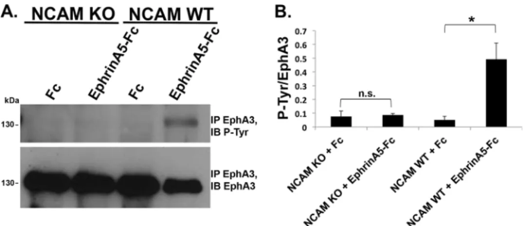
and NCAM KO mice after treatment with preclustered control Fc or ephrin-A5-Fc. EphA3 was immunoprecipitated and subjected to immunoblotting with phosphotyrosine antibody (Tyr(P)-99), followed by stripping and reprobing for total EphA3. The ratio of phospho-EphA3 to total EphA3 was quan-tified by densitometry and compared for significant differences. In WT cultures, ephrin-A5-Fc promoted robust tyrosine phos-phorylation of EphA3 (Fig. 4A). In contrast, NCAM KO cul-tures treated with ephrin-A5-Fc exhibited no increased phos-phorylation compared with Fc controls, indicating that NCAM was required for activation and autophosphorylation of EphA3 kinase.
NCAM-dependent Activation of RhoA GTPase and ROCK Is Required for Ephrin-A5-dependent Growth Cone Collapse—To assess the role of Rho family GTPases in NCAM/EphA3 signal-ing, the activation of RhoA, Cdc42, and Rac1 in cortical neurons was determined by measuring GTP-bound GTPase levels in pulldown assays. Cortical neurons were stimulated with Fc or ephrin-A5-Fc and lysed, and GTP-bound GTPases were pulled down using GST-tagged effector proteins conjugated to gluta-thione-Sepharose beads (GST-Pak binding domain (PBD) for Rac1 and Cdc-42, GST-Rho binding domain of Rhotekin (RBD) for RhoA). The ratio of active (GTP-bound) RhoA, Rac1, or
Cdc-42 to input was determined for each condition (Fc- or eph-rin-A5-Fc-treated) and normalized to Fc-treated cells (n⫽3 for each GTPase). In WT neurons, GTP-bound RhoA increased 5-fold in response to ephrin-A5-Fc, whereas the levels of GTP-bound Rac1 and Cdc-42 were not significantly altered upon treatment (Fig. 5,AandB). In contrast, NCAM KO neurons showed no increase in GTP-bound RhoA upon stimulation with ephrin-A5-Fc, indicating a requirement for NCAM in ephrin-A5-dependent RhoA activation (Fig. 5,AandB).
To determine whether RhoA activation promoted growth cone collapse in cortical GABAergic interneurons treated with ephrin-A5-Fc, cortical neuron cultures were pretreated with the RhoA inhibitor exoenzyme C3 transferase prior to stimula-tion with preclustered Fc or ephrin-A5-Fc. Pretreatment of WT neurons with C3 toxin strongly inhibited ephrin-A5-dependent growth cone collapse in comparison with ephrin-A5-treated cells without C3 pretreatment (two-way ANOVA with Bonfer-roni post hoc testing,p⬍0.001).
The role of RhoA signaling in ephrin-A5-dependent growth cone collapse was further evaluated by inhibition of ROCK1/2, a key downstream effector of RhoA. Cortical neurons were pre-treated with Y-27632, a selective inhibitor of ROCK1/2, prior to stimulation with preclustered Fc or ephrin-A5-Fc, and GABA-positive interneurons were assayed for growth cone collapse. ROCK inhibition significantly decreased the percent of GABAergic cells exhibiting growth cone collapse upon ephrin-A5-Fc treatment compared with cells without Y-27632 pre-treatment (two-way ANOVA with Bonferroni post hoc testing, p⬍0.001). These results supported the conclusion that ephrin-A5-induced growth cone collapse in GABAergic interneurons is dependent on RhoA and its effector ROCK1/2.
Discussion
A novel role for NCAM in clustering and activation of EphA3 receptors necessary for axonal repellent responses in cortical neurons was identified using a combination of molecular modeling, mutagenesis, and functional assays. These studies revealed for the first time that NCAM promotes EphA3 recep-tor clustering in response to ephrin-A5 stimulation through binding between critical determinants in the NCAM Ig2 and EphA3 CRD domains. Although direct binding of NCAM and EphA3 was not demonstrated, molecular modeling and the effects of charge reversal mutations suggested that the interac-tion was likely to be direct. This interacinterac-tion induced NCAM/ EphA3 clustering, EphA3 kinase activation, and growth cone collapse of GABAergic interneurons stimulated by the ligand ephrin-A5. Downstream signaling through RhoA GTPase and one of its principal effectors, ROCK1/2, mediated the collapse response (Fig. 6). This molecular mechanism, as delineated in cortical neuron cultures, may also be important in regulating the density of axonal terminals of GABAergic interneurons in the developing neocortex.
Domain truncation analysis demonstrated that the associa-tion between NCAM and EphA3 was mediated by the NCAM Ig2 domain and EphA3 CRD. An interface involving comple-mentary charged residues in the interacting domains was pre-dicted from molecular modeling and shown by mutagenesis to be critical for association. Charge reversal mutations in either FIGURE 3.NCAM binding to EphA3 promotes ephrin-A5-induced growth
of two basic residues of NCAM Ig2 (R156E and K162D) located in the predicted interface disrupted EphA3 binding. Mutation of either corresponding acidic residue in the EphA3 CRD (Glu-248 and Glu-264) predicted to bind the basic residues blocked the NCAM interaction. In contrast, mutation of R192E in the NCAM Ig2 domain did not significantly perturb EphA3 associ-ation. This residue was located within the amino acid sequence GRILARGE, a motif required for NCAM-NCAM dimerization. Interestingly, the basic residues in NCAM Ig2 (R156E and K162D) mediating EphA3 association were positioned within the heparin-binding site, which is essential for interaction of NCAM with extracellular matrix proteoglycans. It is possible that EphA3 binding to NCAM at the heparin-binding site may disrupt cell matrix adhesion as a necessary step for retraction of axon terminals.
Crystallographic studies of closely related EphA2 and EphA4 receptors identified a leucine zipper-like interface in the CRD comprised of hydrophobic residues that mediates EphA clus-tering in the plasma membrane (31, 41, 47, 53) and an addi-tional LBD interface that promotes large-scale multimerization of EphA2 (31, 47, 53). EphA3 has not been crystallized, but the LBD interface involved in EphA2 dimerization is highly
con-served in EphA3 (31). The predicted NCAM Ig2/EphA3 inter-face is sterically compatible with NCAM binding to the CRD of a dimer of EphA3 interacting through the LBD/LBD interface. It can be speculated that NCAM further enhances the higher-order clustering of EphA3 receptors by side-to-side interac-tions between the NCAM Ig2 domain and EphA3 CRD. This interaction may involve NCAM dimers, which likely form through Ig1-Ig2cisinteractions implicated in the crystal struc-ture of NCAM Ig1–3 (38) (Fig. 6). NCAM formscisandtrans homodimers (32), but results strongly suggest thattrans inter-actions of NCAM are not essential for EphA3 clustering. Func-tional assays of receptor clustering and growth cone collapse were done in NCAM-null cells transfected with NCAM. As individual transfected cells were analyzed, there would be few, if any, NCAMtranshomodimers formed through contact with adjacent untransfected cells (which lack NCAM). Molecular modeling studies of NCAM/EphA3 were consistent withcis interactions, andtransinteractions would likely occur through a different interface because of the altered orientation of the N and C termini of receptors on opposing cells.
In addition, the NCAM cytoplasmic domain directly engages the actin cytoskeleton through spectrin binding (54), which FIGURE 4.NCAM stimulates EphA3 autophosphorylation in response to ephrin-A5 treatment in cortical neurons.A, cortical neuron cultures from WT or NCAM KO mice (DIV 12) were treated with preclustered control Fc or ephrin-A5-Fc, and EphA3 was immunoprecipitated (IP). EphA3 autophosphorylation was assessed using phosphotyrosine antibodies, and total levels of immunoprecipitated EphA3 were assessed by reprobing with EphA3 antibody.IB, immunoblot. B, densitometry ofA. The graph indicates the ratio of phosphotyrosine to EphA3 values for each condition (n⫽3; *,p⬍0.05;n.s., not significant). Student’st test was performed between Fc- and ephrinA5-Fc-treated cells for each genotype.
may stabilize clusters of EphA receptors that are sensitive to adhesive force (55). Clustering of Eph receptors is critical for activating ephrin-dependent signaling (56), and additional fac-tors may contribute, including clustering of ephrin ligands (57), concentration of Eph receptors at the cell surface (58), and seg-regation of ligands into discrete cell surface areas such as lipid rafts (59). Although these molecular modeling and mutagenesis experiments enabled the identification of important interac-tion domains of EphA3 and NCAM, crystal structures of the receptor complex would confirm and extend knowledge of the mechanism of oligomerization. Activation of RhoA down-stream of EphA3/ephrin-A5 was found to be NCAM-depen-dent and required for growth cone collapse. The observed col-lapse of growth cones of axons and dendrites suggested that NCAM/EphA3 signaling may regulate the retraction of both. NCAM and EphA3 are enriched at perisomatic synapsesin vivo (2), supporting a role for this pathway in axons of basket interneurons. Previous studies identified a mechanism of eph-rinA-mediated RhoA activation through the guanine nucleo-tide exchange factor Ephexin (60, 61), but this is the first study to show that NCAM facilitates ephrin-A5-dependent
activa-tion of RhoA in cortical neurons. Selective inhibitors of RhoA or its effector ROCK1/2 substantially decreased the collapse response in GABAergic interneurons in culture. The residual collapse observed in RhoA or ROCK inhibitor-treated cells sug-gested that other signaling effectors may contribute to the repellent response. A limitation of this work is that long-term (DIV 10 –14) cortical neuron cultures were used to study NCAM/EphA3 signaling important for growth cone collapse in GABAergic neurons. Although growth cone collapse responses in vitrocorrelate with inhibitory synapse densities observedin vivoin WT and NCAM-null mice, the culture assay does not distinguish ephrin-A5-induced growth cone repulsion from elimination of nascent synapses. To address this question, live imaging of perisomatic synapse formation and elimina-tionin vivoin mice lacking NCAM and/or EphA3 would be informative.
cleavage of NCAM may enable termini to retract from the opposing cell or adhesive substrate. EphA3 kinase activity is required for ADAM10-mediated NCAM cleavage (62); there-fore, NCAM-mediated clustering and activation of EphA3 are likely upstream of NCAM cleavage. Notably, the interaction interfaces used by ADAM10 and NCAM for binding EphA3 are not overlapping. The EphA3 LBD interacts directly with ADAM10 (64); thus, binding of NCAM to EphA3 may place ADAM10 in a favorable conformation or optimal proximity to cleave NCAM at perisomatic synapses that are eliminated dur-ing development.
These findings support a mechanism in which NCAM stim-ulates ephrin-A5-induced clustering of EphA3 receptors through interactions of NCAM with EphA3 receptors essential for growth cone collapse through RhoA/ROCK activation in cortical GABAergic interneurons (Fig. 6). As clustering of EphA receptors is essential for downstream signaling at local-ized sites in the neuronal membrane, the finding that NCAM can enhance or stabilize the formation of EphA3 clusters reveals an additional level of regulation in modulating growth cone guidance. NCAM and EphA3 are both prominently involved in development of the nervous system as well as in some forms of cancer (46, 56, 65); therefore, understanding how these two receptors interact functionally is relevant to both normal development and disease states.
Experimental Procedures
Mice—The NCAM-null mutant (66, 67) and WT mice were used according to the University of North Carolina Institu-tional Animal Care and Use Committee policies and in accord-ance with National Institutes of Health guidelines. All mice were maintained on the C57BL/6 background.
Immunochemicals and Reagents—The monoclonal antibod-ies used were anti-NCAM (OB11, Sigma-Aldrich), anti-phos-photyrosine (Tyr(P)-99, Santa Cruz Biotechnology), anti-RhoA (610990, BD Transduction Laboratories), anti-Cdc42 (MAb28-10, a gift from Dr. Patrick Brennwald, University of North Car-olina Chapel Hill (68)), and anti-Rac1 (102, BD Transduction Laboratories). The polyclonal antibodies were EphA3 (C-19, Santa Cruz Biotechnology), NCAM (H300, Santa Cruz Biotech-nology), GABA (A2052, Sigma; ab17413, Abcam), and anti-GFP (13970, Abcam). Normal rabbit IgG and goat anti-human IgG were purchased from Jackson ImmunoResearch Laborato-ries. Secondary antibodies purchased from Life Technologies included mouse Alexa 488, mouse Alexa 555, anti-mouse Alexa 647, anti-rabbit Alexa 555, and anti-rabbit Alexa 647. Other secondary antibodies used were anti-rabbit Alexa 405 (Abcam), guinea pig Alexa 405 (Sigma-Aldrich), anti-mouse-HRP (Pierce), and donkey anti-rabbit HRP (Sigma-Al-drich). Recombinant ephrin-A5-Fc and human Fc (R&D Sys-tems) were also used. Human NCAM-Fc proteins (69) were purified from transfected HEK293T cell conditioned media using Protein A-Sepharose (Pierce).
Modeling of NCAM/EphA3 Binding Site—The structure pre-diction server HHpred (70, 71) identified the crystal structure of the ectodomain of human EphA4 (PDB code 4M4P) (41), a closely related homolog sharing 68% sequence identity with mouse EphA3, as a template for predicting a structural model of
the mouse EphA3 LBD and CRD domains. A model of the EphA3 LBD and CRD domains was built using MODELLER (72). The ClusPro protein-protein docking server (40, 73) was used to predict potential interactions between EphA3 and NCAM using the EphA3 CRD domain model and the Ig2 domain of rat NCAM from PDB code 1QZ1 (38), a crystal structure of the rat NCAM Ig1-Ig2-Ig3 domains. Docking poses were screened for compatibility with interaction on the surface of a membrane in acisconformation, lack of steric hindrance between additional domains of the molecules, and the number of hydrogen bonds within the interaction interface. The top candidate was analyzed for residues to mutate and assayed for verification of the predicted interface.
Site-directed Mutagenesis—Mutations were made in the Ig2 domain of rat NCAM 140 (R192E, R156E, and K162D) in pCDNA3 and in the cysteine-rich domain of mouse EphA3 (E248R and E264K). Deletion mutagenesis was used to delete the Ig1 domain from NCAM Ig1–2-Fc cDNA while preserving the signal peptide. All mutations were generated with the Q5 site-directed mutagenesis kit (E0554S, New England Biolabs). The mutagenic primers were as follows: NCAM R192E, 5⬘ -CTGTGAAGGCGAGATCCTGGCCC-3⬘and 5⬘-CG GTAA-GTGCCCTCATCTG-3⬘; NCAM R156E, 5⬘ -ACACAAAGGC-GAAGATGTCATCC-3⬘and 5⬘ -TTCCAGATGATGGTTGGG-3⬘; NCAM K162D, 5⬘ -CATCCTGAAAGACGATGTCCG-GTTCAT AG-3⬘ and 5⬘ -ACATCTCGGCCTTTGTGTTTC-3⬘; EphA3 E248R, 5⬘-CACAGAAGGGAGATGG CTGG-TCC-3⬘ and 5⬘-CTGCAGTACATCCTGGGA G-3⬘; EphA3 E264K, 5⬘-TGGGTATGAAAAA CGAGGTTTCATATG-3⬘ and 5⬘-GCATTGCAAGT GCATTTGC-3⬘; and NCAMIg1-de-letion, 5⬘-GCCA CCGTCAACGTGAAG-3⬘and 5⬘ -CAGGAA-AAAC AAAGTCCAGATGAG-3⬘.
Immunostaining and Colocalization Analysis—For cortical neuron cultures, embryonic day 0.5 (E0.5) was defined as the plug date, and embryonic cultures were made at E15.5. Disso-ciated cortical neurons from NCAM KO mice (E15.5) were plated onto poly-D-lysine- and laminin-coated Lab-Tek II chamber slides (1.5⫻105cells/well) as described previously (74, 76). At 10 DIV, NCAM KO neurons were co-transfected with WT or mutant NCAM140 and pCAG-IRES-EGFP using Lipofectamine 2000, and 48 h later, cells were stimulated with 1
g/ml preclustered Fc or ephrin-A5-Fc for 30 min. Cells were fixed in 4% paraformaldehyde and blocked and permeabilized in 10% horse serum and 0.5% Triton X-100/PBS. Slides were incubated in primary antibodies (anti-NCAM, anti-EphA3, and anti-GABA) overnight at 4 °C. Secondary antibodies (Alexa 405, Alexa 555, and Alexa 647) were added for 1 h and mounted using SlowFade (Life Technologies). Confocal images were obtained with Zeiss LSM700 and LSM710 microscopes using a Plan-Apochromat⫻63 1.4 numerical aperture objective with ⫻2 optical zoom using Zeiss Zen software. Only GFP/GABA double-positive cells were imaged for analysis. Colocalization of NCAM and EphA3 was analyzed using the ImageJ plugins Colocalization_Test and Colocalization_Threshold by T. Col-lins (Wright Cell Imaging Facility, Toronto Western Research Institute, Toronto, ON, Canada) and W. Rasband (Research Services Branch, NIMH, National Institutes of Health, Balti-more, MD). Colocalization was expressed as three parameters: R-coloc (the Pearson correlation coefficient, which varies between⫺1 and 1) and thresholded Manders coefficients (tM1, red channel, EphA3; tM2, green channel, NCAM), both of which represent a fraction between 0 and 1. Each of these values was calculated for pixels above a threshold level determined by the regression algorithm contained in the Colocalization_ Threshold macro. For each condition, an average from mea-surements of at least 45 images, in which individual GFP/ GABA-positive cells had been selected as regions of interest, was reported. Images of colocalized pixels generated by the Colocalization_Threshold plugin were converted to 8-bit images, and the “Fire” lookup table of ImageJ was applied to generate heat maps of colocalization.
RhoA, Rac1, and Cdc-42 GTPase Assays—To measure RhoA, Cdc42, and Rac1 activation, neurons from WT or NCAM-null E15.5 mice were cultured as described previously (76) for 10 days and then treated with preclustered Fc or ephrin-A5-Fc (1
g/ml) (75) for 30 min. The assay of Rac1 activity was per-formed as described previously (77) with modifications. Cells were first rinsed in chilled PBS and lysed in 50 mMTris (pH 7.6), 150 mMNaCl, 1% Triton X-100, 0.5 mMMgCl2, and 1⫻ prote-ase inhibitor mixture (Sigma). GTP-bound activated Rac1 was affinity-precipitated from cell lysates using an immobilized GST fusion of the Rac1 binding domain of murine p65Pak(the PBD) that binds Rac1-GTP but not Rac1-GDP (78). Rac1 that sedimented with the GST-PBD beads was separated using SDS-PAGE, transferred to a polyvinylidene difluoride membrane, and blotted with an antibody against Rac1. Cdc42-GTP binds to the same PBD construct (78), and so the same assay was used to measure Cdc42 activity, except that the blots were probed with an antibody against Cdc42 (MAb28 –10) (68). Essentially the same assay was used to measure RhoA-GTP, except the RBD of
Rhotekin (79) was used as the GST construct. RhoA that sedi-mented with the GST-RBD beads was detected with an anti-body against RhoA. The GST-PBD and GST-RBD beads were kindly provided by the laboratory of Dr. Keith Burridge (Uni-versity of North Carolina Chapel Hill).
Growth Cone Collapse Assay—Dissociated cortical neuron cultures were generated from NCAM-null mice as described above. At 10 DIV, WT or mutant NCAM140 plasmids were transfected along with pCAG-IRES-EGFP using Lipofectamine 2000. After 48 h, cells were treated with preclustered ephrin-A5-Fc or human Fc (1g/ml) for 30 min and fixed, and growth cones were visualized by immunofluorescence of staining for GFP and GABA (2). Growth cones were scored as collapsed by bullet-shaped morphology and non-collapsed by spread mor-phology, and the percentage of collapsed growth cones of GFP/ GABA-positive neurons was compared (10 fields/well,ⱖ2 wells/experiment, ⱖ300 growth cones/condition). For Rho inhibition, neurons were pretreated with 1g/ml C3 (Cytoskel-eton, Inc.) for 4 h prior to treatment with preclustered Fc or ephrin-A5-Fc. For ROCK inhibition, neurons were pretreated with 10MY-27632 for 2 h prior to stimulation with preclus-tered ephrin-A5-Fc or control Fc.
Author Contributions—C. S. S. conducted most of the experiments, analyzed the results, and wrote the manuscript. M. K. made the EphA3 point mutants and performed co-immunoprecipitation experiments with the NCAM point mutants. B. S. T. performed molecular modeling of the NCAM/EphA3 binding site, predicted residues for mutagenesis, and provided advice on manuscript prep-aration. P. F. M. provided advice on experiments and data analysis and edited the manuscript.
Acknowledgments—We thank Dr. Elizabeth Benson and Dr. Keith Burridge for supplying beads conjugated to GST constructs for Rho family GTPase activation assays, Dr. Peter Janes for supplying the EphA3⌬LBD/CRD construct, and Dr. Patrick Brennwald for the Cdc42 antibody. We are also grateful to the University of North Car-olina Microscopy Services Laboratory (Dr. Pablo Ariel, Director) for assistance with this study.
References
1. Maness, P. F., and Schachner, M. (2007) Neural recognition molecules of the immunoglobulin superfamily: signaling transducers of axon guidance and neuronal migration.Nat. Neurosci.10,19 –26
2. Brennaman, L. H., Zhang, X., Guan, H., Triplett, J. W., Brown, A., Demya-nenko, G. P., Manis, P. B., Landmesser, L., and Maness, P. F. (2013) Poly-sialylated NCAM and EphrinA/EphA regulate synaptic development of GABAergic interneurons in prefrontal cortex.Cereb. Cortex23,162–177 3. Tremblay, R., Lee, S., and Rudy, B. (2016) GABAergic interneurons in the
neocortex: from cellular properties to circuits.Neuron91,260 –292 4. Mann, E. O., Kohl, M. M., and Paulsen, O. (2009) Distinct roles of GABAA
and GABABreceptors in balancing and terminating persistent cortical
activity.J. Neurosci.29,7513–7518
5. Isaacson, J. S., and Scanziani, M. (2011) How inhibition shapes cortical activity.Neuron72,231–243
6. Chen, J. L., Villa, K. L., Cha, J. W., So, P. T., Kubota, Y., and Nedivi, E. (2012) Clustered dynamics of inhibitory synapses and dendritic spines in the adult neocortex.Neuron74,361–373
8. Vogels, T. P., Sprekeler, H., Zenke, F., Clopath, C., and Gerstner, W. (2011) Inhibitory plasticity balances excitation and inhibition in sensory path-ways and memory networks.Science334,1569 –1573
9. Keck, T., Scheuss, V., Jacobsen, R. I., Wierenga, C. J., Eysel, U. T., Bonhoef-fer, T., and Hübener, M. (2011) Loss of sensory input causes rapid struc-tural changes of inhibitory neurons in adult mouse visual cortex.Neuron 71,869 – 882
10. Lewis, D. A., and González-Burgos, G. (2008) Neuroplasticity of neocor-tical circuits in schizophrenia.Neuropsychopharmacology33,141–165 11. Seshadri, S., Zeledon, M., and Sawa, A. (2013) Synapse-specific
contribu-tions in the cortical pathology of schizophrenia.Neurobiol Dis.53,26 –35 12. Inan, M., Petros, T. J., and Anderson, S. A. (2013) Losing your inhibition: linking cortical GABAergic interneurons to schizophrenia.Neurobiol Dis. 53,36 – 48
13. Dani, V. S., Chang, Q., Maffei, A., Turrigiano, G. G., Jaenisch, R., and Nelson, S. B. (2005) Reduced cortical activity due to a shift in the balance between excitation and inhibition in a mouse model of Rett syndrome.
Proc. Natl. Acad. Sci. U.S.A.102,12560 –12565
14. Rubenstein, J. L. (2010) Three hypotheses for developmental defects that may underlie some forms of autism spectrum disorder.Curr. Opin. Neu-rol.23,118 –123
15. Marín, O. (2012) Interneuron dysfunction in psychiatric disorders.Nat. Rev. Neurosci.13,107–120
16. Rossignol, E. (2011) Genetics and function of neocortical GABAergic in-terneurons in neurodevelopmental disorders.Neural. Plast.2011:649325 17. McAllister, A. K. (2007) Dynamic aspects of CNS synapse formation.
Annu Rev. Neurosci.30,425– 450
18. Sanes, J. R., and Yamagata, M. (2009) Many paths to synaptic specificity.
Annu. Rev. Cell Dev. Biol.25,161–195
19. Albrecht, A., and Stork, O. (2012) Are NCAM deficient mice an animal model for schizophrenia?Front. Behav. Neurosci.6,43
20. Sullivan, P. F., Keefe, R. S., Lange, L. A., Lange, E. M., Stroup, T. S., Lieber-man, J., and Maness, P. F. (2007) NCAM1 and neurocognition in schizo-phrenia.Biol. Psychiatry61,902–910
21. Arai, M., Itokawa, M., Yamada, K., Toyota, T., Arai, M., Haga, S., Ujike, H., Sora, I., Ikeda, K., and Yoshikawa, T. (2004) Association of neural cell adhesion molecule 1 gene polymorphisms with bipolar affective disorder in Japanese individuals.Biol. Psychiatry55,804 – 810
22. Atz, M. E., Rollins, B., and Vawter, M. P. (2007) NCAM1 association study of bipolar disorder and schizophrenia: polymorphisms and alternatively spliced isoforms lead to similarities and differences.Psychiatr. Genet.17,
55– 67
23. Lewis, C. M., Levinson, D. F., Wise, L. H., DeLisi, L. E., Straub, R. E., Hovatta, I., Williams, N. M., Schwab, S. G., Pulver, A. E., Faraone, S. V., Brzustowicz, L. M., Kaufmann, C. A., Garver, D. L., Gurling, H. M., Lind-holm, E.,et al.(2003) Genome scan meta-analysis of schizophrenia and bipolar disorder, part II: schizophrenia.Am. J. Hum. Genet.73,34 – 48 24. Gray, L. J., Dean, B., Kronsbein, H. C., Robinson, P. J., and Scarr, E. (2010)
Region and diagnosis-specific changes in synaptic proteins in schizophre-nia and bipolar I disorder.Psychiatry Res.178,374 –380
25. Varea, E., Guirado, R., Gilabert-Juan, J., Martí, U., Castillo-Gomez, E., Blasco-Ibáñez, J. M., Crespo, C., and Nacher, J. (2012) Expression of PSA-NCAM and synaptic proteins in the amygdala of psychiatric disorder pa-tients.J. Psychiatr. Res.46,189 –197
26. Wilson, G. M., Flibotte, S., Chopra, V., Melnyk, B. L., Honer, W. G., and Holt, R. A. (2006) DNA copy-number analysis in bipolar disorder and schizophrenia reveals aberrations in genes involved in glutamate signal-ing.Hum. Mol. Genet.15,743–749
27. Ikeda, M., Tomita, Y., Mouri, A., Koga, M., Okochi, T., Yoshimura, R., Yamanouchi, Y., Kinoshita, Y., Hashimoto, R., Williams, H. J., Takeda, M., Nakamura, J., Nabeshima, T., Owen, M. J., O’Donovan, M. C.,et al.(2010) Identification of novel candidate genes for treatment response to risperi-done and susceptibility for schizophrenia: integrated analysis among pharmacogenomics, mouse expression, and genetic case-control associa-tion approaches.Biol. Psychiatry67,263–269
28. Casey, J. P., Magalhaes, T., Conroy, J. M., Regan, R., Shah, N., Anney, R., Shields, D. C., Abrahams, B. S., Almeida, J., Bacchelli, E., Bailey, A. J., Baird, G., Battaglia, A., Berney, T., Bolshakova, N.,et al.(2012) A novel approach
of homozygous haplotype sharing identifies candidate genes in autism spectrum disorder.Hum. Genet.131,565–579
29. Lisabeth, E. M., Falivelli, G., and Pasquale, E. B. (2013) Eph receptor sig-naling and ephrins.Cold Spring Harb. Perspect. Biol.5,1–20
30. Himanen, J. P. (2012) Ectodomain structures of Eph receptors.Semin. Cell Dev. Biol.23,35– 42
31. Seiradake, E., Schaupp, A., del Toro Ruiz, D., Kaufmann, R., Mitakidis, N., Harlos, K., Aricescu, A. R., Klein, R., and Jones, E. Y. (2013) Structurally encoded intraclass differences in EphA clusters drive distinct cell re-sponses.Nat. Struct. Mol. Biol.20,958 –964
32. Soroka, V., Kasper, C., and Poulsen, F. M. (2010) Structural biology of NCAM.Adv. Exp. Med. Biol.663,3–22
33. Rutishauser, U. (2008) Polysialic acid in the plasticity of the developing and adult vertebrate nervous system.Nat. Rev. Neurosci.9,26 –35 34. Bonfanti, L. (2006) PSA-NCAM in mammalian structural plasticity and
neurogenesis.Prog. Neurobiol.80,129 –164
35. Gascon, E., Vutskits, L., and Kiss, J. Z. (2007) Polysialic acid-neural cell adhesion molecule in brain plasticity: from synapses to integration of new neurons.Brain Res. Rev.56,101–118
36. Gómez-Climent, M. Á., Guirado, R., Castillo-Gómez, E., Varea, E., Guti-errez-Mecinas, M., Gilabert-Juan, J., García-Mompó, C., Vidueira, S., San-chez-Mataredona, D., Hernández, S., Blasco-Ibáñez, J. M., Crespo, C., Rutishauser, U., Schachner, M., and Nacher, J. (2011) The polysialylated form of the neural cell adhesion molecule (PSA-NCAM) is expressed in a subpopulation of mature cortical interneurons characterized by reduced structural features and connectivity.Cereb. Cortex21,1028 –1041 37. Persohn, E., Pollerberg, G. E., and Schachner, M. (1989)
Immunoelectron-microscopic localization of the 180 kD component of the neural cell ad-hesion molecule N-CAM in postsynaptic membranes.J. Comp. Neurol. 288,92–100
38. Soroka, V., Kolkova, K., Kastrup, J. S., Diederichs, K., Breed, J., Kiselyov, V. V., Poulsen, F. M., Larsen, I. K., Welte, W., Berezin, V., Bock, E., and Kasper, C. (2003) Structure and interactions of NCAM Ig1–2-3 suggest a novel zipper mechanism for homophilic adhesion. Structure 11,
1291–1301
39. Janes, P. W., Griesshaber, B., Atapattu, L., Nievergall, E., Hii, L. L., Mensinga, A., Chheang, C., Day, B. W., Boyd, A. W., Bastiaens, P. I., Jør-gensen, C., Pawson, T., and Lackmann, M. (2011) Eph receptor function is modulated by heterooligomerization of A and B type Eph receptors.J. Cell Biol.195,1033–1045
40. Comeau, S. R., Gatchell, D. W., Vajda, S., and Camacho, C. J. (2004) Clus-Pro: an automated docking and discrimination method for the prediction of protein complexes.Bioinformatics20,45–50
41. Xu, K., Tzvetkova-Robev, D., Xu, Y., Goldgur, Y., Chan, Y. P., Himanen, J. P., and Nikolov, D. B. (2013) Insights into Eph receptor tyrosine kinase activation from crystal structures of the EphA4 ectodomain and its com-plex with ephrin-A5.Proc. Natl. Acad. Sci. U.S.A.110,14634 –14639 42. Cole, G. J., and Akeson, R. (1989) Identification of a heparin binding
do-main of the neural cell adhesion molecule N-CAM using synthetic pep-tides.Neuron2,1157–1165
43. Kallapur, S. G., and Akeson, R. A. (1992) The neural cell adhesion mole-cule (NCMA) heparin binding domain binds to cell surface heparan sul-fate proteoglycans.J. Neurosci. Res.33,538 –548
44. Soroka, V., Kiryushko, D., Novitskaya, V., Ronn, L. C., Poulsen, F. M., Holm, A., Bock, E., and Berezin, V. (2002) Induction of neuronal differen-tiation by a peptide corresponding to the homophilic binding site of the second Ig module of the neural cell adhesion molecule.J. Biol. Chem.277,
24676 –24683
45. Pasquale, E. B. (2005) Eph receptor signalling casts a wide net on cell behaviour.Nat. Rev. Mol. Cell Biol.6,462– 475
46. Klein, R., and Kania, A. (2014) Ephrin signalling in the developing nervous system.Curr. Opin. Neurobiol27,16 –24
48. Wong, E. V., Kerner, J. A., and Jay, D. G. (2004) Convergent and divergent signaling mechanisms of growth cone collapse by ephrinA5 and slit2.
J. Neurobiol.59,66 – 81
49. Dudanova, I., Gatto, G., and Klein, R. (2010) GDNF acts as a chemoattrac-tant to support ephrinA-induced repulsion of limb motor axons.Curr. Biol.20,2150 –2156
50. Demyanenko, G. P., Siesser, P. F., Wright, A. G., Brennaman, L. H., Bartsch, U., Schachner, M., and Maness, P. F. (2011) L1 and CHL1 coop-erate in thalamocortical axon targeting.Cereb. Cortex21,401– 412 51. Shi, G., Yue, G., and Zhou, R. (2010) EphA3 functions are regulated by
collaborating phosphotyrosine residues.Cell Res.20,1263–1275 52. Davis, T. L., Walker, J. R., Loppnau, P., Butler-Cole, C., Allali-Hassani, A.,
and Dhe-Paganon, S. (2008) Autoregulation by the juxtamembrane region of the human ephrin receptor tyrosine kinase A3 (EphA3).Structure16,
873– 884
53. Seiradake, E., Harlos, K., Sutton, G., Aricescu, A. R., and Jones, E. Y. (2010) An extracellular steric seeding mechanism for Eph-ephrin signaling plat-form assembly.Nat. Struct. Mol. Biol.
54. Puchkov, D., Leshchyns’ka, I., Nikonenko, A. G., Schachner, M., and Syt-nyk, V. (2011) NCAM/Spectrin complex disassembly results in PSD per-foration and postsynaptic endocytic zone formation.Cereb. Cortex21,
2217–2232
55. Salaita, K., Nair, P. M., Petit, R. S., Neve, R. M., Das, D., Gray, J. W., and Groves, J. T. (2010) Restriction of receptor movement alters cellular re-sponse: physical force sensing by EphA2.Science327,1380 –1385 56. Kania, A., and Klein, R. (2016) Mechanisms of ephrin-Eph signalling in
development, physiology and disease. Nat. Rev. Mol. Cell Biol. 17,
240 –256
57. Davis, S., Gale, N. W., Aldrich, T. H., Maisonpierre, P. C., Lhotak, V., Pawson, T., Goldfarb, M., and Yancopoulos, G. D. (1994) Ligands for EPH-related receptor tyrosine kinases that require membrane attachment or clustering for activity.Science266,816 – 819
58. Noren, N. K., Yang, N. Y., Silldorff, M., Mutyala, R., and Pasquale, E. B. (2009) Ephrin-independent regulation of cell substrate adhesion by the EphB4 receptor.Biochem. J.422,433– 442
59. Marquardt, T., Shirasaki, R., Ghosh, S., Andrews, S. E., Carter, N., Hunter, T., and Pfaff, S. L. (2005) Coexpressed EphA receptors and ephrin-A li-gands mediate opposing actions on growth cone navigation from distinct membrane domains.Cell121,127–139
60. Shamah, S. M., Lin, M. Z., Goldberg, J. L., Estrach, S., Sahin, M., Hu, L., Bazalakova, M., Neve, R. L., Corfas, G., Debant, A., and Greenberg, M. E. (2001) EphA receptors regulate growth cone dynamics through the novel guanine nucleotide exchange factor ephexin.Cell105,233–244 61. Sahin, M., Greer, P. L., Lin, M. Z., Poucher, H., Eberhart, J., Schmidt, S.,
Wright, T. M., Shamah, S. M., O’Connell, S., Cowan, C. W., Hu, L., Gold-berg, J. L., Debant, A., Corfas, G., Krull, C. E., and GreenGold-berg, M. E. (2005) Eph-dependent tyrosine phosphorylation of ephexin1 modulates growth cone collapse.Neuron46,191–204
62. Brennaman, L. H., Moss, M. L., and Maness, P. F. (2014) EphrinA/EphA-induced ectodomain shedding of neural cell adhesion molecule regulates growth cone repulsion through ADAM10 metalloprotease.J. Neurochem. 128,267–279
63. Janes, P. W., Wimmer-Kleikamp, S. H., Frangakis, A. S., Treble, K., Griesshaber, B., Sabet, O., Grabenbauer, M., Ting, A. Y., Saftig, P.,
Basti-aens, P. I., and Lackmann, M. (2009) Cytoplasmic relaxation of active Eph controls ephrin shedding by ADAM10.PLoS Biol.7,e1000215 64. Janes, P. W., Saha, N., Barton, W. A., Kolev, M. V., Wimmer-Kleikamp,
S. H., Nievergall, E., Blobel, C. P., Himanen, J. P., Lackmann, M., and Nikolov, D. B. (2005) Adam meets Eph: an ADAM substrate recognition module acts as a molecular switch for ephrin cleavage intrans.Cell123,
291–304
65. Weledji, E. P., and Assob, J. C. (2014) The ubiquitous neural cell adhesion molecule (N-CAM).Ann. Med. Surg.3,77– 81
66. Cremer, H., Lange, R., Christoph, A., Plomann, M., Vopper, G., Roes, J., Brown, R., Baldwin, S., Kraemer, P., and Scheff, S. (1994) Inactivation of the N-CAM gene in mice results in size reduction of the olfactory bulb and deficits in spatial learning.Nature367,455– 459
67. Hata, K., Polo-Parada, L., and Landmesser, L. T. (2007) Selective targeting of different neural cell adhesion molecule isoforms during motoneuron myotube synapse formation in culture and the switch from an immature to mature form of synaptic vesicle cycling.J. Neurosci.27,14481–14493 68. Wu, H., and Brennwald, P. (2010) The function of two Rho family
GT-Pases is determined by distinct patterns of cell surface localization.Mol. Cell. Biol.30,5207–5217
69. Meiri, K. F., Saffell, J. L., Walsh, F. S., and Doherty, P. (1998) Neurite outgrowth stimulated by neural cell adhesion molecules requires growth-associated protein-43 (GAP-43) function and is growth-associated with GAP-43 phosphorylation in growth cones.J. Neurosci.18,10429 –10437 70. Alva, V., Nam, S. Z., Söding, J., and Lupas, A. N. (2016) The MPI
bioinfor-matics toolkit as an integrative platform for advanced protein sequence and structure analysis.Nucleic Acids Res.44,W410 – 415
71. Söding, J., Biegert, A., and Lupas, A. N. (2005) The HHpred interactive server for protein homology detection and structure prediction.Nucleic Acids Res.33,W244 –W248
72. Webb, B., and Sali, A. (2014) Comparative protein structure modeling using MODELLER.Curr. Protoc. Bioinformatics47,5 6 1–32
73. Kozakov, D., Brenke, R., Comeau, S. R., and Vajda, S. (2006) PIPER: an FFT-based protein docking program with pairwise potentials.Proteins65,
392– 406
74. Hinkle, C. L., Diestel, S., Lieberman, J., and Maness, P. F. (2006) Metallo-protease-induced ectodomain shedding of neural cell adhesion molecule (NCAM).J. Neurobiol.66,1378 –1395
75. Yue, X., Son, A. I., and Zhou, R. (2013) Growth cone collapse assay. Meth-ods Mol. Biol.1018,221–227
76. Brennaman, L. H., and Maness, P. F. (2008) Developmental regulation of GABAergic interneuron branching and synaptic development in the pre-frontal cortex by soluble neural cell adhesion molecule.Mol. Cell Neurosci. 37,781–793
77. Bagrodia, S., Taylor, S. J., Jordon, K. A., Van Aelst, L., and Cerione, R. A. (1998) A novel regulator of p21-activated kinases.J. Biol. Chem. 273,
23633–23636
78. Bagrodia, S., Taylor, S. J., Creasy, C. L., Chernoff, J., and Cerione, R. A. (1995) Identification of a mouse p21Cdc42/Rac activated kinase.J. Biol. Chem.270,22731–22737
79. Ren, X. D., Kiosses, W. B., and Schwartz, M. A. (1999) Regulation of the small GTP-binding protein Rho by cell adhesion and the cytoskeleton.