INTRODUCTION
Antineutrophil cytoplasmic antibody (ANCA)-associated vas-culitis (AAV) is a typical systemic vasvas-culitis involving small sized vessels. AAV mainly includes 3 variants, granulomatosis with polyangiitis (GPA), microscopic polyangiitis (MPA), and eosinophilic granulomatosis with polyangiitis (EGPA).1
De-spite several differences in genetic backgrounds, aetiologies, ANCA type and histologic findings between GPA and MPA, these AAV variants exhibit similar clinical manifestations such as kidney, lung, and ear nose throat (ENT) involvements.2-4 Me-anwhile, EGPA exhibits allergic features including asthma and eosinophilia.2,5 In the pathogenesis of AAV, various endogenous and exogenous factors trigger pro-inflammatory environment via activating circulating Th17 cells and macrophages, leading to priming neutrophils.6 Also, natural (homeostatic) ANCA can be converted into pathogenic ANCA by impaired B and T cells as well as enhanced B cell stimulation by ANCA activated neu-trophils.7 Primed neutrophils bound to pathogenic ANCA sub-sequently transmigrate into the vessel-adjacent tissues and pro-voke inflammation.8 At this phase, neutrophil might be consum-ed in the peripheral blood.
As the consumption of neutrophils increases, the number of immature granulocytes may be elevated. This is called the granu-locytic shift to the left which usually reflects the enhanced pro-Received: November 27, 2017 Revised: December 26, 2017
Accepted: January 12, 2018
Corresponding author: Dr. Sang-Won Lee, Division of Rheumatology, Department of Internal Medicine, Institute for Immunology and Immunological Diseases, Yonsei University College of Medicine, 50-1 Yonsei-ro, Seodaemun-gu, Seoul 03722, Korea. Tel: 82-2-2228-1987, Fax: 82-2-393-6884, E-mail: [email protected] •The authors have no financial conflicts of interest.
© Copyright: Yonsei University College of Medicine 2018
This is an Open Access article distributed under the terms of the Creative Com-mons Attribution Non-Commercial License (http://creativecomCom-mons.org/licenses/ by-nc/4.0) which permits unrestricted non-commercial use, distribution, and repro-duction in any medium, provided the original work is properly cited.
Delta Neutrophil Index Is Associated with Vasculitis
Activity and Risk of Relapse in ANCA-Associated
Vasculitis
Juyoung Yoo
1, Sung Soo Ahn
1, Seung Min Jung
1, Jason Jungsik Song
1, Yong-Beom Park
1,2, and Sang-Won Lee
1,21Division of Rheumatology, Department of Internal Medicine, Yonsei University College of Medicine, Seoul; 2Institute for Immunology and Immunological Diseases, Yonsei University College of Medicine, Seoul, Korea.
Purpose: Delta neutrophil index (DNI) represents the immature granulocytes count associated with neutrophil-consumption. We investigated whether DNI might be associated with Birmingham vasculitis activity score (BVAS) at diagnosis and could pre-dict relapse during the follow-up in patients with antineutrophil cytoplasmic antibody-associated vasculitis (AAV).
Materials and Methods: We reviewed the medical records of 97 patients having DNI results. Twenty patients had granulomatosis with polyangiitis (GPA), 58 had microscopic polyangiitis (MPA), and 19 had eosinophilic GPA (EGPA). We collected clinical and laboratory data including BVAS, five factor score (FFS), and DNI. The correlation coefficient and cumulative relapse free survival rate were obtained. The optimal cut-off of DNI was extrapolated by calculating the area under the receiver operator characteristic curve.
Results: DNI was significantly related to cross-sectional BVAS. Furthermore, among continuous variables, only DNI could reflect BVAS of GPA and MPA, but not EGPA. Severe AAV was defined as BVAS ≥20 (the highest quartile). At diagnosis, patients having DNI ≥0.65% had a significantly higher risk of severe GPA and MPA than those having not (relative risk 4.255) at diagnosis. During the follow-up, DNI ≥0.65% could predict the higher relapse rate.
Conclusion: DNI could reflect BVAS at diagnosis and furthermore, DNI ≥0.65% could not only identify severe AAV at diagnosis, but also predict relapse during the follow-up in patients with GPA and MPA.
Key Words: Delta neutrophil index, granulomatosis with polyangiitis, microscopic polyangiitis, vasculitis activity
pISSN: 0513-5796 · eISSN: 1976-2437 Yonsei Med J 2018 May;59(3):397-405
duction of granulocytes in bone marrow.9 In the clinical set-tings, the immature granulocytes count has been widely used for infectious conditions. Leukocyte sub-fraction has been man-ually counted, but currently is being automatically measured by subtracting the fraction of mature polymorphonuclear leu-kocytes from the sum of myeloperoxidase (MPO)-reactive cells, which is called delta neutrophil index (DNI).10,11 DNI was re-ported to be associated with neutrophil-consumption such as disseminated intravascular coagulation (DIC) scores or the mor-tality of patients with sepsis.12,13 Moreover, we previously proved the clinical implication of DNI in differentiating rheumatic dis-eases from infectious disdis-eases.14-16
In the pathogenesis of AAV, neutrophils and antibodies against neutrophils are important participants,7 and the imma-ture granulocytes count can be theoretically elevated like DIC as AAV progress. Thus, it is reasonably speculated that DNI can reflect not only the extent of participation of neutrophil in the AAV pathogenesis, but also cross-sectional vasculitis activity of AAV. Furthermore, because the more neutrophils involved in the early phase of AAV may leave the more serious complica-tions, we expected that DNI can predict poor prognosis of AAV during the follow-up. To the best of our knowledge, however, there was no report on the role of DNI in AAV. In this study, th-erefore, we first investigated whether DNI might be associated with Birmingham vasculitis activity score (BVAS) at diagnosis and could predict relapse during the follow-up in patients with AAV.
MATERIALS AND METHODS
PatientsWe retrospectively reviewed the electronic medical records of 97 patients according to the inclusion criteria as follows: 1) pa-tients who had been first classified as GPA, MPA, and EGPA from January 2010 to January 2017 at Department of Internal Medicine, Yonsei University College of Medicine, Severance Hospital; 2) those who had the results of DNI, which had been reported since January 2010 at our institute; 3) those who met the 1990 American College of Rheumatology criteria for GPA and EGPA, and who were reclassified as the same or another AAV by the 2007 European Medicine Agency algorithm and the 2012 Chapel Hill Consensus Conference criteria for all AAV;1,3-5 4) those who had the results of both MPO-ANCA and proteinase 3 (PR3)-ANCA by the enzyme-linked immunosor-bent assay (ELISA) at diagnosis. Perinuclear (P)-ANCA and cytoplasmic (C)-ANCA were excluded to increase reliability in this study;17 5) those who had documented medical records clear enough to assess BVAS or BVAS for GPA, five factor score (FFS) (1996), and FFS (2009) at diagnosis;18-21 6) those who had no concomitant or previous medical conditions to confuse AAV classification, such as malignancies and serious infec-tions, confirmed by the 10th revised International Classifica-tion of Diseases; 7) those who had not received medicaClassifica-tions
to affect ANCA positivity searched by the Korean Drug Utiliza-tion Review (DUR) system. Twenty patients (20.6%) had GPA, 58 (59.8%) had MPA and 19 (19.6%) had EGPA. This study was approved by the Institutional Review Board of Severance Hos-pital (4-2017-0673).
Clinical data, BVAS, FFS and disease course
We obtained the demographic data such as age, gender, and the follow-up duration. We defined the follow-up duration as the period from diagnosis to the last visit of patients achieving remission without relapse. Meanwhile, we defined it as the period from diagnosis to the first relapse in patients with re-lapse. We collected organ-based items of BVAS or BVAS for GPA as clinical data, assessed BVAS and BVAS for GPA, and calcu-lated FFS (1996) and FFS (2009).18-21 In our study, BVAS implied both BVAS and BVAS for GPA. We also collected medications administered, which could influence the risk of relapse of GPA and MPA such as glucocorticoid, cyclophosphamide, myco-phenolate mofetil, azathioprine, calcineurin inhibitor, metho-trexate, and rituximab, during the follow-up or just prior to re-lapse and compared those between patients with above and below the cut-off of DNI as described in Supplementary Table 1 (only online) by under the Korean Drug Utilization Review (DUR) system. Remission was defined as absence of disease activity attributable to active disease qualified by the need for ongoing stable maintenance immunosuppressive therapy. Re-lapse was defined as recurrence or new onset of disease attrib-utable to active vasculitis after remission.22,23
Laboratory data
We gathered laboratory data including white blood cell, haemo-globin, RDW, prothrombin time (international normalised ra-tio), fasting glucose, blood urea nitrogen, creatinine, protein, serum albumin, alkaline phosphatase, aspartate aminotrans-ferase, alanine aminotransaminotrans-ferase, total cholesterol, erythrocyte sedimentation rate (ESR), and C-reactive protein (CRP). Clini-cal assessments were performed at the same day of blood tests. A specific type of automatic cell analyser (ADVIA 2120, Health-care Diagnostics, Forchheim, Germany) was used to determine DNI as previously described.14-16 Our institution provides DNI as a part of routine complete blood count. MPO-ANCA and PR3-ANCA had been measured with ELISA kit for anti-PR3 and anti-MPO (Inova Diagnostics, San Diego, CA, USA) before 2013, and by the novel anchor coated highly sensitive (hs) Phad-ia ELiA (Thermo Fisher Scientific/PhadPhad-ia, Freiburg, Germany) using human native antigens, performed on a Phadia250 analy-ser (Thermo Fisher Scientific/Phadia) after 2013.24,25
Statistical analysis
All statistical analyses were conducted using SPSS software (version 23 for windows; IBM Corp., Armonk, NY, USA). Con-tinuous variables were expressed as mean±standard devia-tion, and categorical variables were done as number and the
percentage. The correlation coefficient between DNI and oth-er continuous variables was obtained by the univariate Pear-son’s correlation analysis. The standardised correlation coef-ficient between BVAS and other continuous variables was assessed by the multivariate linear regression analysis using variables with significant differences in the univariate analysis. In this study, the highest quartile of BVAS was 20 or greater, and we defined MPA or GPA having BVAS ≥20 as severe AAV. The optimal cut-off of DNI was extrapolated via calculating the area under the receiver operator characteristic (AUROC) curve and selected when its sum of sensitivity and specificity was maximised. The relative risk (RR) of DNI under the initial BVAS ≥20 was evaluated using the contingency tables and the chi square analysis. The Kaplan-Meier survival analysis was used to analyse cumulative relapse free survival rate. p-values less than 0.05 were considered statistically significant.
RESULTS
Baseline characteristics
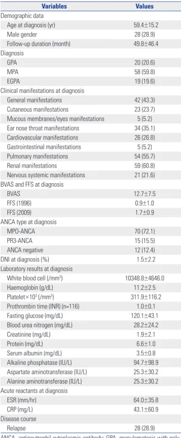
The baseline characteristics are summarised in Table 1. The Table 1. Baseline Characteristics of 97 Patients with ANCA-Associated
Vasculitis
Variables Values
Demographic data
Age at diagnosis (yr) 59.4±15.2
Male gender 28 (28.9)
Follow-up duration (month) 49.8±46.4
Diagnosis
GPA 20 (20.6)
MPA 58 (59.8)
EGPA 19 (19.6)
Clinical manifestations at diagnosis
General manifestations 42 (43.3)
Cutaneous manifestations 23 (23.7)
Mucous membranes/eyes manifestations 5 (5.2)
Ear nose throat manifestations 34 (35.1)
Cardiovascular manifestations 26 (26.8)
Gastrointestinal manifestations 5 (5.2)
Pulmonary manifestations 54 (55.7)
Renal manifestations 59 (60.8)
Nervous systemic manifestations 21 (21.6)
BVAS and FFS at diagnosis
BVAS 12.7±7.5
FFS (1996) 0.9±1.0
FFS (2009) 1.7±0.9
ANCA type at diagnosis
MPO-ANCA 70 (72.1)
PR3-ANCA 15 (15.5)
ANCA negative 12 (12.4)
DNI at diagnosis (%) 1.5±2.2
Laboratory results at diagnosis
White blood cell (/mm3) 10348.8±4646.0
Haemoglobin (g/dL) 11.2±2.5
Platelet×103 (/mm3) 311.9±116.2
Prothrombin time (INR) (n=116) 1.0±0.1
Fasting glucose (mg/dL) 120.1±43.1
Blood urea nitrogen (mg/dL) 28.2±24.2
Creatinine (mg/dL) 1.9±2.1
Protein (mg/dL) 6.6±1.0
Serum albumin (mg/dL) 3.5±0.8
Alkaline phosphatase (IU/L) 94.7±98.9
Aspartate aminotransferase (IU/L) 25.3±30.2 Alanine aminotransferase (IU/L) 25.3±30.2 Acute reactants at diagnosis
ESR (mm/hr) 64.0±35.8
CRP (mg/L) 43.1±60.9
Disease course
Relapse 28 (28.9)
ANCA, antineutrophil cytoplasmic antibody; GPA, granulomatosis with poly-angiitis; MPA, microscopic polypoly-angiitis; EGPA, eosinophilic granulomatosis with polyangiitis; BVAS, Birmingham vasculitis activity score; FFS, five factor score; MPO, myeloperoxidase; PR3, proteinase 3; DNI, delta neutrophil index; INR, international normalised ratio; ESR, erythrocyte sedimentation rate; CRP, C-reactive protein.
Values are expressed as mean±standard deviation and number (%).
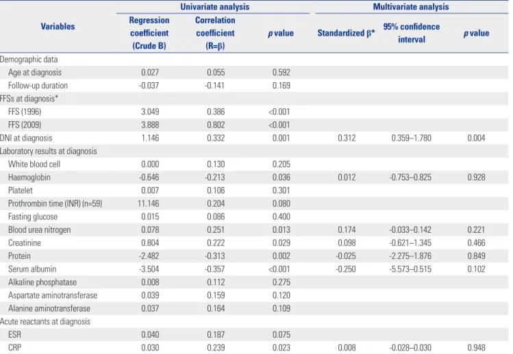
Table 2. Correlation between Delta Neutrophil Index and Other Continuous Variables in 97 Patients with ANCA-Associated Vasculitis at Diagnosis
Variables Correlation
coefficient (R=β) p value
Demographic data
Age at diagnosis -0.026 0.802
Follow-up duration 0.108 0.291
BVAS and FFS at diagnosis
BVAS 0.333 0.001
FFS (1996) 0.191 0.061
FFS (2009) 0.164 0.108
Laboratory results at diagnosis
White blood cell 0.392 <0.001
Haemoglobin 0.141 0.170
Platelet 0.189 0.064
Prothrombin time (INR) (n=116) 0.094 0.424
Fasting glucose 0.226 0.026
Blood urea nitrogen -0.112 0.273
Creatinine -0.033 0.750 Protein -0.153 0.135 Serum albumin -0.139 0.174 Alkaline phosphatase 0.015 0.882 Aspartate aminotransferase 0.071 0.491 Alanine aminotransferase 0.127 0.216
Acute reactants at diagnosis
ESR 0.077 0.467
CRP 0.235 0.025
ANCA, antineutrophil cytoplasmic antibody; BVAS, Birmingham vasculitis ac-tivity score; FFS, five factor score; INR, international normalised ratio; ESR, erythrocyte sedimentation rate; CRP, C-reactive protein.
mean age of 97 patients [28 men (28.9%)] was 59.4 years, and the mean follow-up duration was 49.8 months. The most fre-quent clinical manifestation was renal involvement (60.8%), followed by pulmonary (55.7%), general (43.3%), and ENT (35.1%) manifestations. The mean initial BVAS, FFS (1996), and FFS (2009) were 12.7, 0.9, and 1.7, respectively. MPO-ANCA was detected in 70 patients (72.1%) and PR3-ANCA was found in 15 patients (15.5%). The mean DNI was 1.5%. The mean ESR and CRP were 64.0 mm/hr and 43.1 mg/L, respectively. Twen-ty-eight patients (28.9%) had relapse during the follow-up. Correlation between DNI and other continuous variables in 97 patients with AAV at diagnosis
DNI was significantly related to BVAS at diagnosis (r=0.333, p= 0.001), and DNI was also significantly linked to white blood cell (r=0.392, p<0.001), fasting glucose (r=0.226, p=0.026), and CRP (r=0.235, p=0.025) (Table 2).
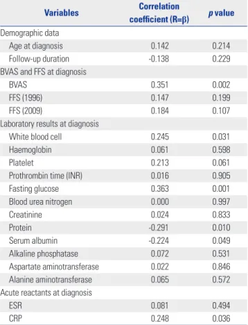
Univariate and multivariate linear regression analyses of BVAS and other continuous variables in 97 patients with AAV at diagnosis
Univariate linear regression analysis showed that BVAS was significantly related to DNI (r=0.332, p=0.001). BVAS was also positively linked to blood urea nitrogen (r=0.251, p=0.013), cre-atinine (r=0.222, p=0.029), and CRP (r=0.239, p=0.023), and was negatively linked to haemoglobin (r=-0.213, p=0.036), pro-tein (r=-0.313, p=0.002), and serum albumin (r=-0.357, p< 0.001). Although BVAS was significantly related to FFS (1996) (r=0.386, p<0.001), and FFS (2009) (r=0.802, p=0.047), FFS was excluded in multivariate linear regression analysis due to the risk of multicollinearity between BVAS and FFS. Multivariate linear regression analysis indicated that only DNI among con-tinuous variables was associated with BVAS with statistical significance in univariate linear regression analysis [β=0.312, 95% confidence interval (CI) 0.359–1.780, p=0.004] (Table 3).
Table 3. Univariate and Multivariate Linear Regression Analyses of BVAS and Other Continuous Variables in 97 Patients with ANCA-Associated Vas-culitis at Diagnosis
Variables
Univariate analysis Multivariate analysis
Regression coefficient (Crude B) Correlation coefficient (R=β)
p value Standardized β* 95% confidence interval p value
Demographic data Age at diagnosis 0.027 0.055 0.592 Follow-up duration -0.037 -0.141 0.169 FFSs at diagnosis* FFS (1996) 3.049 0.386 <0.001 FFS (2009) 3.888 0.802 <0.001 DNI at diagnosis 1.146 0.332 0.001 0.312 0.359–1.780 0.004
Laboratory results at diagnosis
White blood cell 0.000 0.130 0.205
Haemoglobin -0.646 -0.213 0.036 0.012 -0.753–0.825 0.928
Platelet 0.007 0.106 0.301
Prothrombin time (INR) (n=59) 11.146 0.204 0.080
Fasting glucose 0.015 0.086 0.400
Blood urea nitrogen 0.078 0.251 0.013 0.174 -0.033–0.142 0.221
Creatinine 0.804 0.222 0.029 0.098 -0.621–1.345 0.466 Protein -2.482 -0.313 0.002 -0.025 -2.275–1.876 0.849 Serum albumin -3.504 -0.357 <0.001 -0.250 -5.573–0.515 0.102 Alkaline phosphatase 0.008 0.112 0.275 Aspartate aminotransferase 0.039 0.159 0.120 Alanine aminotransferase 0.037 0.164 0.109
Acute reactants at diagnosis
ESR 0.040 0.187 0.075
CRP 0.030 0.239 0.023 0.008 -0.028–0.030 0.948
BVAS, Birmingham vasculitis activity score; ANCA, antineutrophil cytoplasmic antibody; FFS, five factor score; DNI, delta neutrophil index; INR, international normalised ratio; ESR, erythrocyte sedimentation rate; CRP, C-reactive protein.
Correlation between DNI and other continuous variables in 78 patients with GPA and MPA at diagnosis
We conducted sub-group analyses in each variant of AAV. DNI was significantly related to BVAS in patients with GPA (r= 0.730, p<0.001) and MPA (r=0.290, p=0.027), but not in those with EGPA (r=0.415, p=0.078). Therefore, we excluded patients with EGPA and reanalysed the association of DNI with BVAS at diagnosis in 78 patients with MPA and GPA. DNI was signif-icantly related to BVAS (r=0.351, p=0.002). It was also signifi-cantly linked to white blood cell (r=0.245, p=0.031), fasting glu-cose (r=0.363, p=0.001), and CRP (r=0.248, p=0.036), and was inversely linked to protein (r=-0.291, p=0.010) and serum albu-min (r=-0.224, p=0.049) (Table 4).
Univariate and multivariate linear regression analyses of BVAS and other continuous variables in 78 patients with GPA and MPA at diagnosis
Univariate linear regression analysis exhibited that BVAS was significantly related to DNI (r=0.351, p=0.002). BVAS was also positively linked to blood urea nitrogen (r=0.254, p=0.025) and was negatively linked to the follow-up duration (r=-0.270, p= 0.017), protein (r=-0.308, p=0.006), and serum albumin (r= -0.314, p=0.005). However, BVAS was not linked to ESR or CRP. FFS was excluded in multivariate linear regression analysis due to the risk of multicollinearity between BVAS and FFS. Multi-variate linear regression analysis indicated that only DNI among continuous variables was associated with BVAS with statistical significance in univariate linear regression analysis (β=0.276, 95% CI 0.271–2.140, p=0.012) (Table 5).
The optimal cut-off of DNI to identify severe AAV in 78 patients with GPA and MPA at diagnosis
We divided patients with GPA and MPA into the two groups according to the cut-off of severe AAV. Seventeen patients (21.8%) had BVAS ≥20 and belonged to severe AAV group. We calculated the optimal cut-off of DNI to identify severe AAV, and found that 0.65% was a strong measurement of severe AAV (AUROC 0.664, 95% CI 0.516–0.813, p=0.039; sensitivity 70.6% and specificity 63.9%). We also divided 78 patients with GPA and MPA into the two groups according to the optimal cut-off of DNI. There were no significant differences in medications administered during the follow-up or prior to relapse between patients having DNI ≥0.65% and those having DNI <0.65% (Supplementary Table 1, only online). Severe AAV was identi-fied more often in patients having DNI ≥0.65% than those hav-ing DNI <0.65% (35.3% vs. 11.4%, p=0.011) (Fig. 1A). Moreover, patients having DNI ≥0.65% had a significantly higher risk of severe AAV than those not having (RR 4.255, 95% CI 1.325– 13.665).
DNI at diagnosis to predict relapse in 78 patients with GPA and MPA during follow-up
We evaluated whether DNI at diagnosis could predict relapse during the follow-up of patients with MPA and GPA. First, be-cause DNI was significantly related to BVAS at diagnosis, and BVAS at diagnosis could predict poor prognosis during the fol-low-up in previous studies,24,25 we calculated the optimal cut-off of DNI at diagnosis to predict relapse of MPA and GPA, however, we could find no optimal cut-off (AUROC 0.460, p= 0.589). Second, we divided 78 patients with GPA and MPA into the two groups according to the presence of relapse, and com-pared DNI, BVAS, FFS (1996), and FFS (2009). Patients with relapse exhibited the higher mean BVAS and FFS (1996) than those without [17.5 vs. 10.8, p<0.001 for BVAS, and 1.3 vs. 0.7, p=0.013 for FFS (1996)]. However, DNI did not differ between the two groups (1.9 vs. 1.3, p=0.239). Last, because DNI ≥0.65% could identify severe AAV, we investigated its potential as a predictor of relapse using Kaplan-Meier survival analysis. Cu-mulative relapse free survival rate was depicted in Fig. 1B. Ac-cording to DNI ≥0.65% at diagnosis, there was a significant dif-ference in cumulative relapse free survival rates between the two groups (p=0.029).
Table 4. Correlation between Delta Neutrophil Index and Other Continu-ous Variables in 78 Patients with Granulomatosis with Polyangiitis and Microscopic Polyangiitis at Diagnosis
Variables Correlation
coefficient (R=β) p value
Demographic data
Age at diagnosis 0.142 0.214
Follow-up duration -0.138 0.229
BVAS and FFS at diagnosis
BVAS 0.351 0.002
FFS (1996) 0.147 0.199
FFS (2009) 0.184 0.107
Laboratory results at diagnosis
White blood cell 0.245 0.031
Haemoglobin 0.061 0.598
Platelet 0.213 0.061
Prothrombin time (INR) 0.016 0.905
Fasting glucose 0.363 0.001
Blood urea nitrogen 0.000 0.997
Creatinine 0.024 0.833 Protein -0.291 0.010 Serum albumin -0.224 0.049 Alkaline phosphatase 0.072 0.531 Aspartate aminotransferase 0.022 0.846 Alanine aminotransferase 0.065 0.572
Acute reactants at diagnosis
ESR 0.081 0.494
CRP 0.248 0.036
BVAS, Birmingham vasculitis activity score; FFS, five factor score; INR, inter-national normalised ratio; ESR, erythrocyte sedimentation rate; CRP, C-reac-tive protein.
Table 5. Univariate and Multivariate Linear Regression Analyses of BVAS and Other Continuous Variables in 78 Patients with Granulomatosis with Polyangiitis and Microscopic Polyangiitis at Diagnosis
Variables
Univariate analysis Multivariate analysis
Regression coefficient (Crude B) Correlation coefficient (R=β)
p value Standardized β* 95% confidence interval p value
Demographic data Age at diagnosis -0.004 -0.007 0.953 Follow-up duration -0.072 -0.270 0.017 -0.188 -0.106–0.005 0.073 FFSs at diagnosis* FFS (1996) 2.373 0.284 0.012 FFS (2009) 3.647 0.410 <0.001 DNI at diagnosis 1.529 0.351 0.002 0.276 0.271–2.140 0.012
Laboratory results at diagnosis
White blood cell 0.000 0.088 0.445
Haemoglobin -0.643 -0.199 0.080
Platelet 0.005 0.072 0.530
Prothrombin time (INR) (n=59) 10.616 0.201 0.135
Fasting glucose 0.021 0.123 0.281
Blood urea nitrogen 0.076 0.254 0.025 0.058 -0.007–0.123 0.077
Creatinine 0.788 0.222 0.051 Protein -2.559 -0.308 0.006 -0.038 -2.738–2.112 0.798 Serum albumin -3.167 -0.314 0.005 -0.167 -4.449–1.072 0.227 Alkaline phosphatase 0.008 0.110 0.338 Aspartate aminotransferase 0.031 0.131 0.253 Alanine aminotransferase 0.029 0.135 0.237
Acute reactants at diagnosis
ESR 0.041 0.174 0.142
CRP 0.028 0.212 0.074
BVAS, Birmingham vasculitis activity score; FFS, five factor score; DNI, delta neutrophil index; INR, international normalised ratio; ESR, erythrocyte sedimenta-tion rate; CRP, C-reactive protein.
*We did not include FFS (1996) and FFS (2009) in multivariate linear regression analysis due to the risk of multicollinearity between BVAS and FFS.
1.0 0.8 0.6 0.4 0.2 0.0 100 80 60 40 20 0 – – – – – – – – – – – –
Cumulative relapse free survival
% Months 0 DNI<0.65% DNI≥0.65% 20 40 60 80 100 120 DNI<0.65% DNI≥0.65% BVAS<20 (not severe)
BVAS≥20 (severe) p=0.029 88.6% 11.4% 64.7% 35.3% RR 4.255 p=0.011 A B
Fig. 1. DNI identified severe AAV at diagnosis and predicted relapse of GPA and MPA during the follow-up. (A) Patients having DNI ≥0.65% had se-vere AAV (BVAS ≥20) more frequently than those having DNI <0.65% (35.3% vs. 11.4%, p=0.011). (B) In Kaplan-Meier survival analysis, patients having DNI ≥0.65% exhibited lower cumulative relapse free survival rate than those having DNI <0.65% (p=0.029). DNI, delta neutrophil index; AAV, antineu-trophil cytoplasmic antibody-associated vasculitis; GPA, granulomatosis with polyangiitis; MPA, microscopic polyangiitis; BVAS, Birmingham vasculi-tis activity score; RR, relative risk.
DISCUSSION
In this study, we first reported that DNI was associated with BVAS at diagnosis and could predict relapse during the follow-up in patients with AAV. DNI was remarkably related to BVAS and furthermore, among continuous variables, only DNI could reflect BVAS at diagnosis in not only all patients with AAV, but also patients with GPA and MPA. However, because the associ-ation of DNI with BVAS was not apparent in patients with EGPA, we selected the results on patients with only GPA and MPA. In addition, we provided the optimal cut-off of DNI of 0.65% for identifying severe AAV based on BVAS, and found that patients having DNI ≥0.65% had significantly higher risk of severe AAV (GPA and MPA) than those not having (RR 4.255) at diagnosis. On the other hands, we applied DNI ≥0.65% to pre-dict relapse of GPA and MPA during the follow-up, and found a significant difference in cumulative relapse free survival be-tween the two groups according to DNI ≥0.65% (p=0.029). We, therefore, concluded that DNI could reflect BVAS at diagnosis and furthermore, DNI ≥0.65% could not only identify severe AAV at diagnosis, but also predict relapse during the follow-up in patients with GPA and MPA.
How can DNI reflect BVAS? Based on the pathogenesis of AAV, we assume the link between DNI and BVAS as follows: 1) various aetiologies drive T cells and macrophages to produce inflammatory cytokines, which can prime neutrophils, leading to an increase in adhesion molecules and ANCA antigens on their surface;26-28 2) this inflammatory conditions also increase adhesion molecules on endothelial cells;29 3) the ANCA-me-diated interaction between primed neutrophils and activated endothelial cells occurs, and activated neutrophils migrate be-yond vascular walls;30 4) complement pathway also accelerates the ANCA-associated activation of neutrophils;31,32 5) finally, activated neutrophils provoke vasculitis by reactive oxygen radicals and degranulation.7,8,33 Therefore, neutrophils partici-pating in inflammation may be gradually consumed and the number of immature neutrophils increases. With this hypoth-esis, we speculate that DNI can reflect BVAS of AAV.
Although DNI at diagnosis ≥0.65% significantly reduced cu-mulative relapse free survival compared with DNI at diagnosis <0.65% in Kaplan-Meier survival analysis, we failed to obtain the optimal cut-off of DNI at diagnosis to predict relapse of GPA and MPA during the follow-up in AUROC analysis. We recently have reported the predictive value of BVAS at diagnosis for re-lapse of AAV and polyarteritis nodosa.24,25,34 With this concept, we compared the potential of DNI with BVAS at diagnosis for predicting relapse of GPA and MPA during the follow-up. We calculated the optimal cut-off of BVAS at diagnosis to predict relapse using AUROC, and found that 15.5 of BVAS had the strongest predictive value (AUROC 0.746, 95% CI 0.618–0.874, p=0.001; sensitivity 66.7% and specificity 77.2%). Also, we found that patients having BVAS at diagnosis ≥15.5 had the lower cu-mulative relapse free survival than those not having (p<0.001).
Moreover, we conducted a Cox Hazard model using DNI ≥0.65% and BVAS ≥15.5 at diagnosis to predict relapse. Before a Cox Hazard analysis, we investigated the multicollinearity between DNI ≥0.65 and BVAS ≥15.5 using a multivariate linear regres-sion test, and found no significant multicollinearity (variance inflation factor was 1.140). When we performed a Cox Hazard model using those two variables, we found that only BVAS ≥15.5 showed the predictive significance for relapse (odds ratio 6.174, 95% CI 2.268–16.805, p<0.001). Although DNI ≥0.65% showed a significant predictive potential for relapse in Kaplan-Meier survival analysis, it could not surpass that of BVAS at di-agnosis ≥15.5.
We studied the reason of why the predictive potential of DNI could not reach that of BVAS at diagnosis and suggested that DNI is the current progressive type, while BVAS is the current completion type. In the putative sequence of the pathogenesis of AAV, neutrophil priming and activation through loss of toler-ance of T and B cells and ANCA autoimmune response initiates acute injury.7,33 At this phase, most participants are neutrophils. Meanwhile, the next step switches on innate immunity re-sponses, leading to either resolution or sclerotic progress in-cluding granulomatosis formation. At this phase, most partici-pants are lymphocytes and macrophages.8 Because DNI is closely linked to the consumption of mature neutrophils, DNI might be predominantly associated with neutrophil-predom-inant phase, whereas BVAS might be predomneutrophil-predom-inantly associat-ed with irreversible sclerotic phase. In addition, relapse be-gins new and acute autoimmune responses on the basis of established chronic sclerosis, fibrosis, and granulomatosis more often than on that of normal tissues. Not a few patients are classified as AAV at chronic irreversible sclerotic phase rather than at acute neutrophil-predominant phase. Thus, BVAS at diagnosis occupies larger phases in the putative sequence of the pathogenesis of AAV than DNI at diagnosis, and BVAS at diagnosis can predict relapse of GPA and MPA better than DNI at diagnosis. On the contrary, because the higher level of DNI at diagnosis implies more likely phase to heal and return to normal tissues, we suggests that patients having high DNI at diagnosis should receive more immediate and aggressive treatment.
Our present study has a strong advantage in that we first proved the association of DNI with BVAS at diagnosis and provided the optimal cut-off of DNI to predict relapse during the follow-up in patients with GPA and MPA. However, our study has also several limitations. First, we could not clarify the mechanism of the association of DNI and BVAS and its pre-dictive value for relapse. Second, because DNI has been report-ed since January 2010, the number of patients was not large enough to enhance reliability. Last, due to a retrospective de-sign of this study and lack of information on serial BVAS dur-ing the follow-up duration, we could not clarify the link be-tween changes of DNI and BVAS over time. Thus, prospective studies with a larger number of patients are required to obtain
more reliable information on the role of DNI in AAV.
In conclusion, DNI could reflect BVAS at diagnosis and fur-thermore, DNI ≥0.65% could not only identify severe AAV at diagnosis, but also predict relapse during the follow-up in pa-tients with GPA and MPA.
ACKNOWLEDGEMENTS
This research was supported by Basic Science Research Pro-gram through the National Research Foundation of Korea (NRF) funded by the Ministry of Education (2017R1D1A1B03029050).
ORCID
Juyoung Yoo https://orcid.org/0000-0001-8882-1695 Sang-Won Lee https://orcid.org/0000-0002-8038-3341
REFERENCES
1. Jennette JC, Falk RJ, Bacon PA, Basu N, Cid MC, Ferrario F, et al. 2012 revised International Chapel Hill Consensus Conference Nomenclature of Vasculitides. Arthritis Rheum 2013;65:1-11. 2. Millet A, Pederzoli-Ribeil M, Guillevin L, Witko-Sarsat V, Mouthon
L. Antineutrophil cytoplasmic antibody-associated vasculitides: is it time to split up the group? Ann Rheum Dis 2013;72:1273-9. 3. Watts R, Lane S, Hanslik T, Hauser T, Hellmich B, Koldingsnes W,
et al. Development and validation of a consensus methodology for the classification of the ANCA-associated vasculitides and polyar-teritis nodosa for epidemiological studies. Ann Rheum Dis 2007; 66:222-7.
4. Leavitt RY, Fauci AS, Bloch DA, Michel BA, Hunder GG, Arend WP, et al. The American College of Rheumatology 1990 criteria for the classification of Wegener’s granulomatosis. Arthritis Rheum 1990;33:1101-7.
5. Masi AT, Hunder GG, Lie JT, Michel BA, Bloch DA, Arend WP, et al. The American College of Rheumatology 1990 criteria for the clas-sification of Churg-Strauss syndrome (allergic granulomatosis and angiitis). Arthritis Rheum 1990;33:1094-100.
6. de Lind van Wijngaarden RA, van Rijn L, Hagen EC, Watts RA, Gre-gorini G, Tervaert JW, et al. Hypotheses on the etiology of antineu-trophil cytoplasmic autoantibody associated vasculitis: the cause is hidden, but the result is known. Clin J Am Soc Nephrol 2008;3: 237-52.
7. Jennette JC, Falk RJ. Pathogenesis of antineutrophil cytoplasmic autoantibody-mediated disease. Nat Rev Rheumatol 2014;10:463-73. 8. Jennette JC, Falk RJ, Hu P, Xiao H. Pathogenesis of antineutrophil cytoplasmic autoantibody-associated small-vessel vasculitis. Annu Rev Pathol 2013;8:139-60.
9. Honda T, Uehara T, Matsumoto G, Arai S, Sugano M. Neutrophil left shift and white blood cell count as markers of bacterial infec-tion. Clin Chim Acta 2016;457:46-53.
10. Field D, Taube E, Heumann S. Performance evaluation of the im-mature granulocyte parameter on the Sysmex XE-2100 automat-ed hematology analyzer. Lab Hematol 2006;12:11-4.
11. Nigro KG, O’Riordan M, Molloy EJ, Walsh MC, Sandhaus LM. Per-formance of an automated immature granulocyte count as a pre-dictor of neonatal sepsis. Am J Clin Pathol 2005;123:618-24. 12. Nahm CH, Choi JW, Lee J. Delta neutrophil index in automated
immature granulocyte counts for assessing disease severity of pa-tients with sepsis. Ann Clin Lab Sci 2008;38:241-6.
13. Horan TC, Andrus M, Dudeck MA. CDC/NHSN surveillance defi-nition of health care-associated infection and criteria for specific types of infections in the acute care setting. Am J Infect Control 2008;36:309-32.
14. Pyo JY, Park JS, Park YB, Lee SK, Ha YJ, Lee SW. Delta neutrophil in-dex as a marker for differential diagnosis between flare and infec-tion in febrile systemic lupus erythematosus patients. Lupus 2013; 22:1102-9.
15. Park HJ, Ha YJ, Pyo JY, Park YB, Lee SK, Lee SW. Delta neutrophil index as an early marker for differential diagnosis of adult-onset Still’s disease and sepsis. Yonsei Med J 2014;55:753-9.
16. Pyo JY, Ha YJ, Song JJ, Park YB, Lee SK, Lee SW. Delta neutrophil index contributes to the differential diagnosis between acute gout attack and cellulitis within 24 hours after hospitalization. Rheuma-tology (Oxford) 2017;56:795-801.
17. Csernok E, Moosig F. Current and emerging techniques for ANCA detection in vasculitis. Nat Rev Rheumatol 2014;10:494-501. 18. Mukhtyar C, Lee R, Brown D, Carruthers D, Dasgupta B, Dubey S,
et al. Modification and validation of the Birmingham Vasculitis Activity Score (version 3). Ann Rheum Dis 2009;68:1827-32. 19. Stone JH, Hoffman GS, Merkel PA, Min YI, Uhlfelder ML, Hellmann
DB, et al. A disease-specific activity index for Wegener’s granulo-matosis: modification of the Birmingham Vasculitis Activity Score. International Network for the Study of the Systemic Vasculitides (INSSYS). Arthritis Rheum 2001;44:912-20.
20. Gayraud M, Guillevin L, le Toumelin P, Cohen P, Lhote F, Casassus P, et al. Long-term followup of polyarteritis nodosa, microscopic polyangiitis, and Churg-Strauss syndrome: analysis of four pro-spective trials including 278 patients. Arthritis Rheum 2001;44: 666-75.
21. Guillevin L, Pagnoux C, Seror R, Mahr A, Mouthon L, Le Toumelin P; French Vasculitis Study Group (FVSG). The Five-Factor Score revisited: assessment of prognoses of systemic necrotizing vascu-litides based on the French Vasculitis Study Group (FVSG) cohort. Medicine (Baltimore) 2011;90:19-27.
22. Mukhtyar C, Hellmich B, Jayne D, Flossmann O, Luqmani R. Re-mission in antineutrophil cytoplasmic antibody-associated sys-temic vasculitis. Clin Exp Rheumatol 2006;24(6 Suppl 43):S-93-8. 23. Mukhtyar C, Flossmann O, Hellmich B, Bacon P, Cid M,
Cohen-Tervaert JW, et al. Outcomes from studies of antineutrophil cyto-plasm antibody associated vasculitis: a systematic review by the European League Against Rheumatism systemic vasculitis task force. Ann Rheum Dis 2008;67:1004-10.
24. Oh YJ, Ahn SS, Park ES, Jung SM, Song JJ, Park YB, et al. Chest and renal involvements, Birmingham vascular activity score more than 13.5 and five factor score (1996) more than 1 at diagnosis are significant predictors of relapse of microscopic polyangiitis. Clin Exp Rheumatol 2017;35 Suppl 103:47-54.
25. Yoo J, Kim HJ, Ahn SS, Jung SM, Song JJ, Park YB, et al. Clinical and prognostic features of Korean patients with MPO-ANCA, PR3-ANCA and ANCA-negative vasculitis. Clin Exp Rheumatol 2017;35 Suppl 103:111-8.
26. Nakazawa D, Tomaru U, Suzuki A, Masuda S, Hasegawa R, Ko-bayashi T, et al. Abnormal conformation and impaired degrada-tion of propylthiouracil-induced neutrophil extracellular traps: implications of disordered neutrophil extracellular traps in a rat model of myeloperoxidase antineutrophil cytoplasmic antibody-associated vasculitis. Arthritis Rheum 2012;64:3779-87.
27. Schreiber A, Kettritz R. The neutrophil in antineutrophil cytoplas-mic autoantibody-associated vasculitis. J Leukoc Biol 2013;94: 623-31.
28. Kettritz R. How anti-neutrophil cytoplasmic autoantibodies acti-vate neutrophils. Clin Exp Immunol 2012;169:220-8.
29. Ooi JD, Chang J, Hickey MJ, Borza DB, Fugger L, Holdsworth SR, et al. The immunodominant myeloperoxidase T-cell epitope in-duces local cell-mediated injury in antimyeloperoxidase glomer-ulonephritis. Proc Natl Acad Sci U S A 2012;109:E2615-24. 30. Furuta S, Jayne DR. Antineutrophil cytoplasm
antibody-associat-ed vasculitis: recent developments. Kidney Int 2013;84:244-9. 31. Xiao H, Dairaghi DJ, Powers JP, Ertl LS, Baumgart T, Wang Y, et al.
C5a receptor (CD88) blockade protects against MPO-ANCA GN. J Am Soc Nephrol 2014;25:225-31.
32. Jennette JC, Xiao H, Hu P. Complement in ANCA-associated vas-culitis. Semin Nephrol 2013;33:557-64.
33. Sanders JS, Abdulahad WH, Stegeman CA, Kallenberg CG. Patho-genesis of antineutrophil cytoplasmic autoantibody-associated vasculitis and potential targets for biologic treatment. Nephron Clin Pract 2014;128:216-23.
34. Oh YJ, Ahn SS, Park ES, Jung SM, Song JJ, Park YB, et al. Birming-ham vasculitis activity score at diagnosis is a significant predictor of relapse of polyarteritis nodosa. Rheumatol Int 2017;37:685-94.