Open Access
Research article
In silico
characterisation and chromosomal localisation of human
RRH
(peropsin) – implications for opsin evolution
James Bellingham*
1
, Dominic J Wells
1
and Russell G Foster
2
Address: 1Gene Targeting Unit, Department of Neuromuscular Diseases, Division of Neuroscience and Psychological Medicine, Faculty of
Medicine, Imperial College London, Charing Cross Hospital, St. Dunstan's Road, London, W6 8RP, UK and 2Department of Integrative and
Molecular Neuroscience, Division of Neuroscience and Psychological Medicine, Faculty of Medicine, Imperial College London, Charing Cross Hospital, St. Dunstan's Road, London, W6 8RP, UK
Email: James Bellingham* - [email protected]; Dominic J Wells - [email protected]; Russell G Foster - [email protected] * Corresponding author
Abstract
Background: The vertebrate opsins are proteins which utilise a retinaldehyde chromophore in their photosensory or photoisomerase roles in the visual/irradiance detection cycle. The majority of the opsins, such as rod and cone opsins, have a very highly conserved gene structure suggesting a common lineage. Exceptions to this are RGR-opsin and melanopsin, whose genes have very different intron insertion positions. The gene structure of another opsin, peropsin (retinal pigment epithelium-derived rhodopsin homologue, RRH) is unknown.
Results: By in silico analysis of the GenBank database we have determined that the human RRH
comprises 7 exons spanning approximately 16.5 kb and is localised to chromosome 4q25 in the following gene sequence: cen-EGF-RRH-IF-qter – a position that excludes this gene as a candidate for the RP29 autosomal recessive retinitis pigmentosa locus. A comparison of opsin gene structures reveals that RRH and RGR share two common intron (introns 1 and 4) insertion positions which may reflect a shared ancestral gene.
Conclusion: The opsins comprise a diverse group of genes which appear to have arisen from three different lineages. These lineages comprise the "classical opsin superfamily" which includes the rod and cone opsins, pinopsin, VA-opsin, parapinopsin and encephalopsin; the RRH and RGR
group; and the melanopsin line. A common lineage for RRH and RGR, together with their sites of expression in the RPE, indicates that peropsin may act as a retinal isomerase.
Background
The photosensory opsins are a family of membrane bound, heptahelical G-protein coupled receptors (GPCRs) characterised by their ability to covalently bind a vitamin A-based retinaldehyde chromophore via a Schiff base to a lysine residue located in the 7th transmembrane α-helix [1,2]. In the natural environment, the vertebrate vitamin A based chromophore takes the form of either 11-cis-retinal (A1) or 11-cis-3,4-didehydroretinal (A2) [3]. In
the vertebrate retina, the primary events in image detec-tion occur in the outer segments of rod and cone cells, with the opsins located in these lamellar stacks being named after these cell types – rod-opsin in rods and cone-opsins in cones. Absorption of a photon of light by the chromophore located in the retinal binding pocket of an opsin causes its photoisomerisation from the 11-cis to an all-trans conformation. This change of the chromophore induces a conformational change of its surrounding opsin
Published: 24 January 2003
BMC Genomics 2003, 4:3
Received: 18 December 2002 Accepted: 24 January 2003
This article is available from: http://www.biomedcentral.com/1471-2164/4/3
molecule allowing transducin (G-protein) binding and activation of the phototransduction cascade [1,2]. The vertebrate retina generally contains a single rod opsin class and as many as four different cone opsins [4], though the majority of mammals are dichromats being in possession of only two types of cone opsin. In the non-mammals, non-rod, non-cone photosensory opsins have been described. For example, pinopsin (P-opsin) was iso-lated from the avian pineal [5] and vertebrate ancient opsin (VA-opsin) was isolated from the teleost inner reti-na and pineal/deep brain structures [6]. Other opsins have also been described from a variety of vertebrate class-es which either act as a photoisomerase, e.g. RGR-opsin [7]; or have an unknown function, e.g. peropsin [8], para-pinopsin [9] and encephalopsin [10]; or are expressed in photoreceptive cells but have as yet an unknown function, e.g. melanopsin [11]. The one key feature present in all the opsin classes is the presence of a lysine residue in the 7th transmembrane α-helix which is thought to enable retinal attachment in all cases [12].
The first complete vertebrate opsin sequence (bovine rod opsin) was described in 1983 [13], and this opsin has since become the numeration template for critical resi-dues that are conserved throughout all of the opsins. The genomic structure of bovine rod opsin consists of five ex-ons separated by four intrex-ons [13], a structure that has been conserved in all vertebrate rod-opsins including those of the ancient Agnatha [14], with the single excep-tion of the rod opsin in teleost fish which is intronless [15]. Employing the nomenclature proposed by Hunt et al. [16] which correlates the spectral sensitivity (λmax) with amino acid sequence identity, the first cone-opsin se-quences isolated were those of the human blue (ultravio-let/violet sensitive, UVS/VS) (OPN1SW), green and red cone opsins (both longwave sensitive, LWS) (OPN1MW, OPN1LW) [17]. The UVS/VS opsin gene shares the same genomic structure as the rod-opsin gene in that is possess-es four conserved intron insertion positions. The LWS opsin also shares the four conserved introns with the rod-and UVS/VS cone-opsin, but has an additional fifth intron in a 5' prime position to the four conserved introns [17], which we have termed intron 0 (zero) [12]. Sequences from the remaining two visual opsin classes of the verte-brates, shortwave sensitive (SWS) and middlewave sensi-tive (MWS), have been isolated and share the same genomic structure as the rod and UVS/VS cone opsins [18]. Interestingly, the SWS and MWS opsin families found in birds, fish, reptiles and amphibia are absent in the mammalian lineage – a feature thought to be related to the "nocturnal bottleneck" occupied by mammals dur-ing their evolution resultdur-ing in the loss of these two cone classes [19,20].
Many of the more recently described non-rod, non-cone opsins also share a relatively high degree of intron posi-tion conservaposi-tion with the visual opsins, only differing in the position of intron 2 as in pinopsin where it is shifted 14 nucleotides in a 3' direction [21] and VA-opsin where it is shifted 42 nucleotides in a 3' direction [22], or its ab-sence as in parapinopsin [9] and encephalopsin (OPN3) [23]. However, RGR-opsin (retinal G-protein coupled re-ceptor, RGR) [24] and melanopsin (OPN4) [25] do not share intron insertion positions with any other opsins for which a gene structure is known, whilst the gene structure of peropsin is presently unknown.
Little is known about peropsin (retinal pigment epitheli-um-derived rhodopsin homologue, RRH) except that it shares ~26% amino acid identity with the photosensory opsins, is uniquely expressed in the retinal pigment epi-thelium (RPE), and by synteny with mouse will map to human chromosome 4q [8]. To address this issue, we have determined by in silico database searches that the hu-man peropsin gene (RRH) has seven exons and maps to chromosome 4q25. A comparison with the gene struc-tures of other opsins indicates that RRH and RGR share two intron insertion sites. These findings and their phylo-genetic relationships are discussed within the context of the possible function of peropsin. In view of the variety of nomenclature systems that are applied to the various opsin classes [2,12], we have attempted to be as consistent as possible with nomenclature and have deferred to pre-ferred locus symbols accepted by the Gene Nomenclature Committee of The Human Genome Organisation, HUGO – http://www.hugo-international.org/hugo/ and Online Mendelian Inheritance in Man, OMIM – http://www.nc-bi.nlm.nih.gov/omim/. Thus, RRH and peropsin should be considered synonymous.
Results and Discussion
Structure of the RRH gene
(UTR) and the initial 106 bp of coding sequence. We were unable to align the first 16 bp of the RRH cDNA to RP11-602N24 and have concluded that these 16 bp are a cDNA cloning artifact since the breakdown of sequence similar-ity does not occur at an apparent intron acceptor site in the genomic sequence. Exons 2–6 contain the next 793 bp of coding sequence, whilst exon 7 contains the final 115 bp of coding sequence and the 3'UTR. No nucleotide sub-stitutions were observed between the published coding se-quence and that derived from RP11-602N24, but three separate nucleotide substitutions were observed in the 3'UTR. The entire exon/intron structure encompasses ap-proximately 16.5 kilo base pairs (kb) and has been as-cribed GenBank accession number BK000958.
Chromosomal localisation of RRH
In order to map RRH, we subjected the sequence of RP11-602N24 to a BLAST search and found that it contains the I factor (complement) gene, IF, which has been shown to map to 4q25 [28]. Localisation of RP11-602N24 and hence RRH to 4q25 is consistent with the syntenic predic-tion from the mapping data for mouse Rrh [8]. To further refine the localisation of RRH, a BLAST search using a ter-minal 2 kb of sequence (bases 116643–118642) of RP11-602N24 identified BAC clone B200N5 which maps to 4q25 and contains approximately 143 kb of sequence (GenBank: AC005509). A BLAST search using B200N5 in-dicated that this clone contains the epidermal growth fac-tor gene, EGF, which is also known to map to 4q25 in the order cen-EGF-IF-qter [28]. We assembled a mini-contig consisting of approximately 260 kb of sequence from clones RP11-602N24 and B200N5 and have been able to determine the following gene order: cen-EGF~85
kb~RRH~26 kb~IF-qter, and their directions of transcrip-tion (Figure 2). Localisatranscrip-tion of RRH to an interval of ap-proximately 110 kb between EGF and IF on chromosome 4q25 excludes this gene as a causative candidate for the autosomal recessive retinitis pigmentosa locus (RP29) lo-cated on 4q32-q34 [29].
Comparison of opsin gene structures
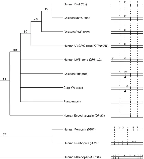
To determine whether any of the intron insertion sites in RRH are conserved with those of other opsins, nucleotide sequences of representatives of the various opsin classes were aligned and intron positions marked (Figure 3). RRH shares two common intron insertion sites (1 and 4) with RGR, whilst the other four intron insertion sites appear novel amongst the opsin families represented. RRH and RGR share two further introns that are present in relatively close proximity. Intron 3 of RRH is located 9 bp in a 3' di-rection of intron 3 in RGR (both introns being in phase +1), whilst intron 5 in RRH is located 3 bp (1 codon) in a 3' direction of the corresponding intron in RGR (both in-trons being in phase 0). Between RRH and members of the other opsin families only two intron positions are in relatively close proximity – intron 6 in RRH is located 1 bp 5' of intron 4 in the rod and cone opsins, and intron 2 of RRH (phase 0) is 8 bp 3' of intron 3 (phase +1) of OPN4.
From the perspective of intron positions, it would appear that the opsins have arisen from three ancestral genes (Fig-ure 4). Considering the clade (81% bootstrap confidence) which includes the visual opsins (rod and cones), brain (pinopsin, VA-opsin, parapinopsin) and encephalopsin, it can be seen that three intron positions (introns 1, 3 and 4) are perfectly conserved throughout (Figures 3 and 4). Figure 1
Exon-intron boundaries of RRH based on GenBank BK000958. Uppercase = exon; lowercase = intron. * Exon sizes exclude the 5' and 3' UTRs.
Exon Size Intron size
Exon (bp) 3' Intron Exon Sequence 5' Intron (bp)
UTR---This would suggest that the ancestral opsin gene giving rise to this clade had introns present at these three posi-tions. Subsequent gene duplication events have given rise to opsin classes that either do not possess intron 2, or pos-sess intron 2 in at least one of three positions (position 2, 2a or 2b), whilst the LWS cone opsin group has accumu-lated a further intron (0) in the visual opsin lineage (Fig-ure 4). Given the organisation of the lamprey (Agnatha) rod opsin gene [14] and pinopsin gene [30] (which has an intron arrangement of the VA-opsin family rather than that of the pinopsin family [22]), these highly conserved gene structures have been in existence for at least 550 mil-lion years. Further support for an ancestral chordate opsin gene with these three conserved introns is provided by an opsin from the sea squirt Ciona intestinalis. This urochor-date possesses Ci-opsin1 whose gene has seven introns, three of which are in positions conserved with those in vertebrate visual opsins [31], corresponding to positions 1, 3 and 4 [12]. Given their common lineage, we have chosen to term this collection of opsins the "classical opsin superfamily".
RRH and RGR form a separate clade (87% bootstrap con-fidence) and share two conserved intron positions with each other, 1 and 4, but none with classical opsin super-family or melanopsin (Figures 3 and 4). This may be in-dicative that RRH and RGR derive from the same ancestral gene which possessed introns at positions equivalent to positions 1 and 4 in RRH and RGR, even though they only share ~25% amino acid identity across the
transmem-brane domains compared with ≥ 40% identity exhibited between the majority of the classical opsin superfamily [12]. An exception being encephalopsin which shares ~30% identity with other members of the classical opsin superfamily [12], but clearly has a highly conserved gene structure. The phenomenon of intron sliding or slippage [32], which has been proposed as a mechanism contribut-ing to the slight variations in position of some introns ob-served between some insect opsins [33], may explain the 1 bp shift between intron 6 of RRH and intron 4 of the classical opsin superfamily, but their proximity may also be due to chance. Whilst single nucleotide intron slippage is an evolutionary phenomenon thought to occur in <5% of introns [34], these two opsin groups do not appear to have a recent common ancestor which probably indicates a random insertion of introns. Similarly, the close prox-imity of introns 2 and 3 of RRH with introns 3 of OPN4 and RGR respectively are likely also to be chance occur-rences given that intron insertion and deletion events are much more common than intron sliding events [32,35,36].
OPN4 (melanopsin) shares no conserved intron positions with any of the other opsin classes, and it has been pro-posed that this opsin class represents a separate line of opsin evolution [12,37]. Collectively the data suggest that three separate lines of evolution have occurred to form the present day vertebrate opsins represented by the structur-ally highly conserved classical opsin superfamily, the Figure 2
Mini-contig showing the arrangement of genes around the RRH locus. Clone names (with associated GenBank accession num-bers) are given as are their sizes and regions of overlap in bp. The direction of transcription and location of translation initia-tion codons (atg) of the various genes are also indicated in bp.
atg
atg
atg
RP11-602N24
(AC126283)
B200N5
(AC005509)
1
1 118642
1993 116643
143391 96226
70169
64892
RRH
EGF
4q25
cen
qter
Figure 3
Human RRH coding sequence and conceptual translation (peropsin) with transmembrane domains (boldface) as predicted by the rod opsin model of Palczewski et al. [48]. The six intron insertion sites in the RRH gene defined in Figure 1 are indicated by black filled circles. Equivalent intron insertion sites for the visual (rod and cone) opsins are indicated by red filled circles – intron positions 1–4 are common to all rod (RHO) and cone opsins, whilst intron 0 is only found in the LWS opsins (OPN1MW,
OPN1LW) [13,17]. The shifted second intron of the pinopsin and VA-opsin families [21,22] are indicated as 2a and 2b respec-tively in red circles. Parapineal opsin [9] and encephalopsin (OPN3) [10] possess only introns 1, 3 and 4 of the rod and cone opsins. Equivalent intron insertion sites for RGR-opsin (RGR) [24] are indicated by open circles, whilst those for melanopsin (OPN4) [25] are indicated by yellow filled circles. Only intron insertion sites 1–7 for melanopsin are indicated due to the extreme length of the melanopsin C-terminus which makes positioning of introns 8 and 9 inaccurate since there is no overlap with any other opsin. Note that intron insertion sites 1 and 4 in RRH are equivalent to intron insertion sites 1 and 4 in RGR.
7
6
5
4
3
2
1
5
6
4
3
2
10 20 30 40 50 60 70 80 90 ATGCTAAGAAATAATTTAGGCAACAGTTCAGACTCTAAAAATGAAGATGGCTCGGTCTTTTCACAGACTGAACACAATATTGTTGCAACT M L R N N L G N S S D S K N E D G S V F S Q T E H N I V A T 30100 110 120 130 140 150 160 170 180 TACTTGATTATGGCAGGTATGATAAGTATTATCAGCAACATAATAGTTCTGGGCATCTTCATTAAGTACAAGGAACTTCGGACACCCACA Y L I M A G M I S I I S N I I V L G I F I K Y K E L R T P T 60
190 200 210 220 230 240 250 260 270 AATGCAATTATTATTAACCTGGCTGTTACTGATATAGGGGTCAGTAGCATTGGCTATCCCATGTCTGCTGCCTCAGATCTGTATGGAAGT N A I I I N L A V T D I G V S S I G Y P M S A A S D L Y G S 90
280 290 300 310 320 330 340 350 360 TGGAAATTTGGATACGCAGGCTGTCAGGTTTATGCTGGATTGAATATTTTTTTTGGAATGGCAAGCATTGGATTACTCACGGTCGTGGCT W K F G Y A G C Q V Y A G L N I F F G M A S I G L L T V V A 120
370 380 390 400 410 420 430 440 450 GTGGACCGATACCTGACCATCTGCCTTCCTGACGTAGGGAGAAGAATGACCACCAACACTTACATCGGCTTGATTCTGGGAGCCTGGATC V D R Y L T I C L P D V G R R M T T N T Y I G L I L G A W I 150
460 470 480 490 500 510 520 530 540 AATGGCCTGTTTTGGGCTTTGATGCCTATCATAGGGTGGGCTAGTTATGCCCCAGATCCTACTGGTGCTACGTGTACCATAAACTGGAGG N G L F W A L M P I I G W A S Y A P D P T G A T C T I N W R 180
550 560 570 580 590 600 610 620 630 AAAAATGATAGATCTTTTGTGTCTTACACCATGACAGTTATTGCGATAAATTTTATTGTGCCCTTGACAGTGATGTTTTACTGCTATTAC K N D R S F V S Y T M T V I A I N F I V P L T V M F Y C Y Y 210
640 650 660 670 680 690 700 710 720 CATGTCACGCTATCCATTAAACATCACACTACCAGTGACTGCACTGAGTCCCTCAACAGAGACTGGTCAGATCAGATAGATGTAACAAAG H V T L S I K H H T T S D C T E S L N R D W S D Q I D V T K 240
730 740 750 760 770 780 790 800 810 ATGTCTGTGATCATGATCTGCATGTTTCTGGTGGCATGGTCCCCTTATTCCATCGTGTGCTTATGGGCTTCTTTTGGTGACCCAAAGAAG M S V I M I C M F L V A W S P Y S I V C L W A S F G D P K K 270
820 830 840 850 860 870 880 890 900 ATTCCTCCCCCCATGGCCATCATAGCTCCACTGTTTGCAAAATCTTCTACATTCTATAACCCCTGCATTTATGTGGTTGCTAATAAAAAG I P P P M A I I A P L F A K S S T F Y N P C I Y V V A N K K 300
910 920 930 940 950 960 970 980 990 TTTCGGAGGGCAATGCTTGCCATGTTCAAATGTCAGACTCACCAAACAATGCCTGTGACAAGTATTTTACCCATGGATGTATCTCAAAAC F R R A M L A M F K C Q T H Q T M P V T S I L P M D V S Q N 330
1000 1010 CCATTGGCTTCTGGAAGAATCTGA P L A S G R I * 337
1
2
3
4
1
4
3
6
2
5
2b 2a
0
Figure 4
Maximum parsimony tree with bootstrap confidence levels based on nucleotide coding sequence of the various vertebrate opsin classes rooted with human melanopsin (see Materials and Methods). Cone opsin class nomenclature after Hunt et al.
[16]. To the right of the tree is a schematic representation of the intron insertion sites in the various opsin genes based on Fig-ure 3. Conserved intron positions between opsin classes are indicated by a dashed vertical line. The relative position of the second rod and cone opsin intron is indicated by an arrow head in the pinopsin and VA-opsin classes to show their shifted sec-ond intron positions (2a and 2b).
Human Rod (RH)
Chicken MWS cone
Chicken SWS cone
Human LWS cone (OPN1LW)
Chicken Pinopsin
Carp VA-opsin
Parapinopsin
Human Peropsin (RRH)
Human RGR-opsin (RGR)
Human Melanopsin (OPN4)
99
46
60
99
81
87
Human UVS/VS cone (OPN1SW)
Human Encephalopsin (OPN3)
1 2 3 4
1 2a 3 4
1 2b 3 4
1 3 4
1 3 4
0 1 2 3 4 1 2 3 4 1 2 3 4 1 2 3 4
1 2 3 4 5 6
1 2 3 4 5 6
more relaxed RRH and RGR grouping, and OPN4 which stands as the single member of the melanopsin class to date.
An ancestral link between RRH and RGR may provide an insight at to the potential function of peropsin. RGR-opsin is known to act as a photoisomerase in the reconver-sion of retinal chromophore from the all-trans to the 11-cis conformation [38,39], and perhaps peropsin also func-tions as an isomerase in some manner given its localisa-tion in the RPE. This suggeslocalisa-tion is supported by recent findings that indicate that an amphioxus (Branchiostoma belcheri) homologue of peropsin acts as an all-trans to 11-cis photoisomerase [40]. Alternatively, peropsin may act as a thermal isomerase since Rgr-/- mice are able to convert all-trans-retinal to the 11-cis conformation under dark ad-aptation [7]. It has been suggested that peropsin may also bind a different retinal isomer, or indeed a non-retinoid ligand [8]. We have recently shown that the rod, non-cone opsins such as RGR-opsin, peropsin, melanopsin and encephalopsin are all expressed early in the embryon-ic development of the mammalian eye. These opsins are expressed by E11.5 in mice and by 8.6 weeks in humans [41], a finding which in all cases predates the expression of the visual rod and cone opsins [42]. This early expres-sion pattern may be supportive of a role in retinoid me-tabolism for these opsins in the developing retina [43], or that these opsins have a role in the embryonic eye that are quite different to those in the adult eye.
Conclusions
Using GenBank database searches we have been able to determine that the human peropsin gene, RRH, comprises seven exons spanning about 16.5 kb. By assembling se-quences from large genomic clones we have determined that RRH localises within an interval of approximately 110 kb between EGF and IF on chromosome 4q25. Of the six introns present in RRH, two are located in positions conserved with introns 1 and 4 of RGR, which given the phylogenetic relationship of RRH and RGR may suggest that these two opsins have arisen from a common ances-tor, and by inference possess a common function e.g. act-ing as a retinal chromophore (photo)isomerase. Our data also strengthens the argument that the present day opsins are represented by genes that have arisen from three differ-ent ancestral genes that have given rise to (1), a classical opsin superfamily cosisting of visual opsins and those opsins who share a highly conserved gene structure with the visual opsins; (2), a peropsin and RGR-opsin family; (3), a melanopsin family.
Methods
Database Searches
The GenBank database was screened using the online BLAST [26] server – http://www.ncbi.nlm.nih.gov/BLAST/
. Searches were carried out using the standard nucleotide-nucleotide BLAST (blastn) option against the "nr data-base" using default values with the low complexity filter off. Subsequent sequence manipulations utilised the on-line BLAST 2 Sequences [44] server – http://www.nc-bi.nlm.nih.gov/blast/bl2seq/bl2.html and MacVector 7.0 (Accelrys Ltd., Cambridge, UK).
Phylogenetic Analysis
Nucleotide coding sequences of: (i) human rod opsin, RHO (U49742); (ii) human red cone (LWS) opsin, OPN1LW (AH005298); (iii) human blue cone (UVS/VS) opsin, OPN1SW (U53874); (iv) chicken green cone (MWS) opsin (M92038); (v) chicken blue cone (SWS) opsin (M92037); (vi) chicken pinopsin (U15762); (vii) carp VA-opsin (AF233520); (viii) catfish parapinopsin (AF028014); (ix) human encephalopsin, OPN3 (AF140242); (x) human peropsin, RRH (AF012270); (xi) human RGR-opsin, RGR (U14910); (xii) human melan-opsin, OPN4 (AF147788), were entered in to DAMBE ver-sion 4.1.15 [45] – http://aix1.uottawa.ca/~xxia/software/ software.htm, and converted to amino acid sequences which were then aligned using the ClustalW [46] option. In order to maintain codon integrity the nucleotide se-quences were then aligned to the amino acid alignment. Phylogenetic analyses were conducted using MEGA ver-sion 2.1 [47] – http://www.megasoftware.net. A maxi-mum parsimony tree with branch confidence values based on 500 bootstrap replicates was constructed and the tree drawn in TreeViewPPC version 1.6.6 – http://taxono-my.zoology.gla.ac.uk/rod/treeview.html. Branches with less than 40% support were collapsed.
Authors' contributions
JB undertook the in silico and phylogenetic analysis, and participated in the preparation of the manuscript. DJW and RGF participated in the preparation of the script. All authors read and approved the final manu-script.
Acknowledgements
This work was supported by The Hammersmith Hospitals Trust Special Trustees (JB and DJW) and BBSRC (RGF).
References
1. Burns ME and Baylor DA Activation, deactivation, and adapta-tion in vertebrate photoreceptor cells.Annu Rev Neurosci 2001,
24:779-805
2. Ebrey T and Koutalos Y Vertebrate photoreceptors.Prog Retin Eye Res 2001, 20:49-94
3. Lythgoe JN The Ecology of Vision. Oxford: Clarendon Press 1979, 4. Okano T, Kojima D, Fukada Y, Shichida Y and Yoshizawa T Primary
structures of chicken cone visual pigments: vertebrate rho-dopsins have evolved out of cone visual pigments.Proc Natl Acad Sci U S A 1992, 89:5932-5936
5. Okano T, Yoshizawa T and Fukada Y Pinopsin is a chicken pineal photoreceptive molecule.Nature 1994, 372:94-97
7. Chen P, Hao W, Rife L, Wang XP, Shen D, Chen J, Ogden T, Van Boemel GB, Wu L, Yang M and Fong HKW A photic visual cycle of rhodopsin regeneration is dependent on Rgr . Nat Genet
2001, 28:256-260
8. Sun H, Gilbert DJ, Copeland NG, Jenkins NA and Nathans J Per-opsin, a novel visual pigment-like protein located in the api-cal microvilli of the retinal pigment epithelium.Proc Natl Acad Sci U S A 1997, 94:9893-9898
9. Blackshaw S and Snyder SH Parapinopsin, a novel catfish opsin localized to the parapineal organ, defines a new gene family.
J Neurosci 1997, 17:8083-8092
10. Blackshaw S and Snyder SH Encephalopsin: a novel mammalian extraretinal opsin discretely localized in the brain.J Neurosci
1999, 19:3681-3690
11. Hattar S, Liao H-W, Takao M, Berson DM and Yau K-W Melanop-sin-containing retinal ganglion cells: architecture, projec-tions, and intrinsic photosensitivity.Science 2002, 295: 1065-1070
12. Bellingham J and Foster RG Opsins and mammalian photoen-trainment.Cell Tissue Res 2002, 309:57-71
13. Nathans J and Hogness DS Isolation, sequence analysis, and in-tron-exon arrangement of the gene encoding bovine rho-dopsin.Cell 1983, 34:807-814
14. Zhang H and Yokoyama S Molecular evolution of the rhodopsin gene of marine lamprey, Petromyzon marinus . Gene 1997,
191:1-6
15. Fitzgibbon J, Hope A, Slobodyanyuk SJ, Bellingham J, Bowmaker JK and Hunt DM The rhodopsencoding gene of bony fish lacks in-trons.Gene 1995, 164:273-277
16. Hunt DM, Wilkie SE, Bowmaker JK and Poopalasundaram S Vision in the ultraviolet.Cell Mol Life Sci 2001, 58:1583-1598
17. Nathans J, Thomas D and Hogness DS Molecular genetics of hu-man color vision: the genes encoding blue, green, and red pigments.Science 1986, 232:193-202
18. Yokoyama S and Yokoyama R Adaptive evolution of photorecep-tors and visual pigments in vertebrates.Annu Rev Ecol Syst 1996,
27:543-567
19. Goldsmith TH Optimization, constraint, and history in the ev-olution of eyes.Q Rev Biol 1990, 65:281-322
20. Jacobs GH The distribution and nature of colour vision among the mammals.Biol Rev Camb Philos Soc 1993, 68:413-471 21. Max M, McKinnon PJ, Seidenman KJ, Barrett RK, Applebury ML,
Taka-hashi JS and Margolskee RF Pineal opsin: a nonvisual opsin ex-pressed in chick pineal.Science 1995, 267:1502-1506
22. Moutsaki P, Bellingham J, Soni BG, David-Gray ZK and Foster RG Se-quence, genomic structure and tissue expression of carp (Cyprinus carpio L.) vertebrate ancient (VA) opsin.FEBS Lett
2000, 473:316-222
23. Blackshaw S and Snyder SH Developmental expression pattern of phototransduction components in mammalian pineal im-plies a light-sensing function.J Neurosci 1997, 17:8074-8082 24. Shen D, Jiang M, Hao W, Tao L, Salazar M and Fong HKW A human
opsin-related gene that encodes a retinaldehyde-binding protein.Biochemistry 1994, 33:13117-13125
25. Provencio I, Rodriguez IR, Jiang G, Hayes WP, Moreira EF and Rollag MD A novel human opsin in the inner retina.J Neurosci 2000,
20:600-605
26. Altschul SF, Madden TL, Schaffer AA, Zhang J, Zhang Z, Miller W and Lipman DJ Gapped BLAST and PSI-BLAST: a new generation of protein database search programs.Nucleic Acids Res 1997,
25:3389-3402
27. Mount SM Genomic sequence, splicing, and gene annotation.
Am J Hum Genet 2000, 67:788-792
28. Shiang R, Murray JC, Morton CC, Buetow KH, Wasmuth JJ, Olney AH, Sanger WG and Goldberger G Mapping of the human com-plement factor I gene to 4q25.Genomics 1989, 4:82-86 29. Hameed A, Khaliq S, Ismail M, Anwar K, Mehdi SQ, Bessant D, Payne
AM and Bhattacharya SS A new locus for autosomal recessive RP (RP29) mapping to chromosome 4q32-q34 in a Pakistani family.Invest Ophthalmol Vis Sci 2001, 42:1436-1438
30. Yokoyama S and Zhang H Cloning and characterization of the pineal gland-specific opsin gene of marine lamprey (Petro-myzon marinus).Gene 1997, 202:89-93
31. Kusakabe T, Kusakabe R, Kawakami I, Satou Y, Satoh N and Tsuda M
Ci-opsin1, a vertebrate-type opsin gene, expressed in the
lar-val ocellus of the ascidian Ciona intestinalis. FEBS Lett 2001,
506:69-72
32. Stoltzfus A, Logsdon JM Jr, Palmer JD and Doolittle WF Intron "slid-ing" and the diversity of intron positions.Proc Natl Acad Sci U S A 1997, 94:10739-10744
33. Bellingham J, Wilkie SE, Morris AG, Bowmaker JK and Hunt DM
Characterisation of the ultraviolet-sensitive opsin gene in the honey bee, Apis mellifera .Eur J Biochem 1997, 243:775-781 34. Rogozin IB, Lyons-Weiler J and Koonin EV Intron sliding in
con-served gene families.Trends Genet 2000, 16:430-432
35. Gotoh O Divergent structures of Caenorhabditis elegans cyto-chrome P450 genes suggest the frequent loss and gain of in-trons during the evolution of nematodes.Mol Biol Evol 1998,
15:1447-1459
36. Rzhetsky A, Ayala FJ, Hsu LC, Chang C and Yoshida A Exon/intron structure of aldehyde dehydrogenase genes supports the "in-trons-late " theory.Proc Natl Acad Sci U S A 1997, 94:6820-6825 37. Bellingham J, Whitmore D, Philp AR, Wells DJ and Foster RG
Ze-brafish melanopsin: isolation, tissue localisation and phyloge-netic position.Molecular Brain Research 2002, 107:128-136 38. Hao W and Fong HKW Blue and ultraviolet light-absorbing
opsin from the retinal pigment epithelium.Biochemistry 1996,
35:6251-6256
39. Hao W and Fong HKW The endogenous chromophore of reti-nal G protein-coupled receptor opsin from the pigment epi-thelium.J Biol Chem 1999, 274:6085-6090
40. Koyanagi M, Terakita A, Kubokawa K and Shichida Y Amphioxus homologs of Go-coupled rhodopsin and peropsin having 11-cis – and all-trans-retinals as their chromophores.FEBS Lett
2002, 531:525-528
41. Tarttelin EE, Bellingham J, Bibb LC, Foster RG, Hankins MW, Grego-ry-Evans K, GregoGrego-ry-Evans CY, Wells DJ and Lucas RJ Expression of opsin genes early in ocular development of humans and mice.Exp Eye Res 2003, 76:393-396
42. Bibb LC, Holt JK, Tarttelin EE, Hodges MD, Gregory-Evans K, Ruther-ford A, Lucas RJ, Sowden JC and Gregory-Evans CY Temporal and spatial expression patterns of the CRX transcription factor and its downstream targets. Critical differences during hu-man and mouse eye development. Hum Mol Genet 2001,
10:1571-1579
43. Maden M Retinoid signalling in the development of the central nervous system.Nat Rev Neurosci 2002, 3:843-853
44. Tatusova TA and Madden TL BLAST 2 Sequences, a new tool for comparing protein and nucleotide sequences.FEMS Microbiol Lett 1999, 174:247-250
45. Xia X and Xie Z DAMBE: software package for data analysis in molecular biology and evolution.J Hered 2001, 92:371-373 46. Thompson JD, Higgins DG and Gibson TJ CLUSTAL W:
improv-ing the sensitivity of progressive multiple sequence align-ment through sequence weighting, position-specific gap penalties and weight matrix choice. Nucleic Acids Res 1994,
22:4673-4680
47. Kumar S, Tamura K, Jakobsen IB and Nei M MEGA2: molecular ev-olutionary genetics analysis software. Bioinformatics 2001,
17:1244-1245