chiropractic management of a 55-year-old man
with cervical radiculopathy
Peter Emary,
BSc, DC** Private practice: Parkway Back Clinic, 201C Preston Parkway, Cambridge, Ontario, N3H 5E8. Phone: 519-653-2101. E-mail: [email protected]
© JCCA 2012
Le présent exposé de cas suit la gestion réussie d’un patient de 55 ans chez qui l’on a diagnostiqué une radiculopathie cervicale au moyen d’une thérapie manipulative et d’étirements de relaxation postisométrique des muscles cervicaux paraspinaux. (JCCA 2012; 56(1):9–17)
m o t s c l é s : Radiculopathie cervicale, relaxation
postisométrique, facilitation neuromusculaire proprioceptive, chiropratique
This case report chronicles the successful management of a 55-year-old patient diagnosed with cervical radiculopathy using spinal manipulative therapy and cervical paraspinal post-isometric relaxation stretches. (JCCA 2012; 56(1):9–17)
k e y w o r d s : cervical radiculopathy, post-isometric
relaxation, proprioceptive neuromuscular facilitation, chiropractic.
Introduction
Cervical radiculopathy (CR) is an impingement or inflam-matory irritation of the cervical spine nerve root(s), re-sulting in pain (or numbness) radiating along nerves of the upper extremity;1,2 the C6 and C7 levels are most
often affected.1,3 Limited research is available on the
inci-dence and prevalence of CR; however, the inciinci-dence rate (in Rochester, Minnesota) has been reported at 83.2 cases per 100,000 people per year (107.3/100,000 for males vs. 63.5/100,000 for females), with peak incidence in those aged 50–54 years.1 A history of physical exertion or major
trauma precedes the onset of symptoms in less than 15% of cases. The most common causes are cervical spondylo-sis and intervertebral disc herniation,1,3 accounting for
ap-proximately 70% and 20% of cases, respectively.1 In the
former, posterior vertebral body osteophytes and/or facet joint/ligamentum flavum hypertrophy encroach upon the intervertebral foramen; posterolateral herniation of disc material results in foraminal encroachment in the latter. In either case, cervical nerve root pain and dysfunction can occur.4
Post-isometric relaxation (PIR) is a technique often used by manual therapists (including some chiropractors) for treating muscle tension and joint dysfunction in myo-fascial pain syndromes;5 however, studies investigating its
effectiveness in the treatment of CR are extremely scarce. This case report chronicles the successful management of a 55-year-old patient diagnosed with CR using spinal manipulative therapy (SMT) and cervical paraspinal PIR stretches.
Case Report
History
generalized weakness and “numbness” in his right hand. Any attempt to lift or reach would shoot a “stabbing” pain down his right arm. Holding the arm (bent at 90°) close to his body was palliative. Coughing, sneezing, or bear-ing down for a bowel movement (i.e. Dejerine’s Triad) did not reproduce the neck, shoulder blade, or right arm pain. The patient also denied any lower extremity or my-elopathy symptoms, and exhibited normal gait. Medically prescribed anti-inflammatories (Naprosyn), muscle relax-ants (Robaxin), heat therapy, and time off work had not provided any relief.
Medical history was remarkable for coronary artery disease, including angioplasty surgery (4 years prior). Medications included Lipitor, Altace, Rhoxal-bisoprolol, and Aspirin. The patient denied any motor vehicle acci-dents, major falls or injuries, and had no previous history of neck problems. He had seen a chiropractor once before because of lower back pain, with good results. He was married with 3 children and had been employed as a ship-per/receiver for the past 7 years. He did not smoke and consumed an average of 7 alcoholic beverages per week. He also walked a total of 2 hours per week for exercise and took a daily multivitamin.
Examination Findings
Blood pressure was normal at 104/68. Postural exam re-vealed severe antalgia, with the patient holding his head forward and tilted to the left. Motion palpation of his spine revealed joint restriction at C2-3 and C3-4 in left rotation, and T5-6 and T6-7 in extension. Static palpation revealed myofascial trigger points within the right rhom-boid muscles, along with hypertonicity of the right para-spinals and localized tenderness of the right C2-3 and C3-4 facet joints. Cervical spine range of motion (ROM) was very painful (with parasthesia) and 90% restricted in extension, 75% in right rotation, and 90% in right lateral flexion. Passive flexion of the patient’s neck produced some cervical facet pain on the right, without signs of myelopathy. The Spurling and Upper Limb Tension Tests provoked the patient’s right-sided radicular pain, while the Cervical Distraction Test relieved it (see Table 1 for orthopedic test descriptions). Depression of the right shoulder while holding the neck in flexion and left rota-tion (i.e. Shoulder Depression Test) also provided relief. Upper extremity neurologic examination was unremark-able for motor, reflex, sensory, and vibratory testing, ex-cept for weakness of the right deltoid muscle (graded as Table 1 Select orthopedic exam procedures for cervical radiculopathy
Test Description of procedure Positive findings
Spurling Patient seated with their neck extended and rotated to the ipsilateral side, and doctor applies a downward pressure through the top of the patient’s head
Radicular symptoms are provoked
Upper Limb Tension Patient supine and doctor performs the following movements to the patient’s upper extremity:
1. scapular depression 2. shoulder abduction
3. forearm supination, wrist and finger extension 4. shoulder external rotation
5. elbow extension
6. ipsilateral/contralateral rotation of the neck
Radicular symptoms are provoked
Cervical Distraction Patient seated and doctor grips under the patient’s mastoids and tractions superiorly
Radicular symptoms are relieved
Valsalva Patient seated and is asked to take and hold a deep breath while bearing down (as if for a bowel movement)
4/5), because of right-sided neck and radicular pain. Cer-vical spine radiographs revealed moderate degenerative disc disease at C6-7, with mild-to-moderate bony foram-inal narrowing at this level on the right (Figure 1A) and mild narrowing on the left (Figure 1B). The patient was diagnosed with acute, right-sided C7 radiculopathy.
Plan of Management & Results
The patient underwent a course of chiropractic treatment consisting of supine cervical and thoracic SMT, soft-tis-sue trigger-point therapy to the right rhomboid muscles, home ice therapy (as needed), cervical spine isometric exercises, and ergonomic instruction (i.e. avoidance of provocative neck positions). To manipulate the right side
of the patient’s neck (i.e. side of radiculopathy), the pa-tient’s head was rotated 90° to the left and then a right lateral flexion “modified rotary break” procedure was used.6 A supine rotary break (with 45° of right rotation
and left lateral flexion) was used on the other side.The initial treatment frequency was 3 times per week for 2 weeks. Outcome measures used were numeric rating scale for pain; subjective changes in neck, shoulder blade, and arm pain; and patient self-rating of outcome (i.e. no, minor, or major improvement). Objective measures used were visual estimation for ROM, as well as orthopedic and neurological examination.
After 2 weeks of treatment, the patient’s neck and shoulder blade pain had improved; each was reduced to Figure 1 Anterior oblique radiographs of the cervical spine showing (A) mild-to-moderate bony foraminal narrowing
at C6-7 on the right (arrow), with mild narrowing at this same level on the left (B).
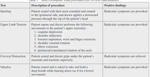
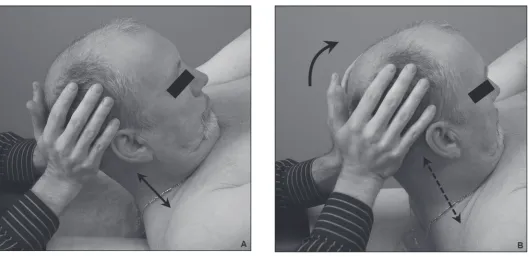
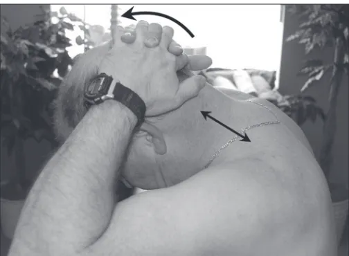
between a 3 (at best) and 5 (at worst) out of 10. The right arm pain, however, remained unchanged. Cervical spine ROM was still painful and 75% restricted in right lateral flexion. Upper extremity neurological exam was normal, and the result of the Upper Limb Tension Test was nega-tive. The Spurling Test was still positive, however, and passive flexion still elicited right-sided facet pain in the neck. At this point, the patient’s self-rated improvement was “minor.” Because of unresolved radicular symptoms, the author decided to include a cervical paraspinal PIR technique with patient treatment (Figure 2). On the next visit, during the application of this technique, the patient experienced immediate, short-term relief of his right arm symptoms. Based on this result, the patient was also in-structed to begin performing cervical paraspinal stretches at home (Figure 3). Using this new protocol, the patient continued to be treated at a frequency of 2 times per week for 3 more weeks.
After 6 weeks and a total of 12 treatments (including 6 with PIR), the right-sided neck, shoulder blade, and arm pain were all reduced to between 1 (at best) and 3 (at worst) out of 10. Cervical ROM was within normal limits and unremarkable, except for right-sided neck and shoul-der blade pain during passive right lateral flexion. Neuro-logic and orthopedic examinations, including the result of the Spurling Test, were normal. The patient’s self-rated improvement at this point was “major.” When asked to subjectively rate his overall percentage improvement on a scale of 0 (no improvement) to 100 (full improvement), he rated it at 75%. Regarding his activities of daily living, the patient’s neck and arm pain were still provoked with prolonged sitting at work (at a computer) or when sleep-ing on his right side at home.
Based on the patient’s overall improvement, the treat-ment frequency progressively decreased to once every 4 weeks. He was also encouraged to continue performing Figure 2 Cervical paraspinal PIR is performed (in this case) with the patient supine, while the doctor slowly lifts
the patient’s head toward the ceiling (A). Once a comfortable stretch is felt, the patient is asked to push their head back (with approximately 10% of their strength), while the doctor resists this movement; thus, creating an isometric contraction. This position is held for 8–10 seconds. The patient is then asked to inhale deeply and, upon exhalation, is instructed to relax while the doctor lifts the patient’s head a little further towards the ceiling (B). After an 8–10 second
his neck stretches on a regular basis (i.e. 1 to 2 times per day). Although the patient’s symptoms had improved, his complaint of recurrent re-aggravation prompted a refer-ral to his family physician for cervical spine magnetic resonance imaging (MRI) and needle electromyography (EMG) studies. MRI examination, performed 4 months after the onset of symptoms, showed dehydration and intervertebral disc bulging at multiple levels, most nota-bly at C6-7; prominent stenosis of the right lateral canal was also evident at this same level (Figure 4).
Electrophysiological studies—including motor and sensory nerve conduction velocity of the radial, ulnar, and median nerves and EMG of the right deltoid, biceps, tri-ceps, and extensor digitorum complex—were interpreted as normal. The attending neurologist did, however, report that the patient’s history was “consistent with right C7 radiculopathy.” He also noted that the patient’s symptoms had “improved considerably [,] with residual cervical and right shoulder blade pain.” Neurological examination was normal for cranial nerve, motor, sensory, reflex, co-ordination, and gait testing, with the exception of “mild weakness of the right tricep[s].” The neurologist told the patient that he had “no permanent damage” and that sur-gery was not indicated. The patient continued with chiro-practic care and, at 6-month follow-up (after a total of 20 treatments, including 14 with PIR), the right-sided neck and radicular pain was completely resolved (graded as
0 out of 10). Cervical spine ROM and upper extremity neurological exam were normal.
After 3 years, the patient’s radicular symptoms con-tinue to be graded as 0 out of 10, and he reports no limita-tions in his activities of daily living. Cervical spine ROM and upper extremity neurological examination remain normal. Only 2 minor episodes of neck pain (i.e. without radiculopathy and graded as 3 out of 10) have been re-ported during this time. Both were attributed to postural strain from sitting at a computer at work. The first episode was self-resolving, while the second was relieved with 1 treatment of manipulation and PIR. In addition, the pa-tient continues to report that he has not used any prescrip-tion or over-the-counter medicaprescrip-tions during the entire course of treatment. The patient has given written con-sent to having his personal health information, including radiographs and photographs of his likeness, published.
Discussion
Examination of patients presenting with CR should in-clude assessment of motor strength, deep tendon reflexes, and dermatomal sensation. In the absence of frank neuro-logic findings, more sensitive (or provocative) exam pro-cedures may be required. In a recent systematic review, Rubinstein et al.7 concluded that when consistent with
history and physical examination findings, the Spurling, Neck Distraction, and Valsalva Tests (given their high specificity), along with the Upper Limb Tension Test (given its high sensitivity) are most useful in establishing a diagnosis of CR, especially in patients without neuro-logical deficits. The scientific literature also supports the use of modern imaging techniques (e.g. MRI) and needle EMG, in diagnosing the cause and site of CR.8 Advanced
diagnostic testing can be expensive, however, and in the case of needle EMG, invasive.7 In addition, MRI findings
of disc herniation may not necessarily correlate with pa-tient symptoms.9 For the chiropractor, proper patient
his-tory and physical examination are the most cost-effective and non-invasive methods for diagnosing CR.
In patients with cervical spondylosis, as in the current case, the possibility of spinal cord compression (myelop-athy) should be considered. Clinical findings may include abnormal gait, clumsiness, bowel or bladder dysfunction, or other upper motor neuron signs (e.g. hyperreflexia, muscle spasticity, Babinski’s sign).2,3 The chiropractor’s
differential diagnosis of CR should also include myofa-Figure 3 Cervical paraspinal stretch (held for 15
scial trigger-point referral, peripheral nerve entrapment syndromes (e.g. thoracic outlet, carpal and/or cubital tunnel), and rotator cuff pathology. In the current case, radiculopathy was suspected over peripheral neuropathy because, in addition to patient history and diagnostic im-aging findings, the patient had positive Spurling, Upper Limb Tension, and Cervical Distraction Tests, as well as limited ipsilateral neck rotation. Wainner et al.10 found
that when these 4 tests are positive, they together identify (with 90% probability) the presence of CR. Less common causes include referred cardiac pain, herpes zoster (shin-gles), and intra- or extraspinal tumours (e.g. Schwan-nomas, Pancoast tumours, lymphomas).2,3
Traditional medical management may include nonster-oidal anti-inflammatories, activity modification, traction (or other physical therapy modalities), epidural steroid injections, and/or surgery (if necessary).1–3 Several
chiro-practic studies have described good outcomes in patients
treated with SMT—either alone or in combination with other conservative therapies.4,11–15 In their case series,
Hubka et al.13 also discuss the importance of the direction
of thrust when treating CR patients using SMT proced-ures. In particular, these authors note that neck manipula-tion is best tolerated by the patient when performed by contacting on the side of radiculopathy, laterally flexing the neck toward the side of radiculopathy, and then rotat-ing the neck away from the side of radiculopathy (fol-lowed by a gentle manipulative thrust). This is similar to the technique used in the current case. In their experience, Hubka et al. have found that manipulation in the opposite direction may provoke the patient’s symptoms, as might prone upper thoracic SMT.13 In the current case, supine
upper thoracic and bilateral cervical manipulations were used, with no adverse effects. In their discussion on the safety of neck manipulation, Murphy et al.4 conclude
that, “when applied by properly trained and experienced Figure 4 (A) T2-weighted sagittal MRI of the patient’s cervical spine showing moderate intervertebral disc
desicca-tion and protrusion at C6-7. Mild degenerative changes are also evident from C2 to C5, with mild disc protrusion at C3-4 and C4-5. (B) T2-weighted coronal MRI showing lateral canal stenosis and intervertebral foraminal
practitioners, [cervical SMT] is potentially a safe option for patients with CR.” Nevertheless, the evidence base for both conservative and surgical management of CR, in-cluding data on its natural history, is limited.4,16,17
PIR Technique
The primary purpose of this article was to showcase a PIR technique that, when combined with SMT, may be useful to chiropractors in treating patients with CR. Historically, PIR has been used as a “muscle energy procedure” for joint mobilization and muscle relaxation.5 The technique
begins by placing the muscle (to be treated) in a stretched position. Lewit5 describes this as “taking up the slack” in
the muscle, by lengthening it, to the point where the first slight resistance (or “barrier”) is felt. Next, the patient is instructed to resist this movement with minimum force, isometrically, for about 10 seconds, and then told to let go (or relax). Lewit stresses the importance of waiting until the patient has indeed relaxed, after which a gentle release is obtained and the muscle lengthens by “spontaneous decontraction” (relaxation). Release may continue for 10 seconds or more, until a new barrier is reached, from which point the procedure can be repeated. If nothing is gained by repetition, the normal physiologic barrier has been reached. In order to improve the patient’s cooper-ation and enhance the effectiveness of PIR, the technique should be combined with other methods of facilitation and inhibition (e.g. patient inhalation and exhalation).5 In
general, inhalation facilitates muscle activity and is there-fore useful during the isometric phase, while exhalation promotes inhibition and therefore helps relaxation. The overall goal of PIR treatment is to reduce muscle tension and relieve the resultant pain and dysfunction by restoring the full stretch length of the muscle.
The terms PIR and proprioceptive neuromuscular fa-cilitation (PNF) are sometimes incorrectly used syn-onymously. The main difference with the PNF technique is that during the isometric contraction phase, the patient exerts against a much greater resistance (i.e. up to 100% of their maximum strength).18 Furthermore, during the
re-laxation phase, the patient’s muscle(s) is more aggressive-ly stretched and the clinician does not necessariaggressive-ly wait to feel the patient’s muscle release. Therefore, practitioners should be cautioned when using PNF as it may result in considerable discomfort to the patient, particularly in an acute pain presentation.
PIR and CR
A paucity of research exists on the effectiveness of PIR for neck pain and/or CR; therefore, it is difficult to com-pare this study with others in the scientific literature. Some authors have compared PIR with SMT in treat-ing neck pain patients (without radiculopathy).19–20 For
instance, Cassidy et al.19 found that 1 treatment of
cer-vical SMT was more effective than mobilization (PIR) in decreasing neck pain intensity, while both treatments increased neck ROM to a similar degree. In a search of PubMed and Index to Chiropractic Literature, no studies were found combining the terms “cervical radiculopathy” and “post-isometric relaxation.” In a hand search of refer-ences retrieved using combinations of the terms “cervical spine,” “radiculopathy,” and “chiropractic,” the author found only 2 case reports relating PIR and CR.14,15 In the
first case by Daub,14 he described the resolution of a C6
radiculopathy in a 44-year-old female following 18 treat-ments (over 7 weeks). Treatment consisted of cervical and thoracic SMT; PIR applied to the levator scapulae, anterior scalene, and suboccipital muscles; manual long axis traction of the cervical spine; and home-based exer-cises. After 1-year follow-up, any mild flare-ups of the patient’s CR symptoms were quickly resolved using the same aforementioned therapies. Whalen’s case15 was a
40-year-old female with CR caused by spondylosis and disc protrusion at C5-6 and C6-7. Resolution of the prob-lem occurred within 3 months (including 20 treatments) and remained after a year. Treatment consisted of cer-vical SMT, along with home-based cercer-vical traction and stretching exercises—including instruction on stretching the upper trapezius muscles using PIR. Whalen did not, however, use PIR to treat the patient directly; nor was it discussed as playing a major role in the patient’s recovery.
In the current case, the patient noted almost immedi-ate relief of radicular symptoms with the application of PIR. PIR has been shown to reduce pain and improve joint function and ROM in the neck.19,21 In addition to
relaxing the paraspinal musculature and mobilizing the facet joints, the technique used in this study incorporated traction (see Figure 2), which altogether may have allevi-ated compression on the neural structures in the patient’s neck. Other studies have demonstrated good results in CR patients when treated with cervical traction or other trac-tion-type techniques (e.g. flexion-distraction).22,23 MRI
both flexion and traction significantly increase the size of the intervertebral foramen in the cervical spine.24,25
Practitioners should be cautioned when using the PIR technique described in this study—especially in patients presenting with acute cervical disc herniation and/or my-elopathy. In cases of cervical myelopathy, this technique is contraindicated—particularly if, on physical examina-tion, flexion of the patient’s neck produces parasthesias and/or electric shock-like sensations that extend down the spine into the lower extremities (i.e. L’Hermitte’s sign). In the current case, care was taken not to cause peripheral-ization of the patient’s symptoms. All treatments (includ-ing both PIR and SMT procedures) were well tolerated by the patient with no reports of complications.
Limitations
Although remaining somewhat unclear, the natural course of CR is considered favourable;1–3 therefore, this
pa-tient’s positive outcome may not have resulted from the treatment(s) delivered. Furthermore, conclusions based on a single, retrospective case study are inherently limited. In light of the paucity of research on its use in the manage-ment of neck pain (with or without radiculopathy), more studies are needed to determine whether PIR (alone or in combination with SMT) is a safe and effective treatment for patients with CR. Future studies should include rigor-ous outcome measures for disability (e.g. Neck Disabil-ity Index, Bournemouth Neck DisabilDisabil-ity Questionnaire), which were lacking in this case.
Summary
Presented here was a patient with acute C7 radiculopa-thy that, despite MRI findings of a C6-7 disc protrusion with right-sided lateral canal stenosis, resolved follow-ing a course of chiropractic treatment that included SMT and cervical paraspinal PIR. The patient’s radiculopathy symptoms did not return in 3 years of follow-up.
Acknowledgement
The author thanks Carolyn Simolo and the staff at the New York Chiropractic College Library for their assistance in retrieving reference articles for this paper.
References
1 Radhakrishnan K, Litchy WJ, O’Fallon WM, Kurland LT. Epidemiology of cervical radiculopathy. A
population-based study from Rochester, Minnesota, 1976 through 1990. Brain. 1994; 117(Pt 2):325–335.
2 Eubanks JD. Cervical radiculopathy: nonoperative management of neck pain and radicular symptoms. Am Fam Physician. 2010; 81(1):33–40.
3 Polston DW. Cervical radiculopathy. Neurol Clin. 2007; 25(2):373–385.
4 Murphy DR, Hurwitz EL, Gregory A, Clary R. A nonsurgical approach to the management of patients with cervical radiculopathy: a prospective observational cohort study. J Manipulative Physiol Ther. 2006; 29(4):279–287. 5 Lewit K. Functional Soft Tissue Examination and
Treatment by Manual Methods. New Perspectives. 2nd ed.
Soft tissue and relaxation techniques in myofascial pain. Maryland: Aspen Publishers, Inc., 1999:479–532. 6 Bergmann TF, Peterson DH. Chiropractic Technique.
Principles and Procedures. The spine: anatomy, biomechanics, assessment, and adjustive techniques. Philadelphia, PA: Churchill Livingstone Inc., 1993:197– 521.
7 Rubinstein SM, Pool JJ, van Tulder MW, Riphagen II, de Vet HC. A systematic review of the diagnostic accuracy of provocative tests of the neck for diagnosing cervical radiculopathy. Eur Spine J. 2007; 16(3):307–319. 8 Nordin M, Carragee EJ, Hogg-Johnson S, Weiner SS,
Hurwitz EL, Peloso PM, et al. Assessment of neck pain and its associated disorders: results of the Bone and Joint Decade 2000–2010 Task Force on Neck Pain and Its Associated Disorders. Spine (Phila Pa 1976). 2008; 33(4 Suppl):S101-S122.
9 Boos N, Rieder R, Schade V, Spratt KF, Semmer N, Aebi M. 1995 Volvo Award in clinical sciences. The diagnostic accuracy of magnetic resonance imaging, work perception, and psychosocial factors in identifying symptomatic disc herniations. Spine (Phila Pa 1976). 1995; 20(24):2613– 2625.
10 Wainner RS, Fritz JM, Irrgang JJ, Boninger ML, Delitto A, Allison S. Reliability and diagnostic accuracy of the clinical examination and patient self-report measures for cervical radiculopathy. Spine (Phila Pa 1976). 2003; 28(1):52–62.
11 Christensen KD, Buswell K. Chiropractic outcomes managing radiculopathy in a hospital setting: a
retrospective review of 162 patients. J Chiropr Med. 2008; 7(3):115–125.
12 Dougherty P, Bajwa S, Burke J, Dishman JD. Spinal manipulation postepidural injection for lumbar and cervical radiculopathy: a retrospective case series. J Manipulative Physiol Ther. 2004; 27(7):449–456.
13 Hubka MJ, Phelan SP, Delaney PM, Robertson VL. Rotary manipulation for cervical radiculopathy: observations on the importance of the direction of thrust. J Manipulative Physiol Ther. 1997; 20(9):622–627.
symptoms: differentiating radicular and referred pain. Chiropr Osteopat. 2007; 15:10.
15 Whalen WM. Resolution of cervical radiculopathy in a woman after chiropractic manipulation. J Chiropr Med. 2008; 7(1):17–23.
16 Hurwitz EL, Carragee EJ, van der Velde G, Carroll LJ, Nordin M, Guzman J, et al. Treatment of neck pain: noninvasive interventions: results of the Bone and Joint Decade 2000–2010 Task Force on Neck Pain and Its Associated Disorders. Spine (Phila Pa 1976). 2008; 33(4 Suppl):S123-S152.
17 Nikolaidis I, Fouyas IP, Sandercock PAG, Statham PF. Surgery for cervical radiculopathy or myelopathy. Cochrane Database Syst Rev 2010(1), doi:10.1002/14651858.CD001466.pub3 Art. No.: CD001466.
18 Hammer WI. Functional Soft Tissue Examination and Treatment by Manual Methods. New Perspectives. 2nd ed.
Muscle imbalance and postfacilitation stretch. Maryland: Aspen Publishers, Inc., 1999:415–445.
19 Cassidy JD, Lopes AA, Yong-Hing K. The immediate effect of manipulation versus mobilization on pain and range of motion in the cervical spine: a randomized controlled trial. J Manipulative Physiol Ther. 1992; 15(9):570–575.
20 Strunk RG, Hondras MA. A feasibility study assessing manual therapies to different regions of the spine for patients with subacute or chronic neck pain. J Chiropr Med. 2008; 7(1):1–8.
21 Buchmann J, Wende K, Kundt G, Haessler F. Manual treatment effects to the upper cervical apophysial joints before, during, and after endotracheal anesthesia: a placebo-controlled comparison. Am J Phys Med Rehabil. 2005; 84(4):251–257.
22 Saal JS, Saal JA, Yurth EF. Nonoperative management of herniated cervical intervertebral disc with radiculopathy. Spine (Phila Pa 1976). 1996; 21(16):1877–1883.
23 Schliesser JS, Kruse R, Fallon LF. Cervical radiculopathy treated with chiropractic flexion distraction manipulation: a retrospective study in a private practice setting. J Manipulative Physiol Ther. 2003; 26(9):E19.
24 Liu J, Ebraheim NA, Sanford CJ Jr, Patil V, Elsamaloty H, Treuhaft K, et al. Quantitative changes in the cervical neural foramen resulting from axial traction: in vivo imaging study. Spine J. 2008; 8(4):619–623.