CHRONIC COMPRESSION OF PERIPHERAL NERVE IN RATS
by
LONG-SUN SAM RO
A thesis submitted for the degree of
DOCTOR OF PHILOSOPHY in the
UNIVERSITY OF LONDON
All rights reserved
INFORMATION TO ALL USERS
The quality of this reproduction is dependent upon the quality of the copy submitted. In the unlikely event that the author did not send a complete manuscript and there are missing pages, these will be noted. Also, if material had to be removed,
a note will indicate the deletion.
uest.
ProQuest U063674
Published by ProQuest LLC(2016). Copyright of the Dissertation is held by the Author. All rights reserved.
This work is protected against unauthorized copying under Title 17, United States Code. Microform Edition © ProQuest LLC.
ProQuest LLC
789 East Eisenhower Parkway P.O. Box 1346
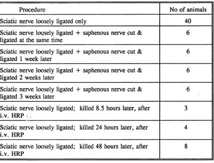
A morphological, behavioural and correlation study was carried out in rats using a method described by Bennett and Xie (1988) to produce neuropathic pain- related behaviour. This involved the placing of multiple loose ligatures around the sciatic nerve.
In this thesis, behavioural tests involving total foot immersion were used to confirm that hyperalgesia, allodynia and possibly spontaneous pain were produced. Section of the saphenous nerve at the time of, or within a week of, tying ligatures resulted in abolition of the early hyperaesthesia, suggesting that this nerve plays an important role in neuropathic pain-related behaviour from 4 to 12 days post operation.
The morphological study included a qualitative and quantitative investigation of the pathological changes produced within the nerve. It showed a variable amount of nerve fibre damage with a preferential loss of large diameter fibres in most cases and some loss of unmyelinated fibres. The variation is a reflection of the difficulty in tying ligatures with a consistent degree of ’tightness’.
The mechanisms of nerve damage caused by this mild lesion were studied from 8 hours post-operation. Morphological changes indicated that ischaemia played a major role within the first 24-48 hours. The presence of endoneurial oedema results in ’self strangulation ’ of the nerve, and compression is the overwhelming effect subsequently.
ACKNOWLEDGEMENTS
First of all I should like to thank my Supervisor, D r Jean M Jacobs, for her incessant help and her considerate guidance in the field which I did not know before; Professor L W Duchen for accepting me into the department and his support and encouragement; Professor Francesco Scaravilli, Dr Stephen Britland and Dr Nazar Alsanjari for many helpful discussions; Henry Goulding for care of the animals; Andrew Beckett and Hilary Ayling for technical support; Steve Durr for help with the photography; Mrs M Beryl Bailey (Librarian) for seeking references; Ms A Petruckevitch for advice on statistical methods; and Miss Janet Simpson for much help in the preparation of the manuscript, tables and figures.
This work was supported by a grant from the Ministry of Education, Taiwan, Republic of China.
ABSTRACT ... 2
ACKNOWLEDGEMENTS
... 4TABLE OF CONTENTS... 5
CHAPTER 1: REVIEW OF LITERATURE... 16
NO RM AL STRUCTURE O F PERIPH ER A L NERVES ... 16
MYELINATED FIBRES ... 17
THE SCHWANN CELL ... 19
UNMYELINATED F IB R E S ... 19
THE A X O N ... 20
AXONAL T R A N S P O R T ... 23
Anterograde tran sp o rt... 23
Slow tra n sp o rt... 23
Fast tr a n s p o r t ... 24
Retrograde transport ... 24
Slow tra n sp o rt... 24
Fast tr a n s p o r t ... 24
THE BLOOD SUPPLY OF PERIPHERAL NERVES ... 27
Ischaemia in peripheral nerves ... 29
PHYSIOLOGICAL CLASSIFICATION O F NERVE F I B R E S ... 30
TRANSMISSION O F TH E NERVE IM PULSE ... 31
AXONAL D E G E N E R A T IO N ... 32
AXON CHANGES IN THE PROXIMAL S T U M P ... 35
VASCULAR PERMEABILITY CHANGES ... 35
NERVE R E G E N E R A T IO N ... 35
NERVE CELL REACTION AFTER NERVE FIBRE D A M A G E ... 38
COM PRESSIVE NERVE INJURY ... 39
ACUTE NERVE I N J U R Y ... 39
Class 1 or N e u rap ra x ia ... 39
Class 2 or Axonotmesis ... 40
Classes 3,4 and 5 or Neurotmesis ... 40
CHRONIC NERVE C O M P R E S S IO N ... 41
TH E RELA TIO N SH IP O F INJURIES O F TH E PERIPH ER A L
NERVOUS SYSTEM W ITH PAIN ... 43
AFFERENT FIBRES ... 43
Large (6-22/xm) myelinated fibres (Aa and A B ) ... 43
Small myelinated (2-5/xm) fibres ( A ô ) ... 44
Unmyelinated afferent (0.3-2.0jum) axons (C f i b r e s ) ... 44
THE EFFECT OF INJURY ON A X O N S ... 45
Early physiological c h an g e ... 46
Morphological change ... 46
Later physiological c h a n g e s ... 46
Ongoing activity ... 46
Mechanical s e n s itiv ity... 47
Sensitivity to adrenalin... 47
Collateral s p ro u tin g ... 47
INJURY OF CENTRALLY DIRECTED A X O N S ... 48
Electrical activity in the dorsal root ganglion ( D R G ) ... 48
Changes in peptides and other substances... 49
THE EFFECT OF PERIPHERAL INJURY ON CELLS IN THE SPINAL C O R D ... 49
Gate control t h e o r y ... 52
Afferents ... 52
Interneurons within the dorsal h o r n... 53
Descending c o n tr o ls... 53
Later changes of spinal c o r d ... 56
CENTRAL PLASTICITY IN NOCICEPTIVE P A I N ... 56
Central plasticity in neuropathic pain ... 56
Phantom p a i n... 56
Denervation hypersensitivity ... 57
Prolonged central changes triggered by nerve im p u ls e s... 57
Expansion o f the receptive fields o f dorsal horn neurons ... 57
Noxious stimulus-induced neurochemical mediators in central p la s tic ity ... 58
C-fibre neuropeptides ... 58
Excitatory amino a c id s... 58
Molecular mechanisms of noxious stimulus-induced CNS p la s tic ity ... 59
Expression o f c-fos and other p ro to -o n co g en es... 59
Relationship between c-fos, central neuroplasticity and hyperalgesia ... 60
S U M M A R Y ... 60
CHAPTER 2: MATERIAL AND METHODS
... 62EX PERIM EN TA L PROCEDURES ... 63
SURGICAL PR O C E D U R E S... 63
A n a esth e sia ... 63
Placing of ligatures ... 63
Saphenous nerve s e c t i o n ... 63
EXAMINATION OF A N IM A L S ... 63
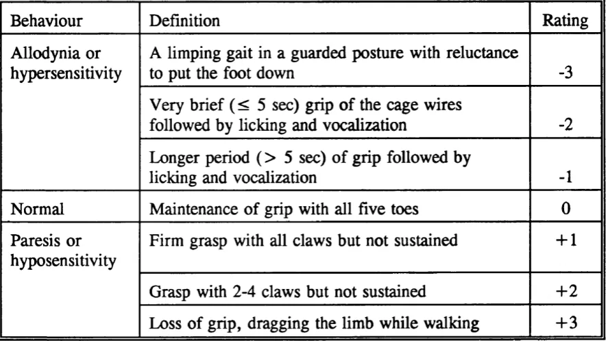
Observation of posture and gait: assessment on a semiquantitative scale ... 64
Tests using thermal stim u latio n ... 65
H e a t... 65
C o ld... 6 6 Measurement ... 67
M O R PH O L O G IC A L M E T H O D S ... 69
FIXATION ... 69
F IX A T IV E S ... 69
PROCESSING ... 70
NERVES FOR TEASING ... 71
STAINING PROCEDURES ... 71
SELECTION OF B L O C K S... 72
TEASED FIBRE PREPARATIONS ... 72
ULTRATHIN SECTIONS ... 73
HRP M E T H O D ... 73
Measurement of number and density of myelinated axons . . . . 74
Measurement of number and density of unmyelinated axons . . 77
Statistical methods and f o r m u la e ... 77
Behavioural s tu d ie s...
77
Morphological co rrela tio n s... 77
CHAPTER 3: RESULTS... 79
C L IN IC A L OBSERV ATION O F N E U R O PA TH IC PA IN FU L BEHAVIOUR AFTER TYING LOO SE LIGATURES ROUND TH E SCIATIC NERVE . 79 OBSERVATION OF GAIT AND PO ST U R E ... 79
Chronic loose ligatures o n l y ... 79
Chronic loose ligatures 4- concurrent saphenous nerve section ... 79
Chronic loose ligatures + saphenous nerve section 7 days l a t e r ... 80
Chronic loose ligatures + saphenous nerve section 14 and 21 days l a t e r ... 80
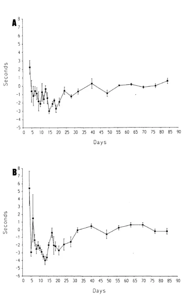
TESTS USING THERMAL STIMULATION ... 81
Chronic loose ligature only ... 81
Thermal stimulation at 48°C ... 81
Thermal stimulation at 48° C ... 81
Thermal stimulation at 6 °C ... 81
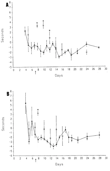
Chronic loose ligatures + saphenous nerve section 7 days l a t e r ... 82
Thermal stimulation at 48°C ... 82
Thermal stimulation at 6 °C ... 82
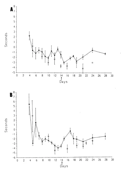
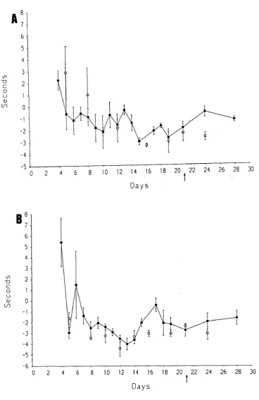
Chronic loose ligatures + saphenous nerve section 14 and 21 days l a t e r ... 82
Sham operated rats ... 82
A u to to m y ... 83
S U M M A R Y ... 83
MORPHOLOGICAL EXAMINATION OF SCIATIC NERVES AFTER PLACING LOOSE L IG A T U R E S ... 92
CHANGES UP TO 48 HOURS p .o ... 92
8.5 hours p .o ... 93
Total fascicular a r e a s... 93
Epineurial c h a n g e s... 93
Changes within the endoneurium ... 93
24 hours p .o ... 96
Total fascicular a r e a s... 96
Epineurial c h a n g e s... 96
Changes within the endoneurium ... 96
48 hours p .o ... 97
Changes within the endoneurium ... 98
CHANGES FROM 5-14 DAYS p .o ...136
Qualitative c h a n g e s ...136
21- 28 days p .o ...137
Sham o p eratio n... 139
Tissue response to the lig a tu re ... 139
H R P ...139
Quantitative c h a n g e s ... 141
Myelinated fibres ...141
Estimation of ’g ’ ratios ... 142
Estimation o f the density o f unmyelinated a x o n s...142
CORRELATION OF NEUROPATHIC PAINFUL BEHAVIOUR WITH SURVIVING NERVE FIBRES ... 179
The correlation o f sural (sensory) nerve fibres with PRB . . . . 179
The correlation o f sciatic nerve (mixed nerve) fibres with P R B... 180
Correlation o f neuropathic painful behaviour with acute myelinated fibre degeneration ...180
C H A P T E R
4:
D I S C U S S I O N ... 190Neuropathic Pain-Related Behaviour ... 190
Relevance of the M o d e l ...192
Comparison with other m o d e ls ... 194
Sham O p e ratio n ...197
The Role of Saphenous N e r v e ... 198
Morphological changes ... 200
Comparison with other studies o f the Bennett and Xie model . . 200
Compressive le s io n s... 205
Tissue response to the lig a tu res...207
Vacuolated fibroblasts ...208
Possible causes o f neuropathic p a i n ... 209
Correlation s t u d i e s ...210
SUGGESTIONS FOR FURTHER STUDY OF THE BENNETT AND XIE M O D E L ...217
C-fos protein and pain-related b e h a v io u r ... 217
Nerve growth factor (NGF) and pain-related su b stan ces 218
CHAPTERS: CONCLUSIONS...220
APPENDICES... 221
REFERENCES
... 234INTRODUCTION
The use o f multiple loose ligatures around the rat sciatic nerve has been proposed as an animal model for the study of causalgia, allodynia and spontaneous pain in man (Bennett and Xie, 1988). Other studies (Attal et al., 1990, Sommer et al., 1993) have confirmed the behavioural changes caused by this procedure.
Pain-related behaviour was examined using a method involving immersion of the whole foot of the rat. This region is innervated not only by the sciatic nerve (the lateral part of the foot), but also by the saphenous nerve (the medial area o f the foot).
It is known that after complete interruption o f the sciatic nerve, the spread of receptive fields of the saphenous nerve begins within a few days (Devor et al., 1979). The spread of sensitization of polymodal nociceptors from a nearby injury is also found (Fitzgerald, 1979). It was thought likely that the saphenous nerve could play a role in pain-related behaviour since the nearby sciatic nerve is injured by the multiple loose ligatures. Therefore, the role of the saphenous nerve in pain-related behaviour formed a major part of the behavioural study.
This thesis includes an account of the investigation of the initial cause of a severe nerve lesion produced by an apparently mild procedure. The ligatures were tied round the nerve loosely enough not to cause compression o f the nerve but sufficiently tightly to restrict blood flow in some epineurial vessels.
A quantitative assessment of nerve damage was made, both to correlate this with the behavioural changes, but also to demonstrate the great variation in amount of damage due to the difficulty in applying ligatures of equal ’tightness*.
The aims of the present study are :
1. To study and quantify the neuropathic painful behaviour caused by placing loose ligatures around the rat sciatic nerve.
2. To study the contribution of the saphenous nerve to pain-related behaviour caused by loose ligatures.
3. To study and quantify the pathology of this lesion.
CHAPTER 1: REVIEW OF LITERATURE
NO RM AL STRUCTURE O F PER IPH ER A L NERVES
Peripheral nerves of vertebrates consist of one or more fascicles containing bundles of nerve fibres enclosed by a perineurial sheath. The fascicles are embedded in a loose connective tissue called the epineurium. Individual nerve fibres lie within an intrafascicular space, the endoneurium, which is largely composed of longitudinally orientated collagen fibrils and of endoneurial fluid. Other cellular components of the intrafascicular compartment include Schwann cells (about 90% of endoneurial cells), fibroblasts (about 5% or less), endothelial cells, smooth muscle cells and pericytes of endoneurial blood vessels and occasional mast cells and macrophages (Oldfors, 1980).
The perineurium is the sheath enclosing the nerve fibres. It is composed of specialized, flattened, circularly orientated cells forming discrete lamellae, each covered by a basal lamina. The number of lamellae varies directly with the size of the fascicle. In between these lamellae are collagen fibrils which are arranged in circular, longitudinal and oblique orientations. Pinocytotic vesicles are a characteristic feature of perineurial cells, and they suggest the presence of a transport system across the cells.
The perineurium is a component of the blood-nerve barrier. Blood vessels in the epineurium are of a non-specialised type, and are freely permeable, whilst those in the endoneurium have tight junctions forming a barrier, similar to but less ’tight’, than that in the brain. The perineurial lamellae, also joined by tight junctions, separate these two regions of differing vascular permeability.
M YELINATED FIBRES
During development, axons of peripheral nerves grow out from neurons in the central nervous system and ganglia in association with Schwann cells which initially multiply as their associated axons lengthen. Some axons become associated with a single Schwann cell and these are destined to become myelinated fibres. The Schwann cells become spaced out along the axons at intervals of 200-300/xm. Myelination begins by elongation of the mesaxon, an extension of the Schwann cell plasma membrane formed by its envelopment of the axon. The elongated mesaxon wraps spirally round the axon, and the membranes compact to form the myelin sheath (Geren, 1954). Mammalian myelinated fibres range in size from about 3-20/im.
is considerably reduced at the node compared with its cross sectional area in the intemodal region.
The myelin sheath is interrupted at intervals along its length by obliquely arranged Schmidt-Lanterman incisures. These are regions at which the myelin lamellae open up at the major dense line to contain small amounts of Schwann cell cytoplasm. Thus a continuous spiral of cytoplasm is formed connecting the cytoplasmic compartments internal and external to the myelin sheath. The number of Schmidt-Lanterman incisures along an intemode is directly related to the size of the fibre (Hiscoe, 1947). The incisures are the site of early changes seen in myelinated fibres undergoing Wallerian degeneration when the sheath segments into ovoids.
The intemodal length of myelinated fibres is directly related to the size of the fibre; in mammalian nerves the range is from about 200-1500^m, the maximum length being determined by the length of the nerve. For example maximum intemodal length in a limb nerve such as the sciatic is much greater than in a short nerve such as the facial.
TH E SCHWANN CELL
The largest concentration of Schwann cell cytoplasm lies in the region of the nucleus; elsewhere along the intemode it is present as very attenuated threads of cytoplasm internal and external to the myelin sheath, joined across the sheath through the Schmidt-Lanterman incisures (see above). In the paranodal region a spiral of cytoplasm extends through the opened out loops o f myelin as they terminate on the axon, and the Schwann cell itself terminates on the axon at the node of Ranvier in a series of finger-like processes. In large fibres a quantity o f cytoplasm occupies the depressions made by the fluting of the myelin sheath in the paranodal region, and these contain large numbers of mitochondria. In fact, in all fibres, the paranodal region can usually be recognised by the increased number o f small mitochondria in Schwann cell cytoplasm. The mitochondria are thought to provide a source of energy for the maintenance of the ionic changes that take place at the node of Ranvier in relation to nerve impulse transmission.
UNMYELINATED FIBRES
generally circular contour; their envelopment by the processes of another cell; the absence of rough endoplasmic reticulum or ribosomes; generally, the more conspicuous content of microtubules.
TH E AXON
The axonal cytoplasm (axoplasm) is a more or less cylindrical extension o f the nerve cell body bounded by a cell membrane, the axolemma. The axoplasm contains microfilaments (5-7nm), neurofilaments (8-1 Inm), neurotubules (hollow cylinders, 23-25nm in diameter), mitochondria, smooth endoplasmic reticulum (SER) and some secondary lysosomes. Dense-core vesicles and coated vesicles may be seen occasionally. Axoplasm lacks ribosomes, rough endoplasmic reticulum and golgi membranes (Lgmdon and Hall, 1976; Thomas and Ochoa, 1984; Thomas, Landon and King, 1992).
Neurofilaments (NF) are longitudinally orientated and of indefinite length. NF content has been correlated with area of axons in the PNS and CNS (Friede and Samorajski, 1970; Friede, Miyagishi and Hu, 1971; Hoffman, Griffin and Price, 1984; Price, Lasek and Katz, 1990; Gold et al., 1991), in unmyelinated and myelinated fibres and in various regions of the same axon. Under normal conditions NF density (100-300//im^) remains relatively stable and axonal area changes proportionately to the NF content of an axon (Friede, Miyagishi and Hu, 1971; Berthold, 1978; Price, Lasek and Katz, 1990).
N F ’s consist of 3 protein subunits which have molecular weights of 6 8KDa (NF-L), 155 KDa (NF-M) and 200 KDa (NF-H) (Hoffman and Lasek, 1975; Sharp, et al., 1982; Mata et al., 1992). These are synthesized in the cell body and rapidly assembled into polymers (Lasek, Oblinger and Drake, 1983). The 200KDa (NF-H) subunits appear to be associated with the sidearms that can be seen extending from neurofilaments. The sidearms may influence the spacing of filaments, and are also potential sites of phosphorylation. Although NFs are poorly phosphorylated in the neuronal perikaryon, they are highly phosphorylated in axons (Bennett and DiLullo,1985; Hart, Nuckolls and Wood, 1987; Lee et al., 1987; Pestronk, Watson and Yuan, 1990).
NFs are delivered to the axon in the slow component of axonal transport (Hoffman and Lasek, 1975; Black and Lasek, 1980) at rates of several mm/day. Although neurofilaments appear to be highly stable polymers, it is possible that they are capable of undergoing reversible assembly and disassembly.
Ultrastructurally they appear to be unbranched 23-25nm hollow cylinders, which according to Bray and Bunge (1981) are lOO-SOO/Ltm in length, although other sources (Thomas, Landon and King, 1992) suggest that they are o f indefinite length. They are composed o f globular subunits of the protein tubulin. This has been shown to exist in two distinct monomeric forms, a and 8, having molecular weights o f 57 and 54KDa respectively, each microtubule being composed of helically arranged chains of alternating a and J1 monomers. The tubulin subunits have a definite orientation, and are arranged so that their (+ ) ends are away from the cell body. Evidence from studies on the effects of agents causing depolymerisation of microtubules, such as colchicine, led to the idea that these organelles were involved in axonal transport. Later, more direct evidence using special microscopic techniques, showed movement of membrane-bound organelles along microtubules. Theories of fast transport mechanisms involve other force-generating proteins, such as kinesin and dynein, which are discussed later.
The number of microtubules in myelinated fibres varies inversely with axon size, ranging from 30-40//>im^ in small axons to 10-15/fcm^ in large axons. Unmyelinated axons contain a proportionately larger numbers of microtubules, with a density of 50-100//xm^ (Friede and Samorajski, 1970). The number of tubules in the terminal branches of some large mammalian axons is many times greater than the number found in the parent axon (Zenker and Hohberg, 1973).
reticulum appears as a continuous system of tubules extending from the perikaryon to the axon terminal. Narrow and wide tubules of SER appear occasionally to be closely apposed to the axolemmal and presynaptic membrane. It is postulated that the exchange of fast axonally transported macromolecules takes place at the contact between the subaxolemmal plate and the SER tubules. At the distal tip of thin tubular branches of SER, synaptic vesicles appear as blebs which may originate either by fission from, or fusion with the SER.
AXONAL TRANSPORT
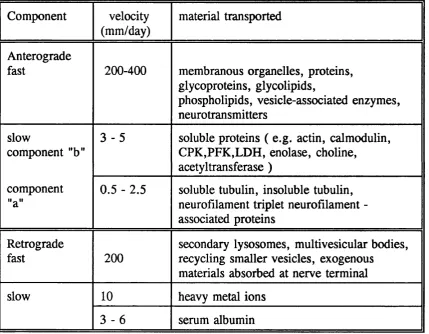
Somatofugal or anterograde transport mechanisms have been the most extensively studied (Schwartz, 1979; Grafstein and Forman, 1980; Ochs, 1982) and have been found to operate at a minimum of two and probably in excess of five different rates which are divided into a slow transport component moving at 0.25- 4mm/day, and a fast transport component which may reach speeds in excess of 400mm/day (Table I.l) . Axonal transport has recently been reviewed by Ochs and Brimijoin (1993).
A nterograde tran sp o rt
Slow transport
may cause alterations in axonal calibre.
Fast transport
Fast anterograde transport is energy utilizing and ATP dependent. It transports mainly mitochondria and transmitter storage vesicles (i.e.transmitter- synthesizing enzymes, glycoprotein and membrane components) (Droz et al., 1979; Griffin et al., 1981).
R etrograde tran sp o rt
Labelled proteins carried anterogradely have been shown to undergo a turnaround at their distal end and are then carried back towards the nerve cell body by retrograde transport. Retrograde transport is of considerable interest in studies of nerve regeneration following injury (Forman, 1983). Their return to the cell body also probably provides a part of the necessary, feedback signal which will enable the cell to modulate the balance of its anabolic and catabolic activity in response to the needs of its far distant axon terminals.
Slow transport.
A slow component of retrograde transport has been described in mammalian peripheral nerve fibres (Gainer and Fink, 1982). The component moves at 3- 6mm/day, and has been tentatively identified as serum albumin. Heavy metal ions may also use this component at a rate of some lOmm/day (Baruah et al., 1981).
Fast transport
absorbed at the nerve terminal (Holtzman, 1971; Bunt and Hasche, 1978). Nerve growth factor also reaches developing sensory dorsal root ganglion cell and sympathetic nerve cell bodies via retrograde fast transport (Hendry et al., 1974; Raivich and Kreutzberg, 1987; Raivich, Hellweg and Kreutzberg, 1991), by active receptor-mediated absorptive pinocytosis (Dumas, Schwab and Thoenen, 1979). Possible mechanisms of fast tran sp o rt
One suggestion is that movement is generated by interactions between actin and myosin, both of which are known to be present within neurons (Trifaro, 1978), and that this may result in cytoplasmic streaming capable of transporting particulate components (Isenberg, Schubert and Kreutzberg, 1980). Support for this is mechanism is provided by the requirement for calcium ions (Ochs, 1983), and by the observations that the substance cytochalasin B is able to inhibit cell movements including neurite growth cones, and axonal fast transport systems.
An alternative explanation is that microtubules are capable of generating movement of adjacent structures, either by interacting with them directly (Schmitt and Samson, 1968) or via an intermediate carrier mechanism (Smith, 1970; Ochs, 1971a, b, 1982). To isolate the essential components, motility assays were developed; kinesin, a protein isolated from mammalian brain and squid neuronal tissue was found to be necessary to support anterograde transport. Kinesin and the related protein, dynein (Sheetz et al., 1987; Schroer and Sheetz, 1991; Toyoshima et al., 1992) have also been shown to be associated with brain microtubules.
(Howard, Hudspeth and Vale, 1989). At the fanned-out tails of heavy chains are folded light chains. A region of bending, the hinge, is found along the main shaft of the kinesin structure. The role of kinesin has been demonstrated in the movement of lysosomes, melanophores and axonal vesicles (Schroer et al., 1988; Urrutia et al.,
1991).
Cytoplasmic dynein is a microtubule-based, mechano-chemical ATPase found in virtually all animal cells (Koonce and McIntosh, 1990; Verde et al., 1991). Ultrastructurally and biochemically similar to axonemal dynein, cytoplasmic dynein is a microtubule-activated ATPase that powers movement toward the minus ends of tubulin filaments (Paschal e ta l., 1987). Dynein may function as a force producer for neurite extension and other microtubule-dependent cytoplasmic rearrangements. How the force-generating proteins actually produce the movement of organelles is as yet speculative. A proposed model is that when the hinge region of the shaft straightens, it rolls the organelle forward to the next kinesin chain which then straightens to hand it on to the next kinesin, and so on (Hirokawa et al., 1989). Another transport model proposes that the carrier is a filament which by bridging cross-arms to microtubules could apply more force to the carrier which would then allow movement of larger structures. Additionally, with several cross-arms, a direction would be given to the movement of the carrier along the microtubules. Cytoplasmic dynein plays a role in axonal transport which is similar to that proposed for kinesin.
aspects of the microtubule. Alternatively, kinesin and cytoplasmic dynein both attach to the same organelle, the direction of transport being determined by some accessory factors activating either kinesin or cytoplasmic dynein.
Table I .l . Different forms of axonal transport (Modified from Tomlinson (1988)).
Component velocity
(mm/day)
material transported
Anterograde
fast 200-400 membranous organelles, proteins, glycoproteins, glycolipids,
phospholipids, vesicle-associated enzymes, neurotransmitters
slow
component "b"
3 - 5 soluble proteins ( e.g. actin, calmodulin, CPK,PFK,LDH, enolase, choline,
acetyltransferase ) component
"a"
0.5 - 2.5 soluble tubulin, insoluble tubulin, neurofilament triplet neurofilament - associated proteins
Retrograde
fast 2 0 0
secondary lysosomes, multivesicular bodies, recycling smaller vesicles, exogenous
materials absorbed at nerve terminal
slow 1 0 heavy metal ions
3 - 6 serum albumin
T H E BLOOD SUPPLY O F PERIPH ER A L NERVES
longitudinally orientated microvessels lying in the endoneurial space of the nerve. An abundant network of interconnecting epineurial and perineurial vessels link the two systems. This rich anastomosis results in the high resistance to ischaemia displayed by peripheral nerves.
Because the peripheral nerve is extremely long and far from its parent cell body, it is exquisitely dependent on nerve microenvironment for its blood supply, oxygenation and nutrition and for the removal of toxic metabolic products. McManis, Low and Lagerlund (1993), in a review of the endoneurial micro-environment, point out several characteristic properties of peripheral nerve vasculature. The first, is a lack of autoregulation; this is normally achieved by varying arteriolar tone and is dependent on myogenic rather than neurogenic mechanisms. The vessels in rat nerve are mainly capillaries of large diameter (Bell and Weddell, 1984a); the arterioles have a poorly developed smooth muscle layer (Bell and Weddell, 1984b). The second feature is the greater intercapillary distance in nerve compared to other tissues such as muscle. This would tend to make perfusion of the endoneurium inefficient. A third feature is based on observations by Nukada, Dyck and Karnes (1985) that capillary density is greater in the superineurial region of a fascicle than in the centre. This morphological difference could account for the relative resistance to ischaemia of the subperineurial region because the shorter diffusion distance provides a greater safety margin.
stores, and the low resting and maximal energy expenditure enable nerve to function well on anaerobically generated high energy phosphate.
Ischaem ia in peripheral nerves
Mild to moderate degrees of ischaemia cause no pathological changes in peripheral nerves. Severe reduction in blood flow in human neuropathies may produce infarction of the centre of fascicles with large diameter fibres, or wedge-shaped infarcts (Dyck, Conn and Okazaki, 1972). In sural nerve biopsies there may be degeneration of all myelinated and unmyelinated fibres.
Experimental models of ischaemic neuropathy require extensive ligation of limb arteries, demonstrating the resistance of peripheral nerve to ischaemia. An effective model was developed by Korthals and Wisniewski (1975) involving tying the abdominal aorta and femoral artery of one limb of the cat. This caused extensive lesions in the distal sciatic nerve and its branches. Sampling of many levels of these nerves showed that between 5 and 12 hours after ligation a zone of abnormal myelinated fibres could be delineated (Korthals, Korthals and WiSniewski, 1978). The axons o f the proximal part of this zone contained large numbers of organelles including mitochondria, dense bodies and vesicles. The distension of axons caused by these organelle accumulations resulted in thinning or loss of the myelin sheaths. More distally was a zone in which fibres showed early changes of Wallerian degeneration; this zone eventually became necrotic, with evidence o f degeneration o f Schwann cells, endoneurial cells and cellular elements of the blood vessels. In some animals, a zone of fibres with axonal organelle accumulations also formed distal to the necrotic region.
it appears to be quite different from that seen near to a crush lesion. Korthals, Korthals and Wisniewski (1978) suggest that initially lack o f energy required for fast axonal transport is the cause of organelle accumulation.
In another study by McManis and Low (1988) individual nutrient branches of the extrinsic system were occluded, causing a regional reduction in nerve blood flow (NBF) with no selectivity for subperineurial or centrifascicular regions. This suggests that longitudinal anastomoses are not as efficient as the regional radial anastomoses. Blunt and Stratton (1956), in a study of the rabbit sciatic nerve, emphasized the importance of regional blood supply at the lower end of the nerve. The abundance of nutrient vessels in the proximal segments of the sciatic nerve provide a head of pressure for the intrinsic system that has decreased by the time it reaches the more distal part of the nerve, which then becomes increasingly dependent upon the regional nutrient arterioles.
PH YSIOLO G ICAL CLASSIFICATION O F NFTIVE FIBRES
Mammalian nerve fibres, on the basis of their electrical properties, have been subdivided into three groups. A, B and C. Group A includes the largest fibres with the fastest conduction velocities (i.e. the myelinated somatic afferent and efferent fibres); Group B includes smaller, myelinated preganglionic fibres; and group C is composed o f the smallest diameter slowly conducting fibres, the unmyelinated visceral and somatic afferent fibres, and the postganglionic autonomic efferent fibres. Group A fibres have been further subdivided into four efferent groups a , B, 7 and ô in order o f descending conduction velocity.
50-100 m/s.
AB: Fusimotor and skeletomotor fibres; 9-15^m: 50-85 m/s.
A 7 : Exclusively fusimotor fibres; 4.5-8.5ptm; 20-40 m/s (Boyd and Davey, 1968).
Later, a second classification system was based on the sensory nerves, subdividing them
into:-Group la: Primary sensory fibres to muscle spindles (12-22/xm). Group lb: Smaller fibres of Golgi tendon organs.
Group II: Secondary sensory terminals of muscle spindles and cutaneous afferent receptors (6-1 2pim).
Group III: Free sensory endings in the connective tissue sheaths around and within muscles (1-6/xm). These appear to be nociceptive, related to ’pressure-pain’ in externally stimulated muscles (Lloyd, 1943).
Group IV: Unmyelinated fibres (0.2-3.9^m); include free endings in muscle, mainly nociceptive. This group also includes C fibres, postganglionic sympathetic and parasympathetic axons.
TRANSMISSION OF THE NERVE IMPULSE
channels are activated, an action potential current can depolarize adjacent segments o f the axolemma, resulting in a continuously propagated wave of depolarization which spreads along the nerve.
In unmyelinated axons the large capacitance of the axonal membrane attenuates forward axial flow of current within the axoplasm, and limits conduction to a speed of around one metre per second. In myelinated fibres, electrical activity is restricted to the nodes of Ranvier, with ionic activity leaping from one node to the next in series; this is described as saltatory conduction.
This mechanism of conduction can enhance conduction velocity to as much as 1 0 0 metres per second, depending on the axon diameter and thickness of the myelin sheath. Loss of one or more myelin segments due to injury or pathology will result acutely in conduction block, but the axon eventually develops the capacity to sustain continuous conduction by a mechanism similar to that occurring in normal unmyelinated axons (Bostock and Sears, 1978; Sears and Bostock, 1981).
AXONAL DEGENERATION
After a peripheral nerve injury such as nerve crush or transection, degeneration of the distal segment of the nerve takes place. Waller (1850) studied the sequence of events in the glossopharyngeal nerve of the frog and was the first to describe them, so giving his name to the process. Nerve degeneration was later described in great detail by Cajal (1928), who made use of silver stains to show both degenerative and regenerative changes.
interruption of the axon and breaking down of the axon into ovoids containing axonal debris. Ovoids were originally thought to be taken up first by a proliferating population of Schwann cells, but more recent studies indicate that the breakdown products are transferred to macrophages which appear in the endoneurium.
The importance of macrophages was demonstrated in elegant studies by Beuche and Friede (1984) who showed that there was no proliferation of Schwann cells and only very slow myelin breakdown if macrophages were prevented from entering an isolated degenerating peripheral nerve. This work also indicated that macrophages are predominantly derived from a blood-borne source.
As the fibres begin to degenerate, macrophages appear and there is Schwann proliferation in the distal part of the nerve. This begins from two to three days after nerve injury and reaches a peak at about three to four weeks (Abercrombie and Johnson, 1946; Bradley and Asbury, 1970). By this time, most of the breakdown products have been removed although small amounts of debris may persist for many weeks. Schwann cell proliferation was observed to occur within the persisting basement membranes, resulting in longitudinal columns of Schwann cells, called the bands of Büngner. The amount of Schwann cell proliferation is a reflection of the size of the degenerating fibres, and a correlation was noted between the degree of proliferation and the extent of myelination (Bradley and Asbury, 1970).
At least a dozen mitogens for Schwann cells have been identified; these include an axolemmal-iich fraction of the axon, myelin-related mitogens and many soluble mitogens such as fibroblast growth factor and platelet-derived growth factor (De Vries, 1993).
myelinated fibres take place in a nerve soon after nerve injury; and Schwann cells of both myelinated and unmyelinated fibres begin to synthesize nerve growth factor (NGF) and nerve growth factor receptor. These changes have been described briefly by Griffin and Hoffman (1993).
By electron microscopy the earliest axoplasmic changes, seen at about 12 hours, are the accumulation of mitochondria, multivesicular bodies and lamellar osmiophilic bodies at nodes of Ranvier (Webster, 1962; Ballin and Thomas, 1969). By 24 hours, the axoplasmic SER breaks up into rows of vesicles, followed by granular disintegration of the microtubules and neurofilaments and swelling of mitochondria (Schlaepfer and Micko, 1978). These changes are advanced at 48 hours, when the axons become filled with clumps of granular debris. The times given are approximate and are known to vary between species (Cajal, 1928).
Axonal disintegration is probably initiated by an increase in axoplasmic calcium concentration, and mediated by calcium-sensitive proteases (Schlaepfer,
1974).
AXON CHANGES IN TH E PROXIM AL STUMP
Immediately proximal to nerve section, reactive axon swellings develop within a few hours, consisting of accumulations of vesicular profiles (Lampert, 1967). A few days later, when axonal sprouting has begun, there may be a reduction in axon size just above the site of injury. Nerve conduction velocity and the size of the largest fibres were decreased in rabbit peroneal nerves proximal to several different types of nerve lesion causing distal fibre degeneration (Gragg and Thomas, 1961).
VASCULAR PERM EABILITY CHANGES
Protein tracers have been used to study permeability changes in nerves undergoing Wallerian degeneration. Mechanical lesions will cause an immediate leakage of vascular tracers from endoneurial blood vessels at the site of injury. At a later stage there is another wave of increased permeability in the distal part of the nerve which appears to follow the front of regenerating axons (Mellick and Cavanagh, 1968; Sparrow and Kieman, 1981). There do not appear to have been any studies on vascular permeability changes in axonal degeneration produced without direct trauma to the nerve. However, studies during the development of a demyelinating lesion, produced without trauma in a mutant mouse (twitcher) (Jacobs and Scaravilli, 1981a) showed that vascular permeability was increased at the first sign of myelin breakdown and the concurrent entry of macrophages into the nerve. It is likely that the process of axonal degeneration, with the entry of macrophages into the nerve, is also associated with increased vascular permeability.
NERVE REGENERATION
end o f the severed axons, or from nodes of Ranvier proximal to the site o f injury (Cajal, 1928). The growing tips of the regenerating axons are known as growth cones which can be recognised ultrastructurally by the large numbers of profiles of endoplasmic reticulum (Bunge, 1973).
When there is a small gap between the severed ends of the nerve, many regenerating axons, accompanied by Schwann cells, grow across the gap into the distal stump and invade the existing basement tubes filled with Schwann cells (bands of Büngner). This appears to be a random process, so that most axons will innervate inappropriate distal bands of Büngner. Regenerating axons will also grow out in all directions, some turning back on themselves and growing in a retrograde direction along the proximal stump (Cajal, 1928). The regeneration process after a nerve crush injury is more effective than after transection of the axons, because the regenerating axons are guided to their correct terminations through the continuous basement membrane lined tubes of Schwann cells in the distal stump.
During regeneration the process of myelination is similar to that during development. However, one proximal axon may give rise to many regenerating axons within the same basement membrane; myelination of more than one of these axons may take place leading to the formation of regeneration clusters.
size and intemodal length (Vizoso and Young, 1948). Schroder (1972) showed that the thickest regenerated fibres were thinner than the largest control fibres, and that 24 months after a nerve crush in the rat the myelin sheaths were still thinner than normal. A linear relationship has been shown between myelin volume and intemodal axonal mantle area (Friede and Bischhausen, 1980; Smith et al., 1982), therefore short intemodes would be expected to have thinner myelin sheaths.
In the regeneration of unmyelinated fibres, large groups of unmyelinated axon sprouts are associated with Schwann cell processes within a common basement membrane. In regenerating nerves with a mixed population of myelinated and unmyelinated axons, King and Thomas (1971) suggested that regenerating unmyelinated axons are diverted and come to be preferentially associated with regenerating myelinated axons.
The study of growth factors is now an important part of the study of nerve regeneration. Macrophages, apart from their function in the removal of debris, also induce the secretion of NGF by Schwann cells. There is a dramatic fifty-fold reexpression of low-affinity nerve growth factor (NGF) receptors on Schwann cells within bands of Büngner in the distal segment during the first two weeks after nerve transection. (Taniuchi, Clark and Johnson, 1986, 1988; Johnson, Taniuchi and DiStefano, 1988). In line with the general concept that growth factors induce the synthesis of their own receptors, it has been suggested that ’reactive’ Schwann cells produce NGF and subsequently sequester it on the induced receptors, providing a favourable substrate over which NGF-dependent (sensory) axons can grow (Taniuchi, Clark and Johnson, 1986; DiStefano and Johnson, 1988). Evidence from in vitro
myelin-derived mitogen for Schwann cells (Baichwal, Bigbee and De Vries, 1988); it is also possible that a product of degraded myelin may induce the secretion of a soluble Schwann cell mitogen by macrophages.
A sequence of events during Wallerian degeneration was suggested by Johnson, Taniuchi and DiStefano (1988) involving firstly the expression of NGF receptors on the Schwann cell surface, and also the release of NGF which is taken up by low affinity receptors and concentrated on the Schwann cell surface (Raivich, Hellweg and Kreutzberg, 1991). As axons of sensory and sympathetic neurons invade bands of Büngner, they are guided over the Schwann cell surface by the binding of NGF. The expression of NGF and NGF receptors is then suppressed by axonal contact, so that the Schwann cell is ahead of the growth cones that possess NGF, giving a directionality to axon elongation.
NERVE CELL REACTION AFTER NERVE FIBRE DAMAGE
there is proliferation of smooth endoplasmic reticulum, Golgi bodies and neurofilaments. The rate of response of the cell body to axon damage is related to the distance between the cell and the axon injury, suggesting the passage of a signal from the injured axon (Kristensson, 1984). Chemical changes were described by Brattgârd, Edstrom and Hydèn (1957) and these led to the interpretation of the morphological changes as regenerative rather than degenerative phenomena.
COMPRESSIVE NERVE INJURY ACUTE NERVE INJURY
Acute nerve injury may be caused by sudden compression, transection or stretching. Several types of injury can be identified and these have been classified by two different systems.
Class 1 or Neurapraxia
Remyelination occurs rapidly and functional recovery is usually complete by about 6 weeks after the injury. Ochoa, Fowler and Gilliatt (1972) concluded that the lesion is caused by mechanical processes.
Class 2 or Axonotmesis
This lesion is associated with percussion and closed crush injuries. The axons are interrupted, but the Schwann cell basement membrane around each fibre remains intact, as does the endoneurial connective tissue (Thomas, 1964). Wallerian degeneration occurs distal to the lesion site, but regeneration proceeds at a rate of about l-2mm per day. All functions are eventually restored since regenerating axons are guided back to their original terminations through the continuous basement- membrane-contained Schwann cells that develop distally following Wallerian degeneration (Haftek and Thomas, 1968).
Classes 3,4 and 5 or Neurotmesis
neuroma at the site of injury (Spencer, 1974). Neuroma formation may be prevented by careful surgical anastomosis of the divided ends of the injured nerve, either as individual fascicles or of the whole nerve.
Clinically, injuries associated with partial or complete nerve transection produce clearly defined areas of total motor, sensory and autonomic dysfunction. Denervated muscle may lose 80% of its bulk, but function can be recovered if reinnervation takes place within three years of transection. Shrinkage of the autonomous sensory zone of a nerve, its area of superficial cutaneous sensation, follows within days of complete nerve section. This phenomenon reflects the overlap of normal innervation; it is important to recognise that this does not reflect regeneration.
CHRONIC NERVE COMPRESSION
Three types of injury are commonly associated with chronic trauma to peripheral nerve.
1. Compression in fibro-osseous tunnels (carpal, cubital or tarsal tunnels) 2. Angulation and stretch over arthritic joints, anomalous fibrous bands, under
ligaments
3. Recurrent external compression e.g. occupational trauma to hands and feet As far as the tunnel syndromes (Type 1) are concerned, histopathological changes in human and experimental animals have been well studied but little is known of Type 2 and 3 injuries.
Neary, Ochoa and Gilliatt, 1975). The morphological features of demyelination in chronic nerve entrapment differ markedly from those in acute compression. A naturally occurring nerve entrapment is found in the guinea pig, and studies of the compressed nerves show that the myelin sheath becomes deformed, with a bulbous swelling at one end of an intemode and tapering of myelin at the opposite end (Ochoa and Marotte, 1973). The bulbous ends are orientated away from the site of compression, indicating the direction of the mechanical force. The myelin sheaths degenerate and bare axons are remyelinated. In older animals, there is eventual axonal degeneration (Fullerton and Gilliatt, 1967). Unmyelinated fibres resist degeneration until late.
Mechanical factors appear to be dominant in the demyelination of chronic compression. The role of ischaemia is less certain, but it may be responsible for the acute attacks of pain suffered in the carpal tunnel syndrome. Short periods of ischaemia can reversibly block conduction in damaged fibres (Fullerton, 1963).
NEUROPATHIC PAIN AND ITS BACKGROUND
Neuropathic pain is pain associated with abnormalities of the peripheral nervous system. This particular aspect of pain will be discussed since it is most relevant to the topic of this thesis. A number of clinical features of neuropathic pain can be recognised in man. These
include:-Pain in the absence of detectable ongoing tissue damage Dysesthesiae, often of burning quality
Paroxysmal shooting or stabbing component Mild innocuous stimuli cause pain (allodynia)
Pronounced summation and after-reaction with repetitive stimuli
In man, not all of these features will be present, but any single one should lead to the suspicion that the pain is neuropathic.
In the rat model of neuropathic pain which is the subject of this thesis, both allodynia and pain in the absence of apparent ongoing tissue damage can be recognised.
Wall (1989) has extensively reviewed the topic of pain, and this is the source of part of this literature overview.
T H E RELA TIO N SH IP O F INJURIES O F T H E PERIPH ER A L NERVOUS SYSTEM W ITH PAIN
Peripheral nerves contain nerve fibres subserving many functions, including motor, autonomic and sensory. It is the latter group which is of the greatest importance in the study of pain; these include large and small myelinated fibres and unmyelinated axons.
AFFERENT FIBRES
Afferent fibres of all sizes may contribute to neuropathic pain. Large (6-22^m) m yelinated fibres (Aa an d AB)
observations which led to the gate control theory (Melzack and Wall, 1965). Support for the role of A a fibres in pain comes from clinical observations that selective electrical stimulation of large diameter primary afferent fibres can give striking relief to the burning pain caused by some forms of peripheral nerve injury (Wall and Sweet, 1967; Meyer and Fields, 1972).
If the electrical stimulus is raised to include the smaller fibres (AÔ) of this group, the inhibition of painful sensation and of the flexor reflex to noxious inputs is replaced by a facilitation (Wilier et al., 1980). In tender areas distant from injury, light mechanical stimuli (A-13 activated) may evoke pain (Kugelberg and Lindblom, 1959; Raja, Campbell and Meyer, 1984; Campbell et al., 1988).
Small myelinated (2-5^m) fibres (A6)
Myelinated nociceptors are included in this group; they respond to noxious mechanical stimuli and are called high threshold mechanoreceptors (HTM’s). A significant proportion (20-50%) of Aô fibres also respond to heat stimuli (mechanothermal nociceptors). Some of these respond to skin temperatures below pain threshold, but all respond as temperatures are raised into the noxious range (45- 47®C), and some also respond to cooling of their receptive fields. Both HTM ’s and mechanothermal nociceptors have the property of sensitization, showing increased sensitivity with repeated stimulation (Fitzgerald and Lynn, 1977; Campbell and Meyer, 1983).
Unmyelinated afferent (0.3-2.0/tm) axons (C fibres)
found to be nociceptive. Most C fibres are polymodal (C-PMN), responding to noxious thermal, mechanical and chemical stimuli applied to the skin. Like the myelinated nociceptors, C-PMN’s sensitize with repeated noxious stimuli. Selective stimulation o f C fibres produces pain (Collins, Nulsen and Randt, 1960; Sinclair, 1981).
There is now good evidence for other functions specific to C fibres beyond the immediate effect of delivering impulses to the spinal cord; these
include:-(a) The axon reflex, in which activation of C fibres in the periphery leads to leakage o f substances (e.g. substance P) from their distal endings, causing vasodilatation, neurogenic oedema and the release of histamine from mast cells.
(b) Long-latency, long-duration, widespread sensitization of spinal cord circuits (See later).
(c) Central consequences of the transport of neuropeptides and other substances. C fibres may respond to the metabolic state and nature of the tissue in which they terminate. They convey information to the central nervous system from their peripheral terminals either by nerve impulses or by the transport of chemicals (Campbell, Meyer and LaMotte, 1979; LaMotte et al., 1982). The many functions of C fibres could help to explain their vast numbers. Loss of C fibres would reveal an absence of peripheral inflammatory responses and of central connectivity control but would leave the rapid painful reaction to injury largely intact, since most of that information could be mediated by Aô fibres.
Early physiological change
Section of a nerve is followed by an immediate, brief and repetitive discharge in all type of axons. This injury discharge activity dies down within seconds and the cut end o f the nerve becomes relatively insensitive for some time (Wall and Gutnick,
1974).
Morphological change
Within a day of the section of an axon, the cut end seals over, growth cones form and sprouts begin to grow (Cajal, 1928). When a nerve is sectioned, there is loss of continuity of the basement membrane surrounding each myelinated fibre, and this has important implications in regeneration. In experimental studies on rat sciatic nerve, even with precise sectioning and immediate careful resuturing, over 25% of axons fail to cross into the distal stump (Gibby, Koeber and Horch, 1983; Pover and Lisney, 1988).
I f axons have been damaged in a simple crush injury, the basement membranes remain intact (Haftek and Thomas, 1968). In this situation, almost 100% of the severed fibres succeed in growing sprouts into distal Schwann cell tubes and reinnervating the area which was previously innervated (Horch and Lisney, 1981). In general more than one sprout is produced, and the total number of axons in the nerve distal to a crush lesion may exceed the number in the proximal part of the nerve (Toft, Fugleholm and Schmalbruch, 1988).
Later physiological changes
The physiological properties of the outgrowing sprouts differ from normal nerve in three respects (Wall and Gutnick, 1974).
As sprouts grow out, all types of sensory fibres begin to generate spontaneous nerve impulses. In the case of crushed nerves, where the sprouts successfully penetrate into distal Schwann cells, this activity rises for about 7 days and then declines to zero. In the cut-ligated nerve where all fibres form a neuroma, the activity reaches a peak at about 2 weeks and declines to a low level over the next 2 weeks. These active sprouts then continue to produce nerve impulses indefinitely (Govrin- Lippmann and Devor, 1978).
(b) Mechanical sensitivity
Normal axons are relatively insensitive to mechanical distortion. However, nerve sprouts become spontaneously active, and they become sensitive to slight mechanical distortion.
(c) Sensitivity to adrenalin
Outgrowing sprouts become extremely sensitive to the alpha receptor action of adrenalin. Stimulation of the sympathetic system releases sufficient noradrenaline in the area of a neuroma to generate a powerful barrage of nerve impulses in sensory afferents. The beta agent, isoprenaline, has no excitatory effect.
Outgrowing axons in a neuroma may establish close contact with other sprouts, without intervening Schwann cell cytoplasm. This may allow impulses to jum p ephaptically from one axon to another. Seltzer and Devor (1979) describe a stable electrical (ephaptic) interaction between pairs of injured sensory and motor axons, when the nerve ends in a neuroma.
Collateral sprouting
investigated behaviourally and electrophysiologically by Devor et al. (1979). It explains the rapid phase of filling in of anaesthetic areas and may contribute to the abnormal sensation associated with the edge of such areas. Recent behavioural, electrophysiological and morphological studies on a sensory field isolated within a large area of denervated skin, have shown that collateral sprouting of cutaneous nerves in this situation is dependent upon nerve growth factor (NGF) (Diamond, Holmes and Coughlin, 1992). This contrasts with regeneration of a damaged nerve which appears to be independent of NGF (Diamond et al., 1992).
Collateral sprouting occurs in C-fibres and AÔ fibres, but not from A a fibres in adult animals.
INJURY OF CENTRALLY DIRECTED AXONS
The injury discharge which results from the damage to peripheral axons will be conducted centrally over the afferent fibres.
When a peripheral sensory nerve is cut, the centrally directed axons, which project to the spinal cord through the dorsal roots, are presumed to remain intact. Some ganglion cells degenerate following damage to their peripheral axons; this occurs particularly in immature animals (see Thomas, Scaravilli and Belai, 1993), but has also been described in adult animals Aldskogius and Arvidsson (1978).
One of the factors causing pain following chronic peripheral nerve lesions may be due to the irreversible but scattered degeneration of spinal cord afferents as a result of DRG cell loss.
Electrical activity in the dorsal root ganglion (DRG)
the DRG (Wall and Devor, 1983). After sciatic nerve section in the rat, this rises to a maximum at about 3 weeks and then declines slowly over subsequent months but never ceases as long as a neuroma exists. There are two different sources of abnormal peripheral afferent nerve impulses in chronic pain, one from the region of injury and one from the DRG cell (see Wall, 1989).
Changes in peptides and other substances
DRG cells synthesise proteins and peptides which are transported to the peripheral endings and to the spinal cord terminals. Each neuropeptide may play some part in neurotransmission, although this is still a matter of uncertainty. When a peripheral nerve is cut, the presence of these substances (e.g. substance P) in the spinal cord afferent terminals in the substantia gelatinosa drops very substantially (Barbut, Polak and Wall, 1981). Whatever may be their function, this type of change could be a link in the slow modification of postsynaptic function. However, after 6 months some of these substances begin to reappear, particularly fluoride-resistant acid phosphatase and substance P. These very slow changes indicate that the central nervous system alterations may continue for many months after a peripheral nerve lesion.
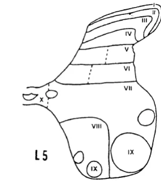
THE EFFECT OF PERIPHERAL INJURY ON CELLS IN THE SPINAL CORD The grey matter of the spinal cord was recognised by Rexed (1952) to be organised intolam inae F ig .I.l); these were defined in terms of the size, orientation and density of the neurons within them.
Lamina I. This marginal layer, contains large flat "marginal" cells and many intermediate sized neurons and is traversed by fibre bundles.
lamina has been given the name ’substantia gelatinosa’. It is a clearly demarcated region containing very small, tightly packed neurons.
Lamina III. The neurons in this lamina are larger and less densely packed than those in lamina II.
Lamina IV. This is the thickest lamina, containing very large neurons with dendrites that spread into the more superficial layers.
Lamina V. The neurons are smaller than those in lamina IV.
V I I
V I I I
Fig. 1.1 Rexed’s scheme for lamination of the spinal grey matter (From Rexed, B., A cytoarchitechtonic atlas of the spinal cord in the cat. J. Comp. Neurol., 96:415-495, 1952).
V.R.A.
Fig.1.2 (taken from McMahon, 1984) shows diagrammatically, the major components of the upper laminae of the spinal cord.
Peripheral nerve injury may produce selective damage to myelinated primary afferents, with sparing of unmyelinated nociceptors. This may be the case, for example, where nerves are subjected to chronic compression as in entrapment neuropathies which are often painful. As stated above, evidence that large fibre loss contributes to the pain in some types of neuropathy comes from clinical experience that stimulation of large fibres can give relief from pain. Observations such as these led Melzack and Wall (1965) to propose a ’gate control’ theory to explain a mechanism of pain.
Gate control theory
The gate control theory has four main components.
(a) Afferents
Unmyelinated nociceptors which terminate in the two outer laminae; and AÔ fibres which also terminate in this area and some extend to a zone in lamina V. Low- threshold AB fibres terminate in the deeper laminae.
The low-threshold afferents may be related to pain through the following findings:
2. Allodynia in some patients can be abolished by selective blockade of large myelinated afferents (Meyer, Campbell and Raja, 1985) which include few if any nociceptors. This indicates that large myelinated afferents can contribute to pain under pathological conditions.
(b) Intemeurons within the dorsal ham
The cells which receive incoming afferents cells are not simply collecting information from particular types of afferents and transmitting these to their destination. They select and compute combinations of the signals received. Some combinations summate signals, others are inhibitory. Such excitatory and inhibitory interaction requires the presence of intemeurons which are present in laminae I and n .
(c) Descending controls
Many regions in the brain project into the dorsal horn and have mainly inhibitory effects on the firing of dorsal horn nociceptive neurons. The inhibition of nociceptive responses in the cord produced by electrical stimulation of some of these regions appears to correlate well with behavioural analgesia. The raphe nuclei and the reticular formation have been emphasized to be the origin of descending control, but other sources (in man) include the periaqueductal grey matter (Fields and Basbaum,
1978).
(d) Transmission cell
spinothalamic tract. However, other projections systems may be cut during that operation.
Although the term ’gate control’ is now restricted to the rapidly acting mechanism, the theory still offers an explanation o f central summation and the phenomenon of allodynia.
According to the gate control hypothesis of Melzack and Wall (1965) (Fig.1.3), the interaction between myelinated and unmyelinated inputs to the spinal cord occurs at two sites: inhibitory neurons in the substantia ^qelaticnsa (SG) (Lanina H )
Output
U
Later changes of spinal cord
Nociceptive pain produced by tissue injury, and neuropathic pain due to damage to peripheral nerve function, each have different peripheral mechanisms but both are influenced by changes in the CNS. When the tissue or peripheral nerve is damaged, the nature of the afferent barrage in the peripheral nerve changes with time, and the receiving mechanisms within the brain and spinal cord also change under conditions of ongoing injury detection. Therefore the relationship of the four main components of the gate control theory will not be as simple as originally proposed now that central plasticity is known to occur. The role of central neuroplasticity in pathological pain has recently been extensively reviewed by Coderre et al., (1993).
CENTRAL PLASTICITY IN NOCICEPTIVE PAIN
This will only be briefly mentioned since the main topic of this review is neuropathic pain. Two types of hyperalgesia can be recognised (Hardy, W olff and Goodell, 1950). Primary analgesia is the increased sensitivity to noxious stimulation at the site o f an injury, and is probably mediated by peripheral mechanisms; secondary analgesia is the increased sensitivity extending beyond the site of injury and is thought to be related to central hyperactivity or sensitization.
Evidence for a central mechanism of hyperalgesia comes from a number of different observations of the phenomenon of referred pain and hyperalgesia, including the fact that pain and hyperalgesia can occur in regions far removed from the injured site.
Central plasticity in neuropathic pain
Phantom pain associated with the removal of limbs and other parts of the body provides striking evidence that central mechanisms are involved since the
’peripheral’ source of the pain no longer exists.
Denervation hypersensitivity
The development of hypersensitivity in the hind paw of a rat after sciatic nerve section occurs at the time of expansion of the projection of the saphenous nerve in the spinal cord (Markus, Pomeranz and Krushelnycky, 1984). This finding will be discussed in more detail elsewhere, since it is pertinent to the topic of this thesis. There are many other examples of nerve injury which are associated with increased activity in the central nervous system.
Prolonged central changes triggered by nerve impulses
In the clinical setting, minor injuries may cause prolonged pain and tenderness that spreads far from the site of injury. Central changes are likely to be implicated in these widespread effects. Studies in decerebrate rats following peripheral thermal injury have shown a prolonged increase in the excitability of both ipsilateral and contralateral flexor reflexes (Woolf, 1983). C fibre activity is essential to produce the central changes. Increased excitability in the contralateral flexor efferent nerve continues after impulses from the injured paw are blocked by local anaesthetic, suggesting that central rather than peripheral changes are implicated.
Expansion o f the receptive fields o f dorsal horn neurons