Case Report
ANCA Negative Vasculitis Presenting with Interstitial Lung Disease
and Pancytopenia
Kriti Gupta
*, Behtash Saeidi, William Pascal
Department of Internal medicine, Maimonides Medical Centre, New York, United States
*
Corresponding author: Kriti Gupta, Department of Internal medicine, Resident physician, Maimonides Medical
Centre, Brooklyn, New York, United States, Tel: 718-283-6000; E-mail: [email protected]
Received: 20 October 2019;Accepted: 06 December 2019;Published: 17 December 2019
Citation: Kriti Gupta, Behtash Saeidi, William Pascal. ANCA Negative Vasculitis Presenting with Interstitial
Lung Disease and Pancytopenia. Archives of Internal Medicine Research 2 (2019): 077-084.
Abstract
Introduction: MPA (or microscopic polyarteritis) is
clinically described as patients with exclusively
nongranulomatous small vessel vasculitis involving the
upper or lower respiratory tract (e.g., alveolar
capillaritis). The most common manifestation of MPA is
necrotizing glomerulonephritis and/or pulmonary
capillaritis. Serologically, more than 90% of MPA
patients have a positive antineutrophil cytoplasmic
antibodies (ANCA). There have been selective case
reports documenting pancytopenia associated with
vasculitis.
Case: Here, we discuss a 56 year old female patient
who presented with pancytopenia, and was found to
have biopsy-proven small vessel vasculitis with
negative ANCA. She presented with 5-6 weeks of
worsening generalized fatigue, cough with hemoptysis,
intermittent fever, and shortness of breath on exertion
and weight loss of 8lbs over this time. On presentation,
she was febrile (102F) and found to have pancytopenia,
with normal kidney function. She was admitted to the
Intensive Care Unit for a presumed viral illness and
underwent a bone marrow biopsy for evaluation of
pancytopenia. Further imaging with computerized
tomography (CT) scan demonstrated ground glass
haziness. Bone marrow biopsy revealed normal results.
She was discharged with the diagnosis of a
pancytopenic viral illness. She returned 4 weeks later
with worsening of her symptoms. CT chest showed
significant interval changes compared to previous CT
with bilateral ground glass opacities. Lung biopsy was
performed which showed small vessel vasculitis. Repeat
CT chest following steroid treatment demonstrated
Discussion: To our knowledge, this is the first reported
case of pancytopenia in small vessel vasculitis not
associated with lupus. In cases of MPA, 96% are ANCA
positive. However, this patient fits into the criteria of
ANCA-negative vasculitis. Although ILD is an
uncommon presentation of MPA, there have been an
increasing number of cases citing association of
vasculitis with ILD. In conclusion, vasculitis associated
with pancytopenia continues to be poorly understood.
ANCA negativity at the time of presentation can give a
false sense of security and delay treatment.
Keywords: pancytopenia; vasculitis
1. Background
Small-vessel vasculitis has been categorized as
granulomatosis with polyangiitis (GPA), microscopic
polyangiitis (MPA), eosinophilic granulomatosis with
polyangiitis and pauci-immune glomerulonephritis.
MPA (or microscopic polyarteritis) is used to describe
patients with exclusively nongranulomatous small
vessel vasculitis involving the upper or lower
respiratory tract (e.g., alveolar capillaritis).
Pathologically it is a pauci-immune necrotizing
vasculitis affecting small vessels, with or without
involvement of medium-sized arteries [1].
The American College of Radiology (ACR) criteria for
small-vessel vasculitis include the following:
Nasal or oral inflammation (painful or painless
oral ulcers, or purulent or bloody nasal
discharge).
Abnormal chest radiograph showing nodules,
fixed infiltrates, or cavities.
Abnormal urinary sediment (microscopic
hematuria with or without red cell casts).
Granulomatous inflammation on biopsy of an
artery or perivascular area.
The presence of two or more of these four criteria
yielded a sensitivity of 88 percent and a specificity of 92
percent. Since the ACR did not establish specific
criteria for MPA, these criteria did not discriminate
GPA from either MPA nor non-vasculitic diseases that
can mimic GPA [2]. The most common manifestation of
MPA is necrotizing glomerulonephritis and/or
pulmonary capillaritis. Serologically, more than 90% of
MPA patients have a positive antineutrophil
cytoplasmic antibodies (ANCA) [3]. Here, we discuss a
56 year old female patient who presented with
pancytopenia, and was found to have biopsy-proven
small-vessel vasculitis with negative ANCA.
2. Case
A 65 year old woman with a past medical history of
Antiphospholipid Antibody (APLA) syndrome,
partial-seizure disorder, hypertension and dyslipidemia
presented with worsening generalized fatigue, cough
with hemoptysis, intermittent fever, shortness of breath
on exertion and weight loss of 8lbs over a period of five
to six weeks. She had been prescribed warfarin for
APLA, however, she had not been compliant with
regular INR measurements. She was sent to the
emergency room by her primary physician. On
presentation to the hospital, she had a fever of
102oFarenhiet (orally) and a normal physical exam.
Laboratory findings on admission were present in Table
1. Chest x-ray (CXR) revealed bilateral upper lobe
opacities of unspecified etiology. Further imaging with
computerized tomography (CT) scan demonstrated
ground glass haziness and interstitial septal thickening
with 6mm and 5mm ground glass nodules in the left
lung base. An echocardiogram performed was normal.
Patient was admitted to the Intensive Care Unit for a
presumed viral illness and underwent a bone marrow
biopsy for evaluation of pancytopenia, which showed
only some mild hypercellularity. Serological studies
hospital stay, her symptoms improved with conservative
management. Her illness was presumed to be viral and
she was discharged to follow up with an out-patient
hematologist. Patient followed up in out-patient clinic,
where she was found to have persistent pancytopenia
and worsening fatigue. She was sent to the emergency
room and found to have bilateral rhonchi, with hypoxia.
Additionally, she was pancytopenic with hemoglobin of
6.2 g/dl and a platelet count of 10 K/ul. CT chest
showed significant interval changes compared to
previous CT with bilateral ground glass opacities. She
was started on broad spectrum antibiotics and was
admitted to the ICU, where she received transfusions of
packed red blood cells (PRBC) and platelets. Over the
course of her stay, she developed respiratory distress
and had to be intubated. Bronchoscopy was performed
and specimens were sent for microbiology and cytology,
which did not reveal any significant findings, including
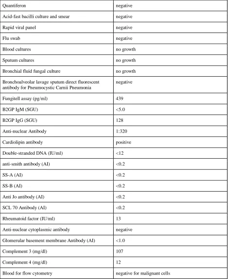
tuberculosis. The laboratory findings over her ICU stay
are presented in Table 3. The Patient continued to
receive broad-spectrum antibiotics, including
anti-fungals, in addition to intravenous steroids. Lung biopsy
was performed which showed small-vessel vasculitis.
Repeat CT chest following steroid treatment
demonstrated significant improvement. Currently, the
patient was followed up at a 6 month interval post
discharge. She experienced repeated exacerbations in
her symptoms with similar presentation which resolved
using immunosuppressants (cyclophosphamide and
rituximab) and plasmapheresis. She has now decided to
pursue further treatment at an out-side hospital.
White Blood Cell Count (K/ul x1000) 3.5
Red Blood cell count (million/ml) 2.57
Hemoglobin (g/dl) 7.9
Platelets (K/ul) 54
Reticulocytes 0.4%
Serum sodium (mmol/L) 137
Serum potassium (mmol/L) 4.1
Blood urea nitrogen (mg/dl) 28
Creatinine (mg/dl) 1.4
International Normalized Ratio 5.5
Lactate Dehydrogenase (IU/L) 177
Haptoglobin (mg/dl) 286
Table 1: Laboratory values at presentation.
Cytomegalo Virus IgG and IgG negative
Epstein-Barr Virus IgG and IgG negative
Human Immunodeficiency Virus 1/2 screening EIA
Table 2: Serological Studies.
Quantiferon negative
Acid-fast bacilli culture and smear negative
Rapid viral panel negative
Flu swab negative
Blood cultures no growth
Sputum cultures no growth
Bronchial fluid fungal culture no growth
Bronchoalveolar lavage sputum direct fluorescent antibody for Pneumocystic Carnii Pneumonia
negative
Fungitell assay (pg/ml) 439
B2GP IgM (SGU) <5.0
B2GP IgG (SGU) 128
Anti-nuclear Antibody 1:320
Cardiolipin antibody positive
Double-stranded DNA (IU/ml) <12
anti-smith antibody (AI) <0.2
SS-A (AI) <0.2
SS-B (AI) <0.2
Anti Jo antibody (AI) <0.2
SCL 70 Antibody (AI) <0.2
Rheumatoid factor (IU/ml) 13
Anti-nuclear cytoplasmic antibody negative
Glomerular basement membrane Antibody (AI) <1.0
Complement 3 (mg/dl) 107
Complement 4 (mg/dl) 12
Blood for flow cytometry negative for malignant cells
Figure 1: CT chest at initial presentation.
Figure 2: CT chest on second presentation.
3. Discussion
Vasculitis has historically been associated with
ANCA-positivitiy and rarely with interstitial lung disease and
pancytopenia. There have been selective case reports
documenting pancytopenia with vasculitis, interstitial
lung disease with vasculitis and ANCA-negativity with
vasculitis. Despite extensive research, we were unable
to find a single case report of a patient presenting with
ANCA-negativity, interstitial lung disease and
pancytopenia. This patient had a negative
double-stranded DNA (ds-DNA) and presented with
pancytopenia which is an unusual presentation. The
chief reason for admission of the patient discussed
above was respiratory failure. Lung involvement is a
common presenting finding of vasculitis. It can initially
manifest like idiopathic pulmonary fibrosis and precede
the full-blown clinical manifestations and diagnosis of
MPA by years [4, 5, 6]. Historically, the incidence of
fibrosis in MPA has been insignificant. A study
published in Arthritis and Rheumatism in 1999 reported
the incidence of lung findings in MPA as 24%, the most
common being alveolarhemorrhage (11.8%),
pneumonitis (10.6%) and pleuritis (5.9%) [7]. The
majority of these patients have a predominant
myeloperoxidase-antineutrophil cytoplasmic antibodies
(MPO-ANCA) positive pattern [7]. However, more
recently, there have been an increasing number of
studies recognizing pulmonary fibrosis in vasculitis. In
2016, a German study that followed the clinical course
of small-vessel vasculitis found that in a series of 144
patients with MPA, 12% presented with fibrosing
interstitial lung disease, which subsequently developed
in 15% over the long term follow-up [8]. Fibrosing ILD
was associated with a poor prognosis in this study.
Interestingly, fibrosing ILD is more commonly
associated with MPO-ANCA than with other subtypes
[9, 10]. This is in contrast to our patient who, being
ANCA-negative, presented with ILD.
It is unclear whether fibrosing ILD significantly impacts
mortality in patients with MPA [4, 10, 11]. Cloe et al, as
a part of a multicentric study, followed up with 49
vasculitis patients for 15 years and reported that chronic
respiratory insufficiency was significantly associated
with mortality, followed by induction therapy with
glucocorticoids alone and initial weight loss. There was
a significant three year mortality benefit in patients
treated with an immunosuppressant as induction therapy
[12]. Over the course of her hospital stay, our patient
was found to have pancytopenia. Pancytopenia is a rare
presentation of vasculitis. A case report of a 65 year old
male patient published in 2007 linked multiple
paraneoplastic syndromes consisting of pancytopenia
leukocytoclastic vasculitis and hypertrophic pulmonary
osteoarthropathy with non-small cell lung carcinoma
[13]. The author attributed the pancytopenia in that case
to the constellation of paraneoplastic syndromes. The
only other case of pancytopenia in small-vessel
vasculitis was of a patient with ANCA-positive
vasculitis and was published in 2003 [14]. To our
knowledge, this is the first reported case of
pancytopenia in ANCA negative small-vessel vasculitis
not associated with lupus. Immunodiagnostically,
though the aforementioned patient was ANCA-negative,
she was diagnosed with MPA based on her lung biopsy
and clinical features. Although a positive ANCA test is
strongly suggestive of vasculitis, false-positive and
false-negative ANCA results may be seen [15].
ANCA-negative vasculitis patients tend to be younger at the
time of diagnosis (mean age 50.0 years old) [3]. In a
study done in 2017, 133 Korean patients with
ANCA-associated vasculitis were classified into 3 categories:
those with MPO-ANCA, anti-PR3 antibody
(PR3-ANCA) and ANCA-negative vasculitis. It was found
that patients with ANCA-negative vasculitis had the
youngest mean age at diagnosis (50 years old) among
the three categories [3]. Literature of cases citing
A case report from the American Journal of Kidney
Disease in 2002, reported a case about an
African-American 11-year-old girl presenting with peri-orbital
edema with an elevated creatinine and was found to
have an increased ANA and anti-dsDNA titer with a
negative ANCA. She was eventually diagnosed with
lupus nephritis and ANCA-negative renal vasculitis
[16]. A large multi-center study done in 1998 found that
in MPA, the diagnostic sensitivity of enzyme-linked
immunosorbent assay (ELISA) based tests namely
anti-PR3 and anti-MPO were 26% and 58% respectively
while the diagnostic sensitivity of the indirect
immunofluorescence (IIF) based tests namely cANCA
and pANCA were 23% and 58% respectively [17].
However, the multi-center study also found that when
the results of the IIF tests were combined with those of
the ELISA tests (cANCA/anti-PR3 positive,
pANCA/anti-MPO positive), the diagnostic specificity
increased to 99%. It is possible that the patient might
have presented with ANCA-positivity if our team had
used a combination of IIF and ELISA.
4. Conclusion
Vasculitis associated with pancytopenia continues to be
poorly understood. ANCA-negativity at the time of
presentation can give a false sense of security and delay
treatment. Any worsening of condition on steroids
warrants the use of immunosuppressants. However, this
has not been required to date and our patient remains
well without evidence of progression in our two month
follow-up.
References
1. Jennette JC, Falk RJ, Bacon PA, Basu N, Cid
MC, Ferrario F, et al. 2012 revised
international chapel hill consensus conference
nomenclature of vasculitides. Arthritis &
Rheumatism 65 (2013): 1-11.
2. Rao Jaya K, Nancy B Allen, Theodore Pincus.
Limitations of the 1990 American College of
Rheumatology classification criteria in the
diagnosis of vasculitis. Annals of internal
medicine 129 (1998): 345-352.
3. Yoo J, Kim HJ, Ahn SS, Jung SM, Song JJ,
Park YB, et al. Clinical and prognostic features
of Korean patients with MPO-ANCA,
PR3-ANCA and PR3-ANCA-negative vasculitis. Clinical
and experimental rheumatology 35 (2017):
111-118.
4. Hui Huang, Yan xun Wang, Chun guo Jiang,
Jia Liu, Ji Li, Kai Xu, et al. "A retrospective
study of microscopic polyangiitis patients
presenting with pulmonary fibrosis in China."
BMC pulmonary medicine 14 (2014): 8.
5. Tzelepis GE, Kokosi M, Tzioufas A, Toya SP,
Boki KA, Zormpala A, et al. Prevalence and
outcome of pulmonary fibrosis in microscopic
polyangiitis. European Respiratory Journal 36
(2010): 116-121.
6. Foulon G, Delaval P, Valeyre D, Wallaert B,
Debray MP, Brauner M, et al.
"ANCA-associated lung fibrosis: analysis of 17
patients." Respiratory medicine 102 (2008):
1392-1398.
7. Guillevin L, Durand-Gasselin B, Cevallos R,
Gayraud M, Lhote F, Callard P, et al.
Microscopic polyangiitis: clinical and
laboratory findings in eighty‐five patients.
Arthritis & Rheumatism: Official Journal of
the American College of Rheumatology 42
(1999): 421-430.
8. Schirmer JH, Wright MN, Vonthein R,
Herrmann K, Nölle B, Both M, et al. Clinical
presentation and long-term outcome of 144
patients with microscopic polyangiitis in a
monocentric German cohort. Rheumatology 55
9. Tanaka T, Otani K, Egashira R, Kashima Y,
Taniguchi H, Kondoh Y, et al. "Interstitial
pneumonia associated with MPO-ANCA:
clinicopathological features of nine patients".
Respiratory medicine 106 (2012): 1765-1770.
10. Arulkumaran N, Periselneris N, Gaskin G,
Strickland N, Ind PW, Pusey CD, et al.
"Interstitial lung disease and ANCA-associated
vasculitis: a retrospective observational cohort
study." Rheumatology 50 (2011): 2035-2043.
11. Fernandez Casares M, Gonzalez A, Fielli M,
Caputo F, Bottinelli Y, Zamboni M.
"Microscopic polyangiitis associated with
pulmonary fibrosis". Clinical rheumatology 34
(2015): 1273-1277.
12. Comarmond C, Crestani B, Tazi A, Hervier B,
Adam-Marchand S, Nunes H, et al. Pulmonary
fibrosis in antineutrophil cytoplasmic
antibodies (ANCA) - associated vasculitis: a
series of 49 patients and review of the
literature. Medicine 93 (2014): 340-349.
13. Cosar-Alas R, Yurut-Caloglu V, Karagol H,
Caloglu M, Yalcin O, Turgut B, et al.
Paraneoplastic syndrome of non-small cell lung
carcinoma: a case with pancytopenia,
leukocytoclastic vasculitis, and hypertrophic
osteoarthropathy. Lung Cancer 56 (2007):
455-458.
14. Letellier E, Cunningham J, Feakins RM,
Hughes GR, D'Cruz DP. "Pancytopenia in
ANCA associated vasculitis: response to
immunosuppressive therapy." Clinical and
experimental rheumatology 21 (2003):
415-416.
15. Gross, W. L., A. Trabandt, and E. Reinhold‐
Keller. Diagnosis and evaluation of vasculitis.
Rheumatology 39 (2000): 245-252.
16. Moeckel, Gilbert W, Kathy Jabs, Agnes B
Fogo. "An 11-year-old African-American girl
with systemic lupus erythematosus and
ANCA-negative renal vasculitis." American
journal of kidney diseases 39 (2002): 433-438.
17. Hagen EC, Daha MR, Hermans J, Andrassy K,
Csernok E, Gaskin G, et al. "Diagnostic value
of standardized assays for anti-neutrophil
cytoplasmic antibodies in idiopathic systemic
vasculitis." Kidney international 53 (1998):
743-753.
This article is an open access article distributed under the terms and conditions of the