1535-9778/08/$08.00⫹0 doi:10.1128/EC.00393-07
Copyright © 2008, American Society for Microbiology. All Rights Reserved.
Abnormal Micronuclear Telomeres Lead to an Unusual Cell Cycle
Checkpoint and Defects in
Tetrahymena
Oral Morphogenesis
䌤
Karen E. Kirk,
1* Christina Christ,
1Jennifer M. McGuire,
1Arun G. Paul,
1Mithaq Vahedi,
1Kathleen R. Stuart,
2and Eric S. Cole
2Department of Biology, Lake Forest College, Lake Forest, Illinois 60045,1and Department of Biology,
St. Olaf College, Northfield, Minnesota 550572
Received 26 October 2007/Accepted 16 April 2008
Telomere mutants have been well studied with respect to telomerase and the role of telomere binding proteins, but they have not been used to explore how a downstream morphogenic event is related to the mutated telomeric DNA. We report that alterations at the telomeres can have profound consequences on organellar morphogenesis. Specifically, a telomerase RNA mutation termed ter1-43AAresults in the loss of germ line micronuclear telomeres in the binucleate protozoan Tetrahymena thermophila. These cells also display a micronuclear mitotic arrest, characterized by an extreme delay in anaphase with an elongated, condensed chromatin and a mitotic spindle apparatus. This anaphase defect suggests telomere fusions and consequently a spindle rather than a DNA damage checkpoint. Most surprisingly, these mutants exhibit unique, dramatic defects in the formation of the cell’s oral apparatus. We suggest that micronuclear telomere loss leads to a “dynamic pause” in the program of cortical development, which may reveal an unusual cell cycle checkpoint.
Telomeres have been implicated in a number of diverse cellular processes. Normal telomeres serve as the means by which chromosomes can be replicated completely, and they function as a cap, thereby protecting the natural ends from inappropriate fusion. Telomeric mutants can be generated by making predictable changes in the RNA template of the en-zyme telomerase, which synthesizes telomeres, such that the corresponding complementary mutation is made at the telo-meres. Although these types of telomeric DNA mutants in complex eukaryotes are poorly studied, particularly with re-spect to the downstream developmental consequences, those in simplified yeasts and the ciliated protozoa are better under-stood.
Ciliated protozoa, such asTetrahymena thermophila, provide excellent model systems for studying telomerase, telomeric DNA mutants, and the downstream developmental conse-quences to changing the telomeric repeat sequence. This is because the components of telomeres and telomerase have been exceptionally well documented (5) and the cells are large enough (roughly 60 to 70 m) to allow a glimpse of their unique developmental processes.
Interestingly, T. thermophila, like other ciliates, bears two nuclei distinct in function and mode of division (reviewed in reference 25). The micronucleus represents the germ line nu-cleus and is transcriptionally silent, whereas the macronunu-cleus serves as the vegetative nucleus of the cell. The micronucleus contains five pairs of chromosomes, which are passed on through mitosis and meiosis. In contrast, the macronucleus contains many subchromosomal fragments, each of which is amplified to⬃45 copies, excluding the ribosomal DNA, which
has thousands of copies. The macronuclear chromosomes are not faithfully inherited, as the macronucleus divides amitoti-cally.
The telomeres of the twoT. thermophilanuclei are dramat-ically different. The macronuclear telomeres, present at a copy number of roughly 40,000 per cell, contain about 0.3 to 0.5 kb of G4T2 repeats (3). Micronuclear telomeres, in sharp con-trast, are present at a copy number of 20 per cell and have much longer telomeric tracts of⬃2.5 kb that contain an inner tract of a variant G4T3 repeat sequence (26). This variant repeat tract is not present in the macronucleus, presumably as it is eliminated with the rest of the micronuclear telomeres during development of the new macronucleus upon conjuga-tion.
A number of telomere mutants have been constructed inT.
thermophila, for example, by mutating the telomerase RNA
and consequently the telomeric DNA itself (40) or by depleting a protein that binds the telomeres (24). The telomeric DNA is mutated in ter1-43AA cells by altering the telomerase RNA template such that it synthesizes a combination of G4T3 re-peats, which are harmless, and G4T4 rere-peats, which are toxic (40). Even with this sequence alteration, the macronuclear telomere length is only subtly altered. Nonetheless, a dramatic arrest appears in micronuclear mitosis, where the chromatin appears elongated during anaphase (27, 40), which takes about 30 times longer than wild-type (WT) anaphase. In a different type of telomere mutant, where the G-strand telomeric binding protein POT1a is depleted, the cells undergo a growth arrest where the macronucleus is enlarged and the macronuclear telomeres become extremely lengthened (24). Thus, there may be more than one kind of cell cycle arrest involved due to telomere defects within the two different nuclei.
Not only isT. thermophilabinucleate, but it exhibits a wealth of unusual developmental landmarks as it progresses through the program of vegetative growth and cell division. It has a remarkable cell cortex, with an oral apparatus (mouth) that
* Corresponding author. Mailing address: Department of Biology, Lake Forest College, 555 N. Sheridan Rd., Lake Forest, IL 60045. Phone: (847) 735-6044. Fax: (847) 735-6194. E-mail: kirk@lakeforest .edu.
䌤Published ahead of print on 9 May 2008.
1712
on September 8, 2020 by guest
http://ec.asm.org/
takes in bacteria and nutrients and a cytoproct (anus) that eliminates waste. Oral development alone is a rich landscape of beautifully choreographed developmental events (12). At a precise location on the cell’s cortex, a field of basal bodies proliferates, forming an “anarchic field.” Waves of morphoge-netic activity then organize this field into a set of uniquely sculpted membranelles, creating the cell’s oral apparatus. There appear to be four parallel programs involving the nuclei and oral apparatus that are coordinated as cells divide: (i) they replicate their micronucleus by mitosis (28, 29), (ii) they rep-licate their macronucleus by amitotic fission (14, 15, 36), (iii) they synthesize and assemble a gallery of cortical organelles associated with the oral apparatus (9, 10), and (iv) they un-dergo cytokinesis in a fashion coordinated with the completion of the other three programs (2, 11, 13, 35, 37).
In this study, we have used the T. thermophilatelomerase RNA template mutantter1-43AAto assess the molecular status of the micronucleus and to correlate its mitotic events with cortical events associated with formation of the oral apparatus. Although it was shown previously that the sequences of the macronuclear telomeres in this mutant are altered (40), the micronuclear telomeres had not been examined. We show here that the micronuclear telomeres are dramatically shortened. Furthermore, many aspects of the blocked micronuclei are consistent with end-to-end fusion during anaphase, specifically the persistence of condensed chromatin and an intact mitotic spindle apparatus. Finally, we demonstrate that the midana-phase arrest associated with the micronucleus in this mutant (27, 40) allows us to create a developmental timeline relating cortical and nuclear events. This work may also cast light upon a potential cell cycle checkpoint that has been largely over-looked.
MATERIALS AND METHODS
Cell lines and culture conditions.Cells were of the CU428 and SB1969 strains, kindly provided by Peter J. Bruns and Eduardo Orias, respectively. Cells were grown at 31°C in a proteose peptone medium (0.25% proteose peptone, 0.25% Difco yeast extract, 0.5% glucose, 0.033 mM FeCl3). Cells were prepared for mating by starving the cells at 3⫻105cells per ml in 10 mM Tris buffer (pH 7.4) at 31°C (23). After 12 to 18 h of starvation, equal numbers of cells were mixed and mated.
Transformation.ter1-43AAwas generated on the vector pRD4-1 and trans-formed as described previously (27).Tetrahymena thermophilastrains CU428 and B2086 or CU428 and SB1969 were starved, mated, and transformed by electro-poration as described in references 16 and 17. Transformed cultures were plated in 96-well plates at dilutions such that clonal lines were obtained from single transformed cells by selection with 120g/ml paromomycin (Sigma). Control cells were transformed by electroporation with the WT telomerase RNA gene (prTer1 [46]). Beginning at 4 days after electroporation,ter1-43AAtransformant cell lines were analyzed for micronuclear telomere length and cytology from a population, or single cells were isolated.
Telomere length analysis.WT and mutant DNAs were isolated from Tetrahy-menacells as described previously (40). DNA was digested with DraI, and samples were dried down. Southern blots of samples were prepared and probed with oligonucleotides that recognize WT telomeres (G4T2), that recognize only WT micronuclear telomeres or mutated micronuclear and macronuclear telo-meres (G4T3) (26), or that recognize only mutated telotelo-meres (G4T4) (40). Macronuclear and micronuclear telomeric regions could be readily distinguished by their dramatic size differences (⬃400 bp versus⬃2.5 kb, respectively) and by virtue of the unique micronucleus-limited G4T3 repeat found in the inner one-third of WT micronuclear chromosomes. (C4A3)4 was used as a probe, following the method used for the G4T3 sequence. The␣-tubulin probe was made by PCR using primers 5⬘AAGGTGGTATCCAAGTCGGTAACGC3⬘and 5⬘TCTGAG GGAGGCAGTCAAAGAGGA3⬘ and labeled with [␣-32P]dATP. The probe
was hybridized to the blot at 37°C and washed at 55°C in Church and Gilbert buffers.
Cytology.Transformants were prepared for microscopy as described previ-ously (43). From pooled transformants, 1.5 ml of cells was collected, washed, and resuspended in 0.1 ml of 60 mM PIPES [piperazine-N,N⬘-bis(2-ethanesulfonic acid)], 25 mM HEPES, 10 mM EGTA, 2 mM MgCl2(PHEM buffer). For Fig. 3, cells were fixed using 0.5 ml 3% paraformaldehyde and 0.25% Triton X-100 in PHEM buffer, then washed twice and resuspended in 0.5 ml 0.1% bovine serum albumin-phosphate buffered saline (BSA-PBS; Sigma), and loaded (0.2 ml) onto poly-L-lysine-coated coverslips and let sit for 20 min. The coverslips were floated cell side down on a drop containing 1% BSA-PBS with 1:100 rabbit anti-phos-phorylated histone H3 (Ser10) mitosis marker (from Upstate Biotechnology, now Millipore Corporation) and 1:50 mouse ascites monoclonal anti-␣-tubulin (acetylated; Sigma) for 1 hour at room temperature or overnight at 4°C. The coverslips were washed and floated cell side down on drops of 1% BSA-PBS secondary antibody solution containing 1:500 Texas Red goat anti-rabbit lecular Probes) and 1:500 goat anti-mouse conjugated to Oregon Green (Mo-lecular Probes) for 1 hour at room temperature. The coverslips were washed once as described above, then incubated in 0.1% BSA-PBS solution containing 0.06g/ml 4,6-diamidino-2-phenylindole (DAPI; Sigma) for 5 min, and then washed twice. Coverslips were loaded onto slides by using VectaShield anti-quenching mounting medium for fluorescence (VectorLabs). These microtubule preparations were analyzed using a Zeiss Axiovert 100 fluorescence microscope in conjunction with MetaMorph imaging software (version 4.5). For Fig. 5, cells were fixed in a triton-ethanol fixative (43). Basal bodies were imaged using a mouse monoclonal antiserum that recognizesTetrahymenabasal bodies (FV4F9; diluted 1:20 courtesy of Joseph Frankel), and fluorescein isothiocyanate-conju-gated goat anti-mouse from Sigma (diluted 1:100). Nuclei were counterstained using propidium iodide. Cells were incubated with 1 mg/ml RNase for 45 min at 30°C and then with 30g/ml propidium iodide for 30 min at room temperature. These basal-body preparations were analyzed using an Olympus Fluo-View laser scanning confocal microscope. For Fig. 6, antibody staining was done as for Fig. 5, and “flattened” preps were imaged using an Olympus BX-50 conventional fluorescence microscope and a SPOT-RT digital camera.
Single-cell isolation.The two transformant wells with the densest growths (C9 and F2) were selected. Single cells from each well were isolated by diluting them to an appropriate concentration and then monitoring them as they passed through a capillary by using a dissecting microscope. One cell was placed into a drop of 30l proteose peptone yeast sequestrine medium with paromomycin and penicillin streptomycin Fungizone, and this was repeated for a total of 48 isolated cells. Cells were grown for 24 h at 30°C in a moist chamber, and the cell number in each well was determined by microscopy as follows. A score of 0 means only a single cell or, in some cases, no cells were observed. A score of 1 means from 2 to 10 cells were observed. A score of 2 means more than 10 cells and fewer than roughly 100 cells. A score of 3 means substantially more than 100 cells. The cells from the densest wells were diluted, and the isolation was repeated.
RESULTS
Micronuclei have lost the majority of telomeric DNA. In order to determine if a telomerase RNA template mutant affects telomere length in bothT. thermophilanuclei, we first tested whether the micronuclear telomeres were altered in the
ter1-43AA mutant. Although the macronuclear telomeres in
this mutant are known to comprise G4T3 and G4T4 mutant repeats in addition to the WT G4T2 repeats (40) (Fig. 1A), the telomeric repeat composition of micronuclear telomeres had not been addressed previously. Therefore, we could not ascribe aspects of the phenotype to changes in the micronuclear telo-meres. We investigated whether there were changes in the lengths of micronuclear and macronuclear telomeric tracts by using Southern blot analysis.
Micronuclear telomeres are difficult to detect due to their relative paucity in the cell. The ratio of micronuclear to mac-ronuclear telomeres is approximately 1:2,000. Thus, we relied on two previous findings regarding WT telomeres (26). First, the micronuclear telomeres are dramatically longer than those of the macronucleus (⬃2.5 kb versus ⬃0.5 kb, respectively)
on September 8, 2020 by guest
http://ec.asm.org/
(Fig. 1B). Second, the micronuclear telomeres have an inner, homogeneous tract of variant G4T3 repeats not present in the macronuclear telomeres yet constituting roughly the inner one-third of the telomeric tract. We used several oligonucleotide probes in our Southern blot assay to assess the lengths and
sequences of the telomeres of both nuclei: a WT probe (G4T2) that recognizes both macronuclear and micronuclear telo-meres, a probe (G4T3) unique to micronuclear telotelo-meres, and a probe unique to mutated telomeres (G4T4).
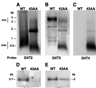
When we probed total DNA that had been digested with a frequently cutting restriction enzyme, we detected macro-nuclear telomeres (⬃0.5 kb) in both the WT and the mutant, using a G4T2 probe (Fig. 2A, lanes WT and 43AA). When the identical blot was reprobed with G4T3 or G4T4 sequences, the results corroborated previous findings that both types of repeat are incorporated intoter1-43AAmacronuclear telomeres (40) (Fig. 2B and C, lanes 43AA) but not into those of the WT (Fig. 2B and C, lanes WT). The macronuclear telomeres in the
ter1-43AA mutant cells are slightly shorter than those in the
WT (Fig. 2A) by roughly 150 bp. As was shown previously, this macronuclear telomere shortening is not responsible for the mutant phenotype (40).
Micronuclear telomeres are not evident in the blot probed with the G4T2 sequence (Fig. 2A), due to the background signal from the macronuclear telomeres. Nonetheless, when the blot was probed with the G4T3 sequence, which recognizes an⬃0.8-kb stretch of the innermost telomere repeats of the micronucleus (Fig. 2B, lane WT), there was clear visualization of micronuclear telomeres in the 3- to 4-kb range of WT cells.
FIG. 1. RNA templates and telomeric repeat lengths in the micro-nucleus and macromicro-nucleus. (A) The WT RNA template synthesizes G4T2 repeats. Theter1-43AA RNA template mutation synthesizes both G4T3 and G4T4 telomeric repeats in the macronucleus, but the mutant micronuclear telomeres had not been studied (40). (B) The WT micronuclear telomeres are⬃2.5 kb long, with the inner⬃0.8 kb composed of G4T3 repeats. The WT macronuclear telomeres are about 0.3 to 0.5 kb long.
FIG. 2. Micronuclear (mic) telomeres are dramatically shortened. (A) A G4T2 probe hybridizes to 40,000 WT macronuclear (mac) telomeres and thus obscures visualization of WT or mutant (ter1-43AA) micronuclear telomeres. In the 43AA telomeres, the strong band below 2 kb is due to a telomere on the transforming plasmid, and a slight shortening of macronuclear telomeres has been described previously (40). (B) Reprobing of the same blot with a G4T3 sequence (characteristic of internal micronuclear telomere tracks) allows clear visualization of micronuclear telomeres in WT cells. Surprisingly, in theter1-43AAmutant micronuclear telomeres, instead of an increase in signal intensity and size due to de novo G4T3 synthesis, there is a decrease in signal intensity and size. (C) No signal was seen when the blot was reprobed with a G4T4 sequence in the micronuclear telomeres for either WT or mutant DNA. (D) Reprobing with the opposite strand (C4A2) indicates that both strands are lost. (E) Loss of the micronuclear telomeres is not due to a general degradation mechanism, since an internal alpha tubulin sequence (ATU1) remains intact.
on September 8, 2020 by guest
http://ec.asm.org/
Surprisingly, we did not observe a larger, more intense signal, due to de novo G4T3 synthesis in the mutant (Fig. 2B, lane 43AA) but instead saw substantially less signal intensity, indic-ative of loss of the micronuclear telomere sequence.
The mutant micronuclear telomere signal was shortened by
⬃0.6 kb. This amount of shortening should not affect the inner G4T3 sequence that is being probed, as this sequence is con-fined to the inner one-third (⬃0.8 kb) of a⬃2.5-kb telomere, as described above. The G4T3 signal intensity would start to diminish only when the telomeres are shortened by more than
⬃1.7 kb. Thus, the loss of signal intensity in this size range, coupled with an absence of noticeable G4T3 signal elsewhere in the lane, strongly suggests some loss of the inner G4T3 repeats. Thus, the loss of signal here most likely corresponds to a loss of most or all of the micronuclear telomeric DNA from a fraction of the telomeres.
We checked for the loading of DNA by probing the same blot with an ␣-tubulin DNA control. The DNA in the ter1-43AAmutant lane appears underloaded, but after determining with gel quantification software that the DNA in this lane represents about 60% of the WT level (Fig. 2E) and normal-izing it to this amount, we determined that roughly 70% of the total telomeric DNA signal is lost from the mutant lane. There-fore, we conclude that a great deal of telomere sequence from the micronucleus has been lost.
We next asked whether in theter1-43AAmutant, the remain-ing, largely intact micronuclear telomeres that were⬃0.6 kb shorter, contained de novo mutant repeats without suffering complete degradation. Alternatively, this signal is due to WT telomeres that were slightly shortened but perhaps had not yet been acted on by the mutant telomerase. If the former is the case, then we should be able to detect the action of mutant telomerase by using the G4T4 probe. The results indicate that there is no obvious G4T4 signal at the mutant micronuclear telomeres around 3 to 4 kb (Fig. 2C, lane 43AA). It is possible that a very low number of repeats are added to the micro-nuclear telomeres and hence are undetectable in this assay, but this seems unlikely given the strong mutant signal at macro-nuclear telomeres. We also tested whether both strands of the micronuclear telomeres inter1-43AAcells are absent by prob-ing with a C4A3 probe to the opposite telomeric strand. The absence of signal indicates that the G-rich strand is also lost (Fig. 2D, lane 43AA). Finally, loss of the micronuclear telo-meres is not due to a general DNA degradation mechanism, since an internal␣-tubulin probe remained intact (Fig. 2E).
We conclude from these results that the majority of micro-nuclear telomeres have been lost in transformed cells
express-ing ter1-43AA. Furthermore, the small percentage of nearly
intact, remaining micronuclear telomeres is most likely WT.
ter1-43AAcells have intact spindles and condensed chroma-tin in anaphase bridges.Tetrahymena thermophilastrains that carryter1-43AA telomerase mutations exhibit a profound cell cycle arrest characterized by enlarged cells and elongate mi-cronuclei characteristic of anaphase. Since this phenotype oc-curs in a cell that expresses both mutant and WT telomerase activities, the mutation clearly behaves in a dominant manner. We used immunofluorescence to examine three markers of cell cycle progression in these mutants to more fully characterize their phenotype. First, we examined whether or not the micro-nuclear histone H3 is in its phosphorylated state. If H3 is
phosphorylated, this indicates that cells have indeed entered mitosis and undergone chromatin condensation. Second, we used acetylated␣-tubulin antibody to visualize whether or not the mitotic spindle was assembled in these arrested mutants. Finally, we employed antibodies against basal-body epitopes to visualize the pattern of cortical ciliature that accompanies oral development during cell division. This marker allows a precise evaluation of when and where in the cell cycle our telomerase mutant was arrested.
The antibody raised specifically against phosphorylated his-tone H3 decorates micronuclear chromatin when it is in its condensed, mitotic configuration (44). Since the macronuclear chromatin does not stain with this antibody, the micronuclei are more clearly visible in theter1-43AAmutant cells as they are not obscured by the large DAPI-stained macronucleus. We were able to identify a much greater proportion of micronuclei that were elongated and stained with this antibody in mutant cells than we had previously detected with DAPI alone (40). In WT log-phase cultures ofTetrahymena(n ⫽1,345), typically 20% of the cells exhibit positive micronuclear labeling, indi-cating mitosis. In contrast, 77% of ter1-43AA transformants (n⫽254) exhibited positive labeling, indicating that they had entered mitosis but failed to exit.
Figure 3A to C show condensed micronuclei at various stages of mitosis in WT cells. The condensed state persists through late anaphase (Fig. 3C). Examples ofter1-43AAcells are shown in Fig. 3D to I. About 56% of the cells in the
ter1-43AApopulation have elongated micronuclei that are
ac-companied by an apparently intact,␣-tubulin-stained spindle apparatus, compared to 17% for the WT population. This apparatus (20.2⫾2.3m) is roughly twice as long (as deter-mined using a measurement tool in MetaMorph) as that ob-served for WT cells (11.6⫾ 0.8m). The persistence of the spindle apparatus and the lengths of the microtubules from pole to pole strongly suggest that the micronuclear chromo-somes are stuck or intertwined and unable to separate.
Micronuclei from these arrested mutants are shown en-larged in Fig. 3G to I. In most cases, the chromatin stretches continuously from one pole to the other, consistent with the hypothesis that bridges have formed between chromosomes via end joining (Fig. 3H). In rare cases, chromatin forms two discrete masses at either end of the mitotic spindle, suggesting that anaphase has segregated two masses of chromatin (Fig. 3I). In this case, there appears to be unequal chromatin seg-regation on each side of a conspicuous gap.
In summary, there are roughly three times the number of
ter1-43AAcells with condensed micronuclear chromatin as WT
cells, and this chromatin is almost always joined from one pole to the other. In the rare cases in which chromatin has dis-jointed, it often appears aneuploid. Furthermore, the mitotic micronuclear spindle apparatus is nearly doubled in length from pole to pole, and the number of cells with mitotic micro-nuclear spindles exceeds the WT number by threefold. These factors support the hypothesis that there is chromosome end joining in this telomeric DNA mutant.
Most cells do not divide after single-cell isolation.Since the micronuclear phenotype was observed within a population, it was difficult to determine the percentage of cells that contin-ued through a cell division or the stage in the cell cycle at which they were blocked. To assess these characteristics of the
on September 8, 2020 by guest
http://ec.asm.org/
FIG. 3. Cells with micronuclear telomere mutations are blocked in midanaphase with chromosome bridges and intact spindle microtubules. Immunofluorescence microscopy of WT and mutantTetrahymenacells showing acetylated tubulin (green), phosphorylated micronucleus-limited histone H3 (red), and DAPI-stained chromatin (blue). WT cells showing early micronuclear anaphase (A), midanaphase (B), and late anaphase (C) configu-rations. Mutantter1-43AAcells showing an elongated micronucleus with bridging chromatin (D) (with the nucleus enlarged in panel G), a mutant cell showing an elongated micronucleus (E) with a more limited region of bridging chromatin material (enlarged in panel H, with an arrow indicating bridging material), and an example mid-late anaphase (F) showing unequal segregation of chromatin (enlarged in panel I, with an arrow indicating a gap). Bar, 10m.
on September 8, 2020 by guest
http://ec.asm.org/
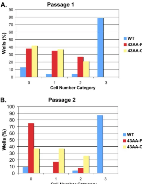
43AAmutant, we performed a type of longitudinal study. As soon as cell populations had divided enough in selective media after transformation, two transformed wells at high density, having coordinates C9 and F2, were arbitrarily chosen. From these wells, single cells were isolated into a drop of medium. This was repeated for a total of 48 isolated cells, which were then allowed to grow for 24 h. The number of cells present in each drop after 24 h was quantified into groups having no apparent cell division (category 0), 2 to 10 cells (category 1), 11 to⬃100 cells (category 2), or⬎⬎100 cells (category 3) (Fig. 4). This was referred to as passage 1 (Fig. 4A). In such a study, the WT cell number is theoretically 256, as the doubling time is roughly 3 h, or 8 doublings in 24 h.
The results showed that after passage 1, about 40% of the isolated cells failed to divide at all, and about 35% went through only approximately three cell divisions (Fig. 4). The remainder of the cells had more vigorous growth, but all grew to fewer than 100 cells, never exhibiting WT growth (category 3). In all wells of the ter1-43AA mutant, regardless of the number of divisions, cells were abnormally large and appeared pear shaped with protrusions, a cellular phenotype that was informally correlated with micronuclear anaphase (data not shown). In addition, the mutant cells had an altered pattern of
swimming, indicative of morphological changes and cellular survival.
Cells isolated from the two transformed wells were equally blocked in cell division and displayed phenotypes similar to those of one another. In contrast, most of the WT cells went through approximately the expected number of divisions, as about 80% of the wells had roughly 100 to 250 cells that appeared normal. In a few of these wells, cells grew less than expected but were phenotypically normal or smaller than usual.
We next determined whether the cells that grew fairly nor-mally (although the population appeared phenotypically mu-tant) continued to divide or whether replication of these cells slowed down. This was done by repeating the passaging of single cells. We isolated single cells from theter1-43AAmutant from the densest-growing wells in passage 1, having 11 to⬃100 cells (category 2). We used WT control cells from category 3. The mutant cells, which appeared phenotypically abnormal and correlated to micronuclear anaphase from the onset, were allowed to grow for an additional 24 h, and the cell number was quantified as before. This was referred to as passage 2 (Fig. 4B). In this analysis, the cells from F2 showed a dramatic increase in the proportion that was blocked. Nearly 75% of these cells did not go through another division. Furthermore, only 18% of the cells went through roughly three divisions in the second 24-hour period (passage 2), and only 8% divided close to normally. This finding was verified by observing cells from passage 1 that had continued to grow for a second 24-hour period. Here, most cells did not increase in numbers, indicating that division was still blocked. Surprisingly, the cells that stayed in category 1, having divided fewer than three times, continued to show abnormal swimming patterns. Thus, these cells went through as few as three divisions and survived for 2 days. These results are consistent with findings that mu-tant cell populations stay in anaphase, on average, 30 times longer than WT populations (27).
Our results show that the majority ofter1-43AAmutants go through very few cell divisions. Even the cells that do go through a number of cell divisions ultimately block with a pronounced cellular phenotype. Thus, even the strongly divid-ing cells give rise to progeny that do not divide.
Telomere mutant pauses during oral development.We took advantage of this severe block in micronuclear mitosis to assess the developmental consequences on cortical development. To our knowledge, there have been no studies correlating mutant telomeres with developmental programs, such as stomatogen-esis, in an organism likeT. thermophila. When Tetrahymena
cells divide, they must not only replicate their nuclear contents but also undergo a dramatic proliferation and patterning of their cortical architecture, resulting in the formation of a new oral apparatus in the posterior division product. The details of stomatogenesis are well documented (12).
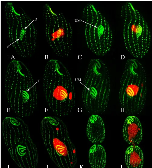
Using an antibody that recognizes basal bodies, we followed the progression of basal-body proliferation and assembly into oral membranelles from WT cells and noted the correspon-dence to stages of micronuclear mitosis by staining with pro-pidium iodide (Fig. 5). Basal bodies in the oral primordium are formed from a uniform anarchic field. Within this field, basal bodies begin aligning into promembranelles as basal-body dou-blets, as the micronucleus migrates away from the
macronu-FIG. 4. Longitudinal study shows that most single cells from ter1-43AAdo not divide. Single cells were isolated into wells and allowed to grow for 24 h (passage 1) (A) or were taken from the densest wells in passage 1 and allowed to grow for 24 h (passage 2) (B). Cell numbers were quantified and placed into the following categories: category 0, representing 0 or 1 cells; category 1, representing 2 to 10 cells; category 2, representing 11 to⬃100 cells; and category 3, representing⬎⬎100 cells.
on September 8, 2020 by guest
http://ec.asm.org/
cleus and elongates (Fig. 5A and B). After promembranelles have formed, basal-body triplet formation is initiated and the undulating membrane (UM) begins to emerge. The elongate micronucleus is no longer associated with the macronucleus
(Fig. 5C and D). The triplet membranelles begin to straighten, and the UM becomes distinct, yet anarchic basal bodies persist at the posterior of the field. At this point, the micronucleus elongates into a midanaphase configuration (Fig. 5E and F).
FIG. 5. Confocal fluorescence imaging of basal-body organization clearly shows the developing oral primordium in the WT. Basal bodies are labeled green, and chromatin is labeled red. Images are paired showing basal-body configurations in the left-hand panels (A, C, E, G, I, K) and composite images of nuclear and cortical features in the right-hand panels (B, D, F, H, J, L). Arrows indicate basal-body singlet (S), basal-body doublet (D), basal-body triplet (T), and UM.
on September 8, 2020 by guest
http://ec.asm.org/
As the UM is completed, the anarchic basal-body field is nearly all incorporated (or resorbed). At this point, the micronucleus is in the late stage of anaphase, where spindle elongation produces two distinct micronuclei (Fig. 5G and H). The UM is completed, and membranelle “sculpturing” takes place. Micro-nuclei now appear round, the macronucleus has begun to elon-gate, and the cytokinetic furrow is just noticeable (Fig. 5I and J). Cytokinesis and macronuclear division are well under way. Development of the oral primordium is complete (Fig. 5K and L).
T. thermophila cells expressing the ter1-43AA telomerase
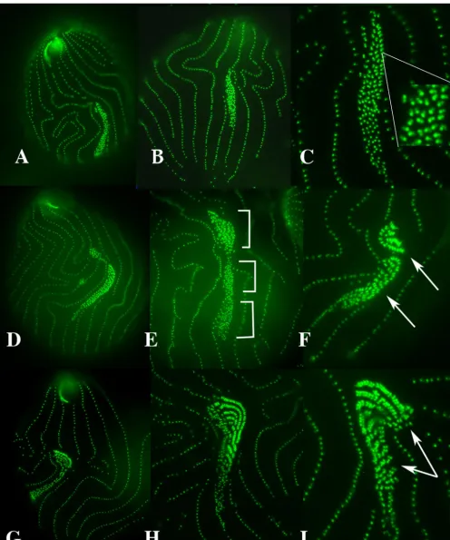
RNA template mutation reveal an extraordinary cortical phe-notype associated with stomatogenesis (Fig. 6). We found that cells arrested in the cell cycle exhibited profoundly elongate oral primordia. Ciliary rows show buckling due to continued basal-body proliferation without cell division (Fig. 6B). The basal bodies within the oral primordia showed one of three configurations, corresponding to early, middle, and late stages of oral development. The early-stage oral primordia (Fig. 6A to C) showed basal bodies proliferating, and at the anterior end of the basal-body field, basal-body doublets appeared to be forming (for example, see basal bodies between arrows in Fig. 6C). Each basal body appears to be nucleating the formation of its basal-body partner. This is particularly clear in Fig. 6C. Here, each doublet appears as a single brightly staining basal body coupled with a faintly staining and diminutive partner (Fig. 6C, insert).
Otherter1-43AAtelomerase mutants appeared to progress a bit further. Figure 6D to F show basal-body doublets that are just beginning to organize into promembranelles (Fig. 6F). Two other features appear in these preparations. First, there appears to be a gradient of development, and to variable ex-tents, these elongate fields appear broken into multiple dis-crete oral primordia. The earliest stage (mere basal-body pro-liferation) occurs in the posterior-most fields and corresponds to stage 1 (Fig. 6E, bottom bracket). More-anterior basal bod-ies become tightly packed, corresponding to stage 2 (Fig. 6E, middle bracket). Even-more-anterior basal-body doublets have formed (Fig. 6E, top bracket), and in the most-anterior fields of some cells, doublets have assembled into promembranelles (linear assemblages of aligned basal-body doublets [Fig. 6F and G to I]). Within the most advanced oral primordia, we could see discrete breaks forming in the abnormally long promembranelles (Fig. 6H and I).
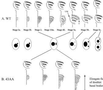
Two events that were conspicuously rare (or nonexistent) were the formation of a flanking UM and the addition of the third row of basal bodies that transform promembranelles into mature membranelles. This allows us to pinpoint the stage in oral development at which these cells become arrested. We have drawn a schematic diagram to indicate this stage (Fig. 7) and to further clarify the stages of oral development linked to nuclear division. The UM first makes its appearance at the same stage that the third row of basal bodies is assembled, namely, stage 4B (Fig. 7). Cells expressing theter1-43AA mu-tant telomerase become arrested at or before stage 4A in oral development, a stage at which the micronucleus shows an elon-gated football form yet has not begun to undergo spindle elongation. The cortical phenotype suggests that while these mutants are prevented from advancing through the cell cycle,
the dynamics of cortical development may be said to be ar-rested in an active, proliferative state.
In summary, the ter1-43AA telomerase template mutation undergoes a “dynamic pause” in oral development. During cell cycle arrest, basal bodies continue to proliferate nucleating synthesis of their doublet partners, and the resulting doublets align into promembranelles, yet no UM is formed, and promembranelles fail to assemble a third row characteristic of a mature membranelle.
DISCUSSION
Here, we have shown that by forcingTetrahymena cells to express a mutant telomerase RNA gene (ter1-43AA), it is the micronuclear telomeres, and not the macronuclear telomeres, which undergo a substantial loss of telomeric DNA. This find-ing is consistent with phenotypic evidence (25, 40; this study), where it was demonstrated that a block in micronuclear ana-phase is the first defect observed. Although it is still possible that the macronuclear telomeres may cause a defect that goes undetected in these mutant cells, the initial cause is most likely due to the shortening of the micronuclear telomeres.
Many characteristics of the micronucleus not seen previously (40) are consistent with end joining at the telomeres. Specifi-cally, over three-quarters of the cells displayed condensed chromatin, most of which remained joined from one pole to another. Most of the micronuclei in anaphase also had an intact mitotic spindle apparatus. In addition, single-cell isola-tion indicated that most cells were unable to undergo even one cell division after 48 h. However, a small percentage of cells divided normally many times but were ultimately blocked. Fi-nally, we have identified an aspect of cortical development that manifests a profound “dynamic pause” during oral develop-ment.
Telomeres and cell division checkpoints in anaphase. Ac-cording to one hypothesis (Fig. 8), the mutated ter1-43AA
template must initially create micronuclear telomere tracks with altered sequences, which are prone to degradation. These shortened telomeres fail to interact properly with end-protec-tive proteins. As the ends erode due to endogenous exonucle-ase activity, they associate with the DNA repair mechanism, and chromosome end joining is promoted. The resulting chro-mosome bridges may trigger a midanaphase checkpoint due to tension induced during centromere fission and chromosome progression. It is possible that the mutated macronuclear telo-meres may have some effect of their own, for example, leading to a high proportion of cells with an elongated macronucleus that is blocked in amitosis, but this has not been observed.
In a related study (24), the telomere protection protein POT1a was depleted in live Tetrahymena cells. POT1a was shown to localize over macronuclei, but not micronuclei, and depletion resulted in lengthened macronuclear telomeres. A cell cycle arrest was associated with these mutants as well, suggesting another type of mitotic checkpoint. This cell cycle arrest appears to be in macronuclear S phase (micronuclear G2), in that cells continue growing and their macronuclear
ploidy increases, yet there are no signs of cortical development indicating entry into mitosis. It would seem that extreme lengthening of macronuclear telomeres may be associated with
on September 8, 2020 by guest
http://ec.asm.org/
FIG. 6. Fluorescence microscopy of basal bodies shows dramatic defect in oral morphogenesis in theter-43AAmutant. Mutant cells arrested with early-stage oral primordia (A to C). Persistent basal-body proliferation is under way, and at the anterior end of the basal-body field, basal-body doublets are forming (note insert). Oral primordia arrested at a later stage: basal-body doublets are organizing into promembranelles (D to F; see arrows in panel F). The bottom bracket in panel E shows the posterior-most field with the earliest stage of development (basal-body proliferation). The middle bracket (E) shows tight packing of the “anarchic field.” The top bracket (E) shows the most anterior fields of some cells, with doublets arranged into promembranelles. The most advanced arrested oral primordia appear in panels G to I. Multiple rows of basal-body doublets appear (I).
on September 8, 2020 by guest
http://ec.asm.org/
an interphase arrest, whereas shortening of micronuclear chro-mosomes results in a midanaphase mitotic arrest.
One question raised by our analysis is how shortened telo-meres lead to a cell cycle arrest. In tumor cells of vertebrates, telomeres are progressively shortened (6) and are subject to instability. Telomere shortening can result in chromosome end joining, leading to the formation of anaphase bridges (20, 21, 33). Indeed, many cancers are characterized by a destructive
cycle of chromosome bridging, fusion, and breakage (6, 42). It has been shown that the frequency of anaphase bridges is directly dependent upon the level of telomerase activity and the subsequent telomere length (41). It is possible that as telomeres shorten to some critical length, the chromosome ends are recognized as “broken ends,” thereby activating the DNA repair machinery, and it is this that results in end-to-end recombination, anaphase bridges, and possibly activation of a cell cycle checkpoint arrest (1, 18, 19).
Several laboratories have explored telomere-associated checkpoints inSaccharomyces cerevisiaeby analyzing telome-rase-defective cell lines. In the telomerase RNA mutanttlc1⌬
ofS. cerevisiae, telomeres gradually shorten, and cells become
arrested somewhere in the G2/M transition (4, 8). These
stud-ies suggest that defective-telomere-induced cell cycle arrest may be mediated through the DNA damage checkpoint mech-anism. In this model, as telomeres become progressively short-ened, the chromosome ends become exposed to the surveil-lance mechanism that detects double-stranded breaks, and this activates the DNA damage checkpoint at the G2/M transition.
If this model is accurate, then the cell division arrest that is triggered in cells undergoing DNA damage should resemble the cell division arrest phenotype seen in telomerase-defective mutants. DNA damage can lead to three different cell division
FIG. 7. A “Dynamic pause” occurs in anaphase-arrested cells compared to WT cells. (A) WT cortical development (stages of basal-body proliferation and stomatogenesis) is linked with both micronuclear and macronuclear events and cytokinesis. Based on data from reference 12. (B) Theter1-43AA mutant undergoes a “dynamic pause.” Three processes continue to operate, although cortical development is arrested somewhere near the end of stomatogenic stage 4A. Basal bodies continue to proliferate. The oral morphogenetic field extends down the postoral ciliary row, recruiting ever-more-posterior basal bodies to initiate replication. Basal bodies within the growing anarchic field nucleate formation of their doublet partners in more-anterior regions of the oral primordium, and doublets align with one another, forming linear arrays or “promembranelles” in the most anterior regions of the oral primordium.
FIG. 8. This schematic summarizes our hypothesis regarding how a mutant telomerase template leads to a midanaphase arrest in Tetrahy-menacells. Shortened telomeres fail to interact properly with protec-tive proteins, ultimately leading to telomere fusions. Bridges formed from these fusions trigger midanaphase arrest of the micronucleus, which normally would go through mitotic division before macronuclear amitosis. This checkpoint mediates arrest of cortex patterning and continuation of the cell division cycle.
on September 8, 2020 by guest
http://ec.asm.org/
arrests inSaccharomyces(30), depending on when DNA dam-age is induced, at G1/S, intra-S, or G2/M. None of these studies
point to a midanaphase arrest, and in fact, most lack the detailed cellular resolution necessary to distinguish a midana-phase arrest from an earlier metamidana-phase/anamidana-phase arrest.
One exception is where researchers demonstrated the exis-tence of a midanaphase checkpoint in yeast that is dependent upon Rad9, part of the DNA damage checkpoint surveillance machinery (45). This could be caused by a chromosome bridge triggered by a dicentric chromosome or, in our case (Fig. 8), by chromosome end joining, Supporting this possibility is a recent finding that mammalian Rad9 has in fact been implicated in telomere stability and its loss associated with end joining (38, 39).
One anaphase checkpoint or two?Little work has been done to characterize the DNA damage/repair mechanisms in
Tetra-hymenacells, yet one study is provocative. It was demonstrated
that RAD51, which participates in double-stranded-DNA re-pair, is necessary for vegetative maintenance of the micronu-cleus (31). Cells bearing the RAD51-null mutation exhibit a mitotic defect in which the micronucleus is arrested in early anaphase while the cell proceeds through cytokinesis (32). This results in segregation of the entire (arrested) anaphase spindle to one daughter cell or the other. This suggests that Tetrahy-menacells exhibit two distinct types of anaphase arrest.
In the telomerase mutant phenotype, end-joining bridges may delay chromosome segregation, leading to an arrest block-ing the cell’s progression into cytokinesis and macronuclear fission. We attempted to determine if the telomeres of the micronucleus had fused to each other by PCR, but we were unable to detect a fusion product (unpublished observations). In Rad51-null mutants, DNA damage is presumably incurred during synthesis and left unrepaired, and this triggers a check-point arrest early during anaphase elongation without blocking the cell’s progression into cytokinesis. It is tempting to propose that the Rad51 defect-induced anaphase arrest occurs prior to spindle elongation and is restricted to the micronuclear divi-sion program. In contrast, the telomere defect-induced ana-phase arrest permits the middle stages of anaana-phase elongation, while blocking progress into the final stages of anaphase elon-gation and subsequent cytokinesis. It is curious that the pro-gram leading to cytokinesis and macronuclear division should be so tightly coupled with the midstage anaphase checkpoint (that triggered by telomerase defect and putative telomere cross-bridges), while being uncoupled from the earlier ana-phase checkpoint (that induced by the Rad51 defect and pu-tative unrepaired DNA damage).
Telomeres affect development of cell cortex.The telomerase phenotype sheds light on the events that accompany normal cell division in Tetrahymena strains. In particular, we have gained several new insights into the events that build an oral primordium during the process of cortical stomatogenesis. First, it has not been perfectly clear whether basal-body dou-blets form by aggregation of existing basal bodies or whether existing basal bodies nucleate de novo synthesis of their part-ners-to-be. The anti-centrin antibody appears to discriminate between two sets of basal bodies: large, brightly staining basal bodies (such as one sees in the mature ciliary row [Fig. 6C]) and more-faintly staining, smaller basal bodies (such as one sees anterior and to the viewer’s right of the mature, ciliary row
basal bodies). The latter clearly represent newly formed struc-tures. If doublets formed through aggregation or recruitment of already mature basal bodies, then one would expect each partner in a doublet to stain equally brightly. The most sensible interpretation of these images is that each mature basal body has nucleated the synthesis of a partner that appears dim and underdeveloped at this stage. Most previous studies on WT oral development have used silver staining or scanning elec-tron microscopy images or an antibody to an uncharacterized basal-body epitope. It is likely that this phenomenon that we observed escaped earlier notice due to differences in imaging techniques and their more exaggerated manifestations unique to this phenotype.
Second, there appears to be a process whereby long promembranelles are severed into shorter segments. In WT cells, this probably occurs only once per membranelle and might easily be missed. In the overelongated membranelles of our mutants, multiple clipping sites can be seen (Fig. 6H and I). Promembranelle severing appears to occur in stage 4A of oral development, prior to triplet assembly. Here, we see sev-ering only after doublet promembranelles are well formed and prior to third-row basal-body addition. This restricts the pos-sibility to early stage 4. Third, given earlier results demonstrat-ing that these telomerase mutants are arrested in midanaphase and our current results demonstrating that such cells are ar-rested in cortical stage 4A, it appears that we can infer that cortical stage 4A corresponds to midanaphase, whereas stage 4B may stand at the gateway to late anaphase and the spindle elongation that occurs as one enters stage 5A.
Finally, it is interesting to note that a cell cycle arrest can be viewed as a “dynamic pause.” That is to say, while progress through the cell division cycle has been arrested, the cortical “subroutines” associated with a given developmental stage continue to operate. Though our telomerase mutants cannot initiate the final stages of spindle elongation associated with late anaphase, nor can they initiate formation of the UM or the proliferation of third-row basal bodies, processes active during midanaphase continue: basal bodies continue to replicate, these then continue to nucleate doublet assembly, and these doublets are organized into ever longer promembranelles, which may then be severed at appropriate lengths. The result is the continuing growth of an exaggerated oral primordium and the continued maturation and patterning of its members up to a set point in development.
This novel study correlates the complexities of telomere-induced nuclear division properties with downstream develop-mental events inTetrahymena thermophila. Future studies with
T. thermophilacould shed light on the development and
mor-phogenesis of human cancer cells with altered telomeres.
ACKNOWLEDGMENTS
We are grateful to Anamitra Bhattacharyya for comments on the manuscript, Joseph Frankel for illuminating discussion of the cortical phenotype, and Daniel Romero for helpful discussions of the Rad51 anaphase arrest phenotype.
This work was supported by grants from the National Science Foun-dation, one entitled “The Gene Stream: from sequence to cell func-tion, MCB-0444700,” awarded to Eric S. Cole, and the other entitled “Telomere Dynamics in Research and Education, MCB-9876126,” awarded to Karen E. Kirk.
on September 8, 2020 by guest
http://ec.asm.org/
REFERENCES
1.Bailey, S. M., and E. H. Goodwin.2004. DNA and telomeres: beginnings and endings. Cytogenet. Genome Res.104:109–115.
2.Berger, J. D.2001. Riding the ciliate cell cycle—a thirty-five-year prospec-tive. J. Eukaryot. Microbiol.48:505–518.
3.Blackburn, E. H.1990. Telomeres: structure and synthesis. J. Biol. Chem.
265:5919–5921.
4.Chen, Q., A. Ijpma, and C. W. Greider.2001. Two survivor pathways that allow growth in the absence of telomerase are generated by distinct telomere recombination events. Mol. Cell. Biol.21:1819–1827.
5.Collins, K.1999. Ciliate telomerase biochemistry. Annu. Rev. Biochem.
68:187–218.
6.Counter, C. M., A. A. Avilion, C. E. LeFeuvre, N. G. Stewart, C. W. Greider, C. B. Harley, and S. Bacchetti.1992. Telomere shortening associated with chromosome instability is arrested in immortal cells which express telomer-ase activity. EMBO J.11:1921–1929.
7. Reference deleted.
8.Enomoto, S., L. Glowczewski, and J. Berman.2002. MEC3, MEC1, and DDC2 are essential components of a telomere checkpoint pathway required for cell cycle arrest during senescence in Saccharomyces cerevisiae. Mol. Biol. Cell13:2626–2638.
9. Reference deleted.
10.Frankel, J.1999. Cell biology ofTetrahymena thermophila, p. 27–125.InD. J. Asai and J. D. Forney (ed.), Methods in cell biology, vol. 62. Academic Press, San Diego, CA.
11.Frankel, J., L. M. Jenkins, and L. E. DeBault.1976. Causal relations among cell cycle processes in Tetrahymena pyriformis. An analysis employing tem-perature-sensitive mutants. J. Cell Biol.71:242–260.
12.Frankel, J., and E. M. Nelsen.2001. The effects of supraoptimal tempera-tures on population growth and cortical patterning in Tetrahymena pyrifor-mis and Tetrahymena thermophila: a comparison. J. Eukaryot. Microbiol.
48:135–146.
13.Frankel, J., E. M. Nelsen, and L. M. Jenkins.1977. Mutations affecting cell division inTetrahymena pyriformis, syngen 1. II. Phenotypes of single and double homozygotes. Dev. Biol.58:255–275.
14.Fujiu, K., and O. Numata.1999. Localization of microtubules during ma-cronuclear division in Tetrahymena and possible involvement of macro-nuclear microtubules in “amitotic” chromatin distribution. Cell Struct. Funct.24:401–404.
15.Fujiu, K., and O. Numata.2000. Reorganization of microtubules in the amitotically dividing macronucleus of tetrahymena. Cell Motil. Cytoskeleton
46:17–27.
16.Gaertig, J., and M. Gorovsky.1992. Efficient mass transformation of Tetra-hymena thermophilaby electroporation of conjugants. Proc. Natl. Acad. Sci. USA89:9196–9200.
17.Gaertig, J., and G. Kapler.2000. Transient and stable DNA transformation of Tetrahymena thermophila by electroporation. Methods Cell Biol.62:485– 500.
18.Gire, V.2004. Dysfunctional telomeres at senescence signal cell cycle arrest via Chk2. Cell Cycle3:1217–1220.
19.Gire, V., P. Roux, D. Wynford-Thomas, J. M. Brondello, and V. Dulic.2004. DNA damage checkpoint kinase Chk2 triggers replicative senescence. EMBO J.23:2554–2563.
20.Gisselsson, D., J. Bjork, M. Hoglund, F. Mertens, P. Dal Cin, M. Akerman, and N. Mandahl.2001. Abnormal nuclear shape in solid tumors reflects mitotic instability. Am. J. Pathol.158:199–206.
21.Gisselsson, D., T. Jonson, A. Petersen, B. Strombeck, P. Dal Cin, M. Hoglund, F. Mitelman, F. Mertens, and N. Mandahl.2001. Telomere dys-function triggers extensive DNA fragmentation and evolution of complex chromosome abnormalities in human malignant tumors. Proc. Natl. Acad. Sci. USA98:12683–12688.
22. Reference deleted.
23.Hamilton, E. P., and E. Orias.2000. Genetic crosses: setting up crosses, testing progeny, and isolating phenotypic assortants. Methods Cell Biol.
62:219–228.
24.Jacob, N. K., R. Lescasse, B. R. Linger, and C. M. Price.2007.Tetrahymena
POT1a regulates telomere length and prevents activation of a cell cycle checkpoint. Mol. Cell. Biol.27:1592–1601.
25.Karrer, K. M.2000. Tetrahymena genetics: two nuclei are better than one. Methods Cell Biol.62:127–186.
26.Kirk, K. E., and E. H. Blackburn.1995. An unusual sequence arrangement in the telomeres of the germ-line micronucleus inTetrahymena thermophila. Genes Dev.9:59–71.
27.Kirk, K. E., B. P. Harmon, I. K. Reichardt, J. W. Sedat, and E. H. Blackburn.
1997. Block in anaphase chromosome separation caused by a telomerase template mutation. Science275:1478–1481.
28.LaFountain, J. R., Jr., and L. A. Davidson.1979. An analysis of spindle ultrastructure during prometaphase and metaphase of micronuclear division in Tetrahymena. Chromosoma75:293–308.
29.LaFountain, J. R., Jr., and L. A. Davidson.1980. An analysis of spindle ultrastructure during anaphase of micronuclear division in Tetrahymena. Cell Motil.1:41–61.
30.Longhese, M. P., M. Foiani, M. Muzi-Falconi, G. Lucchini, and P. Plevani.
1998. DNA damage checkpoint in budding yeast. EMBO J.17:5525–5528. 31.Marsh, T. C., E. S. Cole, and D. P. Romero.2001. The transition from
conjugal development to the first vegetative cell division is dependent on RAD51 expression in the ciliate Tetrahymena thermophila. Genetics157:
1591–1598.
32.Marsh, T. C., E. S. Cole, K. R. Stuart, C. Campbell, and D. P. Romero.2000. RAD51is required for propagation of the germinal nucleus inTetrahymena thermophila. Genetics154:1587–1596.
33.McClintock, B.1941. The stability of broken ends of chromosomes in Zea mays. Genetics26:234–282.
34. Reference deleted.
35.Nagata, K., T. Takemasa, S. Alam, T. Hattori, Y. Watanabe, and Y. Nozawa.
1994. Cloning of cDNAs encoding a cell-cycle-regulatory GTP-binding low-Mr(GBLM) protein, Ran/TC4, from micronucleated Tetrahymena
ther-mophilaand amicronucleatedTetrahymena pyriformis. Gene144:123–125. 36.Numata, O., K. Fujiu, and K. Gonda.1999. Macronuclear division and
cytokinesis in Tetrahymena. Cell Biol. Int.23:849–857.
37.Numata, O., K. Gonda, A. Watanabe, and Y. Kurasawa.2000. Cytokinesis in Tetrahymena: determination of division plane and organization of contrac-tile ring. Microsc. Res. Tech.49:127–135.
38.Pandita, R. K., G. G. Sharma, A. Laszlo, K. M. Hopkins, S. Davey, M. Chakhparonian, A. Gupta, R. J. Wellinger, J. Zhang, S. N. Powell, J. L. Roti Roti, H. B. Lieberman, and T. K. Pandita.2006. Mammalian Rad9 plays a role in telomere stability, S- and G2-phase-specific cell survival, and homol-ogous recombinational repair. Mol. Cell. Biol.26:1850–1864.
39.Pandita, T. K.2006. Role of mammalian Rad9 in genomic stability and ionizing radiation response. Cell Cycle5:1289–1291.
40.Petcherskaia, M., J. M. McGuire, J. M. Pherson, and K. E. Kirk.2003. Loss of cap structure causes mitotic defect in Tetrahymena thermophila telo-merase mutants. Chromosoma111:429–437.
41.Plug-DeMaggio, A. W., T. Sundsvold, M. A. Wurscher, J. I. Koop, A. J. Klingelhutz, and J. K. McDougall.2004. Telomere erosion and chromo-somal instability in cells expressing the HPV oncogene 16E6. Oncogene
23:3561–3571.
42.Stewenius, Y., L. Gorunova, T. Jonson, N. Larsson, M. Hoglund, N. Man-dahl, F. Mertens, F. Mitelman, and D. Gisselsson.2005. Structural and numerical chromosome changes in colon cancer develop through telomere-mediated anaphase bridges, not through mitotic multipolarity. Proc. Natl. Acad. Sci. USA102:5541–5546.
43.Stuart, K. R., and E. S. Cole.1999. Nuclear and cytoskeletal fluorescence microscopy techniques, p. 291–312.InD. J. Asai and J. D. Forney (ed.), Tetrahymena thermophila, vol. 62. Academic Press, San Diego, CA. 44.Wei, Y., C. A. Mizzen, R. G. Cook, M. A. Gorovsky, and C. D. Allis.1998.
Phosphorylation of histone H3 at serine 10 is correlated with chromosome condensation during mitosis and meiosis in Tetrahymena. Proc. Natl. Acad. Sci. USA95:7480–7484.
45.Yang, S. S., E. Yeh, E. D. Salmon, and K. Bloom.1997. Identification of a mid-anaphase checkpoint in budding yeast. J. Cell Biol.136:345–354. 46.Yu, G. L., J. D. Bradley, L. D. Attardi, and E. H. Blackburn.1990. In vivo
alteration of telomere sequences and senescence caused by mutated Tetra-hymenatelomerase RNAs. Nature344:126–132.