The Buoyancy of
Cryptococcus neoformans
Is Affected by
Capsule Size
Raghav Vij,
aRadames J. B. Cordero,
aArturo Casadevall
aaDepartment of Molecular Microbiology and Immunology, Johns Hopkins Bloomberg School of Public Health,
Baltimore, Maryland, USA
ABSTRACT
Cryptococcus neoformans
is an environmental pathogenic fungus with a
worldwide geographical distribution that is responsible for hundreds of thousands of
human cryptococcosis cases each year. During infection, the yeast undergoes a
mor-phological transformation involving capsular enlargement that increases microbial
volume. To understand the factors that play a role in environmental dispersal of
C.
neoformans
and
C. gattii
, we evaluated the cell density of
Cryptococcus
using Percoll
isopycnic gradients. We found differences in the cell densities of strains belonging
to
C. neoformans
and
C. gattii
species complexes. The buoyancy of
C. neoformans
strains varied depending on growth medium. In minimal medium, the cryptococcal
capsule made a major contribution to the cell density such that cells with larger
capsules had lower density than those with smaller capsules. Removing the capsule,
by chemical or mechanical methods, increased the
C. neoformans
cell density and
reduced buoyancy. Melanization of the
C. neoformans
cell wall, which also
contrib-utes to virulence, produced a small but consistent increase in cell density.
Encapsu-lated
C. neoformans
sedimented much more slowly in seawater as its density
ap-proached the density of water. Our results suggest a new function for the capsule
whereby it can function as a flotation device to facilitate transport and dispersion in
aqueous fluids.
IMPORTANCE
The buoyancy of a microbial cell is an important physical
characteris-tic that may affect its transportability in fluids and interactions with tissues during
infection. The polysaccharide capsule surrounding
C. neoformans
is required for
in-fection and dissemination in the host. Our results indicate that the capsule has a
significant effect on reducing cryptococcal cell density, altering its sedimentation in
seawater. Modulation of microbial cell density via encapsulation may facilitate
dis-persal for other important encapsulated pathogens.
KEYWORDS
Cryptococcus neoformans
, buoyancy, capsular polysaccharide, yeast
density
C
ryptococcus neoformans
and
C. gattii
species complexes are important fungal
pathogens that can cause pulmonary and meningeal disease in humans (1). In the
environment,
C. neoformans
is commonly found in soil associated with pigeon excreta,
while
C. gattii
is most commonly found on trees (2, 3).
C. gattii
isolates have been
collected from marine and fresh water environments (4, 5). Cryptococcal infection
occurs via the respiratory tract, where yeast particulates can colonize the lungs (6, 7).
In immunocompromised patients,
C. neoformans
can disseminate from the lungs to
other parts of the body, including the central nervous system, by crossing the blood
brain barrier. The dissemination of
C. neoformans
yeast cells from the lung to the brain
is critical in the development of meningeal disease. The yeast cells can undergo drastic
morphological changes that allow the pathogen to evade host immune response. For
instance, yeast cells can modulate capsule and cell body dimensions in response to
Received27 September 2018Accepted17 October 2018 Published7 November 2018
CitationVij R, Cordero RJB, Casadevall A. 2018. The buoyancy ofCryptococcus neoformansis affected by capsule size. mSphere 3:e00534-18. https://doi.org/10.1128/mSphere.00534-18.
EditorJ. Andrew Alspaugh, Duke University Medical Center
Copyright© 2018 Vij et al. This is an open-access article distributed under the terms of theCreative Commons Attribution 4.0 International license.
Address correspondence to Arturo Casadevall, [email protected].
C. neoformans polysaccharide capsule reduces cell density and may serve as a flotation device. @ACasadevall1
RESEARCH ARTICLE
Host-Microbe Biology
crossm
on September 8, 2020 by guest
http://msphere.asm.org/
environmental conditions such that cell dimensions can range from 1 to 100
m in
diameter (8–11).
The polysaccharide (PS) capsule is composed mostly of water (12). It is formed by a
porous matrix of branched heteropolysaccharides, mainly glucuronoxylomannan, that
extends radially from the cell wall (13). Capsule synthesis is induced under certain
stressful conditions and provides protection against host defense mechanisms by
acting as a physical barrier, interfering with phagocytosis and sequestering reactive
oxygen species (ROS) and drugs (14, 15). The capsule is essential for the virulence of
C.
neoformans
and is of interest for both therapeutic and diagnostic strategies (16).
Melanin is another important virulence factor, such that strains that lack the ability
to melanize are less pathogenic (16). Melanin is formed by the polymerization of
aromatic and/or phenolic compounds, including
L-3,4-dihydroxyphenylalanine (L-DOPA),methyl-DOPA, and epinephrine or norepinephrine (17). In the presence of
catechol-amine precursors found in the human brain,
Cryptococcus
melanizes its inner cell wall
(18). Melanized
C. neoformans
cells are found in the environment (19) and during
mammalian infection (20), suggesting an important role of the pigment in
C.
neofor-mans
biology and pathogenesis. Melanization protects cells against a variety of host
immune mechanisms and antifungal drugs, as well as against radiation, desiccation,
ROS, and temperature stress (21, 22).
Both the polysaccharide capsule and melanin are complex structures that are
difficult to study. Consequently, it is important to apply biophysical methodologies to
gain new insights into the physicochemical properties and biological functions of these
major virulence factors (23). One such property that has not been studied in
crypto-coccal biology is cellular density, presumably a highly regulated characteristic that may
reflect the physiological state of the cell under different conditions (24).
In the first century B.C., Roman writer Vitruvius described a “eureka” moment that
the Greek polymath Archimedes had when, allegedly, he observed the displacement of
water as he sat in a bathtub, which led him to establish the law of buoyancy (25, 26).
In a biological context, Archimedes’ law (law of buoyancy) can be applied to calculate
the ratio of the absolute mass and volume of an organism which could determine
whether it floats or sinks in a fluid of given density. During centrifugation in a
continuous Percoll density gradient, cells equilibrate upon reaching the point at which
the gradient’s density matches their own. This allows us to estimate cell density of
C.
neoformans
and
C. gattii
against bead standards of fixed density.
Cell density is used for the separation of cell populations, but the factors regulating
cell density in microbiology remain understudied, despite the important role that it
may play in the migration and dissemination of microbial and mammalian cells in fluids.
This could be because the cell density depends on many biological and physical factors,
which are often difficult to disentangle. Earlier studies found that cell density was
affected by the osmolality of the medium in which the cells are grown (27, 28), the
encapsulation of bacteria by polysaccharide capsule (29), and the cell cycle stage (30).
Strains of
Porphyromonas gingivalis
with lower cell density were less susceptible to
phagocytosis; however, this could be the result of the correlation between cell density
and cell surface hydrophobicity (31). Other studies have also reported differences in cell
density among different strains of mycobacteria and
Burkholderia
spp. (32, 33). In the
context of eukaryotes,
Saccharomyces cerevisiae
cell density varies at different stages of
cell cycle (34), and quiescent
S. cerevisiae
cell populations can be separated out using
density gradients in a stationary-phase culture of the yeast (35).
The cell density of
C. neoformans
and the factors that affect it have not been
previously investigated. In this study, we used Percoll isopycnic gradients to study the
effect of capsule induction, antibody (Ab) treatment, and melanization on the cell
density of
C. neoformans
.
RESULTS
Comparison of
C. neoformans
and
C. gattii
cell densities.
Cell densities differed
consistently among different serotypes of
C. neoformans
and
C. gattii
species complex
on September 8, 2020 by guest
http://msphere.asm.org/
strains (Fig. 1C). The cell density of replicates showed significant variability in
compar-isons of
C. neoformans
serotype A (strain H99) to serotype D (strain ATCC 24067) and
serotype AD (strain 92.903).
C. gattii
strains showed less variability than the
C.
neofor-mans
isolates (Fig. 1C). To ascertain whether there was a relationship between cell
density and cell dimension, we imaged the cells with an India ink counterstain and
calculated both the capsule radii and cell body radii for the
C. neoformans
and
C. gattii
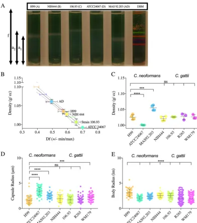
FIG 1 The cell density ofC. neoformansandC. gattiiserotypes. (A) Image representative of 3 to 4 independent repetitions of Percoll density gradients, comparing the cell densities of C. neoformans (serotypes A, D, and AD) and C. gattii (serotypes B and C) to that of density bead markers (DBM). (B) Representative data from 4 independent experiments, depicting the line interpolation of the density factor (minimum [min] and maximum [max]) calculated by pixel area per the following formulae: (f⫺a1/fandf⫺a2/f). The df (min, max) values of the density marker beads are used to estimate
the cell density of the cells run in parallel gradients. (C) Histogram depicting the differences in cell densities of different serotypes ofC. neoformans(serotypes A, AD, and D) andC. gattii(serotypes B and C and variants VGI and VGIIa). The experiment was performed 3 to 4 times, as indicated by the symbols on the bar graph. Error bars represent the SD about the mean. (D) Representative data of capsule (i) and cell body (ii) radii of different serotypes and strains (n⫽3). Error bars represent the SD about the mean. One-way analysis of variance (ANOVA) was used for the comparisons of the cell densities and capsule and cell body radii of different strains and serotypes ofC. neoformansandC. gattiito the respective measurements of strain H99 ofC. neoformans. All comparisons were made to strain H99 ofC. neoformans. The following symbols were used to annotate the statistical significance of the results: ns, P⬎0.05; *, Pⱕ0.05;**, Pⱕ0.01; ***, Pⱕ0.001;****,Pⱕ0.0001.
Buoyancy ofCryptococcus neoformans
on September 8, 2020 by guest
http://msphere.asm.org/
strains. We observed statistically significant differences in the cell body radii of all
strains compared to
C. neoformans
serotype A (strain H99) (Fig. 1E). We also observed
that the capsule radii of
C. neoformans
serotype D and AD and of
C. gattii
VG IIa were
significantly different from those of serotype A (Fig. 1D).
Effect of capsule induction on
C. neoformans
cell density.
In vitro
, the capsule is
induced under stress conditions such as exposure to nutrient starvation medium (36).
Cells grown in minimal medium (MM) had significantly lower density (Fig. 2A to C) than
cells grown under nutrient-rich conditions in Sabouraud dextrose broth (Sab) where
the capsule was significantly smaller. Cells of the acapsular strain
cap59
had
significantly higher density than encapsulated cells with the same genetic
back-ground. Furthermore, we observed no significant differences in the densities of
acapsular mutants grown in minimal versus rich medium, confirming the
contribu-tion of the polysaccharide capsule in determining the cell density in response to
different nutrient conditions.
Previous studies have reported on the molecular composition of the
C. neoformans
capsular polysaccharide by studying the polysaccharide isolated from the cell surface
by dimethyl sulfoxide (DMSO) extraction and gamma irradiation-induced capsule
shed-ding (37). To confirm the effects of the capsule on the cell density, encapsulated H99
cells were treated with gamma radiation and DMSO to remove capsular material
(Fig. 3). We observed a significant increase in cell density when the capsule was
removed by both treatments, indicating that the polysaccharide capsule influences the
cell density.
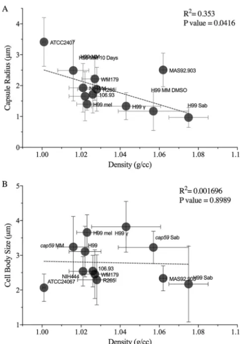
Capsule size correlates with cell density.
Linear regression analysis revealed that
the capsule radius correlates with cell density such that yeast cells with larger capsules
had lower cell density (Fig. 4A). We observed some scatter in the linear relationship
between capsule size and density, which presumably reflects other variables that may
contribute to the density such as differences in lipid, glycogen content, and
polysac-charide composition. There was no significant relationship between cell body size and
cell density (Fig. 4B).
Encapsulated
Cryptococcus neoformans
settles more slowly in seawater.
We
tested whether the lower density of encapsulated
C. neoformans
allowed the fungi to
float in water or seawater. The density of seawater is 1.0236 g/cc at room temperature
(38, 39), and the density of
C. neoformans
in MM is 1.0220
⫾
0.0082 g/cc. When
C.
neoformans
grown in MM was added to a cuvette containing seawater, a large
population of cells became suspended in the seawater, which became turbid (Fig. 5A).
This effect was not seen with phosphate-buffered saline (PBS), where the cells sank to
the bottom within 3 h (Fig. 5B).
Melanization increases
C. neoformans
cell density.
Comparison of melanized and
nonmelanized H99
C. neoformans
cells demonstrated that melanization was associated
with a moderate increase in cell density (Fig. 6). Since the increase in the density was
small and since melanized cells can easily be distinguished visually from nonmelanized
cells, we mixed the melanized and nonmelanized cells in 1:1 ratio before loading the
samples onto the density gradient. While nonmelanized cells displayed a range of
density that overlapped that of melanized cells, the latter tended to have higher
density than nonmelanized cells inoculated from the same Sab preculture. Isolated
melanin “ghosts” (40) had much greater density than the cells, estimated to be
⬎
1.1 g/cc (data not shown). Note that melanized cells also had smaller capsules
(Fig. 6D, panel ii), which might have contributed to the increase in cell density. Thus, we
also compared the densities of melanized and nonmelanized cells after removal of the
capsule by gamma radiation (Fig. 6C, panel ii). Upon capsule removal, we observed that
the density of melanized cells was consistently and significantly higher than that of
nonmelanized cells.
Antibody binding and other conditions that have no significant effect on
C.
neoformans
cell density.
Previous studies showed that capsular antibodies alter the
viscoelastic properties and structure of the capsule (41). Antibody binding also causes
on September 8, 2020 by guest
http://msphere.asm.org/
a change in the hydration state of the PS capsule (12). Treatment of H99
C.
neoformans
with capsular antibodies (18B7 and E1) did not significantly alter the
cell density (see Fig. S1 in the supplemental material). Furthermore, binding of
mouse complement, incubations at different salt concentrations (to induce osmotic
FIG 2 Induction of capsule synthesis decreasesC. neoformanscell density. (A) Image representative of three indepen-dent repetitions of Percoll density gradients, showing the density ofC. neoformansH99 juxtaposed with acapsular mutantcap59, both grown in Sab or MM. (B) Representative data from three independent experiments, depicting a line interpolation of the density factor with the cell densities of the bead standards to calculate the cell densities of the gradients run in parallel. (C) Histogram depicting a decrease in cell density of H99 cells grown in MM compared to Sab, due to capsule induction.cap59mutant cells are significantly denser than normal H99 cells grown in MM. Each data point on the histogram represents an independent replicate (n⫽3); error bars represent SD about the mean. (D) Represen-tative data depict (i) the capsule radii and (ii) the cell body radii ofC. neoformansH99 andcap59grown under different medium conditions (MM or Sab) (n⫽3). Straincap59cells grown in MM and Sab do not have a capsule; therefore, the capsule radii were not quantified. Error bars represent SD about the mean. One-way ANOVA was used for the comparisons of cell density, capsule density, and cell body radii ofC. neoformans cap59and H99 grown under different conditions. The following symbols were used to annotate the statistical significance of the results: ns,P⬎0.05;*, Pⱕ0.05;**,Pⱕ0.01;***,Pⱕ0.001;****,Pⱕ0.0001.
Buoyancy ofCryptococcus neoformans
on September 8, 2020 by guest
http://msphere.asm.org/
FIG 3 Removal ofC. neoformanspolysaccharide capsule increases cell density. (A) (i) Image representative of five independent repetitions of Percoll density gradients, comparing the cell densities of irradiated (␥) and nonirradiatedC. neoformans(H99) grown in MM for 10 days with a standard of colored uniform density beads. (ii) Image representative of four independent Percoll density gradients of encapsulated C. neoformansH99 strains grown in MM for 10 days before and after DMSO extraction. (B) Representative data from independent experiments, depicting a line interpolation of the density factor (df) with the cell densities of the bead standards, to calculate the cell densities ofC. neoformansbefore and after extraction of the capsule, run in parallel. (C) A histogram depicting cell density ofC. neoformansbefore and after capsule extraction by␥rays and DMSO. The experiments were performed 3 to 4 times independently as indicated by the symbols on the bar graph; error
(Continued on next page)
on September 8, 2020 by guest
http://msphere.asm.org/
stress), and incubation in lipid-rich medium had no significant impact on
C.
neoformans
cell density (Fig. S2).
DISCUSSION
In this study, we characterized the cell densities of
C. neoformans
and
C. gattii
under
different conditions. We report minor differences in cell densities between serotypes of
the cryptococcal species complex. Our results also suggest that the capsule plays a
major role in determining the cell density of the yeast such that encapsulated strains
have densities close to that of water. Meanwhile, melanization increased the density
slightly. Changes in buoyancy could influence the dispersal of the yeast in the
envi-ronment and dissemination of the fungal pathogen during infection. Furthermore, the
cell density may be used for the separation of different populations of yeast cells (35)
FIG 3Legend (Continued)
bars represent SD about the mean. One-way ANOVA was used to determine differences between the densities of strains H99 andcap59grown in MM for 10 days with strains treated with DMSO and gamma radiation for capsule removal, respectively. (D) Representative data depict (i) the capsule radii and (ii) the cell body radii ofC. neoformansbefore and after gamma irradiation capsule shedding and DMSO extraction (n⫽3). Error bars represent SD about the mean. One-way ANOVA was used for the comparisons of cell densities and capsule densities and cell body radii ofC. neoformansstrain H99 to those ofC. neoformansstrain H99 after capsule removal by DMSO and gamma irradiation. The following symbols were used to annotate the statistical significance of the results: ns,P⬎0.05;*,Pⱕ0.05;**,Pⱕ0.01;***,Pⱕ0.001;****,Pⱕ0.0001.
FIG 4 The densities ofC. neoformansandC. gattiicorrelate with the capsule radii. (A) Density (in grams per cubic centimeter) significantly correlates with the capsule size (in micrometers). Data points are labeled such that, e.g., “H99 MM 10 days” representsC. neoformansstrain H99 grown in MM for 10 days. (B) No linear relationship was found in comparisons of the cell body size data (radii) to the density data. The density values were collated from all the experiments performed under a specific condition (n⫽3 to 5). The cell size data were taken from a single experiment corresponding to each condition whose results were found to be representative of the replicates.
Buoyancy ofCryptococcus neoformans
on September 8, 2020 by guest
http://msphere.asm.org/
and to characterize
C. neoformans
mutants with capsular defects (16) and for the
isolation of the titan cells (42).
The cell density of a microbe is a fundamental biophysical property that influences
its behavior in aqueous fluids. Depending on its density, a microbe could remain
suspended in a fluid or settle to the bottom. Among other factors, this could influence
the microbe’s access to nutrients, sunlight, and oxygen. Thus, it is not surprising that
marine and freshwater unicellular organisms including phytoplankton, regulate their
cell density via mechanisms that involve the synthesis and storage of gas vacuoles,
polysaccharide mucilage sheaths, and glycogen (43). Interestingly, the polysaccharide
mucilage sheath of these bacteria, which resembles the polysaccharide capsule of
C.
neoformans
, has been characterized as an important factor that decreases the density
of the cell to just about the density of water (44). Our data demonstrate that the
cryptococcal capsule serves a similar function by increasing the volume of the yeast cell
without significantly increasing its mass and thereby reducing its density.
A quantitative parameter used to determine how fast a population of microbial cells
sinks in a fluid of given density is the settling velocity, which is calculated by the Stoke’s
law and depends on the cell density and the size (diameter) for a spherical object such
as a yeast cell (43, 45). In marine bacteria, low cell density (
⬍
1.064 g/cc) correlates with
low settling velocity as calculated by Stoke’s law (46). The variable size (3 to 16
m in
diameter) of
C. neoformans
grown in MM and the low density (
⬃
1.022 g/cc) that we
observed under nutrient starvation conditions in an aqueous environment suggest that
the settling velocity of
C. neoformans
would be similarly low. More importantly, the
FIG 5 EncapsulatedC. neoformanssettles more slowly in seawater. (A) An image representative of cuvettes (3 ml) containing PBS (left) and seawater (right) imaged at different time points shows that H99 grown in MM settles faster in PBS. The relative displacement of cell sedimentation was calculated as (f⫺u)/f, where fis the area of the tube anduis the area from the bottom of the tube to the upper menisci of layers of settling cells. At 5 min, the relative displacement of cell sedimentation in seawater was 0, since all the cells were floating. In contrast, by 5 min, a large population of yeast cells suspended in PBS had already sedimented. At 3 h, the relative displacement value for sedimentation in PBS was 1, as all the cells had completely settled. (B) Plot of the normalized displacement of the upper menisci of cells settling in seawater and PBS. A line is drawn through the mean value of relative displacement at a given time intervals and the error bars represent standard deviation about the mean (n⫽3 independent experiments). At certain time intervals, the error bar is smaller than the symbol and was not been plotted.on September 8, 2020 by guest
http://msphere.asm.org/
encapsulated
C. neoformans
cells would have a lower settling velocity than similarly
sized cells that have no capsule, due to the decrease in density.
We hypothesize that the capsule can function as a flotation device that allows the
yeast cell to float and move along with currents in aqueous environment to access
FIG 6 Effect of melanization onC. neoformanscell density. (A) Image representative of four independent repetitions of Percoll density gradients, comparing the densities of H99 in MM and of H99 in MM withL-DOPA (mel) and a 1:1 mixture of the cells. The white cells (H99) banded slightly above the melanized black cells (mel), as can be seen by the visualization of the gradient that contained the mixture. (B) Representative data from four independent experiments, depicting a line interpolation of the density factor with the cell densities of the bead standards, to calculate the cell densities of the gradients run in parallel. (C) (i) A histogram depicting density of melanized and nonmelanizedC. neoformansto compare the densities of melanized and nonmelanized cultures started from the same Sab preculture. The experiment was performed using replicates (n⫽4), as indicated by the symbols on the bar graph. A pairedttest found the pairing results to be significant (**) and found consistent and significant differences (*) between non-melanized and non-melanized cells. (ii) A histogram depicting the density of non-melanized and nonnon-melanized cells after removal of capsule by gamma radiation. The experiment was performed in pairs, such that the paired cultures were inoculated from the same Sab preculture and were treated with gamma radiation (1,500 Gy) together. A pairedttest found the pairing results to be significant (*) and found consistent and significant differences (*) between non-melanized and non-melanized cells treated with gamma radiation. (D) Representative data depict (i) the cell body radii and (ii) the capsule radii of melanized and nonmelanizedC. neoformanscells (n⫽2). Error bars represent SD about the mean.ttest was used for the comparisons of capsule and cell body radii of melanized and nonmelanized cells of strain H99 ofC. neoformans. The following symbols were used to annotate the statistical significance of the results: ns, P⬎0.05;*,Pⱕ0.05;**,Pⱕ0.01;***,Pⱕ0.001;****,Pⱕ0.0001.
Buoyancy ofCryptococcus neoformans
on September 8, 2020 by guest
http://msphere.asm.org/
nutrients and oxygen (47) and may facilitate the dispersal of the pathogen (48). For
instance, a previous study found that a
C. gattii
clinical isolate survived in filtered ocean
water, distilled water, and saline water (up to 10% of initial inoculum) at room
temperature for up to 94 days (49). The resistance of
Cryptococcus
to different levels of
osmotic stress is consistent with our observations showing that high salt
concentra-tions do not alter cell density. The strains of
C. neoformans
and
C. gattii
have
hetero-geneous global distribution, and the mechanisms of the dispersal are unknown (50).
Possibly, the various densities of the strains influence the differential dispersal of the
fungal pathogen. Thus, in the context of environmental fungal pathogens
C.
neofor-mans
and
C. gattii
, the cell density could play an important role in determining the
dispersal of the yeast in the environment and could affect its ability to infect a wide
range of hosts, including marine mammals such as dolphins (51–53). Estimating the
settling velocity of microbial cells in aqueous fluids will add weight to the hypothesis
that the cell density of
C. neoformans
and
C. gattii
influences environmental dispersal.
We also examined the effect of melanin, another major virulence factor, on the
density of
C. neoformans
. Melanization had a moderate influence on cell density.
Despite the much greater density of melanin ghosts, cellular melanization had a small
effect on cell density, presumably due to the fact that melanin contributes only
approximately 15.4% (mass/mass) of cellular mass (40).
In immunocompromised hosts,
C. neoformans
can disseminate from the lungs to the
brain, where it causes life-threatening meningitis. This multistep process could require
the fungal cell to travel into the draining lymph node and into fluidic blood and lymph
systems to survive and grow outside the lungs. Murine models have shown that capsule
and cell body sizes are different at different sites of infection (9). Our results show that
capsule induction decreased cell density and increased flotation. Thus, capsular
en-largement may be a variable that determines the dissemination of
C. neoformans
in
body fluids.
In summary, the density of
C. neoformans
grown in MM is slightly greater than that
of water. The presence of a capsule reduced the density such that it approached that
of water. Hence, the capsule, by reducing density, also reduces the settling velocity of
C. neoformans
in aqueous solutions, which could favor environmental dispersal. The
establishment of
C. gattii
in the Pacific Northwest is reported to have occurred relatively
recently (50). Clinical isolates of
C. gattii
have been recovered from North America,
although the means by which they reached the continent remain unknown (49). Our
finding that yeast cells have low cell density and a propensity to float suggests that sea
currents could have transported the pathogen between continents. The observation
that the polysaccharide capsule makes a large contribution to reducing density
sug-gests a new role for this structure in the environment as an aid to cell dispersal and
transport in aqueous fluids.
MATERIALS AND METHODS
Yeast cultures.Frozen stocks ofC. neoformansandC. gattiistrains were inoculated into Sab (pH adjusted to 7.4) at 30°C for 48 h. TheC. neoformansandC. gattiistrains used for this manuscript are described in Table 1. Approximately 106cells from the stationary-phase cultures in Sab were washed
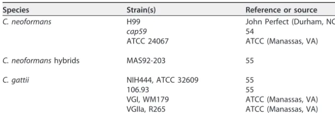
TABLE 1List ofC. neoformansandC. gattiistrains used
Species Strain(s) Reference or source
C. neoformans H99 John Perfect (Durham, NC)
cap59 54
ATCC 24067 ATCC (Manassas, VA)
C. neoformanshybrids MAS92-203 55
C. gattii NIH444, ATCC 32609 55
106.93 55
VGI, WM179 ATCC (Manassas, VA)
VGIIa, R265 ATCC (Manassas, VA)
on September 8, 2020 by guest
http://msphere.asm.org/
twice in MM (10 mM MgSO4, 29.3 mM KH2PO4, 13 mM glycine, 3M thiamine-HCl, and 15 mM dextrose,
with pH adjusted to 5.5). The washed cells were inoculated into MM for capsule induction, MM with
L-DOPA (100 mM) to induce melanization, and Sab for rich medium conditions. Cells were incubated at 30°C for 48 h with rotation at 180 rpm. Cells were washed twice with sterile PBS and were centrifuged for 5 min at 4,700⫻g. Cells were counted using a hemocytometer, and dilutions were made to obtain 1⫻107cells in PBS. The cells were then loaded onto Percoll density gradients with or without treatments
to test the effect of different conditions on the cell density.
Density gradient centrifugation.Percoll is a nontoxic and isotonic alternative to the commonly used sucrose gradient and is composed of polyvinylpyrrolidone-coated colloidal silica particles (56). Percoll has found applications for separation of mammalian blood cells, tumor cells, immune and endothelial cells, and microbial cells due to its ability to form reproducible self-generated continuous gradients (57). Stock isotonic Percoll (SIP) was obtained by addition of 1 part of 1.5 M NaCl to 9 parts of Percoll. The 70% (vol/vol) working solution was obtained by diluting SIP with 0.15 M NaCl to a final density of 1.0914 g/ml. A 3-ml volume of this solution was loaded into polycarbonate ultracentrifuge tubes (13 by 51 mm). Approximately 107cells were pelleted at 4,700⫻gand overlaid in layers directly
or after treatment. All gradients were run in parallel with a standard tube.
For the preparation of the standard tube, 10l of each uniform density bead standard (Cospheric DMB kit), including light orange (orange polyethylene microspheres sized 250 to 300m, with density 1.00 g/cc), fluorescent green (1.02 g/cc), florescent orange (1.04 g/cc), florescent violet (1.06 g/cc), dark blue (1.08) and florescent red (1.099 g/cc), was loaded and mixed with the Percoll.
By adjusting the time and speed of centrifugation, it was found that the most optimal separation of the density gradient beads (which was taken as an indication for the most optimal continuous density gradient formed) occurred at 40,000 rpm for 30 min (acceleration, 9; deceleration, 0), using a TLA 100.3 fixed-angle rotor with an Optima TLX tabletop ultracentrifuge.
Cell density estimation.First, the images of the density gradient were taken under conditions of uniform light and shadow using an Nikon D3000 digital single-lens reflex (DSLR) camera (auto settings). Next, pixel area measurements were taken from the bottom of the tube to the area at the beginning of each band (a1) and ranging to the end of each band (a2) and to the upper meniscus of the tube (f). The
density factor,Df(min, max) was computed in Microsoft Excel with the formula below.
Df
共
min, max兲
⫽再
冉
f⫺a1 f冊
,冉
f⫺a2
f
冊
冎
The averages,Dfaverages (Dfavg), and standard deviations (SD) along the mean were computed in
Microsoft Excel. A standard curve was derived usingDfavg and SD (xaxis) and cell density (in grams per
cubic centimeter) (yaxis) of the density marker beads. A 95% confidence interval was used to interpolate the mean density values of sample cells around a standard deviation, which were run in parallel with the uniform density bead standards.
Although the results from the different Percoll gradient runs followed the same trend, the exact density values differed considerably. This can be attributed to pipetting errors or errors in measurement of density factors.
Gamma irradiation of cells for capsule removal. Gamma irradiation was used to remove the capsule as described earlier (58). Approximately 109cells of melanized and nonmelanized cells were
plated on a 24-well plate. The cells were irradiated with a total dose of 1,500 Gy, using Shepherd Mark 1 at the SKCCC Experimental Irradiator Core at the Johns Hopkins University Sidney Kimmel Compre-hensive Cancer Center. Cells were washed twice in PBS, and approximately 107cells were pelleted at
4,700⫻gand loaded onto the gradient.
DMSO extraction ofC. neoformanscapsule.Approximately 107cells were incubated in 15 ml of
DMSO at 30°C for 30 min to allow capsule extraction. The cells were washed thrice in 1⫻PBS, pelleted, and loaded onto the Percoll density gradient.
Antibody coating ofC. neoformanscapsule.Purified 18B7 and E1 antibodies (kindly provided by Francoise Dromer’s laboratory) were obtained from stock solutions kept at 4°C. The antibodies were serially diluted in PBS to concentrations of 20g, 10g, 1g, and 0.1g/ml. A pellet of 107cells was
suspended with 1 ml of each Ab solution in Eppendorf tubes, subjected to vortex mixing, and incubated at 28°C on a rotating mixer for 1 h.
C. neoformansmelanization.Frozen stocks ofC. neoformansH99 were inoculated into Sab and
incubated at 30°C for 48 h until the cultures reached stationary phase. The cells were counted using a hemocytometer. A total of 106 cells/ml were inoculated into MM (10 mM MgSO
4, 29.3 mM KH2PO4,
13 mM glycine, 3M thiamine-HCl, and 15 mM dextrose, with pH adjusted to 5.5) with and without
L-DOPA (100 mM). The cells were cultured for 10 days at 30°C with rotating at 180 rpm. The cells were washed twice in PBS, and⬃107melanized and nonmelanized cells and a 1:1 mixture of melanized and
nonmelanized cells were loaded onto the gradient. Melanin ghosts were prepared as described previ-ously (40).
Mouse complement deposition in C. neoformans. Frozen stocks of guinea pig complement (1 mg/ml) were thawed. A pellet of 107cells was added and adjusted to 50% (500 g/ml), 20%, 10%, and
1% dilutions with PBS. The cells were incubated with complement for 1 h at 28°C in a rotating mixer. ProvidingC. neoformanswith osmotic stress.C. neoformans(⬃108cells) were incubated with 1 ml
of 10⫻PBS, 1⫻PBS, 0.1⫻PBS, and ultrapure distilled water (MilliQ) for 2 h 30 min in a rotating mixer at 28°C.
Settling ofC. neoformansin seawater.A total of⬃107cells/ml ofC. neoformansgrown in MM were
gently pipetted onto cuvettes containing 3 ml of seawater (Worldwide Imports AWW84130 Live Nutri Buoyancy ofCryptococcus neoformans
on September 8, 2020 by guest
http://msphere.asm.org/
Seawater) and PBS. The settling of the cells was observed by imaging the cuvettes at different time intervals with a Nikon D3000 DSLR camera. The images were analyzed using ImageJ. The relative levels of displacement were measured using the formula (f⫺u)/f, wherefis the area of the tube anduis the area from the bottom of the tube to the upper menisci of cells that are settling.
Cell imaging and yeast size measurements.The cells were visualized and imaged with India ink negative staining under an Olympus AX70 microscope at⫻20 magnification and⫻40 magnification. The capsule and cell body sizes were estimated using automated measurement Python software (59) or by the use of ImageJ when cells were observed to be aggregated.
Statistical analysis.All statistical analysis was performed using GraphPad Prism 7.0 software. The density of cells was estimated by making a standard curve from beads of different densities, using linear regression to estimate the unknown values of a given sample with a 95% confidence interval. Details of the statistical tests applied are provided in the figure legends.
SUPPLEMENTAL MATERIAL
Supplemental material for this article may be found at
https://doi.org/10.1128/
mSphere.00534-18
.
FIG S1
, TIF file, 1.7 MB.
FIG S2
, TIF file, 1.9 MB.
ACKNOWLEDGMENTS
We thank Francoise Dromer (Institut Pasteur) for providing capsular antibody ES1
(IgG).
A.C. was supported by grants 5R01HL059842, 5R01AI033774, 5R37AI033142, and
5R01AI052733.
R.V. designed and conducted the experiments, analyzed the data, and wrote the
manuscript. R.J.B.C. and A.C. contributed to the experimental design, supervised the
experiments, and edited and wrote parts of the manuscript.
REFERENCES
1. Perfect JR. 2000. Cryptococcosis, p 79 –93.InAtlas of infectious diseases. Current Medicine Group, London, United Kingdom.
2. Ellis DH, Pfeiffer TJ. 1990. Natural habitat of Cryptococcus neoformans var. gattii. J Clin Microbiol 28:1642–1644.
3. Emmons CW. 1960. Prevalence of Cryptococcus neoformans in pigeon habitats. Public Health Rep 75:362–364. https://doi.org/10.2307/ 4590800.
4. Bartlett KH, Kidd SE, Kronstad JW. 2008. The emergence of Cryptococcus gattii in British Columbia and the Pacific Northwest. Curr Infect Dis Rep 10:58 – 65.https://doi.org/10.1007/s11908-008-0011-1.
5. MacDougall L, Kidd SE, Galanis E, Mak S, Leslie MJ, Cieslak PR, Kronstad JW, Morshed MG, Bartlett KH. 2007. Spread of Cryptococcus gattii in British Columbia, Canada, and detection in the Pacific Northwest, USA. Emerg Infect Dis 13:42–50.https://doi.org/10.3201/eid1301.060827. 6. Neilson JB, Fromtling RA, Bulmer GS. 1977. Cryptococcus neoformans:
size range of infectious particles from aerosolized soil. Infect Immun 17:634 – 638.
7. Powell KE, Dahl BA, Weeks RJ, Tosh FE. 1972. Airborne Cryptococcus neoformans: particles from pigeon excreta compatible with alveolar deposition. J Infect Dis 125:412– 415.https://doi.org/10.1093/infdis/125 .4.412.
8. Feldmesser M, Kress Y, Casadevall A. 2001. Dynamic changes in the morphology of Cryptococcus neoformans during murine pulmonary infection. Microbiology 147:2355–2365.https://doi.org/10.1099/00221287 -147-8-2355.
9. Charlier C, Chrétien F, Baudrimont M, Mordelet E, Lortholary O, Dromer F. 2005. Capsule structure changes associated with Cryptococcus neo-formans crossing of the blood-brain barrier. Am J Pathol 166:421– 432.
https://doi.org/10.1016/S0002-9440(10)62265-1.
10. Zaragoza O, García-Rodas R, Nosanchuk JD, Cuenca-Estrella M, Rodríguez-Tudela JL, Casadevall A. 17 June 2010. Fungal cell gigantism during mammalian infection. PLoS Pathog https://doi.org/10.1371/ journal.ppat.1000945.
11. Okagaki LH, Strain AK, Nielsen JN, Charlier C, Baltes NJ, Chrétien F, Heitman J, Dromer F, Nielsen K. 2010. Cryptococcal cell morphology affects host cell interactions and pathogenicity. PLoS Pathog 6:e1000953.
https://doi.org/10.1371/journal.ppat.1000953.
12. Maxson ME, Cook E, Casadevall A, Zaragoza O. 2007. The volume and
hydration of the Cryptococcus neoformans polysaccharide capsule. Fun-gal Genet Biol 44:180 –186.https://doi.org/10.1016/j.fgb.2006.07.010. 13. Cordero RJB, Pontes B, Guimarães AJ, Martinez LR, Rivera J, Fries BC,
Nimrichter L, Rodrigues ML, Viana NB, Casadevall A. 2011. Chronological aging is associated with biophysical and chemical changes in the cap-sule of Cryptococcus neoformans. Infect Immun 79:4990 –5000.https:// doi.org/10.1128/IAI.05789-11.
14. Casadevall A, Coelho C, Cordero RJB, Dragotakes Q, Jung E, Vij R, Wear MP. 13 February 2018. The capsule of Cryptococcus neoformans. Viru-lencehttps://doi.org/10.1080/21505594.2018.1431087.
15. Zaragoza O, Rodrigues ML, De Jesus M, Frases S, Dadachova E, Casade-vall A. 2009. The capsule of the fungal pathogen Cryptococcus neofor-mans. Adv Appl Microbiol 68:133–216. https://doi.org/10.1016/S0065 -2164(09)01204-0.
16. Kwon-Chung KJ, Rhodes JC. 1986. Encapsulation and melanin formation as indicators of virulence in Cryptococcus neoformans. Infect Immun 51:218 –223.
17. Garcia-Rivera J, Eisenman HC, Nosanchuk JD, Aisen P, Zaragoza O, Moadel T, Dadachova E, Casadevall A. 2005. Comparative analysis of Cryptococcus neoformans acid-resistant particles generated from pig-mented cells grown in different laccase substrates. Fungal Genet Biol 42:989 –998.https://doi.org/10.1016/j.fgb.2005.09.003.
18. Nosanchuk JD, Stark RE, Casadevall A. 2015. Fungal melanin: what do we know about structure? Front Microbiol 6:1463.https://doi.org/10.3389/ fmicb.2015.01463.
19. Nosanchuk JD, Rudolph J, Rosas AL, Casadevall A. 1999. Evidence that cryptococcus neoformans is melanized in pigeon excreta: implications for pathogenesis. Infect Immun 67:5477–5479.
20. Nosanchuk JD, Rosas AL, Lee SC, Casadevall A. 2000. Melanisation of Cryptococcus neoformans in human brain tissue. Lancet 355:2049 –2050.
https://doi.org/10.1016/S0140-6736(00)02356-4.
21. Casadevall A, Rosas AL, Nosanchuk JD. 2000. Melanin and virulence in Cryptococcus neoformans. Curr Opin Microbiol 3:354 –358.https://doi .org/10.1016/S1369-5274(00)00103-X.
22. Cordero RJB, Casadevall A. 2017. Functions of fungal melanin beyond virulence. Fungal Biol Rev 31:99 –112.https://doi.org/10.1016/j.fbr.2016 .12.003.
23. Frases S, Viana NB, Casadevall A. 2011. Biophysical methods for the study
on September 8, 2020 by guest
http://msphere.asm.org/
of microbial surfaces. Front Microbiol 2:207.https://doi.org/10.3389/ fmicb.2011.00207.
24. Grover WH, Bryan AK, Diez-Silva M, Suresh S, Higgins JM, Manalis SR. 2011. Measuring single-cell density. Proc Natl Acad Sci U S A 108: 10992–10996.https://doi.org/10.1073/pnas.1104651108.
25. Biello D. 2006. Fact or fiction?: Archimedes coined the term “eureka!” in the bath. Sci Amhttps://www.scientificamerican.com/article/fact-or -fiction-archimede/.
26. Kuroki H. 2016. How did Archimedes discover the law of buoyancy by experiment? Front Mech Eng 11:26 –32.https://doi.org/10.1007/s11465 -016-0368-z.
27. Baldwin WW, Myer R, Powell N, Anderson E, Koch AL. 1995. Buoyant density of Escherichia coli is determined solely by the osmolarity of the culture medium. Arch Microbiol 164:155–157.https://doi.org/10.1007/ s002030050248.
28. Baldwin WW, Kubitschek HE. 1984. Evidence for osmoregulation of cell growth and buoyant density in Escherichia coli. J Bacteriol 159:393–394. 29. Lowe BA, Miller JD, Neely MN. 2007. Analysis of the polysaccharide capsule of the systemic pathogen Streptococcus iniae and its implica-tions in virulence. Infect Immun 75:1255–1264.https://doi.org/10.1128/ IAI.01484-06.
30. Kubitschek HE. 1987. Buoyant density variation during the cell cycle in microorganisms. Crit Rev Microbiol 14:73–97.https://doi.org/10.3109/ 10408418709104436.
31. Sundqvist G, Figdor D, Hänström L, Sörlin S, Sandström G. 1991. Phago-cytosis and virulence of different strains of Porphyromonas gingivalis. Scand J Dent Res 99:117–129.
32. Sagripanti J-L, Carrera M, Robertson J, Levy A, Inglis TJJ. 2011. Size distribution and buoyant density of Burkholderia pseudomallei. Arch Microbiol 193:69 –75.https://doi.org/10.1007/s00203-010-0649-6. 33. Vijay S, Nair RR, Sharan D, Jakkala K, Mukkayyan N, Swaminath S,
Pradhan A, Joshi NV, Ajitkumar P. 2017. Mycobacterial cultures contain cell size and density specific sub-populations of cells with significant differential susceptibility to antibiotics, oxidative and nitrite stress. Front Microbiol 8:463.https://doi.org/10.3389/fmicb.2017.00463.
34. Baldwin WW, Kubitschek HE. 1984. Buoyant density variation during the cell cycle of Saccharomyces cerevisiae. J Bacteriol 158:701–704. 35. Allen C, Büttner S, Aragon AD, Thomas JA, Meirelles O, Jaetao JE, Benn
D, Ruby SW, Veenhuis M, Madeo F, Werner-Washburne M. 2006. Isolation of quiescent and nonquiescent cells from yeast stationary-phase cul-tures. J Cell Physiol 174:89 –100.https://doi.org/10.1083/jcb.200604072. 36. Zaragoza O, Casadevall A. 2004. Experimental modulation of capsule size in Cryptococcus neoformans. Biol Proced Online 6:10 –15.https://doi .org/10.1251/bpo68.
37. Frases S, Nimrichter L, Viana NB, Nakouzi A, Casadevall A. 2008. Crypto-coccus neoformans capsular polysaccharide and exopolysaccharide frac-tions manifest physical, chemical, and antigenic differences. Eukaryot Cell 7:319 –327.https://doi.org/10.1128/EC.00378-07.
38. Nayar KG, Sharqawy MH, Banchik LD, Lienhard V JH. 2016. Thermophysi-cal properties of seawater: a review and new correlations that include pressure dependence. Desalination 390:1–24.https://doi.org/10.1016/j .desal.2016.02.024.
39. Sharqawy MH, Lienhard JH, Zubair SM. 2010. Thermophysical properties of seawater: a review of existing correlations and data. Desalination Water Treat 16:354 –380.https://doi.org/10.5004/dwt.2010.1079. 40. Wang Y, Aisen P, Casadevall A. 1996. Melanin, melanin “ghosts,” and
melanin composition in Cryptococcus neoformans. Infect Immun 64: 2420 –2424.
41. Cordero RJB, Pontes B, Frases S, Nakouzi AS, Nimrichter L, Rodrigues ML, Viana NB, Casadevall A. 2013. Antibody binding to Cryptococcus neo-formans impairs budding by altering capsular mechanical properties. J Immunol 190:317–323.https://doi.org/10.4049/jimmunol.1202324. 42. Zaragoza O, Nielsen K. 2013. Titan cells in Cryptococcus neoformans:
cells with a giant impact. Curr Opin Microbiol 16:409 – 413.https://doi .org/10.1016/j.mib.2013.03.006.
43. Reynolds CS, Oliver RL, Walsby AE. 1987. Cyanobacterial dominance: the role of buoyancy regulation in dynamic lake environments. N Z J Mar Freshw Res 21:379 –390. https://doi.org/10.1080/00288330.1987 .9516234.
44. Reynolds CS. 2007. Variability in the provision and function of mucilage in phytoplankton: facultative responses to the environment. Hydrobio-logia 578:37– 45.https://doi.org/10.1007/s10750-006-0431-6.
45. Richardson TL, Jackson GA. 2007. Small phytoplankton and carbon export from the surface ocean. Science 315:838 – 840.https://doi.org/10 .1126/science.1133471.
46. Inoue K, Nishimura M, Nayak BB, Kogure K. 2007. Separation of marine bacteria according to buoyant density by use of the density-dependent cell sorting method. Appl Environ Microbiol 73:1049 –1053.https://doi .org/10.1128/AEM.01158-06.
47. Condie SA, Bormans M. 1997. The Influence of density stratification on particle settling, dispersion and population growth. J Theor Biol 187: 65–75.https://doi.org/10.1006/jtbi.1997.0417.
48. Kidd SE, Bach PJ, Hingston AO, Mak S, Chow Y, MacDougall L, Kronstad JW, Bartlett KH. 2007. Cryptococcus gattii dispersal mechanisms, British Columbia, Canada. Emerg Infect Dis 13:51–57.https://doi.org/10.3201/ eid1301.060823.
49. Kidd SE, Chow Y, Mak S, Bach PJ, Chen H, Hingston AO, Kronstad JW, Bartlett KH. 2007. Characterization of environmental sources of the human and animal pathogen Cryptococcus gattii in British Columbia, Canada, and the Pacific Northwest of the United States. Appl Environ Microbiol 73:1433–1443.https://doi.org/10.1128/AEM.01330-06. 50. Roe CC, Bowers J, Oltean H, DeBess E, Dufresne PJ, McBurney S, Overy
DP, Wanke B, Lysen C, Chiller T, Meyer W, Thompson GR, Lockhart SR, Hepp CM, Engelthaler DM. 2018. Dating the Cryptococcus gattii disper-sal to the North American Pacific Northwest. mSphere 3:e00499-17.
https://doi.org/10.1128/mSphere.00499-17.
51. Gales N, Wallace G, Dickson J. 1985. Pulmonary cryptococcosis in a striped dolphin (Stenella coeruleoalba). J Wildl Dis 21:443– 446.https:// doi.org/10.7589/0090-3558-21.4.443.
52. Migaki G, Gunnels RD, Casey HW. 1978. Pulmonary cryptococcosis in an Atlantic bottlenosed dolphin (Tursiops truncatus). Lab Anim Sci 28: 603– 606.
53. Miller WG, Padhye AA, van Bonn W, Jensen E, Brandt ME, Ridgway SH. 2002. Cryptococcosis in a bottlenose dolphin (Tursiops truncatus) caused by Cryptococcus neoformans var. gattii. J Clin Microbiol 40: 721–724.https://doi.org/10.1128/JCM.40.2.721-724.2002.
54. Coelho C, Souza ACO, da Silveira Derengowski L, de Leon-Rodriguez C, Wang B, Leon-Rivera R, Bocca AL, Gonçalves T, Casadevall A. 2015. Macrophage mitochondrial and stress response to ingestion of Crypto-coccus neoformans. J Immunol 194:2345–2357.https://doi.org/10.4049/ jimmunol.1402350.
55. Cordero RJB, Frases S, Guimaräes AJ, Rivera J, Casadevall A. 2011. Evidence for branching in cryptococcal capsular polysaccharides and consequences on its biological activity. Mol Microbiol 79:1101–1117.
https://doi.org/10.1111/j.1365-2958.2010.07511.x.
56. Pertoft H, Rubin K, Kjellén L, Laurent TC, Klingeborn B. 1977. The viability of cells grown or centrifuged in a new density gradient medium, Per-coll(TM). Exp Cell Res 110:449 – 457.https://doi.org/10.1016/0014-4827 (77)90311-1.
57. Pertoft H. 2000. Fractionation of cells and subcellular particles with Percoll. J Biochem Biophys Methods 44:1–30.https://doi.org/10.1016/ S0165-022X(00)00066-X.
58. Bryan RA, Zaragoza O, Zhang T, Ortiz G, Casadevall A, Dadachova E. 2005. Radiological studies reveal radial differences in the architecture of the polysaccharide capsule of Cryptococcus neoformans. Eukaryot Cell 4:465– 475.https://doi.org/10.1128/EC.4.2.465-475.2005.
59. Dragotakes Q, Casadevall A. 11 January 2018. Automated measurement of Cryptococcal species polysaccharide capsule and cell body. J Vis Exp
https://doi.org/10.3791/56957. Buoyancy ofCryptococcus neoformans