BOSNIAN JOURNAL OF BASIC MEDICAL SCIENCES 2010; 10 (4): 276-281Abstract
Th e purpose of the study was to determine the frequency of expression p and pINKa proteins and bcl- oncoprotein in malignant skin melanoma and to determine their correlation with the proliferative index and tumor thickness. Th e study involved patients: () male and () female. Mitotic index showed a correlation with p protein expression, a negative correlation with pINKa protein expression. Statistically signifi cant correlations were determined between the Breslow tumor thickness, Clark invasion level and p protein expression, as well as Breslow tumor thickness and bcl- oncoprotein expression (p<.), whereas there was no correlation between the pINKa protein expression and melanoma thicknes and Clark invasion level. Overexpression p protein and bcl- oncoprotein, with the loss pINKa pro-tein of expression in the nodular melanoma, confi rms a frequent loss of function of these tumor suppressor gene and oncogene, and indicates a vertical tumor growth phase. Th e loss of tumor suppression function the p protein and bcl- oncoprotein overexpression in cutaneous mela-noma correlates with larger tumor thickness, whereas the overexpression of mutated p protein and loss pINKa protein of expression indicate a higher proliferative tumour potential. Th erefore, these evaluated proteins may be the aggressive biological tumour activity markers.
© Association of Basic Medical Sciences of FBIH. All rights reserved
KEY WORDS: melanoma, skin, cell cycle regulators, bcl-, melanoma progression
p16
ink4a
) and bcl-2 oncoprotein with mitotic index and
thickness of primary cutaneous malignant melanoma
Miloš Kostov1*, Žaklina Mijović2, Dragan Mihailović2, Snežana Cerović3, Miroslav Stojanović4, Marija Jelić5
1 Department of Pathology, Military hospital of Niš, Zorana Djindjića bb, 18000 Niš, Serbia. 2 Institute of Pathology, Faculty of Medicine, University of Niš, Zorana Djindjića 81, 18000 Niš, Serbia. 3 Center for Pathology and Forensic Medicine, Military Medical of Academy Belgrade, Crnotravska 17, 11000 Belgrade, Serbia. 4 Clinic for Surgery, Faculty of Medicine, University of Niš, Zorana Djindjića 81, 18000 Niš, Serbia. 5 Department of Biochemistry, Military hospital of Niš, Zorana Djindjića bb, 18000 Niš, Serbia
Introduction
Malignant melanoma of the skin (MMS) is a melano-cyte system tumor which is produced by the malignant melanocyte transformation generated by the neural crest [, , ]. MMS is characterized by high malignant potential and high mortality rate, as well as a rapid oc-currence rate in the late years, especially in women []. Multistep tumorigenesis is an evolving process that involves the inactivation of tumor suppressor genes, activation of oncogenes and defects of the reparatory genes which can induce inadequate gene reparation []. Th e most common oncogene defect occurs at the control site G/S which me-diates the cell S phase onset [-]. Melanoma is not an ex-ception in the tumor biology and its occurrence is predomi-nantly the result of the gene mutations accumulation whose key role is the regulation of cellular proliferation, diff erentia-tion, apoptosis or some other pathways of cellular death [, ]. Many studies have shown a certain amount of damage
and loss of tumor suppressor genes p and p with MMS patients, which is why they are considered to be responsible for the tumor carcinogenesis process [-]. The tumor suppressor gene p has a central role in the cell cycle and apoptosis regulation. Th e mutation of the p gene, one of the most common genetic defects in human tumors, gener-ates defects in the control site of the cell cycle and genetic instability of some cancerous cells [, , , -]. Th e pro-tein p, the product of cyclin-dependent kinase Na gene, is also known as p INKa, p-MTS or cyclin-dependent
kinase /CDK inhibitor [, , , ]. Th e frequent muta-tions in melanoma are on the CDKNA locus (typical target is pINK) which suggests a key role for this protein in cell
cycle control management in melanocytes [-]. It has been observed that the p INKa protein expression in melanocytic
lesions decreases from benign to malignant and metastatic lesions []. A bcl- oncoprotein overexpression can stop the apoptosis process which induces cell proliferation. Bcl- oncoprotein expression can be noticed in both benign and malignant melanocyte tumors, suggesting that it is not a key role in the malignant melanocyte transformation [, ]. The purpose of this study was to determine the sta-tus of the tumor suppression proteins expression (p and pinka) and bcl- oncoprotein in the primary ma-* Corresponding author: Miloš Kostov, Department of Pathology,
Military hospital of Niš, Zorana Djindjića bb, 18000 Niš, Serbia, tel. +381 18 508 992, fax +381 18 238 191,
e-mail: [email protected]˝
BOSNIAN JOURNAL OF BASIC MEDICAL SCIENCES 2010; 10 (4): 277-281
lignant melanoma of the skin and to analyse their cor-relation with the mitotic index and tumor thickness.
Materials and Methods
The study involved samples of the primary malignant melanoma of the skin. Th e tumors were surgically removed using the elliptic skin incision (excisional biopsy). Th e his-topathological parameters were determined for each tu-mor, including the following: size (maximum diameter in cm), thickness in mm (Breslow), histological cellular tumor type, tumor invasion level (Clark), ulceration, mitotic num-ber/mitotic index and the pathological stage of the disease. The pathological stage /pT/ was determined according to the American Joint Committee on Cancer, . We ana-lyzed the p protein mutation expression level, tumor sup-pressor protein pINKa and bcl- oncoprotein expression.
Th e samples were stored in a neutral, buff ered formaline solution, for - hours, dehydrated in ethanol of progres-sive concentration and embedded in paraffin. From the paraffi n blocks, the tissue was cut into - μm thin samples, stained with the standard hematoxylin-eosine (HE) method and microscopically analysed using Leica DM (Wetzlar, Germany), with digital microscopic camera Leica EC (Wet-zlar, Germany) with Leica LAS EZ imaging software V ...
Immunohistochemical analysis
Th e detection of the target markers - antigen, was performed using commercially available monoclonal human anti-bodies (Table .) and highly sensitive and specifi cally marked streptavidin-biotin complex immunoassays (DAKO LSAB+
-HRP kit). Th e chromogen was .'-diaminobenzidin (DAB), and the slides were lightly counterstained with Mayer he-matoxylin (Merck, Germany). All the reactants were pro-duced by the DAKO Company (Glostrup, Denmark). Dur-ing the tissue stainDur-ing, the positive and negative control samples were simultaneously stained in order to confi rm the specifi city and quality of the immunohistochemical analysis. The mutated p and p and bcl- protein expression evaluation was performed based on the intensity of the im-munohistochemical nuclear (p and pINKa) staining and
cytoplasmic staining (pINKa i Bcl-) and immunoreactive
tumor cell percentage. Th e positive immunohistochemical
reaction was observed as a light and dark brown colouring of the tumor nuclei. Th e p tumor cells were rarely visible - cytoplasmic colouring of light brown. Th e semiquantitative
analysis is calculated as the percentage of the positive cells in relation to the total tumor cells in the fi eld. Immunohis-tochemical staining intensity determine as (+ = mild; + = moderate; + = intensive) and the tumor positive cell per-centage (< no immunoreactive cells; - some positive cells; - many positive cells; > most positive cells).
Morphometric analysis
Tumor thickness measurements
Th e tumor thickness was determined according to Breslow using an ocular micrometer and suitable computer software on a microscope with a digital camera. Th e thickness was de-termined at the widest tumor area starting from the upper line of the granulated epidermal layer or the bottom line of ulcer-ation, vertically into the depth, to the last tumor coast or indi-vidual tumor cell. Th e thickness was measured in millimeters Mitotic activity
The number of mitoses (mitotic index, MI) was deter-mined in the tumor areas with the largest number of the mitotic figures at fields of the highest microscopic amplification (x for each sample), (the total mitoses number/ HighPowerField - HPF). The MI was clas-sified in the following manner: low index - mitotic figures/ HPF; high index > mitotic figures/HPF.
Statistical analysis
The data obtained during the research was filed in a spe-cially created data base. The results were statistically pro-cessed using the descriptive and analytical statistic meth-ods based on the: mean value (x), standard deviation (SD), standard error (SE), Student’s t-test in some groups in order to evaluate the statistical importance of mean value differences, MANOVA and Pearson’s correlation.
Results
The mean age of patients was .±. years; the young-est patient was years old, the oldyoung-est years. There were (.) male and (.) female patients. There were two types of the tumor based on the
cellu-Antibody Catalogue no. Clone Source Company Epitop Retrieval Dilution Hromogen
CINtecTM
p16INK4a
Histology kit
K5334 E6H4 mouse DAKO
Target retrieval Solution, pH 9.0 S2367 MW
KitVisualisation system EnVision
TM+
DAB
p 53 protein M7001 DO-7 mouse DAKO 0.05M citric
buf-fer pH 6.0 MW 1:50 DAB
Bcl-2 oncoprotein M0887 124 mouse DAKO 0.05M citric
buf-fer pH 6.0 MW 1:50 DAB
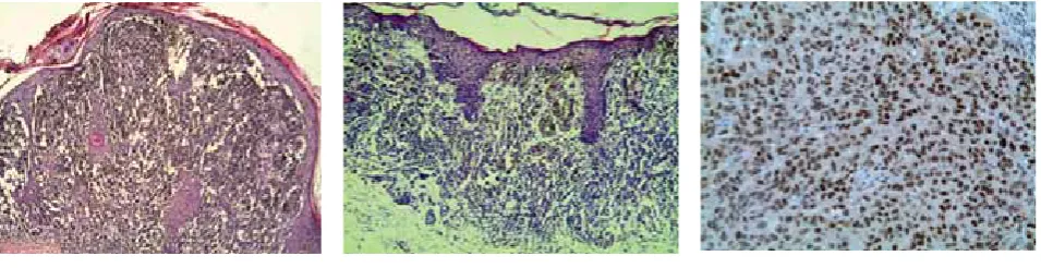
BOSNIAN JOURNAL OF BASIC MEDICAL SCIENCES 2010; 10 (4): 278-281lar type: in thirty-three patients (.) melanomas were nodular type (nodular melanoma, NM), (Figure ), and (.) melanomas were of superficially spreading type (superficially spreading melanoma, SSM), (Figure ).
According to the patient’s sex, the most common MMS anatomical localizations in women were the lower extremi-ties, lower legs - patients (.); the most common ana-tomical localization in men was the back - cases (). Statistically significant differences between NM and SSM were found for the mean age value (p<.), Breslow tumor thickness, invasion level by Clark, WHO tumor stage, ul-ceration, mitosis number (p<.), histological cellular tu-mor type (p<.) and maximum tutu-mor diameter (p<.), whereas the sex variable was not statistically significant.
The p protein mutated form expression was found in () MMS. All () NM showed a positive reac-tion to this protein, while in the SSM it was found in () cases. A positive nuclear reaction was found in over tumor cells in () NM, moderate staining inten-sity (Figure ), while SSM showed a positive reaction mild intensity of nuclear staining (percentage was - of tu-mor cells). Statistically relevant differences between NM and SSM were found for the percentage of p positive cells and p intensity of the immuno reaction (p<.).
The loss of pINKa protein
expres-sion was found in () MMS. A negative immuno reaction to pINKa
protein was found in () NM cases and in () SSM cases. On the other hand, () SSM showed a positive pINKa protein expression,
of tumour cells were immunore-active, and the staining intensity was moderate and intensive. The PINKa
immunoreactive tumour cells MMS also showed the cytoplasmatic light
brown staining (Figure ). Th e immunoreaction intensity for the pINKa protein varied due to the different tumor
seg-ments; a moderate reaction was found in the upper parts of the tumor. Statistically relevant diff erences between the NM and SSM were found in the percentage of the pINKa
positive cells (p<.) and pINKa reaction intensity (p<.).
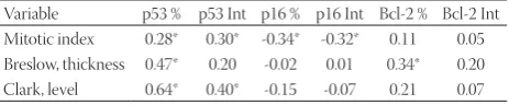
The bcl- oncoprotein expression was found in () MMS. A positive immunoreactivity to this protein was found in () NM and () SSM. With () NM, an immunoreaction was noticed in over of the tumor cells, with signifi cant staining intensity (Figure ). A positive immunoreaction was observed in () SSM with over of the tumor cells of the moderate cytoplas-mic staining level. There were no significant statistical dif-ferences between the NM and SSM for the percentage of the bcl- positive cells and bcl- immunoreaction intensity. However, there were statistically significant correlations between the mitotic number and p positive cell percent-age as well as the mitotic number and p reaction inten-sity. Moreover, there was a statistically significant nega-tive correlation between the mitotic number and pINKa
positive cells percentage and between the mitotic number and pINKa reaction intensity (p < .); whereas there
was no significant difference between the mitotic num-ber and bcl- positive cells percentage, and between the mitotic number and bcl- reaction intensity (Table ).
FIGURE 1. Malignant melanoma of the
skin, nodular type (H&E, x20).
FIGURE 4. Diff use nuclear and
cytoplas-mic immunoreactivity for p16 protein of the tumor cells malignant melanoma of the skin, (IH, x20).
FIGURE 2. Malignant melanoma of the
skin, superfi cially spreading type (H&E, x20).
FIGURE 5. Diff use cytoplasmic
immunore-activity for Bcl-2 oncoprotein of the tumor cells MMS, (IH, x20).
FIGURE 3. Diff use nuclear
BOSNIAN JOURNAL OF BASIC MEDICAL SCIENCES 2010; 10 (4): 279-281
Statistically signifi cant correlations were observed between the Breslow tumor thickness and p positive cell percent-age and between the Breslow tumor thickness and bcl- positive cells percentage. Moreover, there were some sig-nificant correlations between the Clark tumour invasion levels and p positive cells percentage and between Clark tumour invasion level and p reaction intensity (p < .). Other correlations were not signifi cantly relevant (Table ).
Discussion
MMS is one of the most studied tumors due to the vague-ness of the tumor’s etiological and biological aspects [, , , , ]. In our research, the largest amount of the NM () consisted of epithelioid cells only, and then histologically combined tumors, epithelioid and spindle cells, whereas the smallest amount of the nodular melanoma consisted of spindle type cells. However, the largest amount of the SSM () consisted of epithelium cells exclusively. In general, most types of melanoma exhibit a combined histological im-age, with a particular dominant cell type, which was the case with our subjects - the dominant form being the epithelioid cell type []. Th e most aff ected anatomical area in women was the lower extremites area, lower leg, almost of the cases, whereas in men it was the trunk , the back of the cases. Generally speaking, the most common melanoma occurrence in men is on the trunk, in women on the lower extremites, which is confi rmed by the data from our research [, ]. Tumor ulceration as an independent prognosis factor may also characterise cutaneous melanomas with a vertical growth phase in the late disease stage. Th e ulceration was a common characteristics of the NM in our group, and was ob-served in () of the cases, while in the SSM group it was present in only cases (). Our results confi rm a signifi cant diff erence between the NM and SSM compared to the pres-ent ulceration which other studies also confi rm [, , ]. In our group, the NM patients had a higher pathological stage (pT/WHO) compared to the SSM. This histopathological parameter also showed a statistically signifi cant diff erence []. Although it is known that the cutaneous melanoma is a cellular growth and development defi ciency, some genetic mechanisms responsible for its initiation and progression remain unclear [, , ]. Loss of function of p tumor
sup-pressor gene and mutation p tumour supsup-pressor gene in malignant melanoma suggest the importance of their roles in the tumor pathogenesis [, , , ]. Th e results of our study show a high level of nuclear expression of the p protein mutated form with thick skin melanoma, which is also confi rmed by the data from other studies [, ]. Th e correlation between p protein overexpression and MI suggests a high proliferative tumor potential. Our data is confi rmed by a research by Soares de Sa et al. []who be-lieve that an overexpression of the p protein in correlation with a high mitotic rate probably points to the diminish-ing of the S-G control site in high proliferative melanoma. We have encountered a frequent pINKa protein expression
loss in patients with NM, as opposed to the SSM patients who have an overexpression of this protein. Th e permanent loss of the protein expression in NM suggests the lack of its function, late stage, and a possible role in the tumor pro-gression, which is confi rmed by the results from other au-thors [, , , , ]. Th e pINKa protein expression in
melanocyte lesions decreases from benign to malignant and metastatic lesions []. We have observed a statistically rel-evant negative correlation between MI and pINKa protein
expression, while Talve et al. []do not show a correlation between p protein and MI in a group of malignant mela-noma. There is, however, a correlation between the num-ber of Ki- positive cells and pINKa protein expression
loss. Mihic-Probst et al. []have also found a signifi cantly higher proliferative activity with the melanoma of low p protein expression. Th e diff erence in these correlations may be due to a diff erent pINKa protein expression which can
lead to the existence of various pINKa protein mutations
and their impact on the primary MMS proliferative activity. Our study shows a high level of bcl- oncoprotein expression in MMS, which can also be confi rmed by the other research data [, , , ]. Th is protein is expressed with both be-nign and malignant lesions, has no diagnostic signifi cance and is not a key aspect in the malignant melanocyte transfor-mation []. Leiter et al. [] have found an increased bcl- and bcl- related genes bcl-xLm bcl-xS and Bax expression in malignant melanoma during the disease progression as the result of the apoptosis inhibition and higher tumor malignant potential. We didn’t fi nd a correlation between bcl- onco-protein expression and MI which suggests that this onco-protein has no proliferative role in malignant melanoma of the skin. After the tumor thickness, MI is the second statistically relevant prognostic parameter for MMS. The melanoma prognosis is good if the mitotic index is low (< mitoses/ mm), and bad if the MI is high (> mitoses/mm). Many
multivariate analyses have shown a relation of MI with tu-mor thickness in a way that mitosis remains a significant independent variable for the primary cutaneous melanoma
Variable p53 % p53 Int p16 % p16 Int Bcl-2 % Bcl-2 Int Mitotic index 0.28* 0.30* -0.34* -0.32* 0.11 0.05 Breslow, thickness 0.47* 0.20 -0.02 0.01 0.34* 0.20 Clark, level 0.64* 0.40* -0.15 -0.07 0.21 0.07 TABLE 2. Correlation of cell cycle regulation proteins (p53 and p16INK4a) and bcl-2 oncoprotein with mitotic index and Breslow thickness and Clark level MMS
BOSNIAN JOURNAL OF BASIC MEDICAL SCIENCES 2010; 10 (4): 280-281[, , ]. Most of the published studies show a correla-tion of the tumor thickness with other prognostic factors such as ulceration, anatomical localisation, sex and age of patients, inflammatory response, mitotic index, vascular invasion and microscopic satellitosis. Their results show that the tumor thickness is the most powerful prognos-tic parameter for localised cutaneous melanoma [, ]. Both benign melanocyte tumors and malignant melanoma exprimate the p protein. Although the immunohisto-chemical reaction for this protein has no diagnostic rel-evance, numerous data have shown that the p protein expression increases in relation to the MMS thickness sug-gesting the presence of aggressive melanoma forms []. Our study, as well as Yamamoto et al. [], also shows a high mutated p form expression in MMS and a positive correlation with tumor thickness. Compared to our results, other researches show no correlation between the p mu-tation overexpression and tumor size []. Although our study shows a high level of mutated p protein expression in MMS, the possible diagnostic signifi cance of this marker may need to be assessed with a larger subject group. Our re-sults show that there is no correlation between the Breslow tumor thickness and Clark invasion level degree compared to p protein expression, which is confi rmed by other au-thors [, , ]. Moreover, Straume et al. [] and Reed et al. [] in their studies have found a signifi cant connection of the p protein nuclear expression loss and vascular tu-mor invasion, but not so with Breslow tutu-mor thickness or Clark invasion level. In contrast to our results and the stud-ies mentioned above, some other authors suggest that the level of melanoma invasion after Clark is greatly increased with p negative melanoma []. We found a high level of bcl- oncoprotein expression with MMS and a positive correlation with Breslow tumor thickness, which may oc-cur as a result of apoptosis inhibition which favorizes tu-mor cells survival and enables disease progression [, , ]. In addition, we found a combined p and bcl-
pro-tein overexpression with cutaneous melanoma patients, which also causes a higher malignant tumor potential [].
Conclusion
Overexpression mutated p protein and bcl- oncoprotein, with the loss of expression p protein in the nodular melano-ma, as shown by our study, confi rms a common loss of func-tion these tumor suppressor genes and oncogenes, and indi-cates a vertical tumor growth phase. Our results show that the loss of tumor suppression function and the p protein and bcl- oncoprotein overexpression in malignant of mela-noma of the skin correlate with larger tumor thickness value, whereas the overexpression mutated p protein and the
loss of expression pINKa protein indicate a higher
prolifera-tive tumour potential. Th erefore, the evaluated proteins may serve as the aggressive biological tumour activity markers.
Declaration of interest
Authors do not have any commercial affi liations, or potential confl icts of interest associated with this work submitted for publication.
References
[] Herlyn M, Berking C, Li G, Satyamoorthy K. Lessons from melano-cyte development for understanding the biological events in nae-vus and melanoma formation. Melanoma Res ; :-. [] Barnhill RL. Malignant melanoma. In: Barnhill RL, Piepkorn Busam
KJ., editors. Pathology of melanocytic nevi and malignant melanoma.
nd ed. New York: Springer Science; , pp: -.
[] Jiao Z, Zhang ZG, Hornyak TJ. et al. Dopachrome tautomerase (Dct) regulates neural progenitor cell proliferation. Dev Biol ; :-.
[] Boyle P, Levin B. Word cancer Report . Cutaneous melanoma. Lion: IARC Press. .
[] Husein MR. Genetic pathways to melanoma tumorigenesis. J Clin Pathol ; :-.
[] Li W, Sanki A, Karim RZ. et al. Th e role of cell cycle regulatory pro-teins in the pathogenesis of melanoma. Pathology ; :-. [] de Vries E, Bray F, Coebergh JW. et al. In: Le Boit PE, Burg G,
Weedon D, Sarasin A., editors. World Health Organisation classi-fi cation of tumours. Pathology and genetics of skin tumours. Lion: IARC Press; , pp: -.
[] Michalides RJ. Cell cycle regulators: mechanisms and their role in aetiology, prognosis, and treatment of cancer. J Clin Pathol ; :–.
[] Carlson JA, Ross JS, Slominski A. et al. Molecular diagnostics in melanoma. J Am Acad Dermatol ; :-.
[] Crowson AN, Magro C, Miller A, Mihm Jr MC. Th e molecular ba-sis of melanomageneba-sis and the metastatic phenotype. Semin On-col ; :-.
[] Nelson AA, Tsao H. Melanoma and genetics. Clin Dermatol ; :-.
[] Stefanaki C, Stefanaki K, Antoniou C. et al. G cell cycle regulators in congenital melanocytic nevi and melanomas. J Cutan Pathol ; :-.
[] Sekulic A, Haluska P Jr, Miller AJ. et al. Malignant melanoma in st century: the emerging molecular landscape. Mayo Clin Proc ; :-.
[] Straume O, Sviland L, Akslen LA. Loss of nuclear p protein ex-pression correlates with increased tumor cell proliferation (Ki-) and poor prognosis in patients with vertical growth phase mela-noma. Clin Cancer Res ; :-.
[] Straume O, Akslen LA. Alterations and prognostic signifi cance of p and p protein expression in subgroups of cutaneous mela-noma. Int J Cancer ; :–.
[] Sparrow LE, Eldon MJ, English DR, Heenan PJ. p and pWAF-protein expression in melanocytic tumors by
immunohistochem-istry. Am J Dermatopathol ; :–.
[] Talve L, Sauroja I, Collan Y, Punnonen K, Ekfors T. Loss of expres-sion of the pINK/CDKN gene in cutaneous malignant mela-noma correlates with tumor cell proliferation and invasive stage. Int J Cancer ; :–.
BOSNIAN JOURNAL OF BASIC MEDICAL SCIENCES 2010; 10 (4): 281-281
[] Mihic-Probst D, Mnich CD, Oberholzer PA. et al. p expression in primary malignant melanoma is associated with prognosis and lymph node status. Int J Cancer ; :-.
[] O’Leary TJ. Skin. In: O’Leary TJ. editor. Advanced diagnostic meth-ods in pathology: principles, practice, and protocols. Philadelphia: Saunders; , pp: -.
[] Yamamoto M, Takahashi H, Saitoh K, Horikoshi T, Takahashi M. Expression of the p protein in malignant melanomas as a prog-nostic indicator. Arch Dermatol Res ; :-.
[] Talve L, Kainu J, Collan Y, Ekfors T. Immunohistochemical expres-sion of p protein, mitotic index and nuclear morphometry in primary malignant melanoma of the skin. Pathol Res Pract ; :-.
[] Legan M. Cyclooxigenase-, p and glucose transporter- as pre-dictors of malignancy in the development of gallbladder carcino-mas. Bosn J Basic Med Sci ; :-.
[] Radovic S, Babic M, Doric M et al. Non-small cell lung carcinoma: cyclin D, bcl-, p, Ki- and HER- proteins expression in re-sected tumors. Bosn J Basic Med Sci ; :-.
[] Keller-Melchior R, Schmidt R, Piepkorn M. Expression of the tu-mor suppressor gene product pINK in benign and malignant melanocytic lesions. J Invest Dermatol ; :-. [] Fecker LF, Geilen CC, Tchernev G. et al. Loss of proapoptotic
Bcl--related multidomain proteins in primary melanomas is associ-ated with poor prognosis. J Invest Dermatol ; :-.
[] Saenz-Santamaria MC, Reed JA, McNutt NS, Shea CR. Immuno-histochemical expression of BCL- in melanomas and intradermal nevi. J Cutan Pathol ; :–.
[] Elder DE, Elenitsas R, Murphy GF, Xu X. Benign pigmented lesions and malignant melanoma. In: Elder DE. editor. Lever's Histopa-thology of the Skin, th ed. Philadelphia: Lippincott Williams & Wilkins; , pp: -.
[] Crowson AN, Magro C.M., Mihm Jr.M.C. Prognosticators of mela-noma, the melanoma report, and the sentinel lymph node. Mod Pathol ; :S-S.
[] Soares de Sa BC, Fugimori ML, Ribeiro KCB, Neto JPD, Neves RI, Landman G. Proteins involved in pRb and p pathways are dif-ferentially expressed in thin and thick superfi cial spreading mela-nomas. Melanoma Res ; :-.
[] Ilmonen S, Hernberg M, Pyrhonen S, Tarkkanen J, Asko-Seljavaara S. Ki - , Bcl- and p expression in primary and metastatic mela-noma. Melanoma Res ; :-.
[] Leiter U, Schmid RM, Kaskel P, Peter RU, Krähn G. Antiapoptotic bcl- and bcl-xL in advanced malignant melanoma. Arch Derma-tol Res ; :–.