R E S E A R C H A R T I C L E
Open Access
Ensheathing cells utilize dynamic tiling of
neuronal somas in development and injury
as early as neuronal differentiation
Evan L. Nichols
1, Lauren A. Green
1,2and Cody J. Smith
1,2*Abstract
Background:Glial cell ensheathment of specific components of neuronal circuits is essential for nervous system function. Although ensheathment of axonal segments of differentiated neurons has been investigated,
ensheathment of neuronal cell somas, especially during early development when neurons are extending processes and progenitor populations are expanding, is still largely unknown.
Methods:To address this, we used time-lapse imaging in zebrafish during the initial formation of the dorsal root ganglia (DRG).
Results:Our results show that DRG neurons are ensheathed throughout their entire lifespan by a progenitor
population. These ensheathing cells dynamically remodel during development to ensure axons can extend away from the neuronal cell soma into the CNS and out to the skin. As a population, ensheathing cells tile each DRG neuron to ensure neurons are tightly encased. In development and in experimental cell ablation paradigms, the oval shape of DRG neurons dynamically changes during partial unensheathment. During longer extended unensheathment neuronal soma shifting is observed. We further show the intimate relationship of these ensheathing cells with the neurons leads to immediate and choreographed responses to distal axonal damage to the neuron.
Conclusion:We propose that the ensheathing cells dynamically contribute to the shape and position of neurons in the DRG by their remodeling activity during development and are primed to dynamically respond to injury of the neuron.
Keywords:Development, Ensheathment, Neuronal soma, Tiling, Neural niche
Background
Ensheathment of neuronal cells is essential for proper functioning of the nervous system. The role of ensheath-ment is diverse and ranges from functions such as myelin-ation to aid neural transmission [1–3], to metabolic homeostasis and trophic support to maintain neuronal health [4, 5]. This ensheathment typically occurs at an early, yet largely undefined, time in the development of functional neural circuits and is consistently maintained following the initial ensheathment [3, 6,7]. Disruption of ensheathment at any point can result in various patholo-gies depending on the cell types affected [5, 8]. Axonal
ensheathment and myelination have been extensively studied, but an understanding of neuronal soma ensheath-ment in vertebrates remains elusive [1,9,10].
Studies in C. elegans and Drosophila have observed that glial subpopulations extend processes around the soma to completely ensheath the neuron [11, 12]. These soma-ensheathing glia play an important role in devel-opment. Studies inC. elegans demonstrate that glial as-sociation with neurons guides neuronal maturation and axonal outgrowth [13–15]. These ensheathing glia also are closely associated with the neurons and respond to changes within the cell [11]. For example, after neuronal injury these glia have been shown to respond and phagocytize debris [16]. Previous work also demon-strates that disruption of cortex glia, the cells ensheath-ing neuronal cell somas inDrosophila, leads to neuronal death, suggesting an important role of ensheathing glia * Correspondence:[email protected]
1Department of Biological Sciences, University of Notre Dame, 015 Galvin Life
Sciences Building, Notre Dame, IN 46556, USA
2Center for Stem Cells and Regenerative Medicine, University of Notre Dame,
Notre Dame, IN, USA
in neuronal growth and survival [17]. Disruption to cor-tex glia also leads to the disorganization of neuronal and glial cell populations in the DrosophilaCNS [17, 18]. In vertebrates, molecular communication between the neuronal soma and oligodendrocytes ensures the proper ensheathment of specific neuronal compartments, like axons [19]. Collectively, these studies point to tightly controlled and conserved interactions between ensheath-ing glia and sub-compartments of neurons on the macroscopic and microscopic level.
One organizational principle of glia that has emerged is cellular tiling. Tiling ensures comprehensive yet non-redundant coverage of a target area [11,20,21]. This principle appears to be largely universal across cell-types and phylogeny as retinal neurons [22], sensory neurons [20, 21, 23–25], oligodendrocytes [26, 27] and other en-sheathing glia [17,28] have all demonstrated tiling in nu-merous model systems. In glial cells, this tiling behavior has been shown to subdivide neural domains by creating a comprehensive sheathe around the target area without re-dundant commitment of cellular resources [29]. Disrup-tion of glial tiling has been shown to disrupt the overall organization of neural compartments [17,18,30]. A hall-mark of tiling is a space filling response from neighboring cells following perturbation [7,27,31]. For example, oligo-dendrocyte progenitor cells have been reported to exhibit tiling behavior following injury to mature oligodendro-cytes [27]. During development, oligodendrooligodendro-cytes also ex-hibit tiling to space themselves in the spinal cord [26,27]. Oligodendrocytes also tile with other cell types in the ner-vous system [28]. This tiling phenomenon with glia is likely more extensive given that both Drosophila astro-cytes in the brain and cortex glia around cell somas in the CNS also exhibit tiling [18,29]. However, the role of tiling in other glia, like those that ensheath neuronal cell somas has yet to be described in vertebrates.
During early development, neuronal populations are often associated with glial progenitors that tightly ensheath the differentiating neuron and its progenitors [32]. This en-sheathment of progenitor populations has been shown to aid in strict compartmentalization of the nervous system which helps give rise to stereotyped neuronal topographies such asDrosophilabrain lobes [11,17, 30,32]. The dorsal root ganglia of vertebrates (DRG) also exhibits a stereotyped sequestration of neuronal subtypes in a specific spatiotem-poral manner. Mechanoreceptive and proprioceptive neu-rons develop first in the ventrolateral region of the DRG followed by nociceptive neurons in the dorsomedial region of the ganglion [33]. The DRG is also home to a subset of glial cells that ensheathe DRG neuronal somas: satellite glia [9,10]. Throughout life, these cells serve as important pre-cursors for a diverse set of cell types such as terminal glia and melanocytes that locate to the periphery [34]. During differentiation of these cells, resident precursors in the DRG
must migrate through the packed ganglia, exit and then travel along nerves to the periphery. This process of differ-entiation continues through adulthood.
It is currently unclear if and how satellite glia actively and continually contribute to the stereotyped topography of the DRG. However, genetic ablation of glial precursors or disruption of precursor migration to the nascent DRG has been shown to disturb proliferation and cell fate deci-sions, leading to an disorganized or absent DRG [23,35, 36]. DRG neurons can also be ectopically located outside of their normal ganglia location when non-neuronal gan-glia cells are disrupted [23]. These studies highlight the possibility that cellular mechanisms are constantly and dy-namically employed within the ganglia to ensure neurons are stereotypically located while non-neuronal populations retain the capacity to translocate when needed.
Our understanding of these topics is limited because techniques to dynamically image and label glial progeni-tors distinctly from the neuron that they ensheath has been lacking. Moreover, the majority of studies that in-vestigate ensheathment of neuronal cell somas has typic-ally investigated the ensheathment of fully differentiated neurons. To address this, we used single cell photocon-version of glial progenitors within the nascent DRG in zebrafish. Using this approach, we visualized cells ex-tending pericellular processes around neuronal progeni-tors. These sheaths were present throughout the life of the DRG, including during neurite initiation where they remodel to allow for neurite extension. We demonstrate these ensheathing cells exhibit cellular behaviors consist-ent with dynamic tiling in response to disruption of en-sheathment of the neuronal soma in both normal development and disease states. Such pathological dis-ruptions to the neuronal soma ensheathment also led to immediate changes in neuron size and shape, suggesting they could play an active role in restricting neuronal po-sitioning in the ganglia. These ensheathing cells also ex-hibited a consistent and choreographed response to peripheral axonal injury. These data provide an import-ant step-wise visualization of the neuronal soma en-sheathment early in development and provide insight into how cell soma ensheathing glia dynamically ensure DRG neurons retain their stereotypical ganglia location.
Methods
Fish husbandry
All animal experiments were approved by the University of Notre Dame Institutional Animal Care and Use Committee. The zebrafish transgenic lines used in this study were Tg(sox10:eos) [37], Tg(ngn1:gfp) [38], and
In vivo imaging
All embryos were dechorionated at 48 hpf and anesthe-tized with 3-amino-benzoic acid ester. Anestheanesthe-tized em-bryos were mounted in 0.8% low-melting point agarose and mounted on their right side in 35 mm Petri dishes with glass bottoms. For imaging, a spinning disk con-focal microscope from 3i technology© was used. It is equipped with a Zeiss Axio Observer Z1 Advanced Mariana Microscope with X-cite 120LED White Light LED System and filter cubes for GFP and mRFP, a mo-torized X,Y stage, piezo Z stage, 20X Air (0.50 NA) ob-jective with 2 mm working distance, 63X (1.15NA) water objective with 0.66 mm working distance, 40X (1.1NA) water objective with 0.62 mm working distance, CSU-W1 T2 Spinning Disk Confocal Head (50 uM) with 1X camera adapter, andor iXon3 1Kx1K EMCCD cam-era, dichroic mirrors for 446, 515, 561, 405, 488, 561, 640 excitation, laser stack containing 405 nm, 445 nm, 488 nm, 561 nm and 637 nm with laserstack FiberS-witcher that has 250 uS switch time, photomanipulation with vector© high speed point scanner ablations at dif-fraction limited capacity, Ablate!TM© Photoablation System (532 nm pulsed laser, pulse energy 60 J @ 200 HZ). Time lapse images were taken every 5 min over 24 h. Adobe Illustrator and ImageJ were used to process the images and enhance the brightness and contrast.
Eos photoconversion
To label individual progenitor cells, we used Tg(sox10:-eos) fish to express Eos, a photoconvertible protein in DRG cells. Upon exposure to ultraviolet light, Eos tran-sitions from green to red emission. Using the laserstack system described above, single cells were exposed to a 405 nm laser through the z-stack [40]. This laser expos-ure resulted in the photoconversion of the targeted cell from green to red. Pre and post images were acquired to ensure photoconversion of single cells within the DRG was accomplished.
Immunohistochemistry
Zebrafish were fixed and stained as previously reported [28]. Primary antibody Sox10 (rabbit, 1:5000) [28] and secondary antibody Alexa anti-mouse conjugated to 561 (Invitrogen, 1:600) was used. DAPI was also used (Ther-moFisher, 1:1000).
Focal lesioning
To create lesions of individual cells and axons, the Abla-te!TM© Photoablation System described above was utilized. Adjacent, unablated DRG or axons were used as controls. One lesion at a DRG was performed in each animal. Fol-lowing the lesion, time lapse images were taken as de-scribed above. Animals were placed 0.02% 3-aminobenzoic acid ester (Tricaine) in egg water with anesthetic and
mounted with 0.8% low-melting point agarose on a 10 mm glass-coverslip bottom petri dish. Confocal z-stack images of Tg(sox10:eos) at the appropriate age were taken pre-injury. A DRG cell or axon was chosen and brought into a focused ablation window. To ablate we utilized a double-clicked feature which creates an 8 um cursor tool to fire the ablate laser. All laser parameters used are specific to our confocal microscope. Specific parameters include: Laser Power (2), Raster Block Size (4), Double-Click Rectangle Size (8), Double-Click Repetitions (4).
SU6656 treatment
A stock solution of SU6656 (Santa Cruz Biotechnology) was stored at −20 °C at a concentration of 375 μM in DMSO. Treated embryos were manually dechorionated at 36 hpf and incubated at 28 °C in 3μM of SU6656 in egg water until imaging as previously described [41]. Control fish were incubated in 1.25% DMSO in egg water.
Shape descriptors
The shape descriptors (circularity, aspect ratio, round-ness, and solidity) were measured using ImageJ. They were calculated using the following formulas:
Circularity¼ 4πarea perimeter
ð Þ2
Aspect Ratio¼major axis minor axis
Roundness¼ 4area
πðmajor axisÞ2
Solidity¼ area convex area
Quantification of approaching ensheathing processes
To create the intensity profiles found in Figs.1gand 4b, sum z-projection frames from a 24-h timelapse were deconvolved using Autoquant Blind deconvolution in Slidebook software. Intensity profiles were created along a line that transected two approaching processes. These profiles were taken at 75 min intervals as the cellular processes approached to make a coherent sheathe around the neuron soma. All intensity values were nor-malized to the background intensity of the image. These graphs quantify the ensheathment of a single neuron.
Quantification and statistical analysis
plugin for ImageJ. All intensity measurements were taken from sum Z-projections and were normalized to background intensity. GraphPad Prism and Microsoft Excel were used to create all graphs and perform statis-tical analyses. Unpaired Student’s t-tests, paired Stu-dent’s t-test, and unpaired one-way ANOVAs with multiple comparisons (Tukey’s Honest Significant Differ-ence test) were used to calculate statistical significance as noted in the figure legends.
Results
Differentiated dorsal root ganglia neurons (DRG) are encased by a perineuronal glial population known as sat-ellite glia [9, 10]. We visualized this ensheathment in zebrafish using transgenic animals that express a fluores-cent protein in glial populations using sox10 regulatory sequences and expression of a different reporter in neur-onal cells usingneurogenin1,ngn1, regulatory sequences (Fig. 1a,b). We sought to identify how the progenitor
A
B
C
E
D
F
G
H
I
J
populations envelop the neurons during the initial devel-opment of the neuron when it is morphologically plastic.
Neurons are ensheathed during early neuronal differentiation
To dissect how these ensheathing cells develop in rela-tion to the developing neuron, we devised an experimen-tal paradigm that allowed us to mark individual cells of the nascent DRG before they expressed their differenti-ation markers. To do this we utilized Tg(sox10:eos) ani-mals which express the photoconvertible protein, Eos, in DRG precursors under the regulatory sequence ofsox10
[37]. To label individual DRG cells we photoconverted single cells within the nascent DRG by directing UV laser light with our confocal microscope system to an 8μm region in each DRG [40]. Since a typical DRG cell at this age in development is approximately 10 μm in diameter we could reliable photoconvert single cells within the DRG (Fig. 1c). Using this setup, we then could collect confocal z-stacks spanning the DRG every 5 min for 24 h and produce of movie of early DRG de-velopment. In zebrafish at 48 h post fertilization (hpf ), the DRG is comprised of 2–4 cells.
We first hypothesized that the ensheathing progenitor would extend processes around the cell membrane of the developing neuron, like reported in axonal ensheathment by Schwann cells [42]. We further hypothesized that cell soma ensheathment likely occurs after axonal extensions are completed. To test these hypotheses we imaged
Tg(sox10:eos)animals every 5 min for 24 h from 48 to 72 hpf. At the start of these movies, we could reliably visualize two round cells within the ganglia (Fig.1d, Add-itional file1: Movie S1). Using cell tracking software, we determined that after 74.6 ± 21.4 min, one cell remains round (neuronal progenitor) and the other cell within the DRG extends cellular processes that guide along the edge of the other DRG cell (n= 12 ensheathing processes). These processes move at an average speed of 0.029 ± 0.0083μm/min (n = 12 processes). The cellular extensions eventually encircle the entire round DRG cell completing the initial ensheathment of that cell. After ensheathment, the round DRG cell that is ensheathed adopts a more amoeboid-like morphology but continues to be enveloped by the other DRG cell even at these early stages (Fig. 1d,e). Within 24 h, the ensheathed cell produces a dorsally and ventrally projecting axon indicating its neuronal iden-tity. We hypothesized that this ensheathment was occur-ring around the entire neuronal progenitor rather than just encircling it in a single dimension. To dissect this pos-sibility, we rotated the images of the nascent DRG 90° be-fore and after ensheathment so that the x-axis of the image would represent the lateromedial axis of the animal (Fig. 1e). These images show that the ensheathing pro-genitor completely surrounds the neuronal soma in all
three dimensions. Taken together, these results are con-sistent with hypothesis that the neuron is ensheathed even before neuronal projections are produced, indicating that they are ensheathed throughout the neuron’s lifespan.
We next sought to gain a step-wise understanding of neuronal soma ensheathment. We hypothesized that this process is similar to axonal ensheathment where membran-ous sheets extend around the axon [19]. Alternatively, thin, finger-like projections could be initiated by the ensheathing progenitors, more similar to astrocyte processes [43]. To test this hypothesis, we used cell tracking software to deter-mine the dynamic arrangement of ensheathing cells around the soma. In this analysis, we identified that early ensheath-ment occurs as two pericellular extensions independently migrate around the neuronal precursor until they converge and the entire soma is covered by a membrane. Figure1f represents the ensheathment dynamics of a representative DRG by two distinct pericellular extensions. The ensheath-ing extensions travel variable speeds and total distances, as much as 55.6μm or as little as 3.1μm (n= 6 DRG, 12 en-sheathing processes, mean ± SEM: 25.9 ± 6.3 μm). En-sheathing projections that traveled shorter distances still ensured that the entire round neuronal cell is covered by cell membrane. We did not observe the finger-like projec-tions in this analysis but rather the extension of a migrating leading edge around the soma. These results are consistent with the model that neuronal ensheathment occurs by en-sheathing cells initiating projections that wrap around the entirety of the neuronal soma.
analyzed, the profiles return to a single platform (Fig.1g,h). Unfortunately, because of the spatial resolution limits of light microscopy, homotypic contact cannot be definitively concluded. However, together these data are consist-ent with the hypothesis that ensheathing cells likely utilize a self-correction mechanism to completely, but non-redundantly cover their target – the neuronal cell soma.
To investigate if, in addition to changes in the en-sheathment event, the neuronal soma also changed shape during ensheathment, we measured the area of the glia and neuron both before and after the ensheath-ment of the neuron. These measureensheath-ments revealed that the glia greatly expand in size during ensheathment con-sistent with their organization to completely envelop the neuron (Fig. 1j, p= 0.0031, n= 6, mean difference ± SEM: 100.6 ± 18.91 μm2). Conversely, the neuron de-creases in area during ensheathment (Fig. 1i,p= 0.0085, n = 6,−36.07 ± 8.6μm2). These data raise the possibility that early ensheathment event of the DRG neuron may be important for restricting the size, shape, and/or loca-tion of the developing neurons in the ganglia.
Ensheathing progenitors remodel to cover neurons during neurite extension
Given that the DRG neuron must extend processes to the periphery and spinal cord, the complete ensheath-ment of the soma needs to change during the extension of these projections [44]. We reasoned that the en-sheathing cells would rearrange to allow for this process to occur. To test this hypothesis we visualized DRG neuronal differentiation using Tg(ngn1:gfp)[38] in com-bination with Tg(sox10:eos). In these animals, GFP is expressed in newly differentiated neurons while Sox10 is expressed in DRG progenitor cells that ensheath the neurons. To distinguish the two transgenes we photo-converted Eos during each 5 min interval. We first hy-pothesized that the glia could undergo cell death to allow for process extension to occur. However, in our movies we did not visualize cell death during axonal ex-tension. We therefore hypothesized that ensheathing cells dynamically remodel during axonal initiation. To test this we scored the rearrangement of ensheathing cells during axonal extension. In these movies, we visu-alized that these early neurons extend into an oval-like morphology before initiating an axon that extends dor-sally toward the dorsal root entry zone (DREZ). As this extension is initiated, the ensheathed cell/s retracts two projections at the dorsal apex of the neuron (Fig.2a). To measure this, we used cell tracking software to measure the location of these two projections at 5 min intervals. This allowed us to determine the distance between them throughout initiation of the axonal projection. These cal-culations revealed an increase in the distance between
the two ensheathing projections (~ 3 μm) during the period of axonal initiation (Fig.2b,n= 12). We were also able to measure the length of the extending axon at each time point. These tracings reveal that length of the axon remains constant at 1.5 ± 0.68 μm for the initial period that the ensheathing cell rearranges and quickly in-creases after approximately 250 ± 90.8 min of ensheath-ing cell separation (Fig.2b).
To examine the observed changes in the orientation of the DRG cells during the extension of the axon, we used four measures of shape: circularity, aspect ratio, round-ness, and solidity. These measurements were taken of the shape of the ensheathing glia before, during, and after neurite extension. Circularity reflects how closely the shape reflects that of a perfect circle, so a mature, round neuronal soma would have a high circularity value. The circularity of the glia decreases during the process of neur-ite initiation (Fig. 2c, p= 0.0016, n = 12, before: 0.671 ± 0.0214, during: 0.492 ± 0.0425) and then increases to the original level after the extension of the projection is com-pleted (p= 0.6665, n = 12, after: 0.652 ± 0.0289). This de-crease in glial circularity during axonal extension reflects an irregularly shaped ensheathing glia to allow for the physical extension of neuronal processes. Aspect ratio and roundness are measures of the elongation of a shape. Both the aspect ratio (Fig.2d,p= 0.055, n = 12, before: 1.946 ± 0.139, during: 2.278 ± 0.240, after: 2.113 ± 0.173) and roundness of the ensheathing cells (Fig.2e,p= 0.0827, n = 12, before: 0.542 ± 0.0406, during: 0.490 ± 0.0501, after: 0.507 ± 0.0410) do not change during neurite extension. Last, solidity measures how symmetric and regular a shape is. During rearrangement of ensheathing cells dur-ing neurite extension, the solidity of the ensheathdur-ing cells decreases (Fig. 2f, p= 0.025, n = 12, before: 0.904 ± 0.00486, during: 0.841 ± 0.0203) and returns to a more solid shape following axon initiation (p= 0.0008, n = 12, after: 0.920 ± 0.00704). Taken together these data support the hypothesis that the ensheathing cells remodel during neurite extension which has lasting changes on the shape of the neuronal/ensheathing glia unit. These data demon-strate that ensheathment remains plastic as differentiation continues to ensure axonal projections can extend to their appropriate targets.
A
B
G
H
C
D
E
F
K
I
L
J
M
a rearrangement of the soma ensheathing processes where cells parted for neurites to extend resulting in two glial protrusions on each side of the extending axon (Fig.2g). These protrusions are reminiscent “glial horns.” Given this consistent morphology, we hypothesized that the extending axon may interact with one or both of these glial horns. To explore the potential relationship between the axon and the glial horns, we scored the interaction of the extending neur-ite with the glial horns by measuring intensity profiles for
Tg(ngn1:gfp)and convertedTg(sox10:eos)that transected the two glial projections before, during, and after axon initiation. These profiles revealed that before and after axon initiation, theTg(ngn1:gfp)and convertedTg(sox10:eos)intensities were represented with a single peak, suggesting that the glia are ensheathing the neuronal soma before and after neurite initi-ation (Fig.2h,j). During the initiation of the axon, there are two peaks of converted Tg(sox10:eos) intensity, consistent with the observation that the ensheathing cells retract and extend two horns during axon initiation (Fig. 2i). In these profiles, theTg(ngn1:gfp)intensity formed a single peak that was always associated with one of the converted Tg(sox10:-eos)peaks. To ask if there was a preferential association with one of the glial horns we scored the axons that associate with either horn (Fig. 2k-m). However, these nascent axons did not show a preference for either the anterior or posterior glial horn. Interestingly, nascent axons initially extended and retracted along both horns before ultimately selecting a horn to extend along. The other, adjacent horn without an axon, then returned to the neuronal cell body. Taken together, these data are consistent with the hypothesis that the retrac-tion of the ensheathing glial projecretrac-tions not only physically allows for axon initiation but may also provide a substrate for the developing neurite to stabilize on as it grows.
Ensheathment of the cell body is independent of axonal ensheathment
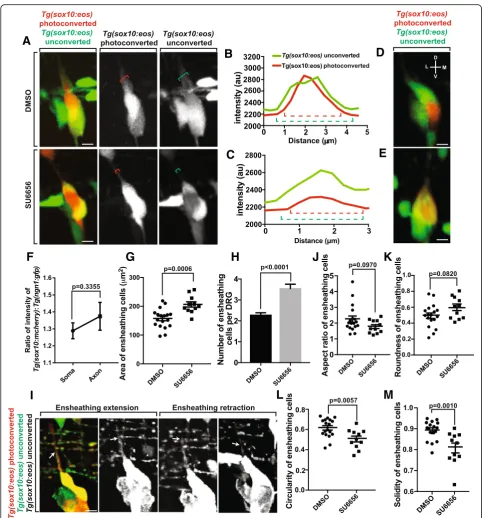
With our data that the ensheathing cell of neuronal soma projects dorsally during axonal initiation, we sought to test whether inhibiting the ensheathment of the axon would affect the ensheathment of the cell soma. To do this, we utilized a drug, Src-family kinase inhibitor SU6656, that inhibits the ensheathment of the axonal pro-jection (Nichols and Smith, unpublished data).
We hypothesized that axonal and neuronal ensheath-ment were two, distinct processes. However, if the cells that ensheath the axon originate as an ensheathing cell of the neuronal soma, disruption of axonal ensheathment could affect neuronal cell soma ensheathment. To test this, we molecularly manipulated axonal ensheathment and measured neuronal soma ensheathment. We treated
Tg(sox10:eos)larvae with SU6656 or DMSO at 36 hpf and imaged them from 48 to 72 hpf, the period when axons navigate dorsally and become ensheathed (Fig.3a). To dis-tinguish between the neurons and the ensheathing glia, we photoconverted the neuron. We then quantified the ensheathment of the axonal projections by measuring the intensity profiles for both converted and unconverted
Tg(sox10:eos)intensities that transect the axon and any as-sociated glia (Fig.3b, c). These intensity profiles revealed a decrease in the width of Tg(sox10:eos) unconverted+ en-sheathing cells around theTg(sox10:eos)converted axon in SU6656 treated animals, suggesting a lack of axonal en-sheathment in these animals (Nichols and Smith, unpub-lished data). To determine the ensheathment of the neuronal cell bodies in both DMSO and SU6656 treated animals we rotated the images of the ensheathed neurons 90° to visualize the association of ensheathing cells with neurons in three dimensions. These rotated images re-vealed that the entire ganglion was ensheathed in both treatment groups (Fig. 3d, e), suggesting that ensheath-ment of the cell body is not affected by SU6656 treatensheath-ment. These data are consistent with the hypothesis that the en-sheathment of the neuronal soma is independent of axonal ensheathment mechanisms.
We sought to further characterize the ensheathment of the neuronal soma and the axon by determining the width of ensheathment in both neuronal compartments. It is possible that the ensheathment of the axon and neuronal soma are entirely independent and exhibit two distinct forms of ensheathment. However, it is also possible that similar biological principles underlie both processes so that similar ensheathment profiles are present in both neuronal compartments. To do this, we used Tg(sox10:m-cherry); Tg(ngn1:gfp)animals to quantify intensity profiles transecting the axon and neuronal soma and calculated the ratios of the width of the Tg(sox10:mcherry) peak
(See figure on previous page.)
A
B
D
C
E
F
G
H
J
K
I
L
M
(ensheathing cells) to the width of theTg(ngn1:gfp) peak. These ratios showed no significant difference between the ensheathment widths of the axon and neuronal soma (Fig. 3f, p= 0.3355, n= 10 DRG, 0.0853 ± 0.0839). This data suggest that similar ensheathment profiles are shared be-tween the axon and the soma during early development. Later, axonal ensheathment likely expands as myelination occurs.
Inhibiting axonal ensheathment leads to increased glia ensheathing the neuron cell body
We reasoned that the observed increase in ensheathing glia coverage in SU6656 animals was due to an axon-ensheathing cell returning to the ganglion after failed axonal ensheathment. Alternatively, failed axonal ensheathment could lead to increased proliferation of ensheathing cells. To test this, we first measured the area of the ensheathing glia at 72 hpf in both DMSO and SU6656 treated animals. These measurements showed an increase in the area of the ensheathing cells in SU6656 treated animals (Fig. 3g, p= 0.0006, n= 18 DMSO, n= 11 SU6656, DMSO: 156.923 ± 7.933 μm2, SU6656: 196.455 ± 13.301μm2). To determine if this in-crease was due to an inin-crease in size of individual glial cells or in the number of glial cells, we counted the number of glial cells that were associated with the neur-onal cell body in both treatments. The SU6656 treated animals had, on average, one extra glial cell present in the ganglia compared to DMSO treated animals (Fig.3h,
p< 0.0001, n = 18 DMSO, n = 11 SU6656, DMSO: 2.278 ± 0.109, SU6656: 3.545 ± 0.207). We were also able to visualize this phenomenon using time-lapse imaging of axonal ensheathment. In SU6656 treated animals, a
Tg(sox10:eos) unconverted+ cell (white arrow) travels dorsally along the nascent axon but returns to the soma before axonal ensheathment can occur (Fig. 3i). These two data sets are consistent with the hypothesis that if axonal ensheathment is inhibited during development, the glial cell that was to ensheath the axon returns back to the ganglia.
From these data, we hypothesized that the presence of an extra glial cell in the DRG could change the shape of entire ganglia. To do this, we determined the shape de-scriptors circularity, aspect ratio, roundness, and solidity for the ensheathing cells in both DMSO and SU6656 treated animals. We detected no significant change in the aspect ratio (Fig.3j,p= 0.0970, n = 18 DMSO, n = 11 SU6656, DMSO: 2.249 ± 0.200, SU6656: 1.776 ± 0.125) or roundness (Fig. 3k, p= 0.0820, n = 18 DMSO, n = 11 SU6656, DMSO: 0.493 ± 0.0344, SU6656: 0.592 ± 0.0312) of the ensheathing cells, consistent with the conclusion that the extra cell did not lead to an elongation of the neuronal/ensheathing glia unit. However, both the circu-larity (Fig. 3l, p= 0.0057, n = 18 DMSO, n = 11 SU6656,
DMSO: 0.622 ± 0.0244, SU6656: 0.506 ± 0.0290) and so-lidity (Fig.3m,p= 0.0010, n = 18 DMSO, n = 11 SU6656, DMSO: 0.891 ± 0.0105, SU6656: 0.807 ± 0.0236) de-creased in SU6656-treated animals. These shape changes are consistent with the conclusion that the extra glial cell from failed axonal ensheathment leads to a larger glial unit and an irregularly shaped, bulky ganglion. It is worth noting that our experiments do not distinguish whether SU6656 impacted these measurements cell-autonomously.
Ensheathing progenitors exhibit space filling potential
Our analysis thus far indicated that these ensheathing cells non-redundantly cover the neuronal cells in the DRG even despite considerable morphological changes. We hypothesized that these ensheathing cells could therefore exhibit continuous tiling potential which en-sures cells' complete but non-redundant coverage of a target area. To first test this tiling hypothesis, we imaged the DRG during the proliferation of the ensheathing cells. In this experiment we observed that the individual neurons were continuously ensheathed. The only point where ensheathment was slightly disrupted was when an ensheathing cell divided and the neuron became mo-mentarily and partially unensheathed (Fig. 4a). In this analysis over a 24-h period, we did not observe any neu-rons that became completely unensheathed. However, if these progenitors do exhibit continuous tiling behavior we would expect that even if a neuron becomes even momentarily unensheathed, the ensheathing cells would respond to eventually fully ensheath the neuron again. To test this, we imaged the projections of the ensheath-ing cells after partial unensheathment (Fig.4a). In doing so, we were able to visualize ensheathing projections travel toward the unensheathed area of the soma. We further hypothesized that the stabilization of this re-ensheathment would recapitulate the first ensheath-ment paradigm where two ensheathing processes con-verged and retracted. To test this, we measured intensity profiles that transected the migrating processes as they approached each other every 75 min. These data showed cellular processes with a single intensity platform which 75 min later separated into two peaks (Fig.4b, represen-tative DRG chosen from 5 assayed DRG). These results are consistent with the hypothesis that these cells likely maintain continuous tiling behavior throughout life by recapitulating developmental ensheathment.
speeds ranged from 0.00515 μm/min to 0.0621 μm/min (mean ± SEM: 0.0286 ± 0.0059μm/min). This data is con-sistent with the hypothesis that in normal tiling, the cells may not respond equally to partial unensheathment but instead one cell responds quickly and travels a further dis-tance to fill space on the neuron.
To continue to provide mechanistic insight into the ability of these cells to tile the neuron we tested our hy-pothesis by manipulating the ensheathing cells experi-mentally to create an unensheathment event. Previous
studies on tiling mechanisms have reported that the remaining ensheathing cells expand to cover the target area following cell ablation. To test the ability of DRG ensheathing cells to do this, we ablated one of the en-sheathing cells and visualized the behavior of the remaining cells. To do this we created small ablations of individual ensheathing cells using our laser ablation sys-tem. In this paradigm, we created a 4μm region of inter-est to ablate individual cells and then captured images every 5 min for 24 h (Fig. 4c, Additional file 2: Movie
A
B
C
D
E
F
S2). First, we confirmed that the ablation resulted in an unensheathment event in the DRG by rotating the image of the ablated DRG 90° to view the ensheathment along the lateromedial axis of the animal at 0 and 24 h post in-jury (hpi). The resulting images revealed a neuron with little glial ensheatment following ablation (Fig. 4d), con-sistent with the idea that the ablation resulted in an unensheathment event. The same DRG 24 h later exhib-ited complete, three-dimensional ensheathment of the neuron (Fig. 4d). This change demonstrated that the remaining ensheathing cells responded to the ablation by filling the area on the neuron where the ablated cell existed. We scored that 90% of all DRG with an ablated glia had been re-ensheathed 24 hpi (Fig. 4e, n= 10 DRG).
Many possibilities, including cell divisions, glial vol-ume expansion and rearrangement, could be responsible for the observed re-ensheathment. We hypothesized that re-ensheathment following injury recapitulated the de-velopmental mechanism and resulted in the rearrange-ment of the remaining ensheathing glia. To test this hypothesis we visualized the movement of the ensheath-ing cells into the ablated site with time-lapse imagensheath-ing. These movies showed that responding glia initiated pro-jections that migrated to the ablated area where they converged, just as in developmental space filling. To fur-ther compare this experimentally induced space filling to that observed in development, we calculated the veloci-ties of the responding glia. Just as in the developmental context, the injury-induced glial response led to asym-metric responses from the ensheathing cells where cells within the same DRG responded to the unensheathment with a wide variability of speeds ranging from 0.00493 μm/min to 0.0206 μm/min (mean ± SEM: 0.0149 ± 0.00155 μm/min). To gain a temporal under-standing of the re-ensheathment events following injury, we recorded the times that the ensheathing cells re-quired to re-ensheath the neuron following experimen-tally induced unensheathment. We found that the ensheathing cells required 679 ± 27.386 min after the in-jury event to re-ensheath the cell body (Fig. 4f, n= 5 DRG). Taken together, these data provide an understand-ing of ensheathment demonstratunderstand-ing re-ensheathment is performed by existing rearrangement of cells within the neuron/ensheathing cell unit. This is consistent with the hypothesis that ensheathing cells in the DRG exhibit con-tinuous tiling capacity throughout life.
Lack of ensheathment perturbs neuronal cell soma shape
Neurons in the DRG exhibit a stereotypical round cell soma morphology and are positioned with precise top-ography in the ganglia [23,33]. Due to their close associ-ation, these ensheathing cells have the potential to
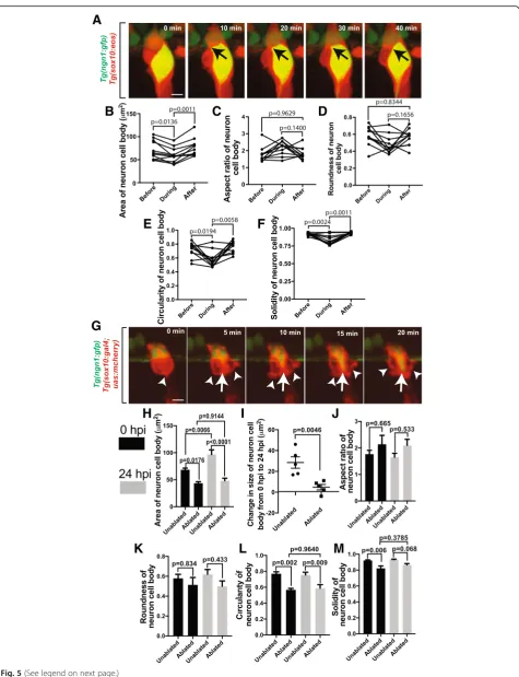
continuously impact neurons morphologically and physiologically. Previous research has demonstrated their physiological role [4,5]. To investigate their poten-tial role in neuronal morphology or spapoten-tial location in the ganglia we first investigated how neuronal shape changed in response to modulations in ensheathing cells. We hypothesized that if these ensheathing cells do con-tinuously impact neuronal shape then even short pertur-bations to the ensheathment would impact neuronal shape. We first tested this possibility by visualizing neu-rons and ensheathing cells inTg(ngn1:gfp); Tg(sox10:eos)
animals from 48 to 72 hpf that have already extended processes. We specifically scored events in which the en-sheathing cells proliferate, causing a partial unensheath-ment event, and then visualized the morphological behavior of the neurons (Fig.5a). These images revealed a bulging of the neuronal soma during a partial unen-sheathment. To quantify this, we measured the area of the neuronal soma before, during, and after proliferation of the ensheathing cells. These measurements showed a dynamic decrease in area of the neuron during the per-turbation of its glial sheath (Fig. 5b, p= 0.0136, n= 11 neurons, before: 71.51 ± 6.24 μm2, during: 59.94 ± 4.89 μm2) which then immediately increased back to its normal level following re-ensheathment (p= 0.0011, after: 76.43 ± 5.78 μm2). Using the shape descriptors for the neuron cell body, we were able to quantify the neur-onal morphology before, during, and after proliferation of the ensheathing cells. The aspect ratio (Fig. 5c, p= 0.1400, n = 11 neurons, before: 1.78 ± 0.131, during: 2.174 ± 0.124, after: 1.82 ± 0.046) and the roundness (Fig. 5d, p= 0.1656, n = 11 neurons, before: 0.585 ± 0.0330, during: 0.478 ± 0.0323, after: 0.565 ± 0.0287) of neurons did not change during the proliferation of ensheathing cells. However, the circularity (Fig. 5e,p= 0.0194, n = 11 neurons, before: 0.722 ± 0.0315, during: 0.585 ± 0.0341) and solidity (Fig. 5f, p= 0.0024, n = 11 neurons, before: 0.924 ± 0.00575, during: 0.826 ± 0.0214) of the neuron decreased during glial proliferation. Both measurements (circularity: p= 0.0058, after: 0.766 ± 0.0256; solidity: p = 0.0011, after: 0.937 ± 0.00444) returned to their normal levels following the completion of glial proliferation, consistent with an overall decrease in area and the ob-served bulging of the neuron during a short perturbation of its glial sheath. These results are consistent with the conclusion that ensheathing cells may continuously pro-vide a physical, restrictive force on DRG neurons.
A
B
C
D
E
F
G
H
I
J
K
L
M
the ablation of an ensheathing cell led to a decrease in the area of the neuron shortly after the ablation compared to neighboring unablated control ganglia (Fig.5h,p= 0.0176,
n= 5 unablated DRG, n = 5 ablated DRG, ablated: 43.52 ± 2.97 μm2, unablated: 68.47 ± 3.01 μm2). This decrease in neuron size persisted following re-ensheathment and was still present 24 hpi (p< 0.0001 unablated DRG, n = 5 ab-lated DRG, abab-lated: 48.29 ± 4.56 μm2, unablated: 69.99 ± 8.34μm2). To further explore this injury-induced perturb-ation of neuronal soma size, we quantified the change in the size of the soma from 0 to 24 hpi. Unablated control DRG neurons increased 26.53 ± 5.52 μm2 (Fig. 5i, n = 5 neurons) during that time period, while the neurons with an ablated glial cell increased only 4.77 ± 2.63μm2(n = 5 neurons, p= 0.0046). To continue to examine the neur-onal morphological changes resulting from an unen-sheathment event, we quantified the shape descriptors for the neuronal cell bodies in DRG with ablated and with unablated ensheathing glia. Just as in the normal develop-mental partial unensheathment, the aspect ratio (Fig.5j,p
= 0.665, n = 5 neurons, unablated at 0 hpi: 1.767 ± 0.145, ablated at 0 hpi: 2.139 ± 0.336, unablated at 24 hpi: 1.654 ± 0.146, ablated at 24 hpi: 2.097 ± 0.230) and roundness (Fig. 5k, p= 0.834, n = 5 neurons, unablated at 0 hpi: 0.579 ± 0.0.0405, ablated at 0 hpi: 0.513 ± 0.0.0744, unab-lated at 24 hpi: 0.621 ± 0.0469, abunab-lated at 24 hpi: 0.500 ± 0.0527) were not affected by unensheathment, suggesting that the neurons did not elongate. However, the circularity (Fig.5l, p = 0.002, n = 5 neurons, unablated at 0 hpi: 0.769 ± 0.0245, ablated at 0 hpi: 0.567 ± 0.0188) and solidity (Fig. 5m, p= 0.006, n = 5 neurons unablated at 0 hpi: 0.921 ± 0.00662, ablated at 0 hpi: 0.822 ± 0.0298) both decreased in neurons with an ablated glia. The circularity of neurons with ablated ensheathing cells remained depressed 24 hpi (p= 0.009, unablated at 24 hpi: 0.756 ± 0.0330, mean ab-lated at 24 hpi ± SEM: 0.588 ± 0.0445). The solidity of these neurons slightly recovered to levels similar to that of DRG neurons without ablated cells (p= 0.068, unablated at 24 hpi: 0.933 ± 0.00530, ablated at 24 hpi: 0.864 ± 0.0185). The simplest explanation for this is that a larger unensheathment event following an injury to the en-sheathing cells results in a persistent change to both the size and shape to the DRG neuron. Overall, these changes
in neuron morphology in both the developmental and in-jury contexts are consistent with the hypothesis that en-sheathing cells could provide continuous structural forces to maintain the morphology of individual DRG neurons.
When DRG glial precursors are genetically ablated, the developing DRG becomes mislocalized at inconsistent lo-cations along the dorsoventral axis of the animal [23]. Based on this observation and our data above that injury to ensheathing cells resulted in prolonged alterations in neuron size and shape, we hypothesized that prolonged unensheathment of neurons could have an impact on the location of the neuron within the ganglia. To address this possibility, we ablated multiple ensheathing cells and traced any movement of the neuron that they once sur-rounded. This quantification was done by measuring the center point of the neuron before and after the cells were ablated. In these experiments, we visualized that ablation of ensheathing glia first caused an immediate ectopic shift-ing of the neuron toward the area that was ablated (Fig.6a, b). To determine if these neurons displayed a consistent direction of movement, we quantified the trajectory of the neurons following ablation and observed that neurons typically shifted either ventrally or dorsally (Fig. 6c). In these movements, the neurons did not move with a con-sistent velocity. Adjacent neurons without any ablated glia moved less. To further explore this injury-induced neur-onal movement, we quantified the total displacement and velocities in these neurons. Neurons with ablated glia were displaced 8.476 ± 0.699μm at a speed of 0.0077 μm/min (Fig. 6d, n = 5 neurons). These measurements were both greater than neurons in unablated DRG which traveled an average distance of 6.0 ± 0.776 μm at a speed of 0.0054μm/min (Fig.6d,p= 0.0429,n= 5 neurons). These tracings are consistent with the possibility that prolonged unensheathment of DRG neurons could cause not only momentary morphological changes (Fig.5) but also shift-ing of the neurons within the ganglia.
Given that ablation of ensheathing cells resulted in misplaced DRG neurons, we next tested if ablation of ensheathing cells resulted in axonal pathfinding defects. However, we did not observe any obvious pathfinding defects. The axon initiated and traveled dorsally, enter-ing the spinal cord at a typical DREZ location (Fig.6e).
(See figure on previous page.)
Proliferation of neurons corresponds with expansion of progenitors
Since these ensheathing cells are present during early gan-glia development, we sought to identify if they represented a terminally defined cell-type or if they were a progenitor population. To first test the progenitor possibility of these ensheathing cells, we imagedTg(sox10:eos)animals at 2 dpf and photoconverted a single ensheathing cell. Twenty-four hours later, we observed multipleTg(sox10:eos) photocon-verted cells in the DRG, suggesting that the ensheathing cells actively divide like a progenitor population (Fig.7a, b)
as previously described [37]. To further explore this possi-bility, we quantified whether they expanded as the neuron number increased. To do this, we imaged the DRG in
Tg(ngn1:gfp); Tg(sox10:eos) animals at two, three, and four dpf and photoconverted the Eos in the entire animal (Fig. 7c). In these images, we quantified the ratio ofsox10+cells and neurons at each time point. The ratios steadily de-creased at each time point (Fig.7d). To further expand this analysis, we quantified the number of ensheathing cells and neurons present in the DRG at each time point (Fig. 7e). From 2 to 4 dpf, the number of neurons increased by one
A
B
C
D
E
on average. The number of glia increased by one from 2 to 3 dpf but then decreased by one by 4 dpf. Overall, these data are consistent with the idea that the proliferation of the ensheathing cells corresponds with an increase in sen-sory neurons and that the new neurons could arise from
sox10+ensheathing progenitor cells.
We next sought to gain a spatial understanding of DRG expansion. We hypothesized that ensheathing cells could ensheath multiple neurons during DRG expansion. How-ever, it is also possible that neurons could be individually encased by individual cells. To test these possibilities, we
used Tg(ngn1:gfp); Tg(sox10:eos) fish and photoconverted Eos at 4 dpf. After photoconverstion, animals were fixed and stained for Sox10 and DAPI. Images of these animals revealed that each neuron was individually encased by multiple ensheathing cells (Fig.7f). While it is difficult to determine exactly how many cells participate in the en-sheathment of an individual neuron, more than one cell nuclei that associated with processes that ensheath indi-vidual neurons at 4 dpf can be visualized, a result that is consistent with recent investigations into adult DRG en-sheathment by mature satellite glia [9].
A
B
D
E
C
F
Fig. 7Proliferation of ensheathing cells is correlated with DRG sensory neuron expansion. (a). Confocal z-projection images of aTg(sox10:eos) zebrafish with a single ensheathing cell photoconverted at 2 dpf. Images were taken at 2 and 3 dpf. Dashed outlines denoteTg(sox10:eos) photoconverted+cells. (b). Schematic summary of the use of photoconversion in the proliferation of ensheathing cells. (c). Confocal z-projection
images ofTg(ngn1:gfp)zebrafish stained with Sox10 at 2–4 dpf. (d). Ratio of the number Sox10+cells to the number of neurons in a DRG at 2 –4 dpf (n= 16 DRG). (e). Number of Sox10+cells and neurons present in the DRG at 2–4 dpf (n= 16 DRG). (f). Confocal z-projection images of a
Ensheathing cells respond to neuronal injury
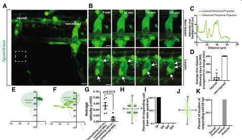
Since these ensheathing cells are closely associated with sensory neurons from as early as we can visualize, we hy-pothesized that they would quickly respond to changes in neuronal homeostasis [10]. Although this phenomenon has been demonstrated, the temporal dynamics of this have not been thoroughly examined. To test this, we imaged DRG neurons that were axotomized distally in the periph-ery and visualized the response of the ganglia cells to that injury. Using Tg(sox10:eos) animals which label cells that ensheath the DRG neurons we performed an 8 μm axot-omy injury approximately 100 μm from the ganglia and then collected images every 5 min for 10 h. In each image, we had an experimental-injured and a control non-injured sensory nerve (Fig. 8a). We first confirmed that our laser parameters created a complete transection of the periph-eral nerve by imagingTg(sox10:eos)intensity profiles of the length of the lesioned axon 1 h following injury and visual-ized a lack of fluorescent signal along the nerve (Fig.8c).
In these animals we visualized that 100% of ganglia with injured peripheral nerves responded by re-arranging ensheathing cells around the neuronal soma (Fig. 8b, Additional file 3: Movie S3, Additional file 4: Movie S4). In control, non-injured, ganglia that were adjacent to the injured nerves we did not visualize this dynamic; within the 10-h period of imaging, 0% of ganglia associated with non-injured nerves re-arranged. We took this analysis fur-ther to dissect the dynamics of re-arrangement in order to investigate the speed of ganglia response to peripheral in-jury. Injured nerves induced a ganglia cell re-arrangement in 72.6 ± 48.22 min with the majority responding within the first hour (Fig.8d,n= 5). These responses occurred in the ganglia 100μm from the injury site. These results are consistent with the hypothesis that the ensheathing cells of the ganglia can respond rapidly to peripheral injury of those neurons.
To expand our understanding of the injury response, we traced the movements of the responding glia in
A
B
C
D
E
F
G
H
I
J
K
DRG with lesioned axons as well as adjacent unlesioned DRG. Ensheathing cells in lesioned DRG dynamically traversed the entire ganglia, while ensheathing cells in unlesioned DRG remained in their original location (Fig. 8e, f). Further, we calculated the velocities of the ensheathing cell projections in both lesioned and unle-sioned DRG. These cells responded to the lesion with a velocity of 0.0519 ± 0.0113 μm/min (Fig. 8g, n = 5 DRG), while cells on unlesioned ganglia were largely stationary (n = 5 DRG, p= 0.0115, 0.0117 ± 0.00492). Since the injury was consistently created in the same portion of the neuron, we hypothesized that the glial response may be similar following each injury. To do this, we determined the location of the cells that re-spond to the peripheral injury by quantifying which quadrant of the ganglia the responding cell originated. We found that 100% of the responding cells on lesioned DRG originated in the dorsal, posterior quadrant of the ganglia (Fig. 8h, i, n = 5 DRG). Given this highly con-sistent spatial response to the injury, we also quantified the final location of the responding cell. We hypothe-sized that the responding cell may travel to the sight of the injury. Instead, 100% of the responding cells even-tually ended their migration at the ventral apex of the neuron cell body where the injured neurite originated (Fig. 8j, k, n = 5 DRG). These data suggest that en-sheathing cells have a strikingly consistent and choreo-graphed response to peripheral axonal injury; they migrate to the extension site of initiation of the injured axon.
Given the effects of unensheathment can have on the neuron and the apparent rearrangement of cells in the ganglia following injury, we next asked if neuronal soma shape was momentarily altered during the injury re-sponse. To ask this, we measured the area and shape de-scriptors for the neuronal cell bodies shortly after the injury and 24 h later. We did not detect any changes in area (p= 0.6830, n = 5 unlesioned neurons, n = 5 lesioned neurons, unlesioned at 0 hpi: 61.199 ± 9.688 μm2, le-sioned at 0 hpi: 73.937 ± 15.863 μm2, unlesioned at 24 hpi: 76.472 ± 12.843μm2, lesioned at 24 hpi: 83.465 ± 13.101 μm2), circularity (p= 0.8453, n = 5 unlesioned neurons, n = 5 lesioned neurons, nonlesioned at 0 hpi: 0.775 ± 0.0600, lesioned at 0 hpi: 0.768 ± 0.0325, nonle-sioned at 24 hpi: 0.765 ± 0.0595, lenonle-sioned at 24 hpi: 0.796 ± 0.0380), aspect ratio (p= 0.2491, n = 5 unlesioned neurons, n = 5 lesioned neurons, nonlesioned at 0 hpi: 1.438 ± 0.188, lesioned at 0 hpi: 1.868 ± 0.125, nonle-sioned at 24 hpi: 1.536 ± 0.131, lenonle-sioned at 24 hpi: 1.713 ± 0.168), roundness (p= 0.2040, n = 5 unlesioned neu-rons, n = 5 lesioned neuneu-rons, nonlesioned at 0 hpi: 0.739 ± 0.0857, lesioned at 0 hpi: 0.545 ± 0.0357, nonlesioned at 24 hpi: 0.671 ± 0.0571, lesioned at 24 hpi: 0.608 ± 0.0650), or solidity (p= 0.6562, n = 5 unlesioned neurons,
n = 5 lesioned neurons, nonlesioned at 0 hpi: 0.914 ± 0.143, lesioned at 0 hpi: 0.930 ± 0.0106, nonlesioned at 24 hpi: 0.918 ± 0.185, lesioned at 24 hpi: 0.921 ± 0.0172). These measurements suggest that the ensheathing glia exhibit a highly-coordinated response throughout the ganglia following peripheral injury that mirrors develop-mental expansion to ensure continued ensheathment of the neuronal cell body as one cell migrates across the ganglion. Only during longer pathological unensheath-ment events does neuronal morphology become altered. Together, these data support the hypothesis that during the lifespan of the DRG, the neuron and its dynamic en-sheathing cells are intimately connected and plastic.
Discussion
Ensheathment of neuronal cells is critical to proper formation and function of complete nerves. Using single-cell photoconversion and time lapse imaging in intact living vertebrates, we demonstrate that the en-sheathment of DRG neuronal cell somas occurs during neuronal differentiation, a distinct event from axonal en-sheathment. We show that these ensheathing cells must rearrange to allow for the initiation of neurites. During development they also exhibit tiling behavior. Perturba-tions of the soma ensheathment causes changes in neur-onal soma morphology and positioning. Using laser induced lesions of the distal peripheral axon, we show that these ensheathing cells respond quickly to neuronal injury in a choreographed manner. Together, our data suggest that ensheathing cells of the DRG neuronal cell soma are closely associated with the neuron starting shortly after initial neuronal differentiation and persist throughout the life of the neuron to maintain stereotyp-ical ganglia positions.
DRG cells exhibit a neural niche
neuronal progenitor differentiation in classical neural niches. In fact, the fate of progenitor populations in neural niches and the DRG may even depend on the level of en-sheathment of a daughter cell by dictating expression of neuronal markers. As a result, future studies on the pre-cise development of the DRG in the context of ensheath-ing cells may yield valuable insights into the dynamics of neural niches and neuronal differentiation.
In addition, glial ensheathment of distinct portions of a mature neuron is critical to the establishment of a functional nervous system [10,48]. While many previous studies have examined the mechanism of axonal ensheath-ment, few have explored the process of neuronal cell soma ensheathment. Our data helps fill this gap by visualizing the ensheathment of DRG neuronal somas in vertebrates. We elucidate the close association between the ensheath-ing cells and the neuron throughout its maturation. Stud-ies inDrosophilahave demonstrated the potential for cells ensheathing the cell soma to interact with and support the neuron which they ensheath. For example, expression of a (DE)-cadherin dominant negative in Drosophila cortex glia, which ensheath neuronal cell bodies, have been shown to lead to misplaced neural progenitors and neur-onal somas as well as disrupt neuron morphology and neurite trajectories [18]. Here, we provide a step wise visualization of the ensheathment of such vertebrate neur-onal somas.
Soma ensheathing cells interact with axons
In a mature DRG, differentiated satellite glial cells en-sheath the sensory neurons located in the ganglia [10]. Interestingly, co-cultures of mature satellite glial cells and neurons lead to inhibited dendrite formation [49]. Our data suggests that the precursors to satellite glial cells are intimately involved in neuronal maturation and morphology, including neurite extension. Given this and the formation of “glial horns” during neurite initiation, the ensheathment of the neuron cell body is likely crit-ical to maturation of the neuron and its neurites. Coupled with increasing evidence for the role of glia in the initiation of neurites [15], our data suggest that re-arrangement of the ensheathing DRG cells may provide a substrate to allow for the initiation or extension of axonal projections. Further, we demonstrate that the ini-tial ensheathing cell of the sensory pioneer axon are pro-duced in the ganglia by cells that ensheath the cell soma. This finding complements recent investigations seeking to characterize satellite glia which identified myelino-genic capabilities of mouse satellite glia in culture as well as robust transciptomic similarities between satellite glia and mature Schwann cells [9]. Our imaging of DRG de-velopment in zebrafish are consistent with these hypoth-eses. The intimate associations of the ensheathing cells with the DRG neurons from as early as we can delineate
suggests they could have a profound influence on neuronal development and homeostasis. But to date, neuronal development and homeostasis are often inves-tigated in the absence of ensheathing cells. Future stud-ies testing these hypotheses may yield important insights in axonal morphology, nerve assembly, and axonal compartmentalization.
Forces that dictate neuronal cell shape and location
Further, our data suggest that neuronal soma ensheath-ment likely plays a conserved function in dictating the size and shape of the soma in development. In zebrafish mutants of Sox10, a gene required for glial differenti-ation, ensheathing cells of the DRG are absent and the DRG are mislocalized [23]. These data are consistent with the hypothesis that ensheathing cells also contrib-ute to the positioning of the DRG. We also observe that the neuronal soma immediately bulges and shifts toward a site of disrupted ensheathment. The totality of these data, in conjunction with previous studies, point to a conserved role of continuous mechanical forces on the neuronal soma from the ensheathing cell membranes imposing a circular shape and potentially precise pos-ition on the neuron within the ganglia [17, 18, 23, 32]. The release of this restraining force in unensheathment events likely causes the neuron to accelerate toward ab-lated area much like a compressed spring.
paradigmatic development. In addition, physical contacts between DRG cell types have been suggested as regulators of cell fate decisions between glia and neurons as well as between different types of neuronal subtypes [23,33, 50]. Taken together with our data, this suggests that the pre-cise forces on neurons by ensheathing cells may aid in the development of the diversity of somatosensory cell types.
Unfortunately, the physiological effects of changes to the shape of neuronal somas are currently unknown. Data comparing the morphology and electrophysiology of CA1 and CA2 hippocampal neurons in Proechimys
rodents suggests that smaller cell somas are correlated with decreased electrical resistance and a longer latency period [52]. However, these recordings were taken from healthy and fully differentiated neuron populations. Cell soma size also correlates with axonal caliber [53]. Stud-ies show axonal caliber impacts ensheathment and mye-lination, pointing to a hypothesis that neuronal size is important in the nervous system [54, 55]. Regardless, given that our data indicate that re-ensheathment itself is not sufficient to restore neuronal morphology follow-ing prolonged unensheathment, the importance of soma shape in neuron electrophysiology, although beyond the scope of this paper, should be investigated, especially given the possibility of unensheathment in disease states.
Ensheathing cells respond to injury
Given the close proximity of ensheathing cells to axonal injury, they represent an important cell type that could respond to neural injury. Previous studies have shown that many ensheathing glial subtypes exhibit tiling be-havior to maintain neuronal ensheathment following in-jury [16]. Our analysis of the space filling behavior by ensheathing processes in both normal development and injury states consistently demonstrated an asymmetric response to unensheathment by individual cells (Fig.4). This response suggests that specific subpopulations of ensheathing glia, at least during development, may be hypervigilant to unensheathment events. We also dem-onstrate that ensheathing cells respond to modulated en-sheathment through tiling behavior throughout the life
of the DRG. Further, we show that neuronal
soma-ensheathing cells display a consistent and choreo-graphed response to neuronal injury to the distal axonal segment by migrating to the initiation site of the injured neurite. This response does not disrupt the ensheath-ment or morphology of the neuron, suggesting a coordi-nated space-filling response by all ensheathing cells following neuronal injury. As a result, our data point to DRG ensheathing glia as a population that can detect and respond to neural injury, a behavior also seen in ma-ture satellite glial cells [10]. This observation is also con-sistent with previous reports inDrosophilawhere cortex glia respond to injury and phagocytize neural debris
[16]. Previous studies have also demonstrated that some DRG cell types are pluripotent stem cells that can differ-entiate into terminal glia, or even skin melanocytes, after migrating down the peripheral nerve to the skin [34]. Given the specific choreographed response by ensheath-ing glia from a consistent and specific neuronal domain following neural injury, it is possible that the responding cell we identified retains a stem-like quality in order to coordinate a cellular response to neuronal injury. Coupled with the hypervigilance of some cells to unen-sheathment, the cells that ensheath DRG neurons could form a heterogeneous population with specific cells primed to respond to various, yet specific disruptions to neuronal homeostasis. Future research will dissect this important topic.
Conclusions
While many studies have been devoted to the mecha-nisms of axonal ensheathment and its role in neuronal homeostasis, mechanisms of ensheathment of neuronal somas has remained elusive. Here we used single-cell photoconversion to visualize the process of soma en-sheathment in zebrafish dorsal root ganglia. Taken to-gether, our data point to the importance of neuronal soma ensheathment in nerve development as early as neuronal differentiation. The close association of the en-sheathing cells with the maturing neuron point to cross-talk as a possible important facet of nerve assem-bly. Elucidating these associated cues, as demonstrated by recent studies, could yield valuable insight into neural niches, neural injury responses and the diversity of soma-ensheathing cells.
Additional files
Additional file 1:Movie S1.Ensheathment of neuronal progenitors occurs soon after neuronal differentiation. Excerpt from a 24-h time lapse of aTg(sox10:eos)animal with a photoconverted neuronal progenitor from 48 to 72 hpf. (Left) ATg(sox10:eos)unconverted cell wraps projections around the neuronal progenitor before neurite extension. (Right) Merged image ofTg(sox10:eos)converted (neuronal progenitor) and unconverted (ensheathing cell). Green arrows denote ensheathing projections. Dark green arrow denotes merged ensheathing projections. Frames in the video were captured every 5 min, and the video plays at 10 frames per second. Supplements Fig.1d. (MOV 4824 kb)
Additional file 2:Movie S2.Soma-ensheathing cells exhibit tiling behavior during injury. Excerpt from a 24-h time lapse of aTg(sox10:eos) animal with a photoconverted neuronal progenitor from 48 to 72 hpf following a laser ablation of an ensheathing cell. (Left)Tg(sox10:eos) unconverted cells respond to the unensheathment of the neuronal soma by sending projections to restore soma ensheathment. (Right) Merged image ofTg(sox10:eos)converted (neuron) and unconverted (ensheathing cell). Green arrows denote ensheathing projections. Dark green arrow denotes merged ensheathing projections. Frames in the video were captured every 5 min, and the video plays at 10 frames per second. Supplements Fig.4e. (MOV 6449 kb)
animal from 72 to 96 hpf. The imaged DRG is adjacent to a DRG neuron with peripheral axonal injury at 72 hpf. Red arrow denotes an ensheathing cell. Frames in the video were captured every 5 min, and the video plays at 10 frames per second. Supplements Fig.8b. (MOV 931 kb)
Additional file 4:Movie S4.Ensheathing cells dynamically remodel following distal axonal injury. Excerpt from a 24-h time lapse of a Tg(sox10:eos)animal from 72 to 96 hpf. The imaged DRG neuron suffered distal axonal injury at 72 hpf. Red arrow denotes an ensheathing cell. Frames in the video were captured every 5 min, and the video plays at 10 frames per second. Supplements Fig.8b. (MOV 1382 kb)
Abbreviations
CNS:Central nervous system; DMSO: Dimethyl sulfoxide; dpf: Days post fertilization; DREZ: Dorsal root entry zone; DRG: Dorsal root ganglia; hpf: Hours post fertilization; hpi: Hours post injury
Acknowledgements
We thank members of the Smith lab and Sarah Kucenas for their helpful comments, Sam Connell and Brent Redford of 3i for fielding imaging questions and Deborah Bang and Karen Heed for zebrafish care. This work was supported by the University of Notre Dame, the Elizabeth and Michael Gallagher Family, the Alfred P. Sloan Foundation, Center for Zebrafish Research at the University of Notre and Center of Stem Cells and Regenerative Medicine at the University of Notre Dame.
Funding
This work was supported by the University of Notre Dame and the Elizabeth and Michael Gallagher Family, Center for Zebrafish Research at the University of Notre, Center of Stem Cells and Regenerative Medicine at the University of Notre Dame and the Alfred P. Sloan Foundation.
Availability of data and materials
All data generated and analyzed for this manuscript are included in this article.
Authors’contributions
ELN, LAG and CJS conducted the experiments. ELN analyzed all the experiments and quantified the data. LAG and CJS created injuries in sensory neurons. CJS conceived the project and ELN and CJS wrote the manuscript with input from LAG. All authors read and approved the final manuscript.
Ethics approval and consent to participate
All experiments were approved by the University of Notre Dame Institutional Animal Care and Use Committee.
Consent to publication
Not applicable.
Competing interests
The authors declare that they have no competing interests.
Publisher’s Note
Springer Nature remains neutral with regard to jurisdictional claims in published maps and institutional affiliations.
Received: 7 February 2018 Accepted: 27 July 2018
References
1. Donaldson HH. 1902. The relation of myelin to the loss of water in the mammalian nervous system with advancing age. Proc Phil Soc Phil mag Wellisch, Amer J Sci Lattey proc R Soc, a Kovarick physic rev * Chaffee 209. 2. Hines JH, Ravanelli AM, Schwindt R, Scott EK, Appel B. Neuronal activity
biases axon selection for myelination in vivo. Nat Neurosci. 2015;18:683–9. 3. Snaidero N, Molbius W, Czopka T, Hekking LHP, Mathisen C, Verkleij D,
Goebbels S, Edgar J, Merkler D, Lyons DA, Nave KA, Simons M. Myelin membrane wrapping of CNS axons by PI(3,4,5)P3-dependent polarized growth at the inner tongue. Cell. 2014;156:277–90.
4. Brown AM, Ransom BR. Astrocyte glycogen and brain energy metabolism. Glia. 2007;55:1263–71.
5. Nave KA. Myelination and the trophic support of long axons. Nat Rev Neurosci. 2010;11:275–83.
6. Jessen KR, Mirsky R. Embryonic Schwann cell development: the biology of Schwann cell precursors and early Schwann cells. J Anat. 1997;191:501–5. 7. Wang H, Moyano AL, Ma Z, Deng Y, Lin Y, Zhao C, Zhang L, Jiang M, He X, Ma
Z, Lu F, Xin M, Zhou W, Yoon SO, Bongarzone ER, Lu QR. miR-219 cooperates with miR-338 in myelination and promotes myelin repair in the CNS. Dev Cell. 2017;40:566–82. e5
8. Bin JM, Rajasekharan S, Kuhlmann T, Hanes I, Marcal N, Han D, Rodrigues SP, Leong SY, Newcombe J, Antel JP, Kennedy TE. Full-length and fragmented netrin-1 in multiple sclerosis plaques are inhibitors of oligodendrocyte precursor cell migration. Am J Pathol. 2013;183:673–80.
9. George D, Ahrens P, Lambert S. Satellite glial cells represent a population of developmentally arrested Schwann cells. Glia. 2018; Available from:http:// doi.wiley.com/10.1002/glia.23320
10. Hanani M. Satellite glial cells in sensory ganglia: from form to function. Brain Res Rev. 2005;48:457–76.
11. Freeman MR. Drosophila central nervous system glia. Cold Spring Harb Perspect Biol. 2015;7
12. Oikonomou G, Shaham S. The glia of caenorhabditis elegans. Glia. 2011;59: 1253–63.
13. Heiman MG, Shaham S. DEX-1 and DYF-7 establish sensory dendrite length by anchoring dendritic tips during cell migration. Cell. 2009;137:344–55. Available from: https://ac.els-cdn.com/S0092867409001597/1-s2.0-S0092867409001597-main.pdf?_tid=8bdd010c-feec-11e7-8c3f-00000aacb361&acdnat=1516567937_ 54b3b51834e8491ad68f0c8e91752af6
14. Procko C, Lu Y, Shaham S, Horvitz HR, Bargmann CI. Glia delimit shape changes of sensory neuron receptive endings in C. Elegans. Development. 2011;138:1371–81. 15. Rapti G, Li C, Shan A, Lu Y, Shaham S. Glia initiate brain assembly through
noncanonical Chimaerin-Furin axon guidance in C. Elegans. Nat Neurosci. 2017;20:1350–60.
16. MacDonald JM, Beach MG, Porpiglia E, Sheehan AE, Watts RJ, Freeman MR. The Drosophila cell corpse engulfment receptor Draper mediates glial clearance of severed axons. Neuron. 2006;50:869–81.
17. Coutinho-Budd JC, Sheehan AE, Freeman MR. The secreted neurotrophin spätzle 3 promotes glial morphogenesis and supports neuronal survival and function. Genes Dev. 2017;31:2023–38.
18. Dumstrei K, Wang F, Hartenstein V. Role of DE-cadherin in neuroblast proliferation, neural morphogenesis, and axon tract formation in Drosophila larval brain development. J Neurosci. 2003;23:3325–35.
19. Redmond SA, Mei F, Eshed-Eisenbach Y, Osso LA, Leshkowitz D, Shen YAA, Kay JN, Aurrand-Lions M, Lyons DA, Peles E, Chan JR. Somatodendritic expression of JAM2 inhibits oligodendrocyte myelination. Neuron. 2016;91:824–36. 20. Gao FB, Kohwi M, Brenman JE, Jan LY, Jan YN. Control of dendritic field
formation in Drosophila: the roles of flamingo and competition between homologous neurons. Neuron. 2000;28:91–101.
21. Grueber WB, Jan LY, Jan YN. Dendritic tiling in Drosophila. Development. 2002;129:2867–78. Available from:https://pdfs.semanticscholar.org/651e/ 308ef39e23a221b50b49aeb0e66fbf1be88d.pdf
22. Huckfeldt RM, Schubert T, Morgan JL, Godinho L, Di Cristo G, Huang ZJ, Wong ROL. Transient neurites of retinal horizontal cells exhibit columnar tiling via homotypic interactions. Nat Neurosci. 2009;12:35–43.
23. Carney TJ, Dutton KA, Greenhill E, Delfino-Machin M, Dufourcq P, Blader P, Kelsh RN. A direct role for Sox10 in specification of neural crest-derived sensory neurons. Development. 2006;133:4619–30. Available from:http:// dev.biologists.org/cgi/doi/10.1242/dev.02668
24. Sagasti A, Guido MR, Raible DW, Schier AF. Repulsive interactions shape the morphologies and functional arrangement of zebrafish peripheral sensory arbors. Curr Biol. 2005;15:804–14. Available from:http://www.ncbi.nlm.nih.gov/ pubmed/15886097
25. Smith CJ, Watson JD, Spencer WC, O’Brien T, Cha B, Albeg A, Treinin M, Miller DM III. Time-lapse imaging and cell-specific expression profiling reveal dynamic branching and molecular determinants of a multi-dendritic nociceptor in C. Elegans. Dev Biol. 2010;345:18–33. Available from:http:// www.ncbi.nlm.nih.gov/pubmed/20537990
26. Hughes EG, Kang SH, Fukaya M, Bergles DE. Oligodendrocyte progenitors balance growth with self-repulsion to achieve homeostasis in the adult brain. Nat Neurosci. 2013;16:668–76.
28. Smith CJ, Morris AD, Welsh TG, Kucenas S. Contact-mediated inhibition between oligodendrocyte progenitor cells and motor exit point glia establishes the spinal cord transition zone. PLoS Biol. 2014;12:e1001961. Available from:http://dx.plos.org/10.1371/journal.pbio.1001961
29. Pereanu W, Kumar A, Jennett A, Reichert H, Hartenstein V. Development-based compartmentalization of the drosophila central brain. J Comp Neurol. 2010; 30. Pereanu W, Spindler S, Cruz L, Hartenstein V. Tracheal development in the
Drosophila brain is constrained by glial cells. Dev Biol. 2007;302:169–80. 31. Radtke C, Wewetzer K, Reimers K, Vogt PM. Transplantation of olfactory ensheathing cells as adjunct cell therapy for peripheral nerve injury. Cell Transplant. 2011;20:145–52.
32. Doyle SE, Pahl MC, Siller KH, Ardiff L, Siegrist SE. Neuroblast niche position is controlled by phosphoinositide 3-kinase-dependent DE-cadherin adhesion. Development. 2017;144:820–9.