Age-Related Hearing Loss & Neurocognitive Function:
Normal and Pathological Processes in Cognitive
Ageing
By
David Loughrey
A dissertation submitted for the degree of Doctor of Philosophy of the
University of Dublin, Trinity College, Dublin 2, Ireland.
i
DECLARATION
I declare that this thesis has not been submitted as an exercise for a degree at this or any other
university and it entirely my own work.
I agree to deposit this thesis in the University’s open access institutional repository or allow the
Library to do so on my behalf, subject to Irish Copyright Legislation and Trinity College Library
conditions of use and acknowledgment.
ii
Abstract
iii
List of Publications
Loughrey, D.G, Kelly, M.E., Kelley, G.A, Brennan, S. & Lawlor, B.A. (2018). The Association of Age-Related Hearing Loss with Cognition Function, Cognitive Impairment and Dementia: A Systematic Review with Meta-Analysis. JAMA Otolaryngol Head Neck Surg, 144(2):115-126
Author Contributions: Loughrey had full access to all the data in the study and takes responsibility
for the integrity of the data and the accuracy of the data analysis. Loughrey oversaw the study concept and design, conducted the statistical analysis and interpretation of results and drafted the manuscript.
Author Affiliations: NEIL (Neuro Enhancement for Independent Lives) Programme, Trinity College
Institute of Neuroscience, Trinity College Dublin, Dublin, Ireland (Loughrey, Kelly, Brennan, Lawlor); School of Medicine, Trinity College Dublin, Dublin, Ireland (Loughrey, Lawlor); Department of Psychology, National University of Ireland Maynooth, Kildare, Ireland (Kelly); Meta-Analytic Research Group, School of Public Health, Department of Biostatistics, Robert C. Byrd Health Sciences Center,West Virginia University, Morgantown (Kelley); Mercer’s Institute for Successful Ageing, St James Hospital, Dublin, Ireland (Lawlor).
Published Abstracts:
Loughrey, D., Coen, R., & Lawlor, B.A. (2017) Age-related Hearing Loss & Temporary Memory Binding. 7th Aging and Speech Communication Research Conference 2017. Florida, USA.
Loughrey, D., Coen, R., & Lawlor, B.A. (2017) Age-related Hearing Loss & Temporary Memory Binding: A Study Protocol. Psychology, Health and Medicine Conference. Royal College of Surgeons in Ireland.
Loughrey, D., Brennan, S., & Lawlor, B.A. (2015). Age-related hearing loss, cognitive decline and dementia: Systematic review of the literature. 6th Aging and Speech Communication Research Conference 2015. Indiana, USA.
Loughrey, D., Brennan, S., & Lawlor, B.A. (2015). Presbycusis and Dementia: A Literature Review of Underlying Factors. The 3rd International Conference on Cognitive Hearing Science for Communication. Linköping, Sweden.
iv
SUMMARY
This thesis investigates age-related hearing loss (ARHL) as a potential biomarker and treatable risk factor for cognitive decline and dementia and attempts to explore and identify a cognitive profile of older adults with ARHL using neuropsychological assessment.
Chapter 1 gives an overview of the literature on cognitive ageing, theoretical perspectives, pathological cognitive ageing, and ARHL. Epidemiological research on the possible link between age-related hearing loss (ARHL) and cognitive decline and dementia has produced inconsistent results and the basis for their possible association remains unclear.
Chapter 2 describes a systematic review and meta-analysis to estimate the association between age-related hearing loss (ARHL) as assessed by pure-tone (PT) audiometry and cognitive function, cognitive impairment and dementia to clarify this association in epidemiological studies. A small but significant association was found for ARHL within all domains of cognitive function. A significant association was found for both cognitive impairment and dementia, but not for Alzheimer’s disease and vascular dementia subgroups. The pattern of results in cognitive ageing indicated a possible mechanistic association via impaired speech perception.
Chapter 3 describes a narrative review of possible pathological processes that represent either a common aetiological cause for both conditions or a mechanistic pathway by which ARHL leads to neurocognitive decline. A review of the evidence suggests that the relationship between ARHL and neurocognitive aging is most likely to be multifactorial with several processes contributing
including behavioural and neuroimaging support for a possible speech-based mechanistic association.
Chapter 4 describes a hypothetical model for a mechanistic association whereby ARHL causes cognitive decline based on the reviews reported in the previous two chapters and the methods used to collect data for the empirical chapters in this thesis. This hypothetical model, termed
Neurocognitive Implicit-Explicit Asymmetric Decline (NIEAD), describes the maintenance of explicit, executive cognitive processes but decline in implicit, automatic cognitive processes in ARHL patients.
Chapter 5 examines differences in markers of processing speed and intra-individual variability between a group of older adults with and without hearing loss and differences in markers of higher cognitive control and neural arousal. It was found that older adults with hearing loss had
v
Chapter 6 describes an analysis based on fluency tasks examining how ARHL may be associated with differences in stored semantic and phonological representations and in retrieval of these representations. The results suggested that the hearing loss group had maintained explicit retrieval but impaired semi-automatic retrieval of representatives in memory.
Chapter 7 presents the results from an analysis of differences in feature binding in short-term episodic memory, a semi-automatic cognitive process. It was found that the HL had maintained short-term memory for single feature items but impaired feature binding in short-term episodic memory.
vi
Acknowledgements
I would like to express my sincere gratitude to my supervisors, Brian Lawlor, Brendan
Lennon and Sabina Brennan, for their advice, support and guidance and for the
opportunity to pursue this project. They have generously shared their extensive experience
and expertise in this field with me. I would like to thank my fellow researchers in Trinity
Institute of Neuroscience for all their help and generous assistance every step of the way. I
also wish to thank them for all the long lunches, amazing food and the parties that have
made the last four years such a pleasure. I also wish to thank my colleagues at DeafHear,
in particular Emma McAuley and Sarah O'Sullivan, for their invaluable help with my
work.
I would like to thank my research assistants Sarah Hourigan, Megan Dibble and Dorota
Wieckowska for their incredible hard work in recruiting participants and data collection
without which this thesis would not be possible. I would like to thank the disability service
in Trinity College Dublin and my disability officer Declan Treanor for their support. I
would like to thank all the people who selflessly volunteered their time to take part in these
studies. I would like to thank Dr Robert Coen, Professor George A. Kelley, Professor
Serguei Pakhomov and Ernest Mihelj for their invaluable help with my analysis. I owe a
special thanks to Michelle Kelly for joining me on the meta-analysis project and for her
support and mentorship throughout my PhD.
vii
TABLE OF CONTENTS
CHAPTER 1
COGNITIVE AGEING AND AGE-RELATED HEARING LOSS
1.1 General Introduction ... 1
1.2 Cognitive Ageing ... 2
Normal cognitive ageing ... 2
Structural changes in cognitive ageing ... 3
Functional changes in cognitive ageing ... 4
Cognitive decline and impairment... 6
Assessment and prediction of pathological cognitive decline ... 6
Treatment and prevention of pathological cognitive decline... 7
1.3 Theoretical perspectives ... 8
Sensory Deficit ... 8
Frontal Lobe ... 9
Speed of Processing ... 9
Resources ... 9
Inhibition ... 10
Brain Reserve & Cognitive Reserve ... 10
1.4 Risk factors & mechanisms ... 11
1.5 ARHL & it’s impact on the health of older adults ... 12
Overview ... 12
Anatomy & pathophysiological processes ... 12
Assessment ... 13
Association with other health concerns in the ageing population... 14
ARHL & cognitive ageing ... 14
Hypotheses & causal factors ... 16
1.6 Summary and conclusions... 17
viii
CHAPTER 2
THE ASSOCIATION OF AGE-RELATED HEARING LOSS WITH
COGNITION FUNCTION, COGNITIVE IMPAIRMENT AND DEMENTIA: A
SYSTEMATIC REVIEW WITH META-ANALYSIS ...
2.1 Introduction ... 19
2.2 Methods ... 20
Data sources and searches ... 20
Study selection criteria ... 20
Data extraction and quality assessment ... 21
Calculation of effect sizes ... 21
Statistical analysis ... 21
Subgroup analyses (moderator and meta-regression analyses) ... 22
2.3 Results ... 23
Hearing loss & cognitive function ... 24
Hearing loss & cognitive impairment ... 25
Hearing loss & dementia ... 26
Subgroup analyses (moderator and meta-regression analyses) ... 27
2.4 Discussion ... 28
Causal mechanisms for ARHL & cognition ... 29
Implications for clinicians, policy makers & future research ... 31
Strengths & limitations... 31
2.5 Conclusions ... 32
CHAPTER 3
AGE-RELATED HEARING LOSS, COGNITIVE DECLINE AND
DEMENTIA: REVIEW OF COMMON AETIOLOGICAL FACTORS AND
MECHANISTIC PATHWAYS
3.1 Introduction ... 413.2 Theoretical views ... 41
3.3 Common aetiologies ... 43
Cardiovascular and cardiometabolic factors ... 43
Oxidative stress and mitochondrial dysfunction ... 48
Immunosenescence and inflammaging... 50
Hormonal & trophic factors ... 53
ix
3.4 Mechanistic pathways ... 59
Loss of perceptual and neurocognitive stimulation ... 59
Neurocognitive compensation and depletion of reserve ... 63
Psychoneuroimmunological pathways ... 67
3.5 Conclusions... 69
CHAPTER 4
METHODS
4.1 Introduction ... 714.2 NIEAD model: a hypothetical framework ... 71
4.3 Methodological Considerations ... 74
4.4 Participants ... 75
Recruitment ... 75
Screening and inclusion/exclusion criteria ... 76
Study process... 76
4.5 Assessment ... 78
Background assessment ... 78
Hearing loss assessment ... 80
Neuropsychological assessment ... 81
Procedure ... 86
Statistical analysis ... 86
4.6 Overall Summary and Objectives ... 88
CHAPTER 5
ARHL & PROCESSING SPEED, INTRA-INDIVIDUAL
VARIABILITY AND TOP-DOWN EXECUTIVE CONTROL
5.1 Introduction ... 905.2 Methods ... 94
Participants ... 94
Procedure ... 95
Statistical analysis ... 95
5.3 Results ... 96
Participant characteristics ... 96
Neuropsychological performance ... 101
Processing speed & executive function ... 102
x
FFT-based model of variability ... 104
Moderator analysis ... 104
5.4 Discussion ... 108
CHAPTER 6
ARHL & FLUENCY
6.1 Introduction ... 1136.2 Methods ... 117
Participants ... 117
Background assessment... 118
Audiological assessment ... 118
Neuropsychological assessment ... 118
Fluency tasks ... 118
Procedure ... 119
Statistical analysis ... 119
6.3 Results ... 119
Participant characteristics ... 119
Neuropsychological performance ... 123
Fluency indices ... 124
Mixed ANOVA on time series analysis for word ... 125
Linear mixed models on time series analysis ... 125
6.4 Discussion ... 130
CHAPTER 7
AGE-RELATED HEARING LOSS, WORKING MEMORY AND
EPISODIC MEMORY: TEMPORARY MEMORY BINDING ...
7.1 Introduction ... 1357.2 Methods ... 139
Participants ... 139
Temporary memory binding test ... 140
Procedure ... 141
Statistical analysis ... 141
7.3 Results ... 142
Participant characteristics ... 142
Neuropsychological performance ... 142
Temporary binding performance ... 146
xi
7.4 Discussion ... 147
CHAPTER 8
DISCUSSION
8.1 Background ... 1528.2 Methods and results ... 153
8.3 Limitations and future directions ... 154
xii List of Tables
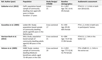
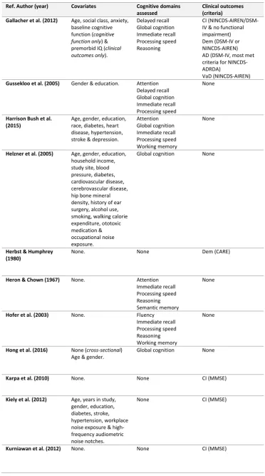
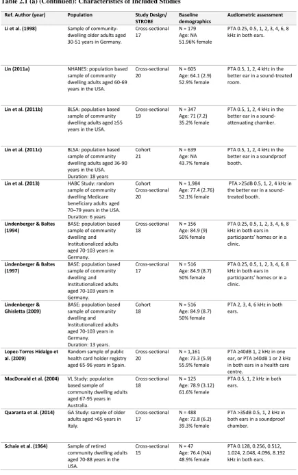
TABLE 2.1:CHARACTERISTICS OF INCLUDED STUDIES 33
TABLE 4.1:DEMOGRAPHIC DATA FOR THE 101 PARTICIPANTS PRIOR TO MATCHING 77
TABLE 5.1:BACKGROUND DATA FOR THE HEARING LOSS AND CONTROL GROUPS 98
TABLE 5.2:NEUROPSYCHOLOGICAL DATA FOR THE HEARING LOSS AND CONTROL GROUPS 99
TABLE 5.3:SPEARMAN’S RHO CORRELATIONS FOR PROCESSING SPEED AND EXECUTIVE FUNCTION FOR THE HEARING LOSS AND CONTROL GROUPS
102
TABLE 5.4:NEUROPSYCHOLOGICAL DATA FOR THE HEARING LOSS AND CONTROL GROUPS 104
TABLE 5.5:NEUROPSYCHOLOGICAL DATA FOR THE HEARING LOSS AND CONTROL GROUPS 105
TABLE 6.1:BACKGROUND DATA FOR THE HEARING LOSS AND CONTROL GROUPS 121
TABLE 6.2:NEUROPSYCHOLOGICAL DATA FOR THE HEARING LOSS AND CONTROL GROUPS 122
TABLE 6.3:FLUENCY INDICES FOR THE TWO GROUPS OF PARTICIPANTS 124
TABLE 6.4:ADDITIONAL FLUENCY DATA FOR THE TWO GROUPS OF PARTICIPANTS 124
TABLE 6.5:LINEAR MIXED EFFECTS MODEL OF WORD GENERATION ACROSS TIME 126
TABLE 6.6:LINEAR MIXED EFFECTS MODEL OF PHONOLOGICAL/SEMANTIC DISCREPANCY ACROSS TIME 127
TABLE 6.7:LINEAR MIXED EFFECTS MODEL OF MANUAL INDICES 128
TABLE 6.8:LINEAR MIXED EFFECTS MODEL OF COMPUTERISED INDICES 129
TABLE 7.1:BACKGROUND DATA FOR THE TWO GROUPS OF PARTICIPANTS 143
TABLE 7.2:NEUROPSYCHOLOGICAL DATA FOR THE TWO GROUPS OF PARTICIPANTS 144
TABLE 7.3:TEMPORARY BINDING DATA FOR THE TWO GROUPS OF PARTICIPANTS 146
TABLE7.6:LINEARMIXEDEFFECTSMODELEXAMININGTHEEFFECTSOFTIME,HEARINGSTATUS,AND
THEIRINTERACTIONONPHONOLOGICALSWT
xiii List of Figures
FIGURE 1.1:CHANGE IN COGNITIVE DOMAINS WITH AGE 3
FIGURE 1.2:PROGRESSION OF AD FROM PRE-SYMPTOMATIC STAGE TO INCIDENT AD 7
FIGURE 1.3:RELATIONSHIP BETWEEN PTA HEARING LOSS AND COGNITIVE DECLINE 15
FIGURE 1.4:RELATIONSHIP BETWEEN PTA HEARING LOSS AND RISK OF INCIDENT DEMENTIA 15
FIGURE 2.1:PRISMAFLOWDIAGRAM 23
FIGURE 2.2:FOREST PLOT OF CORRELATION R VALUES FOR COGNITION/CROSS-SECTIONAL OUTCOMES 24
FIGURE 2.3:FOREST PLOT OF CORRELATION R VALUES FOR COGNITION/COHORT OUTCOMES 25
FIGURE 3.1:THEORETICAL VIEWS OF RELATIONSHIP BETWEEN ARHL AND COGNITIVE DECLINE 43
FIGURE 3.2:MAP OF COMMON AETIOLOGIES BETWEEN ARHL AND COGNITIVE DECLINE 43
FIGURE 3.3:MAP OF MECHANISTIC PATHWAYS BETWEEN ARHL AND COGNITIVE DECLINE 59
FIGURE 4.1:THE NIEAD MODEL. 74
FIGURE 4.2:TIMELINE OF STUDY PROCESS 78
FIGURE 4.3:LIST OF NEUROPSYCHOLOGICAL TEST IN ORDER OF ADMINISTRATION. 87
FIGURE 5.1:DIFFERENCE IN PURE-TONE THRESHOLD BETWEEN THE TWO GROUPS AT EACH FREQUENCY 98
FIGURE 5.2:DIFFERENCE IN MEAN PERFORMANCE BETWEEN THE TWO GROUPS ON EACH COGNITIVE TEST (BASED ON Z-SCORES).
101
FIGURE 5.3:DIFFERENCE IN MEAN PERFORMANCE BETWEEN THE TWO GROUPS ON EACH COGNITIVE DOMAIN (BASED ON Z-SCORES).
101
FIGURE 5.4:BRINLEY PLOT OF SART ERROR SCORE AND SART MEAN REACTION TIME (RT). 103
FIGURE 5.5:BRINLEY PLOT OF SART SLOW AND FAST FREQUENCY VARIABILITY (TOTAL TEST). 106
FIGURE 5.6:BRINLEY PLOT OF SART SLOW AND FAST FREQUENCY VARIABILITY (FIRST HALF). 106
FIGURE 5.7:BRINLEY PLOT OF SART SLOW AND FAST FREQUENCY VARIABILITY (SECOND HALF). 107
FIGURE 6.1:PTA FOR HL AND NH GROUPS ACROSS FREQUENCIES 120
FIGURE 6.2:DIFFERENCE IN MEAN PERFORMANCE BETWEEN THE TWO GROUPS ON EACH COGNITIVE TEST (BASED ON Z-SCORES).
123
FIGURE 7.1:THE TWO CONDITIONS (SHAPES AND SHAPES-COLOURS) OF THE TEMPORARY MEMORY BINDING TEST.
140
FIGURE 7.2:DIFFERENCE IN PURE TONE THRESHOLD BETWEEN THE TWO GROUPS AT EACH FREQUENCY 142
FIGURE 7.3:DIFFERENCE IN MEAN PERFORMANCE BETWEEN THE TWO GROUPS ON EACH COGNITIVE TEST (BASED ON Z-SCORES).
145
FIGURE 7.4:DIFFERENCE IN MEAN PERFORMANCE BETWEEN THE TWO GROUPS ON EACH COGNITIVE DOMAIN (BASED ON Z-SCORES).
xiv
List of Abbreviations
AD ADHD
Alzheimer's Disease
Attention Deficit Hyperactivity Disorder ANOVA
ANSI
Analysis of Variance
American National Standards Institute
APOE Apolipoprotein E
ARHL Age-related Hearing Loss
Aβ Beta Amyloid
BBB Blood-Brain Barrier
BDNF Brain-derived neurotrophic factor
BLB blood-labyrinth barrier
BMI Body Mass Index
BNT Boston Naming Test
BSA British Society of Audiology
CAEP CAMCOG CAMDEX CESD CI
Cortical Auditory Evoked Potentials Cambridge Cognitive Examination
Cambridge Mental Disorders of the Elderly Examination Center for Epidemiologic Studies Depression
Confidence Interval
CMA Comprehensive Meta-Analysis
CNS Central Nervous System
CRP C-reactive protein
CRT Choice Reaction Time
CV Coefficient of Variation
DAN dB
Dorsal Attention Network Decibel
DMN Default Mode Network
DTI Diffusion Tensor Imaging
EEG Electroencephalogram
ELU Ease of Language Understanding
FCSRT Free and Cued Selective Reminding Test
FFT Fast Fourier Transform
FFV Fast Frequency Variability
fMRI Functional Magnetic Resonance Imaging
FPCN Frontoparietal Control Network
GCs Glucocorticoids
GM-CSF GRM GSTs HADS
Granulocyte-macrophage Colony-stimulating Factor Glutamate Receptor, Metabotropic
Glutathione S-transferases
Hospital Anxiety and Depression Scale
HAROLD Hemispheric Asymmetry Reduction in Older Adults HHIE-S
HLG
Hearing Handicap Inventory for the Elderly-Screening Hearing Loss Group
HPA hypothalamic-pituitary-adrenal
HR IIV IL IGF Hazard ratios Intra-individual variability Interleukin
Insulin-like growth factor ISD
ISHAA
Individual Standard Deviation
Irish Society of Hearing Aid Audiologist ISI
kHz
Inter-stimulus Interval Kilohertz
MAPK Mitogen-activated Protein Kinases
xv
MCI Mild Cognitive Impairment
MCS MMSE
Mean Cluster Size
Mini-Mental State Examination
MoCA Montreal Cognitive Assessment
MOOSE Meta-analysis Of Observational Studies in Epidemiology MTHFR
NART
Methylenetetrahydrofolate Reductase National Adult Reading Test
NEIL Neuro-Enhancement for Independent Lives
NFT Neurofibrillary Tangles
OR Odds Ratios
OXPHOS Oxidative Phosphorylation
PAR Population Attributable Risk
PASA Posterior-Anterior Shift in Ageing
PET Positron emission tomography
PFC Prefrontal Cortex
PON1 Paraoxonase/arylesterase
PRISMA Primary Reporting Items for Systematic Reviews and Meta-analyses PSQI
PSS PTA
Pittsburgh Sleep Quality Index Perceived Stress Scale Pure-tone Average
RNS Reactive Nitrogen Species
ROS Reactive Oxygen Species
RT Reaction Time
SAM Sympathetic-adrenal-medullary
SART Sustained Attention to Response Task
SD SFV
Standard Deviation Slow Frequency Variability
SHAA Irish Society of Hearing Aid Audiologists
SHARE Survey of Health, Ageing and Retirement in Europe SPSS Statistical Package for Social Sciences
STROBE Strengthening the Reporting of Observational Studies in Epidemiology
TILDA The Irish Longitudinal Study on Ageing
TNF Tumour Necrosis Factor
VaD Vascular Dementia
VEGF Vascular Endothelial Growth Factor
VEP Visual Evoked Potentials
VRFs Vascular Risk Factors
WHO WMHs
World Health Organisation White Matter Hyperintensities
1
Cognitive ageing and age-related hearing loss
1.1
General Introduction
This thesis aims to explore the potential association between age-related hearing loss (ARHL) and neurocognitive decline with a view to explicating the causal pathways that underpin this
association. A demographic shift towards an increasingly larger proportion of older adults in the human population is projected to lead to an exponential increase in the prevalence of dementia (Brookmeyer, Johnson, Ziegler-Graham, & Arrighi, 2007; Ferri et al., 2005; Suzman & Beard, 2011). In the absence of an effective treatment for dementias such as Alzheimer’s disease (AD) (Thies & Bleiler, 2013), there is a strong need to identify potential risks factors that influence and modify the rate of age-associated cognitive decline and the onset of dementia (Norton, Matthews, Barnes, Yaffe, & Brayne, 2014; Sperling, Mormino, & Johnson, 2014). Cohort studies indicate that age-related hearing loss (ARHL) may precede the onset of clinical dementia by five to 10 years, and is therefore a possible non-invasive biomarker (Albers et al., 2015). It has been further
suggested that modification or prevention of sensory decline may offer a treatment pathway to alter clinical outcomes (Albers et al., 2015; Lin & Albert, 2014).
The importance of clarifying the association between ARHL and cognitive decline is profound. It is estimated that dementia affects 46·8 million people worldwide and is projected, due to
demographic trends and longer lifespan, to increase in global prevalence to approximately 131·5 million in 2050 (Prince et al., 2015). The global cost of dementia in 2015 was US$818 billion and this is projected to increase to US$2 trillion by 2050 (Prince et al., 2015). Current pharmaceutical approaches which target neuropathologies such as Alzheimer’s disease (AD) offer limited benefit with symptom modifying effects at best (Thies & Bleiler, 2013). Reduction of risk factors such as social isolation may contribute to a cognitive reserve which in turn moderates the impact of neuropathology to maintain optimal cognitive function (Stern, 2009, 2012; Valenzuela & Sachdev, 2006; Wilson et al., 2013). This approach may be more beneficial than pharmacological
2
1.2
Cognitive Ageing
Normal cognitive ageing
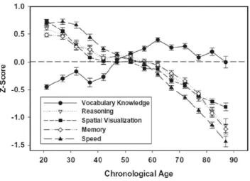
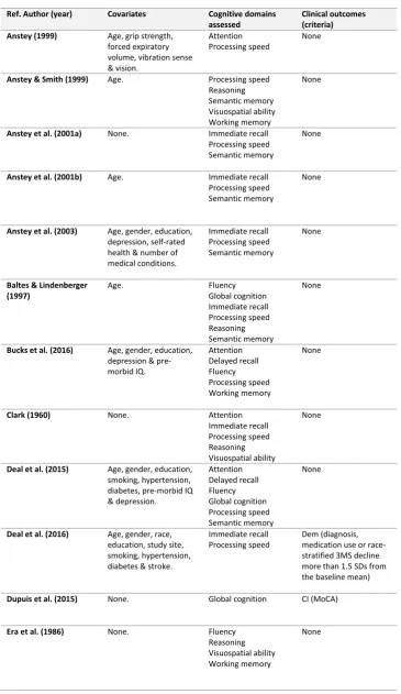
One of the earliest findings in cognitive ageing research has been that normal cognitive ageing is associated with differential decline across domains of cognitive functions (See Figure 1.1)
3
Figure 1.1: Change in cognitive domains with age (Salthouse, 2010b).
Vocabulary, considered a form of crystallised intelligence, increases into late middle age then gradually declines. In contrast, fluid intelligence linearly declines from young adulthood.
Structural changes in cognitive ageing
Modern brain imaging research approaches, such as task-evoked brain response studies, have contributed further to understanding by exploring how non-pathological cognitive ageing is mediated by changes in the neuroanatomical structure and function of the brain (Dennis & Cabeza, 2011; Fjell & Walhovd, 2010). Volumetric reductions in regional brain grey matter, particularly in the prefrontal cortex and medial temporal lobe regions, are associated with decline in executive function and memory respectively (Buckner, 2004; Dennis & Cabeza, 2011; Fjell, McEvoy, Holland, Dale, & Walhovd, 2013; Hedden & Gabrieli, 2004; Jagust, 2013; Yuan & Raz, 2014). This loss is more likely due to shrinkage of neurons, reductions of synaptic spines and loss of synapses rather than neuronal loss (Fjell & Walhovd, 2010). Studies assessing functional
connectivity report decline in white matter integrity associated with slower information processing speed and consequent body mass index (BMI).
4
differential rates of decline (Dennis & Cabeza, 2011). In non-pathological ageing, the hippocampus undergoes substantial atrophy whereas the entorhinal cortex does not (Raz et al., 2005) which is of interest as the entorhinal cortex is one of the first affected by AD neuropathology (Braak & Braak, 1997). Shrinkage in the entorhinal cortex was also found to be associated with episodic memory decline over a five-year period while the hippocampus was not, suggesting that the entorhinal cortex may be a sensitive marker of memory decline in normal ageing (Rodrigue & Raz, 2004). The perirhinal and entorhinal areas are selectively involved in familiarity-based recognition while the hippocampus is associated with more explicit recollection of stimuli (Bowles et al., 2007; Bowles et al., 2010; Martin, Bowles, Mirsattari, & Kohler, 2011).
Cellular and molecular neuroscience examining neurochemical and metabolic influences on cognitive ageing have contributed further to understanding (Dennis & Cabeza, 2011). For example, the neurotransmitter norepinephrine (Robertson, 2013) and its substrate dopamine (Backman, Lindenberger, Li, & Nyberg, 2010) underpin alterations in brain metabolic pathways, influence neurogenesis and modify the deteriorative effects of pro-inflammatory mediators and
neuropathological substrates on neurons. Consequently, their decline is associated with deficits in episodic memory, executive functions and processing speed and clinical expression of AD (Backman et al., 2010; Dennis & Cabeza, 2011; MacDonald, Karlsson, Rieckmann, Nyberg, & Backman, 2012; Robertson, 2013). Altered neurometabolism, particularly glucose regulation and oxygen metabolic rate, is also associated with cognitive decline and AD (Dennis & Cabeza, 2011; Lourenco, Ledo, Dias, Barbosa, & Laranjinha, 2015). Dysfunction in cellular processes leads to production and accumulation of neuropathological substrates (Joshi & Pratico, 2014). Beta amyloid (Aβ) peptide which forms neuritic plaques and hyperphosphorylated tau-based neurofibrillary tangles (NFT) accumulating in medial temporal lobe and limbic regions are characteristic signatures of AD (Braak & Braak, 1991; Hyman et al., 2012) although they are also observed in samples exhibiting normal cognition (Aizenstein et al., 2008; Mintun et al., 2006; Oh et al., 2011; Rowe et al., 2007). While their accumulation is still associated with loss in neurons and
connectivity between brain regions, leading to disruption in neurocognitive networks in the normal ageing population (Dennis & Thompson, 2014; Oh et al., 2011), the presence and quantity of these substrates alone is not predictive of clinical outcomes (Stern, 2009, 2012).
Functional changes in cognitive ageing
5
specificity and increased recruitment of higher order cognitive processes to complete tasks (Grady, 2012; Stern, 2012). This is supported in neurocognitive studies which find that such shifts in neural activation are reflected behaviourally in decreased reaction times for older adults but comparable levels of accuracy compared to young adult controls (Grady, 2012; Grady et al., 1994). Studies examining changes in neural activity with age have generally noticed two neural patterns across a wide range of domains (Dennis & Cabeza, 2011). The first is referred to as Posterior-Anterior Shift in Ageing (PASA) which posits that older adults compensate for decrease in neural activity in posterior regions by recruiting prefrontal cortex regions (Grady et al., 1994). The second is referred to as the Hemispheric Asymmetry Reduction in OLDer Adults (HAROLD) and describes how older adults show less hemispheric lateralisation in prefrontal cortical activity (Cabeza, 2002). This was originally conceptualised by Cabeza as a compensatory mechanism. An alternative
explanation, the dedifferentiation hypothesis, suggests that this more diffuse activation is due to an age-related difficulty in engaging specialised neural mechanisms (de Frias, Lovden, Lindenberger, & Nilsson, 2007). However, research has generally found stronger support for the HAROLD model (Batterham, Christensen, & Mackinnon, 2011; Cabeza, Anderson, Locantore, & McIntosh, 2002; Reuter-Lorenz et al., 2000; Rosen et al., 2002).
6
MCI and with performance on the Montreal Cognitive Assessment (MoCA) (Chand, Wu, Hajjar, & Qiu, 2017).
Cognitive decline and impairment
With increasing age, comes a higher risk for developing age-related cognitive pathologies or dementia, the most common of which is Alzheimer’s disease (AD) (Salthouse, 2010a). Interposed between these two conditions is an intermediate stage of cognitive impairment, usually termed Mild Cognitive Impairment (MCI), which represents a decline in cognitive function greater than expected for the person’s age but which does not yet meet criteria for dementia (Petersen, 2004). While it has been hypothesised that forms of dementia such as AD might reflect simply an
accelerated ageing process, qualitative differences in cognitive and neuro-imaging profiles suggest that these conditions, particularly AD, represent a distinctive, separate condition (Toepper, 2017). Mild cognitive impairment was originally characterised by a decline in memory which had no identifiable aetiology and did not interfere with daily function (Petersen et al., 1999). This concept has been expanded to include other variations of cognitive impairment and to reflect subtle changes in function seen in this condition (Morris et al., 2001) such as amnestic and nonamnestic forms of impairment and within one or multiple cognitive domains (Petersen & Morris, 2005; Winblad et al., 2004). Originally used to describe a prodromal phase of AD, subsequent work has found that MCI precedes other age-associated conditions such as VaD (Hayden & Welsh-Bohmer, 2012). Though estimates vary according to diagnostic criteria, MCI has an increasing prevalence with age in the older adult population and is associated with an increased risk of conversion to dementia (Busse, Bischkopf, Riedel-Heller, & Angermeyer, 2003; de Souza-Talarico, de Carvalho, Brucki, Nitrini, & Ferretti-Rebustini, 2016).
Assessment and prediction of pathological cognitive decline
7
expensive and inefficient for large-scale screening assessments (Albert et al., 2011; Logie, Parra, & Della Sala, 2015).
In contrast, neuropsychological assessment is comparably easier to administer and studies suggest that it has good if not greater predictive value compared to biomarkers (Belleville et al., 2014; Landau et al., 2010; Logie et al., 2015). Identification of patterns of strengths and weaknesses can aid in differentiating between different forms of cognitive impairment and dementias and in earlier identification of those at risk (Jacobson, Delis, Bondi, & Salmon, 2002; Nestor, Scheltens, & Hodges, 2004; Twamley, Ropacki, & Bondi, 2006). In normal and pathological ageing, there is decline in memory, executive function and processing speed (Toepper, 2017). However, prodromal AD is associated with deficits in episodic and semantic memory and with subtle deficits in
executive function, whereas decline with normal ageing is more accentuated in executive functions and processing speed but is associated with maintained semantic memory (Buckner, 2004;
[image:25.595.103.473.364.642.2]Toepper, 2017).
Fig 1.2: Progression of AD from pre-symptomatic stage to incident AD (Caldwell et al., 2015).
Treatment and prevention of pathological cognitive decline
8
Folch et al., 2015; Karran, Mercken, & De Strooper, 2011; Thies & Bleiler, 2013). As early diagnosis is a factor in successfully managing dementia, the long prodromal stage of AD offers an opportunity for earlier identification and prevention and may provide the most effective avenue to reduce the burden and prevalence of AD (Bateman et al., 2012; Lin, Yang, et al., 2014; Sperling et al., 2014; Villemagne et al., 2013). However, overlap between cognitive deficits and
pathophysiological substrates due to AD pathology and the benign effects of ageing make it
challenging to distinguish prodromal AD cases from the normal ageing population (Belleville et al., 2014; Bondi et al., 2008). Investigative efforts are increasingly focused on identifying the unique biomarkers and cognitive signatures of this disease and the modifiable factors which delay AD incidence (Caldwell et al., 2015; Norton et al., 2014) and may also allow for more successful cognitive ageing among the normal ageing population (Beydoun et al., 2014). Switching to a strategy of earlier identification and prevention would significantly reduce prevalence and the associated cost (Lin, Yang, et al., 2014). Interventions that delay the onset of dementia by one year would lead to a decrease of more than 10% in the global prevalence of dementia in 2050
(Brookmeyer et al., 2007). ARHL may be one such modifiable risk factor or a predictive marker for age-associated neurocognitive decline and disease (Deal et al., 2015; Gallacher et al., 2012; Lin, Metter, et al., 2011; Panza, Solfrizzi, & Logroscino, 2015).
1.3
Theoretical perspectives
There has been much debate as to what factors are most fundamental in explaining age-related variance in cognitive function with some researchers attempting to explain this phenomenon in terms of a single factor or a small number of factors (Dennis & Cabeza, 2011). Multiple theories have been proposed attempting to account for cognitive ageing - a few of the main ones are described briefly below.
Sensory Deficit
9
Frontal Lobe
The frontal lobe hypothesis describes how age-related atrophy in the prefrontal cortex leads to decline in the executive functions: monitoring, sequencing, initiation of action, inhibiting pre-potent responses, formulating goals, focusing attention and generating response alternatives (Miller & Cohen, 2001; West, 2000). As these functions mediate performance between age and general cognitive capacities (Salthouse et al., 2003), it is argued that age-related cognitive decline may arise from impaired or inefficient deployment of cognitive control processes due to age-related degeneration of frontal lobe structures (Braver & Barch, 2002; Crawford, Smith, Maylor, Della Sala, & Logie, 2003; Glisky, 2007; Greenwood, 2000; Rodriguez-Aranda & Sundet, 2006; West, 2000). This view is supported by neurological evidence which finds that the frontal lobes typically demonstrate the fastest rate of decline compared to other regions (Raz et al., 2005; Resnick, Pham, Kraut, Zonderman, & Davatzikos, 2003).
Speed of Processing
This theory posits that cognitive deficits in older adults can be attributed to a reduction in the speed of processing (Salthouse, 1996). There is strong evidence to support this view because processing speed shares a large proportion of variance with age related deficits in cognitive measures and declines steadily with age. A possible neural mechanism for this decline is white matter
deterioration because several studies have found a correlation between decline in processing speed and WMHs (white matter hyperintensities) and DTI (diffusion tensor imaging) measures of white matter integrity (Dennis & Cabeza, 2011). Performance on tasks assessing speed of processing, executive functioning and visual detection reaction time were found to have a significant
relationship with DTI measures in frontal and frontal striatal areas (Madden et al., 2004; O'Sullivan et al., 2001; Persson et al., 2006). Periventricular WMHs were associated with slower motor speed (Soderlund, Nyberg, Adolfsson, Nilsson, & Launer, 2003). Older adults may compensate for these deficits by recruiting PFC regions and increasing speed performance on tasks (Reuter-Lorenz et al., 2000).
Resources
Craik and colleagues (Craik, 1986; Craik, a. Routh, & Broadbent, 1983; Craik & Byrd, 1982) propose that a decline in the available attentional resources leads to age-related deficits in cognitive performance. This has been supported by behavioural studies which have found that when
10
that they will compensate for this deficit by recruiting contralateral PFC regions thus tapping into other cognitive resources. Neuroimaging studies have found that older adults typically show reduced activation in PFC regions normally engaged by younger adults on visuospatial working memory, and on episodic encoding and retrieval tasks (Grady et al., 1994; Schiavetto, Kohler, Grady, Winocur, & Moscovitch, 2002). Supporting the second proposal, studies have also found that older adults recruit contralateral regions to support performance on tasks assessing multiple cognitive domains (Cabeza et al., 2002; Reuter-Lorenz et al., 2000).
Inhibition
This theory proposes that decline in inhibitory control in working memory allows goal-irrelevant information to interfere with working memory processes, leading to deficits in cognitive
performance (Hasher & Zacks, 1988; Zacks, Hasher, & Li, 2000). Extending these assumptions to a neural context, Dennis and Cabeza (2011) propose that older adults should demonstrate weaker neural activity in inhibitory control regions and greater activity in inhibited regions. These assumptions have been supported by neuroimaging research (Cabeza et al., 1997; Gazzaley, Cooney, Rissman, & D'Esposito, 2005; Jonides et al., 2000). Additionally, older adults show bilateral PFC activations during inhibitory tasks compared to younger adults suggesting that they compensate for this deficit by recruiting other neural regions (Nielson, Langenecker, & Garavan, 2002).
Brain Reserve & Cognitive Reserve
Brain reserve posits that those with a greater brain size and number of neurons are able to tolerate greater damage to the brain, either from age-related changes, injury or neuropathology before manifesting clinical symptoms (Stern, 2012). With the advent of normal ageing or neuropathology such as Alzheimer’s disease, comes a volumetric shrinkage in various brain structures (Desikan et al., 2009; Raz et al., 2005). Brain reserve is essentially a passive model, presuming that the clinical manifestation of these neuropathological or age-related neuronal changes exists once a threshold of neuronal damage has been reached. Research has found links between the clinical effects of
neuropathology and brain size, head circumference and number of synapses (Bigio, Hynan, Sontag, Satumtira, & White, 2002; Mori et al., 1997; Mortimer, Snowdon, & Markesbery, 2003).
11
2012; Zarahn, Rakitin, Abela, Flynn, & Stern, 2007). Proxies of cognitive reserve have been found to be associated with cognitive function independently of neuropathology (Wilson et al., 2013) and atrophy (Vaughan et al., 2014; Vuoksimaa et al., 2013).
1.4
Risk factors & mechanisms
Another trend that has emerged in the literature is that there is considerable variability in the level of cognitive functioning between individuals of the same age, even within the normal cognitive ageing population (Salthouse, 2010a) and in fluid intelligence and memory rather than in crystallised intelligence (Christensen, 2001). Based on evidence from a meta-analysis by Verhaeghen and Salthouse (1997) in population samples ranging from 18 to 80 years of age, Salthouse (2010a) points out that age accounts for between only 4% to 36% of the variance in cognitive function. This differential decline may be mediated and determined by multiple biological, environmental and lifestyle factors which modify the trajectory of cognitive ageing (Beydoun et al., 2014; Bozzali et al., 2015; Deary et al., 2009; Depp, Harmell, & Vahia, 2012; Hayden & Welsh-Bohmer, 2012; Stern, 2012) and modify risk of incident dementia (Barnes & Yaffe, 2011; Norton et al., 2014; Sperling et al., 2014).
This variance has led to a diverse range of research approaches assessing various biomarkers which may aid prediction of cognitive decline and potential risk factors that can be modified or treated to alter outcomes. For example, research on specific genetic markers have found associations between cognitive health and genetic markers regulating cardiovascular health (e.g. PON1, APOE), cell metabolism and oxidative stress (e.g. SIRT3), inflammatory processes (e.g. IL6, IL10) (Depp et al., 2012; Glatt, Chayavichitsilp, Depp, Schork, & Jeste, 2007; Zubenko, Hughes, Zubenko, & Maher, 2007).
Apart from biological processes, there are also several lifestyle factors such as education and social network size that influence cognitive ageing (Depp et al., 2012) and that can predict clinical outcomes and contribute significantly to the onset of dementias, including AD, independently of neuropathologic substrates (Bennett, Schneider, Tang, Arnold, & Wilson, 2006; Norton et al., 2014; Stern, 2012). These modifiable factors possibly impart a ‘reserve’ against neuropathology (Stern, 2009, 2012) and provide a potential avenue for interventions to reduce prevalence (Norton et al., 2014). Additionally, cardiovascular risk factors related to lifestyle, such as atherosclerosis, produce grey and white matter lesions contributing significantly to cognitive decline and dementia pathology including AD (Qiu & Fratiglioni, 2015). Chronic stress influences a network of
12
restriction has been linked to significantly improved memory performance in older adults (Witte, Fobker, Gellner, Knecht, & Floel, 2009).
1.5
ARHL & its impact on the health of older adults
Overview
ARHL generally begins at the higher frequencies in young adulthood and progresses gradually, bilaterally and symmetrically towards the lower frequencies (<3 kilohertz (kHz)), becoming more noticeable in older age when severe enough to affect speech understanding (Gates & Mills, 2005). As hearing loss progresses in severity, it leads to impairment of social (Gopinath et al., 2012) and daily function (Lopez-Torres Hidalgo et al., 2009). It is a highly prevalent condition in the older adult population with at least a mild hearing loss affecting roughly 25-30% of older adults aged between 50-59 years increasing to more than 50% of adults aged over 60 years with significant increases per decade (Agrawal, Platz, & Niparko, 2008; Gopinath, Rochtchina, et al., 2009; Lin, Thorpe, Gordon-Salant, & Ferrucci, 2011; Nash et al., 2011; Raynor et al., 2009; Wilson et al., 1999). The World Health Organisation estimates that one-third of adults over the age of 65 years have a moderate or disabling hearing loss (World Health Organisation, 2015). Acquired hearing loss most likely represents a mixture of pathophysiological processes - primarily genetic factors and environmental exposures such as noise and ototoxic factors (Fetoni, Picciotti, Paludetti, & Troiani, 2011; Gates & Mills, 2005; McMahon, Kifley, Rochtchina, Newall, & Mitchell, 2008; Viljanen et al., 2007; Wingfield et al., 2007; Yamasoba et al., 2013). Regardless of specific pathophysiology, functional outcomes are similar and are characterised by an increase in hearing thresholds and poorer frequency resolution, initially experienced as a loss of perception of speech in noisy backgrounds (Barrenas & Wikstrom, 2000; Dubno et al., 2008; Gates & Mills, 2005; Yamasoba et al., 2013).
Anatomy & pathophysiological processes
Decline in peripheral hearing function is primarily due to dysfunction of the cochlea in the inner ear, the most complex part of the peripheral hearing structure, which transduces incoming
mechanical sound into neurochemical signals for processing in the auditory cortex (Gates & Mills, 2005; Yamasoba et al., 2013). Primary sites of ARHL pathology in the cochlea are the stria vascularis, auditory hair cells and spiral ganglion neurons (Fetoni et al., 2011; Kamogashira, Fujimoto, & Yamasoba, 2015; Ohlemiller, 2004; Schmiedt, 2010; Schuknecht & Gacek, 1993; Yamasoba et al., 2013).
The stria vascularis is a highly vascularised epithelium with an intense aerobic metabolism that lines the outer wall of the cochlear duct (scala media) where the organ of Corti is located (Gates & Mills, 2005; Schmiedt, 2010). It contains Na+ K+ ATPase pumps and produces endolymph,
13
consequently leads to loss of endocochlear potential with a flat increase in decibel threshold across low frequencies and a sloping increase on high frequencies (Schmiedt, Lang, Okamura, & Schulte, 2002; Schuknecht & Gacek, 1993; Suzuki et al., 2006).
The outer and inner auditory hair cells are located in the organ of Corti, a sensory epithelium on the basilar membrane of the cochlea (Gates & Mills, 2005; Schmiedt, 2010; Yamasoba et al., 2013). Displacement of outer hair cells by sound waves propagating in the endolymph amplifies the waves and extends the frequency range. Outer hair cells are more susceptible to damage than inner hair cells and their loss, termed ‘sensory presbycusis’ (Schuknecht & Gacek, 1993), results in a rise in hearing decibel threshold progressing from the higher frequencies downwards and poorer
frequency selectivity (Davis, Ahroon, & Hamernik, 1989). Displacement of inner hair cells opens transduction ion channels allowing an influx of potassium (K+) and calcium (Na+) ions into the hair
cell, depolarising the cell and triggering the release of neurotransmitters, thereby transducing mechanical sound waves into neurochemical signals. These signals are transmitted to the auditory cortex through release of the neurotransmitter glutamate by inner hair cell synapses to afferent spiral ganglion neurons. Little is known about the behavioural consequences of inner hair cell deterioration, the loss of which does not seem to substantially lower hearing threshold (Lobarinas, Salvi, & Ding, 2013).
The spiral ganglion, formed by the cell bodies of the cochlear (auditory) nerve, transports signals from the hair cells to the auditory cortex in the temporal lobe (Gates & Mills, 2005; Schmiedt, 2010; Yamasoba et al., 2013). Deterioration in the afferents, termed ‘neural presbycusis’
(Schuknecht & Gacek, 1993), is associated with poorer temporal and frequency coding and speech perception in noise (Lopez-Poveda, 2014; Sergeyenko, Lall, Liberman, & Kujawa, 2013;
Yamasoba et al., 2013).
Deterioration in hearing function can also originate in the central auditory cortex independently of observed changes in the peripheral structure (Ouda, Profant, & Syka, 2015). Decline in these structures leads to poorer auditory perceptual function as characterised by poorer understanding of degraded or rapid speech without apparent deterioration in peripheral pathways (Gates & Mills, 2005). This may be due to age related neurodegeneration or neuropathology (Gates, Anderson, McCurry, Feeney, & Larson, 2011; Sinha, Hollen, Rodriguez, & Miller, 1993; Wong et al., 2009). Data from epidemiological ageing studies suggest that disabling central auditory dysfunction independent of peripheral function has a low rate of prevalence in the adult population (Gates & Mills, 2005). Central auditory dysfunction usually occurs secondary to longer-term peripheral dysfunction in the later stages of the pathological process (Gates & Mills, 2005).
Assessment
14
frequencies to measure pre-tone thresholds (Bagai, Thavendiranathan, & Detsky, 2006). However, it does not detect decline in a multitude of peripheral hearing functions such as frequency
selectivity or localising of sound sources which also contribute to hearing status and may precede threshold elevation (Bharadwaj, Masud, Mehraei, Verhulst, & Shinn-Cunningham, 2015; Kujawa & Liberman, 2015; Sergeyenko et al., 2013; Wayne & Johnsrude, 2015). Future research using multiple alternative assessments of peripheral functions, such as speech-in-noise perception or temporal processing (Humes, Busey, Craig, & Kewley-Port, 2013; Humes, Kidd, & Lentz, 2013), may give further insight into whether these functions have unique associations with cognitive function independent of pure-tone audiometry (Wayne & Johnsrude, 2015).
Association with other health concerns in the ageing population
Hearing loss has been associated with a wide range of health and neuropsychiatric conditions typically reported with ageing and dementia. These include poorer health-related quality of life (Appollonio, Carabellese, Frattola, & Trabucchi, 1996; Dalton et al., 2003; Genther, Frick, Chen, Betz, & Lin, 2013; Sugawara et al., 2011), functional impairment (Appollonio et al., 1996; Chen, Genther, Betz, & Lin, 2014; Dalton et al., 2003; Marsiske, Klumb, & Baltes, 1997; Strawbridge, Wallhagen, Shema, & Kaplan, 2000; Wahl et al., 2013), reduced social participation (Appollonio et al., 1996; Chen, Genther, et al., 2014; Gopinath et al., 2012; Kramer, Kapteyn, Kuik, & Deeg, 2002; Marsiske et al., 1997; Strawbridge et al., 2000), apathy (Sugawara et al., 2011), depression (Jayakody, Friedland, Eielboom, Martins, & Sohrabi, 2017; Kramer et al., 2002), anxiety
(Jayakody et al., 2017), frailty (Kamil, Li, & Lin, 2014), sleep dysfunction (Nakajima, Kanda, Hosobuchi, & Suwa, 2014), change in personality (reduced extraversion) (Berg & Johansson, 2013), increased risk of falls (Lin & Ferrucci, 2012), increased numbers of hospitalisations (Genther et al., 2013) and greater risk of mortality (Appollonio et al., 1996; Karpa et al., 2010; Wahl et al., 2013).
ARHL & cognitive ageing
15
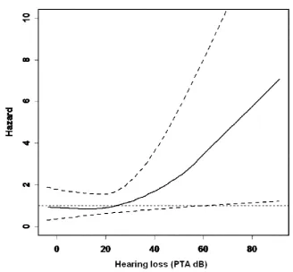
[image:33.595.99.428.377.681.2]between audiometric acquired hearing loss and cognition (Humes, Busey, Craig, & Kewley-Port, 2009; Lin et al., 2004). Likewise, some studies have associated hearing loss with cognitive impairment (Karpa et al., 2010; Kiely, Gopinath, Mitchell, Luszcz, & Anstey, 2012; Lin et al., 2013; Lopez-Torres Hidalgo et al., 2009; Quaranta et al., 2014) while others have not (Gates et al., 1996; Kurniawan et al., 2012; Tay, Kifley, et al., 2006).
Figure 1.3: Relationship between hearing loss and cognitive decline.
Note: tasks assess a) global cognition, b) processing speed (Lin et al., 2013)
16
Variance in findings may be due to suboptimal audiometric assessment, limited audiometric criteria (excluding higher speech frequencies) or cognitive tests using auditory stimuli (Gallacher et al., 2012). However, studies that aimed to assess decline in cognitive function using visual tests have reported significant decline in executive function and episodic memory (Gallacher et al., 2012; Jayakody et al., 2017). Neuroimaging studies researching both conditions have found further support for an association, finding that pure-tone thresholds were linked with increased atrophy of both regional (Eckert, Cute, Vaden, Kuchinsky, & Dubno, 2012; Husain, Medina, et al., 2011; Lin, Ferrucci, et al., 2014) and global brain grey matter (Lin, Ferrucci, et al., 2014) and white matter alterations (Eckert et al., 2013; Husain, Medina, et al., 2011). Additionally, a small number of intervention trials have reported improved cognitive outcomes following audiological rehabilitation (Acar, Yurekli, Babademez, Karabulut, & Karasen, 2011; Mosnier et al., 2015; Mulrow et al., 1990).
Hypotheses & causal factors
Hypotheses on the cause of this association include a common aetiology, such as alterations in the vascular system affecting cochlear and neural function (Lin, Ferrucci, et al., 2014; Lindenberger & Baltes, 1994; Malgrange, Varela-Nieto, de Medina, & Paillasse, 2015) or a more general
association as part of frailty syndrome (Panza, Solfrizzi, & Logroscino, 2015). Other hypotheses suggest that the association may be mechanistic with hearing loss affecting cognition (Lin, Ferrucci, et al., 2014; Lin, Metter, et al., 2011; Lindenberger & Baltes, 1994). Potential
17
in long-term episodic and semantic memory systems through disuse compared to recruited cognitive functions such as working memory and immediate recall (Ronnberg et al., 2011; Ronnberg, Hygge, Keidser, & Rudner, 2014; Ronnberg et al., 2013).
1.6
Summary and conclusions
There is projected to be a significant increase in the prevalence of dementia in the next few decades (Brookmeyer et al., 2007; Ferri et al., 2005; Suzman & Beard, 2011). However, there is currently no effective treatment for these dementias such as AD (Thies & Bleiler, 2013). Conditions such as ARHL may offer predictive biomarkers for dementia, assisting with development of public health policy and with selection for future clinical trials. Furthermore, modification of risk factors such as hearing loss may influence the rate of age-associated cognitive decline and the time of onset of dementia (Albers et al., 2015; Norton et al., 2014; Sperling et al., 2014). Neuropsychological assessment is the primary method used by researchers and clinicians to assess and predict the development of dementia (Belleville et al., 2014; Landau et al., 2010; Logie et al., 2015). Further research is required to continue the development of neuropsychological tests that detect subtle changes in cognition to promote earlier diagnosis of cognitive impairment. In particular, more research is needed to assess how different risk factors may contribute to differences in patterns of cognitive decline. Given the prevalence of ARHL, the identification of a pattern of
neuropsychological changes associated with it may benefit diagnosis and facilitate intervention. Additionally, it may give insight into the causal factors underlying the possible association between ARHL and cognitive decline.
1.7
Thesis outline
This thesis took an exploratory approach to assessing the association between ARHL and cognitive function and the possible causal mechanism underpinning this association. Chapter 2 describes a systematic review and meta-analysis of this association in epidemiological studies to examine and estimate the extent of the association between age-related hearing loss (ARHL) and cognitive function, cognitive impairment and dementia. Epidemiological research on the possible link between age-related hearing loss (ARHL) and cognitive decline and dementia has produced inconsistent results. Chapter 3 describes another review of possible pathological processes that represent either a common aetiological cause for both conditions or a mechanistic pathway by which ARHL leads to neurocognitive decline. Chapter 4 describes a hypothetical model for a mechanistic association whereby ARHL causes cognitive decline and outlines the methods used to collect data for the empirical chapters in this thesis. Chapter 5 describes differences in markers of processing speed and intra-individual variability between a group of older adults with and without hearing loss and differences in markers of higher cognitive control and neural arousal. Chapter 6 describes an analysis based on fluency tasks examining how ARHL may be associated with differences in stored semantic and phonological representations and in retrieval of these
18
19
The Association of Age-Related Hearing Loss
with Cognition Function, Cognitive Impairment and Dementia:
A Systematic Review with Meta-Analysis
2.1
Introduction
This chapter reports on a systematic review and meta-analysis conducted to examine the
association between age-related hearing loss (ARHL) and cognitive decline and dementia. Meta-analysis is a powerful tool to examine associations between potential risk factors and health
outcomes as it is based on a larger sample size than an individual cohort study (Borenstein, Hedges, Higgins, & Rothstein, 2011). Further statistical analysis using moderator analysis and
meta-regression allows for exploration of potential confounders that may explain the association between risk factors and outcomes as well as the influence of potential biases (e.g. publication bias). Pooling data from multiple independent studies could allow for a more robust estimate of the strength of the association between ARHL and cognitive decline, thus potentially informing design of future cohort studies as well as randomised controlled trials. Compared to other risk factors for dementia, there has been very little research examining the effects of ARHL on cognitive health outcomes despite its prevalence (Lin & Albert, 2014). Approximately one-third of adults over 65 experience a disabling hearing loss (Wilson, Tucci, Merson, & O'Donoghue, 2017; World Health
Organisation, 2015). It is easily diagnosed and treated and half of all cases are preventable; it would therefore be a serviceable risk factor (Albers et al., 2015; Lin & Albert, 2014). Prior reviews have either not included a meta-analysis of this association or have included different measures of hearing impairment and studies of different designs (Cherko, Hickson, & Bhutta, 2016; Gennis, Garry, Haaland, Yeo, & Goodwin, 1991; Schmulian Taljaard, Olaithe, Brennan-Jones, Eikelboom, & Bucks, 2015).
20
This chapter reports on a systematic review and meta-analysis to investigate the association
between ARHL and cognitive function, cognitive impairment and dementia in cohort observational studies. Qualitatively different search and audiometric criteria were used compared to other
reviews of this topic (Cherko et al., 2016; Gennis et al., 1991; Schmulian Taljaard et al., 2015). To reduce conceptual heterogeneity, only observational cross-sectional and cohort studies that
assessed hearing loss using pure-tone audiometry (the criterion standard) were included. Subgroup analyses were conducted to investigate the effects of various study, demographic, audiometric and lifestyle factors and to explore possible explanations for heterogeneity. An examination of whether cognitive reserve mediated cognitive outcomes for ARHL was completed.
2.2
Methods
This systematic review was performed according to an a priori established protocol (PROSPERO: CRD42015026052) and adhered to the Primary Reporting Items for Systematic Reviews and Meta-analyses (PRISMA) Statement (Liberati et al., 2009). It also met the Meta-analysis Of
Observational Studies in Epidemiology (MOOSE) guidelines (Stroup et al., 2000). Six a priori
meta-analyses were planned across two levels of study design and three levels of cognitive
outcome: (1) cross-sectional studies of ARHL & cognitive function; (2) cohort studies of ARHL & cognitive function; (3) cross-sectional studies of ARHL & cognitive impairment; (4) cohort studies of ARHL & cognitive impairment; (5) cross-sectional studies of ARHL & dementia; and (6) cohort studies of ARHL & dementia. All analyses were conducted using Comprehensive Meta-Analysis (CMA; version 3).
Data sources and searches
Studies published up to August 26, 2015 were retrieved from four electronic databases: (1) PubMed; (2) Cochrane Library; (3) EMBASE; and (4) SCOPUS. Keywords included: ‘hearing’, ‘cognition’, ‘dementia’ and ‘Alzheimer’s disease’ (several papers included had been published online prior to publication of the print version). Search terms and strategy are provided in Appendix A. Results were updated on April 15, 2016. Cross-referencing for potentially eligible papers was conducted using retrieved study papers and the author’s personal files.
Study selection criteria
21
Primary outcomes of interest were cognitive function, cognitive impairment and dementia. Cognitive function was a continuous variable and was sub-divided into 10 cognitive domains including attention, delayed recall, fluency, global function, immediate recall, processing speed, reasoning, semantic memory, visuospatial ability and working memory (Lezak, 2004). Cognitive impairment and dementia were dichotomous variables. A secondary outcome of interest was any data that examined subgroups (e.g. AD) among dementia studies.
Data extraction and quality assessment
Two researchers independently screened for eligible studies and conducted data extraction using a codebook. If consensus could not be reached, one author acted as arbitrator for study inclusion. The same author was consulted regarding data extraction. Multiple publication bias was avoided by using data from the most recently published study. Data from different papers that examined the same cohort were included provided they were for different cognitive outcomes and were treated as separate studies in analysis. Priority was given to outcomes that were maximally adjusted for covariates. The first and second author independently assessed the quality of reporting for each paper using the Strengthening the Reporting of Observational Studies in Epidemiology (STROBE) instrument (Vandenbroucke et al., 2014). Using Cohen’s kappa coefficient (Cohen, 1968),
agreement was excellent (0.91) prior to correcting discrepant items.
Calculation of effect sizes
Pearson’s r correlation coefficient was chosen as the effect size of the linear association between pure-tone audiometric hearing loss and cognitive function. Negative scores indicated that greater hearing loss was associated with poorer cognitive functioning. Odds ratios (OR) were used for cognitive impairment and dementia.
If the required outcome metric was not reported in the paper, r or OR values were calculated using available data. Where the predictor variable was continuous, unstandardized Beta (β) values were standardised by dividing them by the standard error. Where the standard error was not available, the β values, provided they were within +0.5, were converted to r using the Peterson and Brown formula (Peterson & Brown, 2005). Standardised β values were converted to r by dividing them by the square root of the sample size. If the predictor variable was categorical, β values were entered into CMA as either raw mean differences or as Cohen’s d as appropriate. Hazard ratios (HR), where the rate of incidence of outcome was less than 10%, were interpreted as OR (Zhang & Yu, 1998). If this rate exceeded 10%, HR were still treated as OR and a sensitivity analysis with the study deleted from the model was conducted to see if it had a significant impact on the overall results. Other effect sizes, (e.g. Chi-square, mean scores, etc.) were converted in CMA.
Statistical analysis
22
outcomes from each study were converted to either Fisher’s Z or log ORs for analysis purposes and then converted back to the original metric i.e. r and OR respectively. For the meta-analyses of cognitive function, multiple tests of the same cognitive domain from the same study were collapsed into one effect size and subgroups were analysed independently as separate effect sizes.
Heterogeneity was examined using the Q test and any p-value ≤0·10 was considered statistically significant (Higgins, Thompson, Deeks, & Altman, 2003). Inconsistency was examined using I2
and the following grades were applied: <25% (very low), 25% to <50% (low), 50% to <75% (moderate) and >75% (large) (Higgins et al., 2003).
Small-study effects were examined using funnel plots and the regression-intercept approach of Egger and colleagues (Egger, Davey Smith, Schneider, & Minder, 1997) provided there were at least ten effect sizes (Egger et al., 1997; Sterne et al., 2011). To examine the effects of each result on the overall findings, outcomes were analysed by deleting each study from the model once. Cumulative meta-analysis, ranked by year, was used to examine the accumulation of evidence over time (Lau, Schmid, & Chalmers, 1995). An ad hoc analysis was conducted to project the effect of hearing loss treatment and prevention on dementia prevalence using the population attributable risk (PAR) formula (Barnes & Yaffe, 2011; Levin, 1953), the WHO estimate of hearing loss prevalence in older adults (one-third) (World Health Organisation, 2015), and our cohort dementia OR (the more conservative estimate). The number of cases of dementia potentially prevented with 10%-25% reduction in ARHL was also calculated (Barnes & Yaffe, 2011).
Subgroup analyses (moderator and meta-regression analyses)
Subgroup analyses were conducted to examine whether heterogeneity between studies was caused by differences in study samples and methods. Planned variables included (1) study characteristics, (2) subject characteristics, (2) Audiometric factors, (3) cognitive measures and (4) statistical analysis (see Appendix A for a list of each planned variable). For continuous variables, simple weighted least squares meta-regression (random-effects, method of moments approach) were used (Borenstein et al., 2011). Missing data for different variables from different studies was anticipated; therefore, only simple meta-regression was planned and performed. Meta-regression was
performed only on covariates for which there were at least four effect sizes.
Between-group differences (Qb) in effect size for categorical variables were examined using mixed
23
meta-regression and moderator analyses were considered to be exploratory (Littell, Corcoran, & Pillai, 2008).
2.3
Results
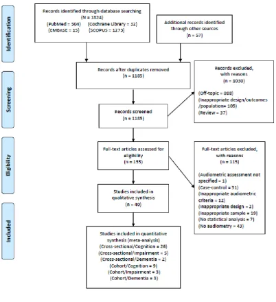
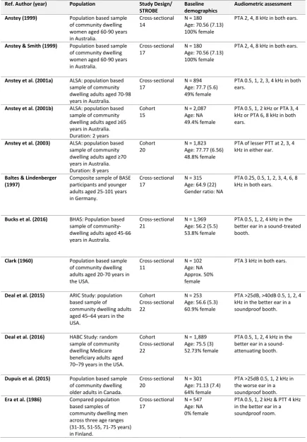
[image:41.595.104.507.251.692.2]The characteristics of included studies are shown in Table 2.1. Of the 1,185 citations reviewed, 40 studies met the inclusion criteria (Figure 2.1). An excluded studies table is available upon request. Study quality results are shown in Table 2.1 and Appendix A. More than 80% of the included papers met the criteria for 16 out 22 STROBE items.
24
Hearing loss & cognitive function
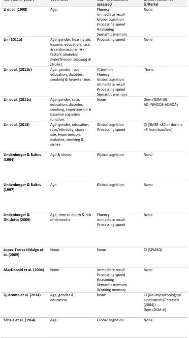
Twenty-six studies with 15,620 participants were included in the cross-sectional/cognitive function analysis (Anstey, 1999; Anstey, Luszcz, & Sanchez, 2001a; Anstey & Smith, 1999; Baltes & Lindenberger, 1997; Bucks et al., 2016; Clark, 1960; Deal et al., 2016; Deal et al., 2015; Dupuis et al., 2015; Era, Jokela, Qvarnberg, & Heikkinen, 1986; Gussekloo, De Craen, Oduber, Van Boxtel, & Westendorp, 2005; Harrison Bush, Lister, Lin, Betz, & Edwards, 2015; Helzner et al., 2005; Heron & Chown, 1967; Hofer, Berg, & Era, 2003; Hong, Mitchell, Burlutsky, Liew, & Wang, 2016; Li, Jordanova, & Linberger, 1998; Lin, 2011; Lin, Ferrucci, et al., 2011; Lin et al., 2013; Lindenberger & Baltes, 1994, 1997; MacDonald, Dixon, Cohen, & Hazlitt, 2004; Schaie, Baltes, & Strother, 1964; Sugawara et al., 2011; Thomas et al., 1983; Valentijn et al., 2005; van Boxtel et al., 2000). Nine studies with 8,233 participants were included in the cohort/cognitive function analysis (Anstey, Hofer, & Luszcz, 2003; Anstey, Luszcz, & Sanchez, 2001b; Deal et al., 2016; Deal et al., 2015; Gallacher et al., 2012; Hong et al., 2016; Lin et al., 2013; Lindenberger & Ghisletta, 2009; Valentijn et al., 2005). The cohort studies had a follow-up length ranging from two to 23 years (mean 10·4 years).
There was a small but statistically significant association between ARHL and all of the ten cognitive domains of interest in cross-sectional studies including; global cognition (r, -0.15, p<0.001), executive functions (r, -0.08 to -0.18, p<0.001), episodic memory (r, -0.1 to -0.14, p≤0.002), processing speed (r, -0.13, p<0.001), semantic memory (r, -0.14, p<0.001) and
25
Figure 2.2: Forest plot of correlation r values for cognition/cross-sectional outcomes.
The black squares represent the r value while the lines represent the corresponding 95% confidence intervals. The middle of the black diamond represents the overall r value while the left and right extremes of the diamond represent the corresponding 95% confidence intervals.
Figure 2.3: Forest plot of correlation r values for cognition/cohort outcomes.
Heterogeneity was significant in all domains except fluency and visuospatial ability. Inconsistency across studies ranged from very low to large. Qualitative analysis of small-study effects
demonstrated moderate to no asymmetry across studies (Appendix A). Quantitative analysis with Egger’s Test of the Intercept found statistically significant small-study effects for cross-sectional semantic memory (Appendix A). With each included study deleted from the model once, results remained statistically significant across all deletions for all domains with the exception of Clark (1960) (Clark, 1960) for cross-sectional visuospatial ability (Appendix A). The difference between the largest and smallest values, having deleted each group once, ranged from 16.6% to 60.2%. Cumulative meta-analysis demonstrated that ARHL has been significantly related to cognitive function since between 1960 and 2012 (Appendix A).
Hearing loss & cognitive impairment
Five studies with 6,582 participants were included in the cross-sectional/cognitive impairment analysis (Dupuis et al., 2015; Karpa et al., 2010; Kiely et al., 2012; Kurniawan et al., 2012; Lopez-Torres Hidalgo et al., 2009; Quaranta et al., 2014; Tay, Wang, et al., 2006). Three studies with 7,817 participants were included in the cohort/cognitive impairment analysis (Gallacher et al., 2012; Kiely et al., 2012; Lin et al., 2013). The cohort studies had a follow-up length ranging from six to 18 years (mean 11.7 years).
26
Small-study effects were not examined because there were less than ten effect sizes. With each group deleted from the model once, results remained statistically significant across all deletions (Appendix A). The difference between the largest and smallest values with each group deleted was 0.45 (20.1%) for cross-sectional studies and 0.04 (3.4%) for cohort studies. Cumulative meta-analysis demonstrated that cognitive impairment has been significantly related to ARHL since the completion of the first cross-sectional study in 2009 and cohort study in 2012 (Appendix A).
Hearing loss & dementia
Two studies with 741 participants were included in the cross-sectional/dementia analysis (Herbst & Humphrey, 1980; Quaranta et al., 2014). One assessed dementia (Herbst & Humphrey, 1980) and the other assessed AD (Quaranta et al., 2014). Three studies with 3,585 participants were included in the cohort/dementia analysis (Deal et al., 2016; Gallacher et al., 2012; Lin, Metter, et al., 2011). All three reported incident dementia outcomes, two for an AD subset (Gallacher et al., 2012; Lin, Metter, et al., 2011) and one for a vascular dementia (VaD) subset (Gallacher et al., 2012). The follow-up period ranged from nine to 18 years (mean 15 years).
There was a significant association between ARHL and dementia in both cross-sectional (OR 2.42, 95% CI 1.24-4.72, p=0.01) and cohort studies (OR 1.28, 95% CI 1.02-1.59, p=0.03) (Appendix A). There was no statistically significant association between ARHL and AD for cross-sectional (OR 1.80, 95% CI 0.58 – 5.60, p=0.31) or cohort studies (OR 1.69, 95% CI 0.72-4.00, p=0.23). Forest plots are shown in Appendix A. No statistically significant heterogeneity or inconsistency was observed for cross-sectional studies. For cohort studies, statistically significant heterogeneity was observed as well as a moderate amount of inconsistency. Adults with dementia totalled