1068
http://dx.doi.org/10.1107/S2056989016010781 Acta Cryst.(2016). E72, 1068–1073research communications
Received 1 July 2016 Accepted 4 July 2016
Edited by W. T. A. Harrison, University of Aberdeen, Scotland
Keywords:crystal structure; polymorph; gold; thiocarbamate; Hirshfeld surface analysis.
CCDC reference:1489737
Supporting information:this article has supporting information at journals.iucr.org/e
A monoclinic polymorph of [(
Z
)-
N
-(3-chlorophen-yl)-
O
-methylthiocarbamato-
j
S
](triphenylphos-phane-
j
P
)gold(I): crystal structure and Hirshfeld
surface analysis
Chien Ing Yeo, Sang Loon Tan and Edward R. T. Tiekink*
Research Centre for Crystalline Materials, Faculty of Science and Technology, Sunway University, 47500 Bandar Sunway, Selangor Darul Ehsan, Malaysia. *Correspondence e-mail: [email protected]
The title compound, [Au(C8H7ClNOS)(C18H15P)], is a monoclinic (P21/n,Z0= 1;
form) polymorph of the previously reported triclinic form (P1,Z0= 1; form) [Tadbuppa & Tiekink (2010).Acta Cryst. E66, m664]. The molecular structures of both forms feature an almost linear gold(I) coordination geometry [P—Au— S = 175.62 (5) in the title polymorph], being coordinated by thiolate S and phosphane P atoms, a Z conformation about the C N bond and an intramolecular Au O contact. The major conformational difference relates to the relative orientations of the residues about the Au—S bond: the P—Au— S—C torsion angles are8.4 (7) and 106.2 (7)in formsand, respectively. The molecular packing of formfeatures centrosymmetric aggregates sustained by aryl-C—H O interactions, which are connected into a three-dimensional network by aryl-C—H contacts. The Hirshfeld analysis of forms and shows many similarities with the notable exception of the influence of C—H O interactions in form.
1. Chemical context
Interest in the chemistry of phosphanegold(I) N-aryl-O -alkylthiocarbamates, i.e. compounds of general formula
R3PAu[SC(OR0) NR00] (R, R0 = alkyl, aryl; R00 = aryl)
continues owing to their recently disclosed exciting biological activities. Thus, various triphenylphosphane derivatives display excellent cytotoxicity profiles against HT-29 colon cancer cells, a particularly virulent form of cancer, and mechanistic studies have shown these to induce both intrinsic and extrinsic pathways of cell death leading to apoptosis (Yeo, Ooi et al., 2013; Ooi et al., 2015). Further, species with R00 =
p-tolyl have proven to exhibit impressive in vitro potency against Gram-positive bacteria (Yeo, Simet al., 2013). It was during another synthesis of the title compound, (I), for further biological studies, that crystals of a new polymorph were isolated from its methanol solution. This is called form to distinguish it from the earlier triclinic form, form (Tadbuppa & Tiekink, 2010). Herein, the crystal and mol-ecular structures of form of (I) are described along with a comparison with the parameters characterizing form . Further, a Hirshfeld surface analysis of both polymorphic forms of (I) is presented.
2. Structural commentary
The molecular structure of the new monoclinic form of (I), form, is shown in Fig. 1, and selected geometric parameters
are collected in Table 1. The gold(I) atom is coordinated in an approximately linear configuration by phosphane-P and thiolate-S atoms. Confirmation of the ‘thiolate’ assignment is readily seen in the relatively long C1—S1 bond length and the significant -character in the C1—N1 bond when the geometric parameters are compared with structures of related thiocarbamide molecules (Hoet al., 2005; Kuan et al., 2007); the crystal structure of the thiocarbamide precursor in (I) is not available for comparison. As is invariably observed in this class of compound, the Au—S bond length is longer than the Au—P bond. The small deviation from ideal linearity for the P—Au—S bond is related to the close approach of the oxygen atom to the gold(I) atom,i.e. 3.052 (3) A˚ . The pattern of bond angles about the quaternary carbon atom, C1, follow the expected trends with the widest angle involving the sulfur and doubly bonded nitrogen atom and with the narrowest angle involving the single-bonded atoms. The conformation about the formal C1 N1 bond, Table 1, isZ.
Formcrystallizes in the monoclinic space groupP21/nwith
Z0 = 1. The earlier polymorph, by contrast, crystallizes in triclinic space groupP1, also withZ0= 1. A comparison of the key geometric parameters is given in Table 1. From these data, it is clear that there is experimental inequivalence in the bond lengths involving the gold(I) atoms, with the Au—S and Au— P bond lengths in formbeing marginally longer. The intra-molecular Au O separation in formis also longer than the comparable separation in form, and this is correlated with a smaller deviation from a linear geometry about the gold(I) atom in . By contrast, the bond angles are, by and large, equivalent within experimental error. A significant confor-mational difference is evident in the molecular structures of
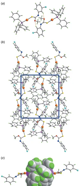
formsandof (I). As seen from the overlay diagram shown in Fig. 2, this difference occurs as a result of a twist about the Au—S bond as seen in the values of the P1—Au—S1—C1 torsion angles of 8.4 (7) and 106.2 (7) in forms and, respectively.
3. Supramolecular features
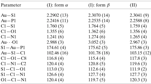
Supramolecular dimers feature in the molecular packing of form of (I), which are sustained by N-aryl-C— H O(methoxy) interactions, Fig. 3aand Table 2. The dimers are connected into a three-dimensional architecture by a network of C—H interactions, Fig. 3band Table 2. Within this arrangement, centrosymmetrically related Ph3P ligands
align to form a so-called six-fold phenyl embrace (6PE) (Dance & Scudder, 1995) featuring edge-to-face phenyl-C— H (phenyl) interactions, Fig. 3c. While the interactions are too long to be considered as significant in terms of the criteria inPLATON(Spek, 2009), there are a number of such inter-actions,i.e. 2[3.22, 3.26 and 3.29 A˚ ], that serve to reinforce the 6PE embrace with one pair of rings accepting two inter-actions each. In formof (I), the most prominent feature of the molecular packing is the formation of supramolecular chains mediated by C—H interactions (Tadbuppa & Tiekink, 2010). Further analysis of the molecular packing in polymorphic (I) is given in the following Section.
research communications
Acta Cryst.(2016). E72, 1068–1073 Yeoet al. [Au(C
[image:2.610.313.565.92.232.2]8H7ClNOS)(C18H15P)]
1069
Figure 1The molecular structure of polymorphic formof (I), showing the atom-labelling scheme and displacement ellipsoids at the 70% probability level.
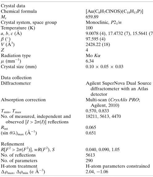
Table 1
Geometric data (A˚ ,) for (I), formsa
and, and (II)b.
Parameter (I): form (I): form (II)
Au—S1 2.2902 (13) 2.3070 (14) 2.3041 (9) Au—P1 2.2416 (11) 2.2535 (14) 2.2588 (8) C1—S1 1.760 (5) 1.764 (5) 1.759 (4) C1—O1 1.355 (6) 1.362 (6) 1.356 (4) C1—N1 1.241 (6) 1.274 (6) 1.265 (4) Au O1 2.988 (3) 3.052 (3) 2.967 (3) S1—Au—P1 174.61 (4) 175.62 (5) 175.86 (3) Au—S1—C1 102.46 (16) 101.78 (18) 103.15 (12) C1—O1—C8 116.8 (4) 115.4 (4) 117.8 (3) C1—N1—C2 120.4 (4) 120.8 (5) 119.6 (3) S1—C1—O1 113.0 (3) 112.6 (4) 111.9 (2) S1—C1—N1 126.6 (4) 127.7 (4) 127.7 (3) O1—C1—N1 120.4 (4) 119.7 (5) 120.3 (3)
[image:2.610.67.275.292.399.2]Notes: (a) Tadbuppa & Tiekink (2010); (b) Tadbuppa & Tiekink (2009).
Figure 2
[image:2.610.46.297.580.723.2]4. Analysis of the Hirshfeld surfaces
The non-covalent interactions present in the pair of poly-morphs of (I), i.e. forms and , were studied through Hirshfeld surface analysis by mapping on the normalized contact distance (dnorm) upon computation of the inner (di)
and outer (de) distances of the Hirshfeld surface to the nearest
nucleus (Spackman & Jayatilaka, 2009; McKinnonet al., 2007). All computation as well as generation of two-dimensional fingerprint plots were performed usingCrystal Explorer 3.1
(Wolffet al., 2012). Distances involving hydrogen atoms were normalized by default to the standard neutron-diffraction bond lengths.
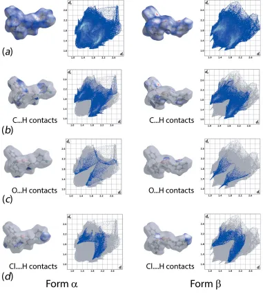
As evident from Fig. 4 and Table 3, forms and of (I) exhibit relatively similar percentage contributions of the indicated intermolecular interactions to their Hirshfeld surfaces. However, the specific contributions to their inter-action profiles are distinct as evidenced from the overall and decomposed two-dimensional fingerprint plots shown in Fig. 5. As mentioned above in Supramolecular features, C—H interactions feature in both structures. To a first approxima-tion the decomposed fingerprint plots look similar, as seen from Fig. 5b. However, relatively shorter contacts are found in form cf. form , i.e. 2.62 vs 2.68 A˚ . The clear distinction between the two forms is readily noted from the decomposed fingerprint plots for the O H/H O contacts with very
1070
Yeoet al. [Au(C [image:3.610.42.298.70.671.2]8H7ClNOS)(C18H15P)] Acta Cryst.(2016). E72, 1068–1073
research communications
Table 2
Hydrogen-bond geometry (A˚ ,).
Hydrogen-bond geometry (A˚ ,), form,Cg1,Cg3 andCg4 are the centroids
of the C2–C7, C21–C26 and C31–C36 rings, respectively.
D—H A D—H H A D A D—H A
C3—H3 O1i 0.95 2.47 3.315 (7) 148
C5—H5 Cg4ii 0.95 2.85 3.492 (6) 126 C12—H12 Cg1ii 0.95 2.64 3.450 (6) 143
C14—H14 Cg3iii 0.95 2.80 3.570 (6) 139 C23—H23 Cg1iv 0.95 2.65 3.435 (6) 140
Symmetry codes: (i)xþ1;yþ2;zþ1; (ii)x;yþ2;zþ1; (iii)x1;y;z; (iv)xþ1
2;y12;zþ12.
Figure 4
[image:3.610.311.566.117.186.2] [image:3.610.313.564.549.710.2]Percentage contribution of different close contacts to the Hirshfeld surface of formsandof (I).
Figure 3
Molecular packing in formof (I): (a) view of the supramolecular dimer sustained by C—H O contacts, shown as orange dashed lines, (b) view of the unit-cell contents shown in projection down theaaxis, highlighting the C—H interactions as purple dashed lines, (c) image of the sixfold phenyl (6PE) between centrosymmetrically related Ph3P ligands,
distinct spikes evident for form, Fig. 5c, correlating with the C—H O interactions leading to dimer formation. While beyond the sum of their respective van der Waals radii (Spek, 2009), Cl H/H Cl interactions make contributions to the Hirshfeld surfaces of both forms and, with the contacts, again, being shorter in form,i.e. 2.76vs3.00 A˚ , leading to more the distinct forceps in Fig. 5d.
In general, the observation of generally shorter contacts in formmay indicate greater crystal-packing efficiency (Lloyd
et al., 2005). Table 4 collates various molecular/crystal struc-ture descriptors for the polymorphic forms. Immediately evident is that the calculated unit-cell densities are identical but the crystal-packing efficiency (KPI; Spek, 2009) for form is marginally greater. Computation on the area-to-volume ratio between formsandrevealed very little difference as did the globularity (G) and asphericity () indices. All these
indicators suggest that the polymorphs arise as a result of a simple interplay between molecular conformation and crystal-packing effects.
5. Database survey
The most closely related structure to (I) in the crystallographic literature (Groomet al., 2016), is theR0= OEt analogue,i.e. (II), (Tadbuppa & Tiekink, 2009). Key geometric parameters for this structure are also included in Table 1. Non-systematic variations in parameters are noted,e.g. the Au—S bond length in (II) is intermediate between those found in the polymorphic forms of (I), and the Au—P bond length is the longest of the three structures. However, differences are small and probably can be ascribed to the influences of crystal-packing effects.
research communications
Acta Cryst.(2016). E72, 1068–1073 Yeoet al. [Au(C
[image:4.610.108.489.281.710.2]8H7ClNOS)(C18H15P)]
1071
Figure 5Comparison of the (a) complete Hirshfeld surface and full fingerprint plots between formand formpolymorphs (top row) and the corresponding
As indicated in theChemical context, biological considera-tions motivate ongoing investigaconsidera-tions into the chemistry of phosphanegold(I) N-aryl-O-alkylthiocarbamates. This notwithstanding, the relative ease of growing crystals have prompted several crystal engineering studies. Thus, correla-tions between Au Au (aurophilic) and solid-state lumines-cence responses have been made for the series of compounds,
R3PAu[SC(OMe) NC6H4NO2-p] (R = Et, Cy and Ph), and
bidentate phosphane analogues, Ph2P–(CH2)n–PPh2forn= 1–
4 and when the bridge is Fc (ferrocenyl) (Hoet al., 2006). In another study, the influence ofRandYsubstituents upon the molecular packing of compounds of the general formula [(Ph2P(CH2)4PPh2){AuSC(OR0) NC6H4Y-p}2] for R0 = Me,
Et or iPr and Y = H, NO2 or Me was undertaken (Ho &
Tiekink, 2007). Besides the anticipated linear P—Au—S configuration, a common feature of all the analysed structures until then was the presence of intramolecular Au O inter-actions, as illustrated in Fig. 1. This changed in another systematic study, this time ofR3PAu[SC(OMe) NR00], forR=
Ph, o-tol, m-tol or p-tol, and R0’ = Ph,o-tol, m-tol,p-tol or C6H4NO2-p, where it proved possible to induce a
conforma-tional change in the molecule so that an intramolecular Au interaction formed rather than Au O (Kuanet al., 2008); Au interactions are well documented in the crys-tallographic literature (Tiekink & Zukerman-Schpector, 2009; Caracelli et al., 2013). For example, having R = R00 = p-tol simultaneously activated the gold atom, making it amenable to form an Au interaction with the comparatively
electron-rich aryl ring. Recently, bipodal forms of the thiocarbamide ligands were prepared and complexed with phosphanegold(I) species yielding binuclear molecules also with intramolecular Au interactions (Yeo et al., 2015). Computational chem-istry showed the Au interactions to be more favourable, by ca 12 kcal mol1, than the putative Au O interaction (Yeoet al., 2015).
Such interplay between substituents in crystal engineering endeavours, along with the observation that biological activ-ities are acutely sensitive to substitution patterns, ensures this area of research will continue to attract significant attention.
6. Synthesis and crystallization
All chemicals and solvents were used as purchased without purification. All reactions were carried out under ambient conditions. Melting points were determined on a Biobase auto melting point apparatus MP300. IR spectra were obtained on a Perkin Elmer Spectrum 400 FT Mid-IR/Far-IR spectro-photometer from 4000 to 400 cm1; abbreviation: s, strong.
Preparation of (I): NaOH (Merck; 0.25 mmol, 0.01 g) in MeOH (Merck; 1 ml) was added to a suspension of Ph3PAuCl
(0.25 mmol, 0.12 g) in MeOH (Merck; 15 ml), followed by addition of the thiocarbamide, MeOC( S)N(H)C6H4Cl3
(0.25 mmol, 0.05 g), prepared following literature precedents (Hoet al., 2005), in MeOH (15 ml). The resulting mixture was stirred for 2 h at 323 K. The solution mixture was left for slow
1072
Yeoet al. [Au(C [image:5.610.313.561.91.378.2]8H7ClNOS)(C18H15P)] Acta Cryst.(2016). E72, 1068–1073
research communications
Table 5
Experimental details.
Crystal data
Chemical formula [Au(C8H7ClNOS)(C18H15P)]
Mr 659.89
Crystal system, space group Monoclinic,P21/n
Temperature (K) 100
a,b,c(A˚ ) 9.0078 (4), 17.4732 (7), 15.5641 (7)
() 97.595 (4)
V(A˚3) 2428.22 (18)
Z 4
Radiation type MoK
(mm1) 6.34
Crystal size (mm) 0.100.050.03
Data collection
Diffractometer Agilent SuperNova Dual Source diffractometer with an Atlas detector
Absorption correction Multi-scan (CrysAlis PRO; Agilent, 2010)
Tmin,Tmax 0.570, 0.833
No. of measured, independent and observed [I> 2(I)] reflections
18211, 5613, 4470
Rint 0.065
(sin/)max(A˚
1) 0.651
Refinement
R[F2> 2(F2)],wR(F2),S 0.040, 0.090, 1.05
No. of reflections 5613 No. of parameters 290
H-atom treatment H-atom parameters constrained
max, min(e A˚
3) 2.04,1.06
[image:5.610.44.296.102.259.2]Computer programs:CrysAlis PRO (Agilent, 2010),SHELXS97 (Sheldrick, 2008), SHELXL2014(Sheldrick, 2015),ORTEP-3 for Windows(Farrugia, 2012),QMol(Gans & Shalloway, 2001),DIAMOND(Brandenburg, 2006) andpublCIF(Westrip, 2010). Table 4
Physiochemical properties for formsandof (I).
Parameter Form Form
Volume,V(A˚3) 596.42 596.78
Surface area,A(A˚2) 518.92 511.11
A:V(A˚1) 0.87 0.86
Globularity,G 0.660 0.671
Asphericity, 0.165 0.172
Density (g cm1) 1.805 1.805
Packing index (%) 66.9 67.4
Table 3
Percentage contribution of the different intermolecular contacts to the Hirshfeld surface in formsandof (I).
Contact % Contribution form % Contribution form
Au Cl 0.2 0.6
Au C 0.3 0.2
Au H 4.2 2.8
Cl C 2.7 0.3
Cl H 7.6 9.8
Cl S 0.0 0.2
S C 0.1 0.0
S H 6.6 6.3
O H 2.5 3.2
N H 1.9 1.7
N C 0.0 0.3
C C 0.4 0.8
C H 27.8 30.6
H H 45.6 43.2
[image:5.610.44.296.305.389.2]evaporation at room temperature, yielding colourless prisms of the title compound after 3 weeks. Yield: 0.134 g (81%). M.p. 431–433 K. IR (cm1): 1434 (s) (C N), 1180 (s) (C—O), 1098 (s)(C—S).
7. Refinement
Crystal data, data collection and structure refinement details are summarized in Table 5. The carbon-bound H atoms were placed in calculated positions (C—H = 0.95–0.98 A˚ ) and were included in the refinement in the riding-model approximation, with Uiso(H) set to 1.2–1.5Ueq(C). The maximum and
minimum residual electron density peaks of 2.04 and 1.06 e A˚3, respectively, were located 1.01 and 0.77 A˚ from the Au atom.
Acknowledgements
Intensity data were provided by the University of Malaya Crystallographic Laboratory.
References
Agilent (2010). CrysAlis PRO. Agilent Technologies Inc., Santa Clara, CA, USA.
Brandenburg, K. (2006).DIAMOND. Crystal Impact GbR, Bonn, Germany.
Caracelli, I., Zukerman-Schpector, J. & Tiekink, E. R. T. (2013).Gold
Bull.46, 81–89.
Dance, I. & Scudder, M. (1995).J. Chem. Soc. Chem. Commun. pp. 1039–1040.
Farrugia, L. J. (2012).J. Appl. Cryst.45, 849–854.
Gans, J. & Shalloway, D. (2001).J. Mol. Graphics Modell.19, 557–559. Groom, C. R., Bruno, I. J., Lightfoot, M. P. & Ward, S. C. (2016).Acta
Cryst.B72, 171–179.
Ho, S. Y., Bettens, R. P. A., Dakternieks, D., Duthie, A. & Tiekink, E. R. T. (2005).CrystEngComm,7, 682–689.
Ho, S. Y., Cheng, E. C.-C., Tiekink, E. R. T. & Yam, V. W.-W. (2006).
Inorg. Chem.45, 8165–8174.
Ho, S. Y. & Tiekink, E. R. T. (2007).CrystEngComm,9, 368–378. Kuan, F. S., Mohr, F., Tadbuppa, P. P. & Tiekink, E. R. T. (2007).
CrystEngComm,9, 574–581.
Kuan, F. S., Ho, S. Y., Tadbuppa, P. P. & Tiekink, E. R. T. (2008).
CrystEngComm,10, 548–564.
Lloyd, G. O., Bredenkamp, M. W. & Barbour, L. J. (2005).Chem.
Commun. pp. 4053–4055.
McKinnon, J. J., Jayatilaka, D. & Spackman, M. A. (2007).Chem
Commun. pp. 3814–3816.
Ooi, K. K., Yeo, C. I., Ang, K.-P., Akim, A. Md., Cheah, Y.-K., Halim, S. N. A., Seng, H.-L. & Tiekink, E. R. T. (2015). J. Biol. Inorg.
Chem.20, 855–873.
Sheldrick, G. M. (2008).Acta Cryst.A64, 112–122. Sheldrick, G. M. (2015).Acta Cryst.C71, 3–8.
Spackman, M. A. & Jayatilaka, D. (2009).CrystEngComm,11, 19–32. Spek, A. L. (2009).Acta Cryst.D65, 148–155.
Tadbuppa, P. P. & Tiekink, E. R. T. (2009).Acta Cryst.E65, m1663. Tadbuppa, P. P. & Tiekink, E. R. T. (2010).Acta Cryst.E66, m664. Tiekink, E. R. T. & Zukerman-Schpector, J. (2009).CrystEngComm,
11, 1176–1186.
Westrip, S. P. (2010).J. Appl. Cryst.43, 920–925.
Wolff, S. K., Grimwood, D. J., McKinnon, J. J., Turner, M. J., Jayatilaka, D. & Spackman, M. A. (2012).Crystal Explorer. The University of Western Australia.
Yeo, C. I., Khoo, C.-H., Chu, W.-C., Chen, B.-J., Chu, P.-L., Sim, J.-H., Cheah, Y.-K., Ahmad, J., Halim, S. N. A., Seng, H.-L., Ng, S., Otero-de-la-Roza, A. & Tiekink, E. R. T. (2015). RSC Adv. 5, 41401– 41411.
Yeo, C. I., Ooi, K. K., Akim, A. Md., Ang, K. P., Fairuz, Z. A., Halim, S. N. B. A., Ng, S. W., Seng, H.-L. & Tiekink, E. R. T. (2013).J.
Inorg. Biochem.127, 24–38.
Yeo, C. I., Sim, J.-H., Khoo, C.-H., Goh, Z.-J., Ang, K.-P., Cheah, Y.-K., Fairuz, Z. A., Halim, S. N. B. A., Ng, S. W., Seng, H.-L. & Tiekink, E. R. T. (2013).Gold Bull.46, 145–152.
research communications
Acta Cryst.(2016). E72, 1068–1073 Yeoet al. [Au(C
supporting information
sup-1
Acta Cryst. (2016). E72, 1068-1073
supporting information
Acta Cryst. (2016). E72, 1068-1073 [https://doi.org/10.1107/S2056989016010781]
A monoclinic polymorph of [(
Z
)-
N
-(3-chlorophenyl)-
O
-methylthiocarbamato-κ
S
](triphenylphosphane-
κ
P
)gold(I): crystal structure and Hirshfeld surface
analysis
Chien Ing Yeo, Sang Loon Tan and Edward R. T. Tiekink
Computing details
Data collection: CrysAlis PRO (Agilent, 2010); cell refinement: CrysAlis PRO (Agilent, 2010); data reduction: CrysAlis PRO (Agilent, 2010); program(s) used to solve structure: SHELXS97 (Sheldrick, 2008); program(s) used to refine structure: SHELXL2014 (Sheldrick, 2015); molecular graphics: ORTEP-3 for Windows (Farrugia, 2012), QMol (Gans & Shalloway, 2001) and DIAMOND (Brandenburg, 2006); software used to prepare material for publication: publCIF
(Westrip, 2010).
[(Z)-N-(3-Chlorophenyl)-O-methylthiocarbamato-κS](triphenylphosphane-κP)gold(I)
Crystal data
[Au(C8H7ClNOS)(C18H15P)]
Mr = 659.89
Monoclinic, P21/n
a = 9.0078 (4) Å
b = 17.4732 (7) Å
c = 15.5641 (7) Å
β = 97.595 (4)°
V = 2428.22 (18) Å3
Z = 4
F(000) = 1280
Dx = 1.805 Mg m−3
Mo Kα radiation, λ = 0.71073 Å Cell parameters from 5344 reflections
θ = 2.3–27.5°
µ = 6.34 mm−1
T = 100 K Prism, colourless 0.10 × 0.05 × 0.03 mm
Data collection
Agilent SuperNova Dual Source diffractometer with an Atlas detector Radiation source: SuperNova (Mo) X-ray
Source
Mirror monochromator
Detector resolution: 10.4041 pixels mm-1
ω scan
Absorption correction: multi-scan (CrysAlis PRO; Agilent, 2010)
Tmin = 0.570, Tmax = 0.833
18211 measured reflections 5613 independent reflections 4470 reflections with I > 2σ(I)
Rint = 0.065
θmax = 27.6°, θmin = 2.3°
h = −11→9
k = −22→22
l = −17→20
Refinement
Refinement on F2
Least-squares matrix: full
R[F2 > 2σ(F2)] = 0.040
wR(F2) = 0.090
S = 1.05 5613 reflections
290 parameters 0 restraints
Hydrogen site location: inferred from neighbouring sites
supporting information
sup-2
Acta Cryst. (2016). E72, 1068-1073
w = 1/[σ2(F
o2) + (0.0282P)2]
where P = (Fo2 + 2Fc2)/3
(Δ/σ)max < 0.001
Δρmax = 2.04 e Å−3
Δρmin = −1.06 e Å−3
Special details
Geometry. All esds (except the esd in the dihedral angle between two l.s. planes) are estimated using the full covariance matrix. The cell esds are taken into account individually in the estimation of esds in distances, angles and torsion angles; correlations between esds in cell parameters are only used when they are defined by crystal symmetry. An approximate (isotropic) treatment of cell esds is used for estimating esds involving l.s. planes.
Fractional atomic coordinates and isotropic or equivalent isotropic displacement parameters (Å2)
x y z Uiso*/Ueq
Au 0.19155 (2) 0.79563 (2) 0.43242 (2) 0.02135 (8) Cl1 0.35621 (18) 1.26890 (8) 0.33228 (10) 0.0342 (4) P1 0.11209 (17) 0.67786 (8) 0.46382 (9) 0.0209 (3) S1 0.25415 (18) 0.91846 (8) 0.39637 (9) 0.0263 (3)
O1 0.3390 (4) 0.9118 (2) 0.5632 (2) 0.0231 (8)
N1 0.2776 (5) 1.0330 (2) 0.5172 (3) 0.0238 (10)
C1 0.2894 (6) 0.9623 (3) 0.4991 (3) 0.0206 (11)
C2 0.2162 (7) 1.0859 (3) 0.4530 (4) 0.0240 (12)
C3 0.3063 (6) 1.1426 (3) 0.4247 (3) 0.0222 (12)
H3 0.4107 1.1438 0.4442 0.027*
C4 0.2408 (7) 1.1975 (3) 0.3675 (4) 0.0237 (12)
C5 0.0901 (7) 1.1990 (3) 0.3373 (4) 0.0231 (12)
H5 0.0489 1.2371 0.2975 0.028*
C6 0.0000 (7) 1.1425 (3) 0.3670 (4) 0.0270 (13)
H6 −0.1046 1.1423 0.3478 0.032*
C7 0.0618 (6) 1.0863 (3) 0.4249 (4) 0.0262 (13)
H7 −0.0007 1.0484 0.4452 0.031*
C8 0.3796 (7) 0.9450 (3) 0.6480 (3) 0.0294 (13)
H8A 0.4077 0.9042 0.6903 0.044*
H8B 0.2941 0.9735 0.6647 0.044*
H8C 0.4645 0.9798 0.6466 0.044*
C11 −0.0850 (6) 0.6656 (3) 0.4249 (3) 0.0208 (12) C12 −0.1829 (7) 0.7251 (3) 0.4386 (4) 0.0258 (13)
H12 −0.1448 0.7711 0.4657 0.031*
C13 −0.3350 (7) 0.7173 (3) 0.4127 (4) 0.0316 (14)
H13 −0.4014 0.7575 0.4228 0.038*
C14 −0.3908 (7) 0.6504 (3) 0.3719 (4) 0.0301 (14)
H14 −0.4953 0.6451 0.3544 0.036*
C15 −0.2952 (7) 0.5923 (3) 0.3569 (4) 0.0289 (13)
H15 −0.3340 0.5474 0.3277 0.035*
C16 −0.1436 (6) 0.5982 (3) 0.3838 (3) 0.0252 (12)
H16 −0.0788 0.5569 0.3747 0.030*
C21 0.2049 (5) 0.6011 (3) 0.4174 (3) 0.0157 (11)
C22 0.2091 (6) 0.6018 (3) 0.3269 (3) 0.0222 (12)
H22 0.1637 0.6431 0.2935 0.027*
supporting information
sup-3
Acta Cryst. (2016). E72, 1068-1073
H23 0.2804 0.5458 0.2248 0.031*
C24 0.3423 (6) 0.4830 (3) 0.3338 (4) 0.0251 (12)
H24 0.3879 0.4427 0.3057 0.030*
C25 0.3412 (6) 0.4803 (3) 0.4227 (4) 0.0258 (13)
H25 0.3868 0.4386 0.4554 0.031*
C26 0.2729 (6) 0.5389 (3) 0.4639 (4) 0.0243 (12)
H26 0.2725 0.5366 0.5248 0.029*
C31 0.1333 (6) 0.6589 (3) 0.5800 (3) 0.0197 (11)
C32 0.0283 (6) 0.6151 (3) 0.6158 (3) 0.0233 (12)
H32 −0.0569 0.5951 0.5804 0.028*
C33 0.0503 (7) 0.6009 (3) 0.7051 (4) 0.0274 (13)
H33 −0.0207 0.5709 0.7303 0.033*
C34 0.1734 (7) 0.6299 (3) 0.7572 (4) 0.0293 (14)
H34 0.1883 0.6192 0.8176 0.035*
C35 0.2751 (7) 0.6748 (3) 0.7201 (4) 0.0318 (14)
H35 0.3587 0.6961 0.7557 0.038*
C36 0.2558 (7) 0.6890 (3) 0.6314 (4) 0.0286 (13)
H36 0.3265 0.7192 0.6063 0.034*
Atomic displacement parameters (Å2)
U11 U22 U33 U12 U13 U23
supporting information
sup-4
Acta Cryst. (2016). E72, 1068-1073
C31 0.020 (3) 0.016 (3) 0.023 (3) −0.002 (2) 0.005 (2) −0.005 (2) C32 0.027 (3) 0.015 (3) 0.026 (3) −0.002 (2) −0.003 (2) 0.000 (2) C33 0.031 (3) 0.026 (3) 0.026 (3) −0.001 (3) 0.005 (3) 0.002 (2) C34 0.039 (4) 0.026 (3) 0.021 (3) 0.001 (3) 0.000 (3) 0.003 (2) C35 0.030 (4) 0.040 (3) 0.024 (3) −0.013 (3) −0.004 (3) −0.002 (3) C36 0.031 (4) 0.033 (3) 0.023 (3) −0.004 (3) 0.006 (3) 0.001 (2)
Geometric parameters (Å, º)
Au—P1 2.2535 (14) C13—H13 0.9500
Au—S1 2.3070 (14) C14—C15 1.372 (8)
Cl1—C4 1.756 (6) C14—H14 0.9500
P1—C21 1.783 (5) C15—C16 1.378 (8)
P1—C11 1.811 (6) C15—H15 0.9500
P1—C31 1.824 (5) C16—H16 0.9500
S1—C1 1.764 (5) C21—C26 1.402 (7)
O1—C1 1.362 (6) C21—C22 1.413 (7)
O1—C8 1.443 (6) C22—C23 1.390 (7)
N1—C1 1.274 (6) C22—H22 0.9500
N1—C2 1.418 (7) C23—C24 1.383 (7)
C2—C3 1.389 (8) C23—H23 0.9500
C2—C7 1.402 (8) C24—C25 1.386 (8)
C3—C4 1.387 (7) C24—H24 0.9500
C3—H3 0.9500 C25—C26 1.394 (7)
C4—C5 1.378 (8) C25—H25 0.9500
C5—C6 1.395 (8) C26—H26 0.9500
C5—H5 0.9500 C31—C36 1.379 (7)
C6—C7 1.398 (7) C31—C32 1.389 (7)
C6—H6 0.9500 C32—C33 1.399 (7)
C7—H7 0.9500 C32—H32 0.9500
C8—H8A 0.9800 C33—C34 1.380 (8)
C8—H8B 0.9800 C33—H33 0.9500
C8—H8C 0.9800 C34—C35 1.389 (8)
C11—C12 1.398 (8) C34—H34 0.9500
C11—C16 1.409 (7) C35—C36 1.391 (8)
C12—C13 1.383 (8) C35—H35 0.9500
C12—H12 0.9500 C36—H36 0.9500
C13—C14 1.391 (8)
P1—Au—S1 175.62 (5) C15—C14—C13 120.2 (6)
C21—P1—C11 105.5 (2) C15—C14—H14 119.9
C21—P1—C31 105.8 (2) C13—C14—H14 119.9
C11—P1—C31 106.2 (2) C14—C15—C16 120.8 (5)
C21—P1—Au 114.80 (17) C14—C15—H15 119.6
C11—P1—Au 111.17 (17) C16—C15—H15 119.6
C31—P1—Au 112.75 (17) C15—C16—C11 119.8 (5)
C1—S1—Au 101.78 (18) C15—C16—H16 120.1
supporting information
sup-5
Acta Cryst. (2016). E72, 1068-1073
C1—N1—C2 120.8 (5) C26—C21—C22 117.0 (5)
N1—C1—O1 119.7 (5) C26—C21—P1 124.8 (4)
N1—C1—S1 127.7 (4) C22—C21—P1 118.2 (4)
O1—C1—S1 112.6 (4) C23—C22—C21 121.8 (5)
C3—C2—C7 119.6 (5) C23—C22—H22 119.1
C3—C2—N1 119.9 (5) C21—C22—H22 119.1
C7—C2—N1 120.1 (5) C24—C23—C22 119.4 (5)
C4—C3—C2 118.8 (5) C24—C23—H23 120.3
C4—C3—H3 120.6 C22—C23—H23 120.3
C2—C3—H3 120.6 C23—C24—C25 120.6 (5)
C5—C4—C3 123.2 (5) C23—C24—H24 119.7
C5—C4—Cl1 118.5 (4) C25—C24—H24 119.7
C3—C4—Cl1 118.2 (4) C24—C25—C26 119.8 (5)
C4—C5—C6 117.6 (5) C24—C25—H25 120.1
C4—C5—H5 121.2 C26—C25—H25 120.1
C6—C5—H5 121.2 C25—C26—C21 121.5 (5)
C5—C6—C7 120.8 (5) C25—C26—H26 119.3
C5—C6—H6 119.6 C21—C26—H26 119.3
C7—C6—H6 119.6 C36—C31—C32 120.8 (5)
C6—C7—C2 119.9 (5) C36—C31—P1 118.4 (4)
C6—C7—H7 120.0 C32—C31—P1 120.7 (4)
C2—C7—H7 120.0 C31—C32—C33 118.8 (5)
O1—C8—H8A 109.5 C31—C32—H32 120.6
O1—C8—H8B 109.5 C33—C32—H32 120.6
H8A—C8—H8B 109.5 C34—C33—C32 121.0 (5)
O1—C8—H8C 109.5 C34—C33—H33 119.5
H8A—C8—H8C 109.5 C32—C33—H33 119.5
H8B—C8—H8C 109.5 C33—C34—C35 119.1 (5)
C12—C11—C16 119.0 (5) C33—C34—H34 120.5
C12—C11—P1 118.1 (4) C35—C34—H34 120.5
C16—C11—P1 122.9 (4) C34—C35—C36 120.7 (5)
C13—C12—C11 120.2 (5) C34—C35—H35 119.7
C13—C12—H12 119.9 C36—C35—H35 119.7
C11—C12—H12 119.9 C31—C36—C35 119.5 (5)
C12—C13—C14 120.0 (6) C31—C36—H36 120.2
C12—C13—H13 120.0 C35—C36—H36 120.2
C14—C13—H13 120.0
C2—N1—C1—O1 −175.5 (5) C12—C11—C16—C15 0.8 (8)
C2—N1—C1—S1 6.8 (8) P1—C11—C16—C15 179.0 (4)
C8—O1—C1—N1 −2.1 (7) C11—P1—C21—C26 −110.0 (5)
C8—O1—C1—S1 176.0 (4) C31—P1—C21—C26 2.3 (5)
Au—S1—C1—N1 −153.4 (5) Au—P1—C21—C26 127.3 (4)
Au—S1—C1—O1 28.7 (4) C11—P1—C21—C22 69.0 (4)
C1—N1—C2—C3 −113.5 (6) C31—P1—C21—C22 −178.7 (4)
C1—N1—C2—C7 73.4 (7) Au—P1—C21—C22 −53.7 (4)
C7—C2—C3—C4 −1.4 (8) C26—C21—C22—C23 −0.2 (7)
supporting information
sup-6
Acta Cryst. (2016). E72, 1068-1073
C2—C3—C4—C5 0.3 (8) C21—C22—C23—C24 0.8 (8)
C2—C3—C4—Cl1 −180.0 (4) C22—C23—C24—C25 −1.1 (8)
C3—C4—C5—C6 0.7 (8) C23—C24—C25—C26 0.7 (8)
Cl1—C4—C5—C6 −179.0 (4) C24—C25—C26—C21 −0.1 (8)
C4—C5—C6—C7 −0.6 (8) C22—C21—C26—C25 −0.2 (8)
C5—C6—C7—C2 −0.6 (8) P1—C21—C26—C25 178.8 (4)
C3—C2—C7—C6 1.6 (8) C21—P1—C31—C36 90.7 (5)
N1—C2—C7—C6 174.7 (5) C11—P1—C31—C36 −157.5 (4)
C21—P1—C11—C12 −169.0 (4) Au—P1—C31—C36 −35.5 (5)
C31—P1—C11—C12 79.0 (5) C21—P1—C31—C32 −89.3 (5)
Au—P1—C11—C12 −44.0 (5) C11—P1—C31—C32 22.4 (5)
C21—P1—C11—C16 12.7 (5) Au—P1—C31—C32 144.4 (4)
C31—P1—C11—C16 −99.3 (5) C36—C31—C32—C33 −1.1 (8)
Au—P1—C11—C16 137.7 (4) P1—C31—C32—C33 179.0 (4)
C16—C11—C12—C13 0.6 (8) C31—C32—C33—C34 0.2 (8)
P1—C11—C12—C13 −177.7 (4) C32—C33—C34—C35 1.1 (9)
C11—C12—C13—C14 −0.8 (9) C33—C34—C35—C36 −1.7 (9)
C12—C13—C14—C15 −0.3 (9) C32—C31—C36—C35 0.6 (9)
C13—C14—C15—C16 1.7 (9) P1—C31—C36—C35 −179.5 (5)
C14—C15—C16—C11 −1.9 (8) C34—C35—C36—C31 0.8 (9)
Hydrogen-bond geometry (Å, º)
Hydrogen-bond geometry (Å, °) for (I), Form β, Cg1, Cg3 and Cg4 are the centroids of the C2–C7, C21–C26 and C31–C36 rings, respectively.
D—H···A D—H H···A D···A D—H···A
C3—H3···O1i 0.95 2.47 3.315 (7) 148
C5—H5···Cg4ii 0.95 2.85 3.492 (6) 126
C12—H12···Cg1ii 0.95 2.64 3.450 (6) 143
C14—H14···Cg3iii 0.95 2.80 3.570 (6) 139
C23—H23···Cg1iv 0.95 2.65 3.435 (6) 140