VIRUSES AND THE INTERFERON (IFN) RESPONSE :
METHODS TO IMPROVE PRODUCTION AND TO RAPIDLY
SELECT IFN-SENSITIVE VIRUSES FOR VACCINE
DEVELOPMENT
Claire Emma Stewart
A Thesis Submitted for the Degree of PhD at the
University of St Andrews
2017
Full metadata for this item is available in St Andrews Research Repository
at:
http://research-repository.st-andrews.ac.uk/
Viruses and the Interferon (IFN) Response:
Methods to Improve Production and to Rapidly
Select IFN-sensitive Viruses for Vaccine
Development
Claire Emma Stewart
A thesis submitted for the degree of PhD
at the
University of St Andrews
Abstract
Manipulation of a virus’s capacity to circumvent the interferon (IFN) response
aids both fundamental studies as well as many practical applications including
the design of live-attenuated vaccines. However, these IFN-sensitive viruses are
often difficult to grow to high titer in cells that produce and respond to IFN. In
the first part of this study we further characterised the use of the IFN inhibitor,
Ruxolitinib (Rux) for its ability to block the IFN response and subsequently
enhance replication of IFN-sensitive viruses. This study has shown that i) Rux
could provide a more rapid and therefore more efficient alternative for the
growth of IFN-sensitive viruses than the current default option, growth in Vero
cells and ii) addition of Rux can increase growth of multiple viruses in numerous
cell-lines. These results indicate that as well as aiding fundamental studies the
addition of Rux could become a valuable technique in a number of virological
applications including live attenuated vaccine production and techniques to
isolate newly emerging viruses. In the second part of this study we developed a
novel method to isolate IFN-sensitive viruses from Paramyxoviruses, using PIV5
(Parainfluenza virus 5) as an experimental model system to obtain selection
parameters. We successfully isolated three mutant viruses (rPIV5mCh-α,
rPIV5mCh-β and PIV5 W3-γ) that each contain mutations within the IFN
antagonist V protein and the P protein which is essential for RNA replication.
Subsequently, both rPIV5mCh-α and PIV5 W3-γ were shown to contain
non-functional V proteins and exhibit IFN-sensitivity. Ultimately, this study is the first
step towards creating a general method to isolate various types of IFN-sensitive
Declarations
I, Claire Stewart, hereby certify that this thesis, which is approximately
50000 words in length, has been written by me, and that it is the record of work
carried out by me, or principally by myself in collaboration with others as
acknowledged, and that it has not been submitted in any previous application for
a higher degree.
Date ……… Signature of candidate ………
I was admitted as a research student in September, 2012 as a candidate
for the degree of Doctor of Philosophy (PhD) in Molecular Virology; the higher
study for which this is a record was carried out in the University of St Andrews
between 2012 and 2016.
Date ……… Signature of candidate ………
We hereby certify that the candidate has fulfilled the conditions of the
Resolution and Regulations appropriate for the degree of Doctor of Philosophy at
the University of St Andrews and that the candidate is qualified to submit this
thesis in application for that degree.
Date……… Signature of supervisor ……….
Dr. C.S. Adamson
Date……… Signature of supervisor ……….
In submitting this thesis to the University of St Andrews I understand that
I am giving permission for it to be made available for use in accordance with the
regulations of the University Library for the time being in force, subject to any
copyright vested in the work not being affected thereby. I also understand that
the title and the abstract will be published, and that a copy of the work may be
made and supplied to any bona fide library or research worker, that my thesis
will be electronically accessible for personal or research use unless exempt by
award of an embargo as requested below, and that the library has the right to
migrate my thesis into new electronic forms as required to ensure continued
access to the thesis. I have obtained any third-party copyright permissions that
may be required in order to allow such access and migration, or have requested
the appropriate embargo below.
The following is an agreed request by candidate and supervisor regarding
the publication of this thesis:
Access to printed copy and electronic publication of thesis through the
University of St Andrews
Date ……… Signature of candidate ………
Date ……… Signature of supervisor ……….
Dr. C.S. Adamson
Date ……… Signature of supervisor ……….
Acknowledgements
I am extremely grateful to both of my supervisor’s Dr Catherine
Adamson and Prof Richard Randall for their continued advice,
support and encouragement throughout my PhD studies. I would also
like to express my warmest gratitude to the numerous colleagues in
the Adamson and Randall labs, past and present, who I have met
throughout my studies, with special thanks to Dr Lena Andrejeva and
Mr Dan Young for your continued advice and support. I am also
sincerely thankful to my fellow PhD student’s and friend’s Dr Stacey
Bell, Dr Andri Vasou, Dr Lee Sherry, Dr Zoe Gage-Walker, Elizabeth
Wignall-Fleming and Francisco Dominguez. We have been through so
much together and I wish you all the best for whatever the future
holds.
My final and biggest thanks goes to my family, with special
thanks to my mum and dad, brother Gareth and my husband Fraser.
You have always kept me grounded and pushed me to achieve goals
which I never thought I could achieve. Without your continued
support, encouragement and understanding I would never have
made it to this point. It has been a long and emotional journey but we
got there in the end!
List of Figures ... X List of tables ... XII Abbreviations ... XIII
1 Introduction ... 1
1.1 The Interferon (IFN) response ... 1
1.1.1 Classes and subtypes of IFN ... 1
1.2 IFN-β induction ... 3
1.2.1.1 TLR3 and RLR (RIG-I and mda-5)-dependent induction cascade. ... 3
1.2.1.2 TLR7 and TLR9-dependent induction cascade ... 6
1.3 IFN-β signalling ... 10
1.3.1 IFN-β signalling cascade ... 10
1.4 Viral IFN antagonism ... 13
1.4.1 Pleiotropic nature of viral IFN antagonists ... 13
1.4.2 Bunyamwera virus (BUN WT) and the IFN antagonist NSs ... 14
1.5 IFN-sensitive viruses as Vaccines ... 15
1.5.1 Live Attenuated vaccine development ... 15
1.5.2 Traditional methods to obtain attenuated viruses ... 17
1.5.3 Rational design of live attenuated vaccines ... 18
1.5.4 Vaccine production ... 19
1.5.5 Experimental Objectives ... 19
1.6 Paramyxoviridae family ... 20
1.7 Paramyxovirus virion structure ... 22
1.8 Paramyxovirus replication cycle ... 24
1.9 Additional proteins encoded by paramyxoviruses ... 26
1.10 Parainfluenza virus 5 (PIV5) ... 28
1.11 PIV5 proteins ... 29
1.11.1 Nucleoprotein (NP) ... 29
1.11.2 V protein (V) ... 30
1.11.3 Phosphoprotein (P) ... 33
1.11.4 Matrix protein (M) ... 34
1.11.5 Fusion protein (F) ... 34
1.11.6 Small hydrophobic protein (SH) ... 35
1.11.7 Haemagglutinin-neuraminidase protein (HN) ... 35
1.11.8 Large protein (L) ... 36
1.12 PIV5 strains ... 36
1.12.1 PIV5 W3 and rPIV5mCh ... 36
1.12.2 PIV5 CPI+/CPI- ... 36
1.13 Research aims ... 37
1.13.1 Aim 1: Enhancing virus replication of IFN-sensitive viruses using the IFN inhibitor, Rux ... 37
1.13.2 Aim 2: Development of a novel method to rapidly select IFN-sensitive viruses using FACS ... 37
2 Materials and Methods ... 38
2.1.2 Wild-type and recombinant viruses ... 40
2.1.3 Antibodies ... 41
2.1.4 IFN inhibitors ... 41
2.2 Cell culture and Virological methods ... 42
2.2.1 Cell maintenance ... 42
2.2.2 Cryopreserving and resuscitation of cells ... 42
2.2.3 Preparation of virus stocks ... 43
2.2.4 Virus infections ... 43
2.2.5 Plaque assays ... 43
2.2.6 Multistep viral growth curves ... 45
2.3 IFN Signalling GFP reporter assay to assess the stability of the IFN inhibitors Rux ... 45
2.4 Generation of the A549.pr(ISRE).GFP/ISG56-/BVDV Npro cell-line using pdl’shISG56.blast and pdl’BVDV Npro.puro lentivirus ... 46
2.4.1 Generation of pdl’shISG56.blast and pdl’BVDVNpro.puro lentiviruses ... 47
2.4.2 ii) Transduction of A549.pr(ISRE).GFP cells with pdl’shISG56.blast and pdl’BVDV Npro.puro lentivirus ... 48
2.4.3 Characterization of A549.pr(ISRE).GFP/ISG56-/Npro cells using western blot analysis ... 48
2.5 Florescence activated cell sorting (FACS) analysis ... 50
2.5.1 Preparation of cells for FACS analysis ... 50
2.5.2 Set up of the BD FACSJazzTM cell sorter and FACS analysis ... 51
2.6 Isolation of IFN-sensitive mutants from rPIV5mCh using flow cytometry .. ... 54
2.6.1 Selection and sorting of potentially IFN-sensitive rPIV5mCh mutants into 96 well plates ... 54
2.6.2 Preparation of working stocks of potentially IFN-sensitive rPIV5mCh mutant viruses ... 54
2.6.3 Confirmation of potentially IFN-sensitive rPIV5mCh mutant viruses using flow cytometry ... 55
2.6.4 Sequencing of the V/P gene of potentially IFN-sensitive mutant viruses .... 56
2.6.4.1 RNA Extraction using TRIzol ... 56
2.6.4.2 First strand cDNA synthesis ... 57
2.6.4.3 Polymerase Chain reaction (PCR) ... 57
2.6.4.4 Gel Electrophoresis ... 58
2.6.4.5 Ligation and transformation ... 59
2.6.4.6 Colony PCR ... 59
2.6.5 Adaption of the method to isolate IFN-sensitive viruses from the wild-type non fluorescent virus PIV5 W3 ... 60
2.6.6 Methods required for characterization of IFN-sensitive viral mutants following isolation ... 62
2.6.6.1 i) FACS analysis of potentially IFN-sensitive mutants at different MOI ... 62
2.6.6.2 ii) Western blot analysis of STAT1 ... 63
2.6.6.3 iii) IFN-β induction and IFN signalling luciferase reporter assays to analyse mutant V protein activity ... 63
2.6.6.5 v) DAPI staining to assess fusogenicity and apoptosis of the IFN-sensitive
mutants rPIV5mCh-α and PIV5-W3-γ ... 66
2.6.6.6 vi) Full genome sequencing of each mutant virus ... 67
3 Chapter 3 ... 69
3.1 Inhibitors of the IFN response enhance virus replication in vitro ... 69
3.1.1 Introduction and aims ... 69
3.1.2 Monitoring the stability of Rux in vitro ... 72
3.1.3 Analysis of growth kinetics of the IFN-sensitive virus, BUNΔNSs, in A549 naïve and Vero cells supplemented with the IFN inhibitor, Rux ... 74
3.1.4 Effects of Rux on replication of various viruses from the Bunyaviridae family in a range of cell-lines derived from different mammalian species ... 82
3.1.4.1 Analysis of the effects of the IFN inhibitor, Rux, on BUNΔNSs growth in MRC5 cells ... 83
3.1.4.2 Analysis of the effects of the IFN inhibitor, Rux, on BUNΔNSs growth in a number of different mammalian cell-lines ... 85
3.1.4.3 Analysis of the effects of IFN inhibitor, Rux, on BUN WT infection in A549, MRC5 and five other commonly used mammalian cell-lines ... 85
3.1.4.4 Effects of the IFN inhibitor, Rux, on several viruses from the Bunyaviridae family ... 91
3.1.5 Conclusions ... 99
4 Chapter 4 ... 101
4.1 Development of A Novel Method to Isolate IFN-Sensitive Viruses Using FACS ... 101
4.1.1 Introduction and aims ... 101
4.1.2 Method concept ... 102
4.1.3 Optimisation of the method concept ... 104
4.1.3.1 Optimising the time of IFN treatment following infection ... 104
4.1.3.2 Generation of the A549/pr(ISRE).GFP/ISG56-/BVDV Npro to create a more suitable environment to propagate IFN-sensitive viruses ... 106
4.1.3.3 Use of rPIV5mCh virus to distinguish between uninfected cells and cells infected with a virus unable to block IFN signalling ... 109
4.1.3.4 Use of neutralising antibody to inactivate progeny viruses released from infected cells ... 111
5 Chapter 5 ... 115
5.1 Isolation of potentially IFN-sensitive mutant viruses using FACS ... 115
5.1.1 FACS analysis and selection of potentially IFN-sensitive mutant viruses from rPIV5mCh ... 115
5.1.2 Selection of potentially IFN-sensitive mutants ... 118
5.1.3 Confirmation of potentially IFN-sensitive rPIV5mCh mutants using FACS analysis ... 120
5.1.4 Sequencing of the V/P gene from rPIV5mCh mutants ... 123
5.1.5 Adapting the method to isolate viruses from PIV5 W3 ... 129
6 Chapter 6 ... 136
6.1 Analysis of PIV5 mutants rPIV5mCh-α, rPIV5mCh-β and PIV5 W3-γ ... 136
6.1.1.1 FACS analysis of mutant PIV5 viruses at different MOI ... 136
6.1.1.2 Analysis of mutant V protein ability to cause STAT1 degradation ... 140
6.1.1.3 Analysis of mutant V protein activity independent of virus infection ... 142
6.1.2 Analysis of IFN sensitivity of PIV5 mutant viruses. ... 149
6.1.2.1 Comparison of plaque development of PIV5 mutant viruses in the presence and absence of an active IFN response. ... 149
6.1.2.2 Analysis of PIV5 mutant virus growth in the presence and absence of an active IFN response using a multistep viral growth curve ... 153
6.1.3 Further analysis of the IFN-sensitive mutant viruses rPIV5mCh-a and PIV5 W3-g ... 156
6.1.3.1 Analysis of the ability of the IFN-sensitive mutant viruses to regain V protein function ... 156
6.1.3.2 Analysis of IFN-sensitive mutant virus fusogenicity in Vero cells ... 159
6.1.3.3 Analysis of the induction of apoptosis by the IFN-sensitive mutants rPIV5mCh-α and rPIV5mCh-γ in A549 naïve cells -/+ Rux ... 161
6.1.4 Full genome sequencing of PIV5 mutant viruses ... 164
6.1.5 Overall conclusions ... 167
7 Discussion ... 168
7.1 Analysis of the IFN inhibitor Rux and its ability to enhance viral replication in vitro ... 168
7.1.1 Rux and its array of potential applications ... 168
7.1.2 Fundamental studies initiated during this study ... 169
7.1.2.1 BUN WT growth is suppressed in MDBK bovine cells ... 169
7.1.2.2 Other host cell constraints aside from the IFN response limit infection of the Bunyaviridae virus family ... 171
7.1.3 Other potential uses of Rux not explored in this study ... 174
7.1.3.1 The use of Rux in the application of oncolytic viruses ... 174
7.1.4 Potential Drawbacks ... 175
7.1.5 Concluding remarks ... 176
7.2 Isolation of IFN-sensitive viruses using FACS ... 177
7.2.1 Potential drawbacks to the method ... 177
7.2.2 Cell-line development ... 178
7.2.2.1 Increasing the number of IFN-sensitive mutant viruses available for selection ... 179
7.2.2.2 Development of an automated analysis method ... 180
7.2.3 Analysis of PIV5 mutants ... 180
7.2.4 PIV5 W3-γ ... 182
7.2.5 rPIV5mCh-α ... 183
7.2.5.1 Increased apoptosis and fusogenicity ... 185
7.2.5.2 The development of rPIV5mCh-α as a viral vaccine vector ... 186
7.2.6 Concluding remarks ... 188
List of Figures
Figure 1.1: Overview of the IFN-β induction cascade. ... 9
Figure 1.2: Overview of the IFN signalling cascade ... 12
Figure 1.3: Paramyxovirus virion structure ... 23
Figure 1.4: Paramyxovirus replication cycle ... 25
Figure 1.5: Accessory proteins produced by RNA editing and leaky scanning during paramyxovirus transcription and translation respectively ... 27
Figure 1.6: PIV5 W3 gene organisation. ... 29
Figure 1.7: Summary of properties attributed to A) the V protein and B) the P protein of PIV5 ... 32
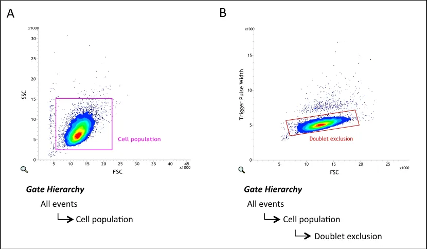
Figure 2.1: Cell sorter gating applied to all samples to exclude debris and doublets ... 53
Figure 3.1: Analysis of the stability of the IFN inhibitor Rux in vitro. A549 naïve cells were cultured in the presence of media supplemented with Rux (4μM) or equivalent volume of DMSO ... 73
Figure 3.2: BUNΔNSs plaque development in A549 naïve, A549/PIV5-V and Vero cells supplemented with Rux ... 75
Figure 3.3: Analysis of the effect of the IFN inhibitor, Rux, on BUNΔNSs growth in Vero cells using a multistep viral growth curve ... 77
Figure 3.4: Comparison of growth of the IFN-sensitive virus, BUNΔNSs, in A549: naïve cells supplemented with Rux compared to growth in Vero and A549/PIV5-V cells using a multistep viral growth curve. ... 79
Figure 3.5: Analysis of the effect of pre-treatment of A549: naïve cells with the IFN inhibitor, RUX, on BUNΔNSs growth, using a multistep viral growth curve ... 81
Figure 3.6: Effects of IFN inhibitor, Rux, on BUNΔNSs and BUN WT plaque formation in a range of mammalian cell-lines ... 84
Figure 3.7: BUN WT growth in MDBK bovine cells ... 88
Figure 3.8: Viral growth curves of BUN WT in the presence of IFN inhibitor Rux (4μM) or equivalent volume DMSO ... 90
Figure 3.9: Plaque development of seven different viruses from the Bunyaviridae family (ANAV, BWAV, CVV, KRIV, MDV and SBV) in six mammalian cell-lines (A549 naïve, BalB/C, MDBK, MDCK, NBL-6 and RK.13) ... 95
Figure 3.10: Plaque development of Cache Valley virus in A549 naïve cells ... 95
Figure 4.1: Method concept to isolate IFN-sensitive viruses using FACS ... 103
Figure 4.2: Determining the optimum time to treat cells with IFN following infection ... 105
Figure 4.3: A549/pr(ISRE).GFP/ISG56-/BVDV Npro cell-line analysis. ... 108
Figure 4.5: Addition of neutralising antibody to inactivate progeny viruses
released by infected cells ... 113
Figure 4.6: Optimised method concept for isolation of IFN-sensitive viruses from rPIV5mCh via FACS ... 114
Figure 5.1: FACS analysis and selection of potentially IFN-sensitive mutant viruses from rPIV5mCh ... 117
Figure 5.2: Examples of the three potential outcomes following sorting ... 119
Figure 5.3: FACS analysis of potentially IFN-sensitive mutant viruses ... 123
Figure 5.4: Nucleotide and amino acid mutations in V and P genes/proteins of rPIV5mCh-α and β. ... 126
Figure 5.5: Mapping the positions of amino acid mutations rPIV5mCh-α and rPIV5mCh-β to the wild-type V protein structure ... 128
Figure 5.6: PIV5 W3 method workflow for the isolation of potentially IFN-sensitive mutant viruses using FACS. ... 131
Figure 5.7: FACS analysis of PIV5 W3 virus. ... 133
Figure 5.8: Mutations in V and P proteins of PIV5 W3-γ ... 134
Figure 5.9: Mapping the position of amino acid mutation found in PIV5 W3- γ to wild-type V protein structure ... 134
Figure 6.1: FACS analysis of isolated PIV5 mutants at different MOI ... 140
Figure 6.2: Analysis of STAT1 expression following infection by isolated PIV5 mutant viruses ... 141
Figure 6.3: Analysis of mutant V proteins ability to block the IFN signalling pathway using an IFN signalling luciferase reporter assay ... 144
Figure 6.4: Analysis of mutant V protein ability to block IFN induction via an IFN induction luciferase reporter assay ... 147
Figure 6.5: Comparison of mutant virus plaque development in A549 naïve -/+ Rux and Vero cells. ... 151
Figure 6.6: Quantification of virus plaque size.. ... 152
Figure 6.7: Multistep viral growth curve analysis of PIV5 mutants ... 154
Figure 6.8: rPIV5mCh-α and PIV5 W3-γ serial passage. ... 158
Figure 6.9: Analysis of mutant fusogenicity ... 160
List of tables
Table 1.1: Paramyxoviridae subfamily and genus classification ... 21
Table 2.1: List of mammalian cell-lines used in this study ... 38
Table 2.2: List of mammalian cell-line derivatives used in this study ... 39
Table 2.3: List of viruses used in this study ... 40
Table 2.4: List of Primary and Secondary antibodies used within this study ... 41
Abbreviations
% [v/v] Percentage concentration (volume per volume) % [w/v] Percentage concentration (weight per volume) AKT1 RAC-alpha serine/threonine kinase
ANAV Anopheles A virus
ATF-2 Activating Transcription Factor 2 BD Becton-Dickinson Company
BRSV Bovine Respiratory Syncytial Virus BUN WT Bunyamwera wild-type virus
BUNΔNSs Bunyamwera with deleted NSs protein BVDV Bovine Viral Diarrhoea Virus
BWAV Bwamba virus
CARDIF CARD adaptor inducing interferon-β CBP CREB-binding protein
CD46 Cluster of Differentiation 46
cGAS Cyclic guanosine monophosphate-adenosine monophosphate (GMP-AMP) cGAMP synthetase
CPE Cytopathic effect
CPI+/- Canine parainfluenza virus cPPT central Polypurine Tract CV Crystal Violet
CVV Cache Valley virus
DAPI 4’,6-diamindino-2-phenylindole DDB1 Damage DNA binding protein 1 DMEM Dulbecco’s Modified Eagle Medium DMSO Dimethyl sulfoxide
dNTPs deoxynucleotide dsRNA double stranded RNA
EDTA Trypsin/ethylenediaminetetraacetic acid F Fusion protein
FACS Fluorescence Activated Cell Sorting FBS Foetal bovine serum
G Glycoprotein
GFP Green fluorescent protein
H Hemagglutinin
HCV Hepatitis C HeV Hendra virus
HN Hemagglutinin- Neuraminidase HSV-1 Herpes Simplex Virus-1
IFN Interferon
IFNAR1 Interferon-alpha/beta receptor alpha chain IFNAR2 Interferon-alpha/beta receptor beta chain IFNλR1 Interferon- gamma receptor 1
IKK-α IκB kinase-α IKK-b IκB kinase-b
IPA Isopropyl alcohol
IRF Interferon regulatory factor ISG Interferon stimulated gene ISGF3 IFN-stimulated gene factor 3 ISRE IFN-stimulated response element IκB Inhibitor of κB
JAK Janus Activated Kinases KIRV Kairi virus
L Large protein
L Segment Large Segment LB Luria-Bertani broth
LGP2 Laboratory of genetics and Physiology 2 LTRs Long terminal repeat regions
M Matrix protein
M Segment Medium Segment
M-MLV RT Moloney Murine Leukemia Virus Reverse Transcriptase Mda-5 Melanoma Differentiation-Associated protein 5
MDV Main Drain virus
MED8 Mediator complex subunit 8 MeV Measles virus
MOI Multiplicity of Infection MuV Mumps Virus
MyD88 myeloid differentiation factor 88 NEMO NF-κB essential modulator NF-κB Nuclear factor-κB
NIBSC The National Institute for Biological standards and controls NiV Nipah virus
NLR Nucleotide oligomerization domain (NOD)-like receptors NLRP3 NACHT, LRR and OYD domains-containing protein 3
NP Nucleoprotein
NP0 Soluble Nucleoprotein
NS1 Non-structural protein 1 NS2 Non-structural protein 2 NSm Non-structural protein NSs Non-structural protein
OAS 2'-5'-oligoadenylate synthetase ORF Open reading frame
P Phosphoprotein
PAC puromycin-N-Acetyl-trasnferase
PAMPs pathogen associated molecular patterns PBS Phosphate buffered Saline
PCR Polymerase Chain reaction PFU Plaque forming units
PIV1-4 human parainfluenza virus 1-4 PIV5 Parainfluenza virus 5
PKR Protein kinase R PLK1 Polo-like kinase 1
RIP1 Receptor-interacting protein 1 RLR RIG-I like receptor
RNAP II RNA polymerase II
rPIV5mCh recombinant parainfluenza virus 5 mCherry RRE Rev response element
RSV human Respiratory Syncytial Virus RT Room Temperature
Rux Ruxolitinib
RVFV Rift Valley Fever Virus S Segment Small Segment
SBV Schmallenberg Virus
SDS-PAGE Sodium Dodecyl Sulphate Polyacrylamide Gel Electrophoresis SeV Sendai virus
SFFV Spleen focus-forming virus SH Small hydrophobic protein
SLAM Signalling lymphocyte activation molecular (SLAM) ssRNA single stranded RNA
STAT Signal Transducers and Activators of Transcription SV5 Simian Virus 5
TAB2/3 TAK1-binding protein 2/3 TAK1 TGF-β activated kinase 1 TBE Tris-Boric acid-EDTA buffer TBK-1 TANK binding protein -1 TGS Tris-Glycine-SDS buffer TLR Toll-like receptor
TRAF6 Tumour necrosis factor receptor-associated factor 6 TRIF TIR domain-containing adaptor inducing interferon-β TRIM25 Tripartite motif containing 25
Tyk2 Tyrosine kinase 2
USP3 Ubiquitin specific Protease 3
V V protein
Introduction
1
Introduction
1.1 The Interferon (IFN) response
The Interferon (IFN) response is a vital defence against viral infection, without
which we would not be able to survive. As an immediate response to infection,
cells synthesize and release IFNs, a group of widely expressed cytokines, which
can then communicate in an autocrine or paracrine manner to induce an
antiviral response that limits the spread of infection. Despite the majority of
viruses having known mechanisms to circumvent the IFN response, it remains
critical to slow viral infection to allow the adaptive immune response to develop
(reviewed in Iwasaki 2012, Randall and Goodbourn 2008, Schneider et al 2014)
1.1.1 Classes and subtypes of IFN
There are three classes of IFN, type I, II and III, grouped according to their
similarity in amino acid sequence. The type I group consists of fourteen IFN-α
subtypes, a single IFN-β subtype and the less well characterized IFN- ω, ε, τ, δ, κ
subtypes, with IFN-α and -β playing the most well defined roles in the antiviral
response (Schneider et al. 2014). It is known that IFN-α/β act through the same
heterodimeric receptor, composed of interferon-alpha/beta receptor alpha chain
(IFNAR1) and IFNAR2, to trigger what is termed the IFN signalling pathway (Kim
et al 1997, Piehler et al 2000). This receptor is thought to be expressed
ubiquitously throughout all tissues however hematopoietic cells are thought to
be the main producers of IFN-α and fibroblasts of IFN-β (de Weerd et al 2007).
Introduction
stimulated genes (ISGs) and subsequently forms an antiviral state within the cell
and its neighbours.
The type III IFNs comprise IFN- λ1, λ2, λ3, which again are released in response
to viral infection in a similar manner to type I IFNs (Onoguchi et al. 2007).
However, unlike the type I IFNs, they exhibit high tissue specificity due to the
expression of their receptor, Interferon-lamda receptor 1 (IFNλR), on specific
cell types such as epithelial cells (Lazear et al. 2015). Latterly, a 4th member of
the type III IFNs has been discovered; namely λ4, however frameshift mutations
render the gene inactive in a large proportion of the human population.
Surprisingly, this inactivation has been shown to be associated with an increased
chance of clearance of Hepatitis C virus (HCV) indicating that its suppression is
somewhat beneficial despite it showing strong antiviral activity in vitro
(Hamming et al. 2013). Type II IFN contains only one member, IFN-γ, and unlike
the other types is secreted by natural killer cells and activated T cells as oppose
to direct response to viral infection (Schroder et al. 2004; Billiau & Matthys
2009)
This study focuses mainly on the pathways associated in response to IFN-α/β.
Predominately, IFN-β is used as an example as this pathway is better
understood. The basic pathway is generally divided into two parts the IFN-β
induction cascade and the IFN-β signalling cascade, both of which are outlined in
Introduction
1.2 IFN-β induction
The IFN-β induction cascade is mediated by the recognition of molecular
pathogen-associated molecular patterns (PAMPS), these include single stranded
RNA (ssRNA), double stranded RNA (dsRNA), genomic DNA, or viral proteins
(reviewed in Iwasaki 2012). These PAMPS are recognised by pattern recognition
receptors (PRRs) namely i) the Toll-like receptors (TLRs) such as TLR3, TLR7
and TLR9, ii) the RIG-I-like receptors (RLRs) Retinoic acid-inducible gene 1
(RIG-I) and Melanoma Differentiation-Associated protein 5 (Mda-5) iii) the nucleotide
oligomerization domain (NOD)-like receptors (NLRs) such as NACHT, LRR and
OYD domains-containing protein 3 (NLRP3) (Poeck et al. 2010), and the recently
discovered range of cytosolic nucleic acid sensors such as cGAMP synthase
(cGAS) (Sun et al. 2013; Cai et al. 2014). Each group recognises distinct PAMPs
such as dsRNA in endosomes, recognised by TLR3, or ssRNA, recognised by TLR7
and TLR9, thereby allowing for recognition of many types of viral infection
(reviewed in Broz and Monack 2013). For the purpose of this study we will
focus on the pathways mediated by the cytoplasmic and endosomal recognition
of RNA viruses, which is predominantly mediated by RLRs and the TLRs TLR3,
TLR7 and TLR9. Aside from the recognition stage, the TLR3 and RLR dependent
induction pathways are very similar, as demonstrated in Figure 1.1A. By
contrast, the TLR7 and TLR9 dependent induction pathway exhibits greater
differences and is represented in Figure 1.1B.
1.2.1.1 TLR3 and RLR (RIG-I and mda-5)-dependent induction cascade.
Both TLR3 and the RLRs recognise substrates at different stages during viral
Introduction
TLR3 can recognise dsRNA in endosomes or extracellular dsRNA at the cell
surface (Randall & Goodbourn 2008). By contrast, the RLRs can only recognise
infection once inside the cellular cytoplasm (Goubau et al 2013). Specifically,
both RIG-I and Mda-5 are activated by dsRNA, however, RIG-I is also activated by
short blunt ended dsRNA with a 5’triphosphate and is therefore indispensable
for the recognition of many viruses such as Influenza A (Loo et al. 2008; Hornung
et al. 2006). Interestingly, RIG-I but not Mda-5 activation has been shown to be
dependent on ubiquitination and is regulated by the E3 ligases Tripartite motif
containing 25 (TRIM25), TRIM4 and Riplet, and the Deubiquitylation enzymes
Ubiquitin specific protease 3 (USP3) and ubiquitin C-terminal hydrolase (Heaton
et al. 2016; Cui et al. 2014; Friedman et al. 2008; Yan et al. 2014; Gack et al.
2007). Irrespectively, once activated each receptor must then interact with an
adaptor to confer activation of the induction pathway. In particular, TLR3 and
the RLRs use the adaptors TIR domain-containing adapter inducing interferon-β
(TRIF) and CARD adapter inducing interferon-β (CARDIF), respectively, both of
which act as a scaffold for the recruitment of a number of other factors (Kawai et
al. 2005; Meylan et al. 2005). Notably it appears that engagement of CARDIF by
PRRs such as RIG-I results in a conformational change that recruits inactivated
CARDIF and results in a large-scale amplification of the signalling cascades (Hou
et al. 2011). This results in a highly sensitive mechanism to detect small amounts
of viral RNA with evidence suggesting that less than 20 molecules of
5’triphosphate is sufficient to activate the RIG-I-CARDIF pathway (Zeng et al.
2010). From this point on, the signalling pathways are almost identical as the
adaptors can activate both of the so-called ‘arms’ of the induction pathway,
Introduction
(IRF) ‘arm’ (Figure 1.1A). The NF-κB ‘arm’ initiates with the recruitment of
tumour necrosis factor associated factor 6 (TRAF6) and
receptor-interacting protein 1 (RIP1) to the adaptor (either TRIF or CARDIF depending on
the initial receptor)(Sato et al. 2003). At this stage the TRAF6-RIP1-adaptor
complex can then interact with a complex called TAK1 that consists of three
subunits namely TAK1-binding protein 2/3 (TAB2/3), TAB1 and TGF-β activated
kinase 1 (TAK1)(Jiang et al 2004, Meylan et al 2004, Sato et al 2003). This
interaction then promotes the interaction with a second complex namely the IKK
complex, which also consists of three subunits: NF-κB essential modulator
(NEMO), IκB kinase-α (IKK-α) and IKK-β. Now that these two complexes (TAK1
and IKK) are in close proximity the TAK1 subunit of the TAK1 complex can then
phosphorylate the IKK-β subunit of the IKK complex leading to its activation.
Here it must be noted that NF-κB, one of the molecules that is required for the
activation of the IFN-β promoter and thus IFN-β up-regulation, is held in an
inactive state within the cytoplasm by the molecule inhibitor of κB (IκB)(Zandi et
al. 1997; Alexopoulou et al. 2001). In view of this, the now active IKK-β subunit
can phosphorylate the IκB subunit, subsequently leading to its ubiquitination
and degradation (Jiang & Chen 2012). This then releases NF-κB from its
inhibition and allows for its uptake into the nucleus where it assembles on the
IFN-β promoter (Jiang & Chen 2012; Randall & Goodbourn 2008). This factor
alone however does not result in IFN-β up-regulation, because the activation of
both the NF-κB and IRF ‘arms’ of the pathway are required. In a similar manner
to the NF-κB ‘arm’, a number of factors are recruited to the adaptor protein to
Introduction
the initial receptor). TRAF3 can then also bind to a factor known as TANK, which
in turn binds to TANK-binding protein 1 (TBK-1) and/or IKKε (Figure1.1A)(Paz
et al. 2011). Primarily TBK-1 and/or IKKε can then phosphorylate IRF3 directly,
allowing it to migrate to the nucleus, however, in certain circumstances it is
known that IRF7 is also activated in a similar manner (Trinchieri 2010). In this
case, both IRFs can migrate to the nucleus to assemble on the IFN-β promoter.
Ultimately, with both the κB and IRF ‘arms’ of the pathway switched on,
NF-κB and IRF3 (and IRF7) are now assembled on the IFN-β promoter. This in turn
allows other factors to assemble on the promoter namely activating
transcription factor 2 (ATF-2)/c-jun, CREB-binding protein (CBP)/p300 and RNA
polymerase II which function collectively to up-regulate the production and
secretion of IFN-β (Figure1.1A) (Randall & Goodbourn 2008; Bhoj & Chen 2009;
Takeuchi et al. 2010).
1.2.1.2 TLR7 and TLR9-dependent induction cascade
In addition to the TLR3 and RLR dependent pathway, TLR7 and TLR9 receptors
can also trigger the induction of IFN-β. Notably, TLR7 and TLR9 recognise
different PAMPs compared to the previous method such as ssRNA and CpG
(unmethylated) DNA that has been engulfed by endosomes, respectively (Heil et
al. 2004; Tabeta et al. 2004). This property thereby increases the number of
ways in which viral infection can be recognised, allowing for greater protection
of the cell. In a similar manner to the TLR3 and RLR-dependent pathway, the
TLR7 and TLR9-dependent pathway uses an adaptor to recruit factors that
Introduction
More specifically, once TLR7 and TLR9 are activated through recognition of viral
PAMPs both can recruit the adaptor myeloid differentiation factor 88 (MyD88).
This adaptor can then recruit two factors namely interleukin 1
receptor-associated kinase 4 (IRAK4) and IRAK1 (Kawagoe et al. 2008). Subsequently,
IRAK4 and IRAK1 then lead to activation of the NF-κB and IRF ‘arms’ of the
pathway, respectively. To initiate the activation of NF-κB, IRAK4 interacts with
the factor TRAF6 (forming the MyD88-IRAK1-IRAK4-TRAF6 complex), which
then interacts with a number of factors namely RIP1 and the TAK1 complex (Kim
et al 2007). From this stage onwards, the activation cascade is exactly the same
as in the TLR3/RLR-dependent pathway (Figure 1.1A and B). In brief, the TAK1
complex interacts with the IKK complex phosphorylating the IKK-β subunit. This
active subunit can then phosphorylate the IκB subunit that inhibits NF-κB
leading to its ubiquitylation and degradation, thus releasing NF-κB to relocate to
the nucleus where it assembles on the IFN-β promoter (Figure 1.1B)(Zandi et al.
1997; Alexopoulou et al. 2001). Again, this alone does not result in IFN-β
up-regulation, as it also requires the activation of the IRF ‘arm’ of the pathway.
Unlike the previous method that contained an IRF ‘arm’ that activates IRF3 (and
IRF7) this cascade offers another route to IFN-β induction using only IRF7. The
MyD88-IRAK1-IRAK4-TRAF6 complex, described previously, has been shown to
bind directly to IRF-7 (Kawai et al. 2004). IRF-7 is then polyubiquitinated by
TRAF6 in the presence of polyubiquitinated RIP1 (Konno et al. 2009). IRF-7 is
subsequently phosphorylated by IRAK-1 allowing the translocation of the whole
complex into the nucleus, where it binds to the IFN-β promoter (Figure
Introduction
NF-κB on the IFN-β promoter, this along with a number of other co-factors such
as ATF-2/c-jun, CBP/p300 and RNA polymerase II leads to increased
transcription and secretion of IFN-β (Figure 1.1B)(Randall & Goodbourn 2008).
A
Introduction
Figure 1.1: Overview of the IFN-β induction cascade. A) TLR3 and the RLR
(RIG-I and Mda5)-dependent induction cascade. Both TLR3 and the RLRs
recognise substrates at different stages during viral infection. In particular, TLR3
can recognise dsRNA in endosomes or extracellular dsRNA at the cell surface. By
contrast, the RLRs can only recognise infection once inside the cell. Specifically,
both RIG-I and Mda-5 are activated by dsRNA however RIG-I is also activated by
5’triphosphoRNA and is therefore indispensable for the recognition of many
viruses such as Influenza A (Loo et al., 2008, Hornung et al., 2006). After
recognition of the infection both TLR3 and the RLRs mediate the same induction
cascade through the adaptors TRIF and CARDIF respectively, hence they are
considered together in A. B) TLR7 and TLR9-dependent induction cascade.
TLR7 and TLR9 recognise ssRNA and CpG (unmethylated) DNA that has been
engulfed by endosomes, respectively. After recognition of the viral PAMPs both
TLRs mediate the same induction cascade through interaction with the adaptor
Introduction
1.3 IFN-β signalling
As outlined above there are a number of methods by which viral infection is
recognised however ultimately they all lead to the up-regulation of production of
IFN-β. Once produced, this cytokine is released by the cell into the surrounding
area, this importantly signals to neighbouring cells that it has been infected and
thus triggers them to up-regulate the production of many protective genes that
are collectively known as interferon stimulated genes (ISGs). This so-called
‘antiviral state’ consequently reduces the spread of infection to surrounding cells
allowing a greater chance of cell survival (reviewed in Ivashkiv and Donlin
2014). The process by which this ‘antiviral state’ is produced by IFN-β signalling
is outlined in the following section.
1.3.1 IFN-β signalling cascade
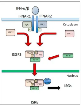
The IFN-β signalling cascade initiates with the activation of the type I IFN
receptor by IFN-β. This receptor is found at the cell surface and consists of two
chains namely IFNAR1 and IFNAR2 (Kim et al 1997). The interaction with IFN-β
results in autophosphorylation of the receptor itself and subsequently activation
of two janus activated kinases (JAKs) namely tyrosine kinase 2 (Tyk2) and JAK1
which are found attached to the cytoplasmic domain of the receptor of IFNAR1
and IFNAR2, respectively (Figure 2)(Velazquez et al. 1994; Müller et al. 1993;
Gauzzi et al. 1996). Once activated, these JAKs can then phosphorylate two signal
transducers and activators of transcription (STAT), namely, STAT1 and STAT2
on tyrosine 701 and tyrosine 690, respectively (Stark et al. 1998).
Introduction
nucleus where STAT1 is further phosphorylated by cyclin-dependent kinase 8 on
Serine 727 (Wen et al. 1995; Uddin et al. 2002; Bancerek et al. 2013). This
heterodimer subsequently forms a heterotrimer complex with interferon
regulatory factor 9 (IRF-9) that is collectively termed IFN-stimulated gene factor
3 (ISGF3)(Fu et al. 1990; Ivashkiv & Donlin 2014). ISGF3 then translocates to the
nucleus and binds to a specific sequence within target promoters, the
IFN-stimulated response element (ISRE). This subsequently stimulates the
transcription of hundreds of ISGs, thereby producing the ‘antiviral state’ (Figure
1.2)(Schoggins & Rice 2011; Stark & Darnell 2012). As well as regulation by
post-translational modification such as phosphorylation, STAT- and ISGF3 mediated
gene transcription is also regulated by cooperation with other transcription
factors such as IRF1, IRF7, IRF8 and IRF9 (van Boxel-Dezaire et al. 2006),
chromatin remodelling which is mediated by STAT1 and STAT2 and
IRF-mediated recruitment of nucleosome-remodelling enzymes and histone
acetyltransferases (HATs)(Tartey & Takeuchi 2015), and through the interaction
of STATs with co-activators and co-repressors (Ivashkiv & Donlin 2014;
Au-Yeung et al. 2013). Examples of ISGs upregulated by this pathway include protein
kinase R (PKR) and 2'-5'-oligoadenylate synthetase (OAS), PKR prevents
initiation of transcription in the presence of dsRNA and OAS degrades cellular
and viral RNAs in the presence of dsRNA (Carlos et al. 2007). Another important
example includes ISG56, which has been shown recently to specifically inhibit
translation of mRNA from Rubulaviruses such as PIV5 but not other members of
the Paramyxoviridae family due to the lack of methylation at a particular position
Introduction
to gain a ‘head start’ in producing the proteins that allow protection against viral
infection. Understandably, this mechanism of defence has driven viruses to gain
numerous ways in which to circumvent this response to tip the balance in favour
of viral infection. Crucially, by studying these interactions we can gain an insight
into how to weaken the virus to tip the favour back towards the host. This could
ultimately aid in the production of attenuated viruses, which are the essential
[image:28.595.91.355.271.614.2]component of numerous vaccines.
Figure 1.2: Overview of the IFN signalling cascade. IFN-β can trigger the
activation of the JAK-STAT signalling pathway, inducing an ‘antiviral state’ in
surrounding cells through the activation of many hundreds of Interferon
stimulated genes (ISGs)(Fleming 2016).
Cytoplasm
Introduction
1.4 Viral IFN antagonism
Viruses have developed an astonishing variety of IFN antagonists to counteract
the induction, signalling or antiviral actions of the IFN system. Over 170 viral IFN
antagonist has been described from 93 distinct viruses with most demonstrating
a multifunctional role targeting multiple steps within the IFN response (Versteeg
et al. 2010; Haller et al. 2006). Although each is unique, IFN antagonists
counteract the cellular IFN response using common strategies that include i)
broad-spectrum inhibition of cellular gene expression and ii) sequestration,
proteolytic cleavage and proteasome-mediated degradation of key components
of the IFN system (Versteeg et al. 2010).
1.4.1 Pleiotropic nature of viral IFN antagonists
The pleiotropic nature of IFN antagonists is dictated by restrictions on the viral
genome. More specifically, RNA viruses have a relatively limited genome capacity
in comparison to large dsDNA viruses, which drives a high-degree of
multifunctionality of the IFN antagonists. This evolutionary pressure has
resulted in the conservation of IFN antagonists within related RNA viruses
whereas there is limited conservation in large dsDNA viruses (Versteeg et al.
2010). A prime example of this is the Non-structural, NS1, protein, which is
conserved among Influenza A and B, and exhibits a plethora of different
strategies to antagonise the IFN system (Hale et al. 2008). A second example of
this is the Non-structural protein, NSs, protein which is a multifunctional IFN
antagonist conserved among the Phleboviruses and Orthobunyaviruses (Elliott &
Introduction
1.4.2 Bunyamwera virus (BUN WT) and the IFN antagonist NSs
As BUN WT of the Orthobunyaviruses genus and the recombinant virus
BUNΔNSs are used within a major part of this study we will discuss these viruses
further, with particular focus on the functions of the IFN antagonist NSs.
Bunyamwera virus is a prototypical virus within the Bunyaviridae family which
contains pathogens of serious concern such as Rift Valley fever virus (RVFV) and
Schmallenberg (SBV) (Fields et al. 2013; Garigliany et al. 2012). It possesses a
tri-segmented negative sense RNA genome consisting of the Large (L), Medium
(M) and Small (S) segments. The L segment encodes the viral RNA polymerase,
the M segment encodes a polyprotein precursor, which is co-translationally
cleaved to obtain the glycoproteins Gn and Gc and a non-structural protein NSm,
and the S segment encodes the N protein and the NSs IFN antagonist in
overlapping reading frames (Elliott and Blakqori 2011). For BUN WT, NSs is a
non-essential small hydrophobic protein of 101 amino acids that is expressed
from an internal, +1-shifted reading frame within the N gene of the S segment.
The main function of this protein is attributed to globally inhibiting host mRNA
transcription by blocking RNA polymerase II (RNAP II) through an interaction
with Mediator complex subunit 8 (MED8), a complex required for mRNA
production (Thomas et al. 2004; Leonard et al. 2006). Generation of a
recombinant virus, BUNΔNSs, which does not express NSs demonstrated
reduced plaque size and growth in IFN competent cells to approximately 10-fold
lower titers compared to the wild-type virus (BUN WT). Furthermore, when
inoculated intracerebrally into BALB/c mice, BUNΔNSs killed the mice in a
slower time course than BUN WT (Bridgen et al. 2001). Indicating that although
Introduction
deficient virus is more sensitive to IFN and therefore attenuated compared to the
wild-type virus.
1.5 IFN-sensitive viruses as Vaccines
With most, if not all, viruses encoding an IFN antagonist this opens up the
opportunity for prophylactic intervention through the development of
‘IFN-sensitive’ viruses as live attenuated virus vaccines. A concept which has now
been successfully verified for many viruses including Influenza A and B, RVFV
and more recently Respiratory syncytial virus (RSV) (Bird et al. 2008; Jang &
Seong 2012; Meng et al. 2014).
Live attenuated virus vaccines contain a mutant strain of a wild-type virus,
whereby its ability to inhibit the hosts immune response has been weakened.
This impairment can be associated with loss of a viruses IFN antagonist function
or mutations within other proteins that slow viral replication such that it can be
controlled by the IFN response. This attenuation does not prevent host infection
but rather prevents the cause of severe disease. As the virus can still replicate
within the host, it can produce numerous antigens causing a wide range of
immune responses that later protect the host from subsequent infection from the
wild-type virus.
1.5.1 Live Attenuated vaccine development
One of the first live attenuated vaccines to be developed was that against
smallpox (variola virus)(reviewed in Minor 2015). In the 18th century it was well
Introduction
subsequently resistant to smallpox. In 1976, Edward Jenner experimented by
inoculating a young boy, James Phipps, with material from a cowpox-infected
milkmaid. Seven weeks later he then injected the boy with material from a
smallpox lesion and Phipps survived with no indication of infection (Jenner
1798). These initial investigations subsequently led to compulsory vaccination in
the UK however debate over the quality of the production of the vaccine led to
compulsion being dropped. Subsequently, in 1959 the World Health
Organisation developed criteria for the production of a safer vaccine tackling
major problems such as the possibility of bacterial contamination of the vaccine
and also the transfer of syphilis (WHO 1959). Unbeknown to many the original
constituent of the smallpox vaccine had mutated into what is now known as
vaccinia virus. Whether this virus derived from cowpox or variola virus is
unknown however its effectiveness is unquestionable (Elwood 1989; Petersen et
al. 2016). In 1980 smallpox was declared eradicated making it one of the most
successful human vaccines to date. Given that the infectious agent we now know
as a virus was not described until 1898, it is not surprising that there was little
influence of virology on the development of smallpox vaccines first investigated
in 1798 (Minor 2015). This was not the case for the polio vaccine. This vaccine
was obtained through a better understanding of viral pathogenesis and the
virology of the poliomyelitis. Albert Sabin developed the Sabin type 1 vaccine
from the serial passage of the wildtype virus through monkey testis cells (Sabin
& Ward 1941). The type 2 and 3 strains were then isolated from clinical cases
(Sabin & Boulger 1973). Many years later, following the introduction of the
vaccines, sequence analysis of the vaccine strains then determined mutations
Introduction
the success of this vaccine in decreasing polio to an unprecedented level it is not
surprising that similar techniques including serial passage are still used today to
try to obtain attenuated viruses for use as live attenuated vaccines.
1.5.2 Traditional methods to obtain attenuated viruses
Several approaches are used today to produce live attenuated viruses. One such
technique is to passage the virus within a foreign host. Mutants generated with
increased virulence in the foreign host typically lose virulence in humans and
can therefore be analysed as a potential vaccine strain. This method has been
shown to be a useful method with RNA viruses as they have a particularly high
mutation rate. Alternatively, the virus can be grown in a different temperature to
the human host, this can cause mutations to occur which adapt the virus to this
new temperature but slow the viral growth in human cells enough for the
adaptive immune response to fight infection (Baxter 2007; Minor 2015). A final
approach is chemical mutagenesis which has been used to create potential
vaccines for dengue and tuberculosis (Blaney et al. 2001; Collins 2000). These
forward genetics approaches, firstly finding a mutated strain and then
identifying what is making that strain attenuated have been a successful method
to identify numerous vaccines against viruses such measles (MeV), mumps
(MuV) and seasonal and pandemic strains of influenza (Fiore et al. 2009;
Suguitan et al. 2006; Bankamp et al. 2011; Rubin et al. 2008). However, this
process can be somewhat lengthy, as the virus needs to be passaged multiple
times and then followed by pairing of genomic nucleotide changes with
Introduction
1.5.3 Rational design of live attenuated vaccines
Due to the difficulties associated with traditional empirical methods, the
development of vaccines is currently moving towards rational design. Using this
method known IFN antagonists can be directly mutated to attenuate the virus.
This is typically completed by mutating the gene code to prevent the expression
of the IFN antagonist or by editing specific sites to allow the expression of a
non-functional protein (Rueckert et al. 2012). A prime example of this is the BovelaÒ
attenuated virus vaccine produced by mutation of the IFN antagonists Npro
Protease and ERNS RNase of Bovine Viral Diarrhoeal virus (BVDV). This vaccine is
now used successfully to prevent persistent infection of bovine with BVDV
(Meyers et al. 2007). Recently, codon-pair de-optimisation has presented a novel
approach to the attenuation of viral IFN antagonists. This method was first used
to tackle the issue of genetic instability of live-attenuated poliovirus vaccines by
incorporating the rarest codons in the human genome to lower translation of the
capsid protein resulting in virus attenuation (Mueller et al. 2006). More recently
this method has been used to attenuate RSV by codon de-optimization of the
nonessential IFN antagonists NS1 and NS2 to create a potential live attenuated
vaccine candidate (Meng et al. 2014). Importantly, this approach offers increased
safety by decreasing the likelihood that the attenuated strain can revert to regain
IFN antagonist function as well as improved immunogenicity. Despite the
successes of attenuating a virus in this way, this process is by no means simple.
Firstly, it relies on the notion that the IFN antagonist of the virus is already
known which is not always the case. Secondly, it is particularly complicated for
Introduction
all rationally designed attenuations result in a viable live attenuated vaccine
strain.
1.5.4 Vaccine production
Whether a virus has been developed by traditional methods or by rational design
it is important to consider that the more attenuated the virus the more difficult it
will be to produce for clinical use (Jang & Seong 2012). Currently, the default
option for growth of IFN-sensitive viruses is limited to a select number of
cell-lines such as Vero cells, that do not have an intact IFN system, however, as
viruses exhibit host cell specificity not all viruses can be grown in such cells
(Desmyter et al. 1968; Mosca & Pitha 1986). Previously we have developed
cell-lines expressing IFN antagonists that enable blockage of the IFN response and
can subsequently relieve host cell constraints on the virus, allowing virus growth
(Young et al. 2003). However, development of these cell-lines is time consuming
and creates regulatory problems during vaccine development, as each cell line
has to be approved for use during production.
1.5.5 Experimental Objectives
To tackle this issue, it was hypothesised that blocking the IFN induction and
signalling pathways using a small molecule inhibitor would offer a simple and
flexible solution to increase viral growth, as an inhibitor could easily supplement
the tissue culture medium of cell-lines of choice. To test this, eight inhibitors
known to target different components of the IFN response (TBK-1, IKK-b and
JAK1/2) were tested for their ability to block their corresponding pathways. Of
Introduction
response (Stewart et al. 2014). Consequently, the first objective of my study was
to further characterize the use of this IFN inhibitor, Rux, with respect to
increasing the growth of IFN-sensitive viruses.
The second objective of my study then aimed to speed the process of traditional
methods to identify attenuated viruses and allow selection of viable vaccine
strains by developing a method to rapidly isolate potentially IFN-sensitive
viruses from Paramyxoviruses, using fluorescent activated cell sorting (FACS).
Following isolation, we could then employ the IFN inhibitor Rux to aid us in
characterisation of potential IFN-sensitive viruses. As Paramyxoviruses are the
main focus of this study, I will now outline the basic properties of the
Paramyxoviridae family with particular focus on the prototypical virus
Parainfluenza virus type 5 (PIV5) used throughout our experimental research.
1.6 Paramyxoviridae family
The family Paramyxoviridae are responsible for a range of acute respiratory
diseases in both humans and animals. As a result, they are linked to a substantial
mortality rate and cause a significant economic burden. The family is divided
into the subfamilies Paramyxovirinae and Pneumovirinae (Table 1.1). The
subfamily, Paramyxovirinae, is the largest group divided into 7 genera:
Aquaparamyxovirus, Avulavirus, Ferlavirus, Henipavirus, Morbillivirus,
Respirovirus and Rubulavirus. It contains many clinically important viruses such
as MeV and MuV virus, which cause highly contagious viral diseases in human
infants, in addition to, the newly emerging viruses Hendra (HeV) and Nipah virus
Introduction
latter smaller subfamily, the Pneumovirinae, is divided into 2 genera the
Metapneumovirus and the Pneumovirus and it contains viruses such as human
RSV, which is the major cause of lower respiratory tract infection in infants
[image:37.595.84.519.255.686.2](reviewed in Audsley 2013, Fields et al 2013).
Table 1.1: Paramyxoviridae subfamily and genus classification. (adapted
from Audsley 2013, King et al 2012)
Subfamily Genus Virus
Paramyxovirinae
Aquaparamyxovirus Atlantic salmon paramyxovirus
Avulavirus Avian paramyxoviruses 2-9 Newcastle disease virus Ferlavirus Fer-de-Lance paramyxovirus
Henipavirus Hendra virus (HeV) Nipah virus (NiV)
Morbillivirus Canine distemper virus Measles virus (MeV)
Respirovirus
Sendai virus (SeV)
Human parainfluenza virus 1 and 3 (PIV1/3)
Rubulavirus
Mumps virus (MuV)
Parainfluenza virus 5 (PIV5)
Human parainfluenza virus 2 and 4 (PIV2/4)
Pneumovirinae
Metapneumovirus Human Metapneumovirus Avian Metapneumovirus
Introduction
1.7 Paramyxovirus virion structure
Despite classification of paramyxoviruses into several genera, the basic
paramyxovirus virion structure is consistent across all subtypes (Figure 1.3)
(reviewed in Chang and Dutch 2012, Fields et al 2013, El Najjar et al 2014). It
consists of pleomorphic (spherical or filamentous), enveloped particles ranging
typically from 150 to 300nm in diameter (Kalica et al. 1973; Goldsmith et al.
2003; Loo et al. 2013). These particles encapsidate a non-segmented, negative
sense, single stranded RNA that encodes for the necessary proteins required for
replication of the virus. Genomic RNA is wrapped with nucleoprotein (NP), the
phosphoprotein (P) and the large (L) protein forming what is termed a
nucleocapsid structure, which is essential for RNA replication as well as
transcription (Horikami et al. 1992; Noton & Fearns 2015). The L protein is the
RNA dependent RNA polymerase allowing transcription and replication of the
genomic RNA, aided by the P protein. NP is important for protecting the viral
genome from cellular responses by encapsidating the RNA during RNA
replication. Together this nucleocapsid interacts with the matrix (M) protein, a
protein important in virion assembly, which lines the virion envelope. This
envelope contains two surface glycoproteins, a trimeric fusion (F) protein and a
tetrameric attachment protein- HN, H or G depending on the virus. The
Aquaparamyxoviruses, Avulaviruses, Ferlaviruses, Respiroviruses and
Rubulaviruses share a common attachment protein- Hemagglutinin–
neuraminidase (HN) which has both Hemagglutinin (sialic acid binding) and
neuraminidase (sialic acid cleaving) activity. These viruses can therefore use
Introduction
Morbilliviruses contain only Hemagglutinin (H) protein so can bind sialic acid but
lack the ability to cleave it. Subsequently it has been shown that these viruses
use cellular proteins such as signalling lymphocyte activation molecule (SLAM;
also known as CD150) to gain entry to cells (Ono et al. 2001). The remaining
families the Henipaviruses and both the Metapneumoviruses and the
Pneumoviruses of the Pneumovirinae subfamily contain a G protein which
facilitates attachment to cellular proteins such as Ephrin B2/B3 in the case of
HeV and NiV (Negrete et al. 2006; Bonaparte et al. 2005), or nucleolin in the case
of RSV (Tayyari et al. 2011).
Figure 1.3: Paramyxovirus virion structure. All paramyxoviruses contain a
nucleocapsid that comprises the negative sense RNA genome wrapped in
nucleocapsid protein (NP), phosphoprotein (P) and the large (L) polymerase
protein. This nucleocapsid is then enclosed in a lipid envelope that is lined with
matrix proteins, which play an important role during virion assembly. The lipid
envelope of the virion contains two surface glycoproteins: the attachment
proteins (HN, H or G) and the fusion protein (F). These function to allow binding
Introduction
1.8 Paramyxovirus replication cycle
As paramyxoviruses, like all viruses, are obligate parasites they must infect a
host cell to replicate and produce new progeny virions (Figure 1.4). This cycle is
initiated by binding of the attachment protein (HN, H or G) to the receptor on the
host cell. The F protein then permits fusion of the envelope with the plasma
membrane and allows release of the nucleocapsid into the cytoplasm (Bose et al.
2013; Chang & Dutch 2012). Once inside, transcription, protein synthesis and
replication of the viral genome can occur within the cytoplasm of the host cell.
Upon entry, the RNA polymerase, L protein, carried as part of the nucleocapsid
can initiate transcription of the viral genome to form individual mRNAs in a
transcription gradient that are subsequently transcribed to proteins (Abraham &
Banerjee 1976). The most renowned model for switching to RNA replication
then proposes that when enough unassembled nucleocapsid protein is present,
the polymerase can then switch to transcribe full-length positive-sense
anti-genomic RNA templates (Kolakofsky et al. 2004; Noton & Fearns 2015). This can
then be used as a template to synthesise full length genomic RNAs. Notably most
Paramyxoviruses obey the ‘Rule of 6’, i.e. their genome length must be exactly
divisible by 6 to replicate efficiently due to restrictions on the wrapping of RNA
with NP(Kolakofsky et al. 2004; Alayyoubi et al. 2015). Together with L, NP and
P, the progeny RNA’s form nucleocapsids, which associate with the M protein at
the plasma membrane along with the viral glycoproteins (HN, H or G)(Ghildyal et
al. 2006). Mature virions can then bud from the plasma membrane and exit to
Introduction
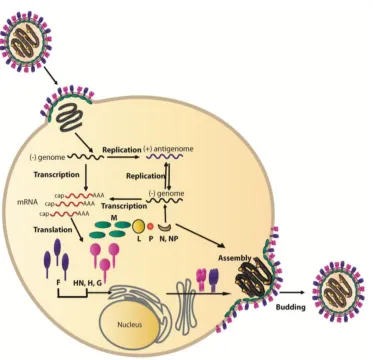
Figure 1.4: Paramyxovirus replication cycle. Entry to the cell is initiated by
binding of the attachment protein to the cell (HN, H or G). The fusion protein can
then instigate fusion of the viral envelope to the plasma membrane and allow
release of the nucleocapsid to the cell cytoplasm. Inside the host the viral
polymerase initiates transcription of viral mRNAs, which are then translated to
proteins. The polymerase can also replicate the negative sense genome to a
positive sense antigenome, which can then be used as a template to synthesise
new progeny genomes. Together with L, P and NP, these newly synthesised RNA
genomes form nucleocapsids, which by exploiting M protein can assemble with
newly synthesised viral glycoproteins found at the cellular membrane into new
[image:41.595.97.470.67.427.2]Introduction
1.9 Additional proteins encoded by paramyxoviruses
Interestingly, in addition to the aforementioned proteins, a number of the
Rubulaviruses and Avulaviruses and the entire Pneumovirinae subfamily produce
a small hydrophobic (SH) protein that is thought to play a role in preventing
apoptosis or protecting against the immune response (Li et al. 2013; Wilson et al.
2006). Furthermore, most paramyxoviruses can express a number of proteins
from the P gene due to a number of overlapping open reading frames (ORFs).
The Morbillivirus, Respiroviruses, Henipaviruses and Avulaviruses transcribe P
mRNA however a process termed ‘RNA editing’ can allow the insertion of a G
nucleotide into the transcript (Figure 1.5). This insertion creates a frameshift
mutation in the downstream sequence, which thereby produces an mRNA that
translates into a protein termed the V protein. This protein therefore shares a
common N-terminus with the P protein but has a unique C terminus (Goodbourn
& Randall 2009). The V protein is a cysteine rich protein termed as an interferon
(IFN) antagonist as it is able to block the IFN immune response and allows the
virus to propagate more easily within the cell (Andrejeva et al 2004, Didcock et
al 1999, reviewed in Parks and Alexander-Miller 2013, Poole et al 2002). A
second G residue insertion creates an mRNA that encodes proteins with different
C-termini termed W, D or I. Intriguingly, it is the V protein of the Rubulaviruses
such as PIV5 that is genomically templated and two G residues are inserted into
the mRNA to produce the P protein. Furthermore, the addition of 1 or 4 G
residues creates the I protein in certain viruses (reviewed in Parks and