organic papers
Acta Cryst.(2006). E62, o1469–o1470 doi:10.1107/S1600536806009573 Wang and Tang C
11H13N3O3H2O
o1469
Acta Crystallographica Section E Structure Reports Online
ISSN 1600-5368
(
E
)-4-(Isonicotinoylhydrazono)pentanoic
acid monohydrate
Yong-Tao Wang* and Gui-Mei Tang
Department of Chemical Engineering, Shandong Institute of Light Industry, Jinan, Shandong 250100, People’s Republic of China
Correspondence e-mail: [email protected]
Key indicators
Single-crystal X-ray study
T= 293 K
Mean(C–C) = 0.004 A˚
Rfactor = 0.080
wRfactor = 0.185
Data-to-parameter ratio = 13.9
For details of how these key indicators were automatically derived from the article, see http://journals.iucr.org/e.
Received 28 February 2006 Accepted 14 March 2006
#2006 International Union of Crystallography All rights reserved
The title compound, C11H13N3O3H2O, was synthesized by the
reaction of acetopropanoic acid and isonicotinoylhydrazide in ethanol. A three-dimensional network of intermolecular O—
H O, O—H N and N—H O hydrogen bonds stabilizes
the crystal structure.
Comment
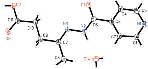
Hydrazine and its derivatives exhibit biological and pharma-cological activities (Vicini et al., 2002; Maccari et al., 2005). These compounds are also used as synthetic intermediates (Rollaset al., 2002). The title compound, (I), was synthesized in our laboratory by the reaction of acetopropanoic acid and isonicotinoylhydrazide in an ethanol medium. We present here its crystal structure (Fig. 1).
The bond lengths (Table 1) and angles show normal values. The uncoordinated water molecule plays an important role in the formation of a three-dimensional network of
inter-molecular O—H O and N—H O hydrogen bonds
(Table 2), which stabilize the crystal structure (Fig. 2). O— H N interactions are also present.
Experimental
A mixture of acetopropanoic acid (1.16 mg, 10 mmol) and isonicoti-noylhydrazide (1.37 mg, 10 mmol) was refluxed in ethanol for 3 h. After cooling, the mixture was filtered and dried. The title compound was recrystallized from a mixed solvent of methanol and water (1:5) in 85% yield (200 mg). Block-shaped colourless single crystals suitable for X-ray diffraction were obtained. Analysis found (%): C 52.03, H 5.96, N 16.66; C11H15N3O4requires (%): C 52.17, H 5.97, N
16.59.
Crystal data
C11H13N3O3H2O
Mr= 253.26 Monoclinic,P21=c
a= 9.7834 (14) A˚
b= 12.4798 (17) A˚
c= 10.0924 (15) A˚
= 92.447 (3) V= 1231.1 (3) A˚3
Z= 4
Dx= 1.366 Mg m3
MoKradiation Cell parameters from 6637
reflections
= 2–52
= 0.11 mm1
Data collection
Bruker SMART CCD area-detector diffractometer
’and!scans
Absorption correction: multi-scan (SADABS; Sheldrick, 1996)
Tmin= 0.969,Tmax= 0.979
6637 measured reflections
2425 independent reflections 1674 reflections withI> 2(I)
Rint= 0.038
max= 26.0
h=5!12
k=15!14
l=12!12
Refinement
Refinement onF2
R[F2> 2(F2)] = 0.080
wR(F2) = 0.185
S= 1.23 2425 reflections 175 parameters
H atoms treated by a mixture of independent and constrained refinement
w= 1/[2(F
o2) + (0.0716P)2
+ 0.1162P]
whereP= (Fo2+ 2Fc2)/3
(/)max< 0.001
max= 0.28 e A˚
3
min=0.29 e A˚
3
Table 1
Selected bond lengths (A˚ ).
O1—C6 1.224 (3) O2—C11 1.321 (4) O3—C11 1.204 (3) N1—C1 1.325 (4)
[image:2.610.312.566.75.193.2]N1—C5 1.331 (4) N2—C6 1.342 (3) N2—N3 1.396 (3) N3—C7 1.273 (3)
Table 2
Hydrogen-bond geometry (A˚ ,).
D—H A D—H H A D A D—H A
O1W—H1WA O1i 0.86 (5) 1.92 (5) 2.769 (4) 171 (4) N2—H2 O1W 0.86 2.09 2.869 (3) 151 O1W—H1WB O2ii
0.86 (5) 2.04 (5) 2.887 (3) 168 (4) O2—H2C N1iii 0.85 (4) 1.81 (4) 2.647 (4) 170 (4)
Symmetry codes: (i)x;yþ1
2;zþ12; (ii)x;yþ12;zþ12; (iii)x1;y;z1.
All H atoms were initially located in a difference Fourier map. The O-bound H atoms were refined isotropically. All other H atoms were placed in geometrically idealized positions and refined as riding, with N—H = 0.86 A˚ , C—H = 0.93–0.97 A˚ and Uiso(H) = 1.2Ueq of the
parent atom.
Data collection:SMART(Bruker, 2001); cell refinement:SAINT (Bruker, 2001); data reduction: SAINT; program(s) used to solve structure: SHELXS97(Sheldrick, 1997); program(s) used to refine structure: SHELXL97 (Sheldrick, 1997); molecular graphics:
SHELXTL(Sheldrick, 1999); software used to prepare material for publication:SHELXTL.
YTW acknowledges the financial support of the Starting Fund of Shandong Institute of Light Industry.
References
Bruker (2001).SMARTandSAINT, Bruker AXS Inc., Madison, Wisconsin, USA.
Maccari, R., Ottana`, R. & Vigorita, M. G. (2005).Biol. Med. Chem. Lett.15, 2509–2513.
Rollas, S., Gulerman, N. & Erdeniz, H. (2002).Il Farmaco,57, 171–174. Sheldrick, G. M. (1996).SADABS. University of Go¨ttingen, Germany. Sheldrick, G. M. (1997). SHELXS97 and SHELXL97. University of
Go¨ttingen, Germany.
Sheldrick, G. M. (1999).SHELXTL. Bruker AXS Inc., Madison, Wisconsin, USA.
Vicini, P., Zani, F., Cozzini, P. & Doytchinova, I. (2002).Eur. J. Med. Chem.37, 553–564.
Figure 1
[image:2.610.316.565.237.366.2]View of (I), showing displacement ellipsoids drawn at the 30% probability level. H atoms are represented by circles of arbitrary size.
Figure 2
[image:2.610.44.294.416.472.2]supporting information
sup-1
Acta Cryst. (2006). E62, o1469–o1470
supporting information
Acta Cryst. (2006). E62, o1469–o1470 [https://doi.org/10.1107/S1600536806009573]
(
E
)-4-(Isonicotinoylhydrazono)pentanoic acid monohydrate
Yong-Tao Wang and Gui-Mei Tang
(E)-4-(Isonicotinoylhydrazono)pentanoic acid monohydrate
Crystal data
C11H13N3O3·H2O Mr = 253.26
Monoclinic, P21/c
Hall symbol: -P 2ybc a = 9.7834 (14) Å b = 12.4798 (17) Å c = 10.0924 (15) Å β = 92.447 (3)° V = 1231.1 (3) Å3 Z = 4
F(000) = 536 Dx = 1.366 Mg m−3
Mo Kα radiation, λ = 0.71073 Å Cell parameters from 6637 reflections θ = 2–52°
µ = 0.11 mm−1 T = 293 K Block, colourless 0.30 × 0.30 × 0.20 mm
Data collection
Bruker SMART-CCD area-detector diffractometer
Radiation source: fine-focus sealed tube Graphite monochromator
φ and ω scans
Absorption correction: multi-scan (SADABS; Sheldrick, 1996) Tmin = 0.969, Tmax = 0.979
6637 measured reflections 2425 independent reflections 1674 reflections with I > 2σ(I) Rint = 0.038
θmax = 26.0°, θmin = 2.1° h = −5→12
k = −15→14 l = −12→12
Refinement
Refinement on F2
Least-squares matrix: full R[F2 > 2σ(F2)] = 0.080 wR(F2) = 0.185 S = 1.23 2425 reflections 175 parameters 0 restraints
Primary atom site location: structure-invariant direct methods
Secondary atom site location: difference Fourier map
Hydrogen site location: inferred from neighbouring sites
H atoms treated by a mixture of independent and constrained refinement
w = 1/[σ2(F
o2) + (0.0716P)2 + 0.1162P]
where P = (Fo2 + 2Fc2)/3
(Δ/σ)max < 0.001
Δρmax = 0.28 e Å−3
Special details
Experimental. IR (KBr, ν cm-1): 3455, 2899, 2446, 1768, 1611, 1567, 1533, 1423, 1265, 1201, 1021, 841.
Geometry. All e.s.d.'s (except the e.s.d. in the dihedral angle between two l.s. planes) are estimated using the full
covariance matrix. The cell e.s.d.'s are taken into account individually in the estimation of e.s.d.'s in distances, angles and torsion angles; correlations between e.s.d.'s in cell parameters are only used when they are defined by crystal symmetry. An approximate (isotropic) treatment of cell e.s.d.'s is used for estimating e.s.d.'s involving l.s. planes.
Refinement. Refinement of F2 against ALL reflections. The weighted R-factor wR and goodness of fit S are based on F2,
conventional R-factors R are based on F, with F set to zero for negative F2. The threshold expression of F2 > σ(F2) is used
only for calculating R-factors(gt) etc. and is not relevant to the choice of reflections for refinement. R-factors based on F2
are statistically about twice as large as those based on F, and R- factors based on ALL data will be even larger.
Fractional atomic coordinates and isotropic or equivalent isotropic displacement parameters (Å2)
x y z Uiso*/Ueq
supporting information
sup-3
Acta Cryst. (2006). E62, o1469–o1470 Atomic displacement parameters (Å2)
U11 U22 U33 U12 U13 U23
O1 0.0672 (16) 0.0384 (13) 0.0456 (14) 0.0098 (11) −0.0281 (12) −0.0115 (10) O2 0.0642 (16) 0.0372 (13) 0.0568 (16) 0.0076 (11) −0.0361 (14) −0.0052 (11) O3 0.0624 (16) 0.0416 (13) 0.0621 (16) 0.0027 (11) −0.0353 (13) 0.0074 (11) N1 0.0431 (15) 0.0483 (16) 0.0351 (15) 0.0078 (13) −0.0163 (12) −0.0035 (12) N2 0.0382 (14) 0.0317 (13) 0.0340 (14) 0.0013 (11) −0.0149 (12) −0.0044 (11) N3 0.0379 (14) 0.0369 (14) 0.0325 (14) 0.0021 (11) −0.0160 (12) 0.0000 (11) C1 0.0359 (17) 0.0425 (18) 0.050 (2) −0.0037 (15) −0.0127 (15) −0.0077 (15) C2 0.0371 (18) 0.0371 (17) 0.0458 (19) −0.0010 (14) −0.0139 (15) 0.0010 (14) C3 0.0269 (15) 0.0393 (17) 0.0288 (16) 0.0033 (13) −0.0016 (13) −0.0018 (13) C4 0.046 (2) 0.0409 (18) 0.0376 (18) −0.0008 (15) −0.0117 (15) −0.0027 (14) C5 0.059 (2) 0.0395 (18) 0.043 (2) 0.0038 (16) −0.0153 (17) 0.0046 (15) C6 0.0337 (16) 0.0362 (17) 0.0283 (16) 0.0002 (14) −0.0063 (13) −0.0007 (13) C7 0.0363 (16) 0.0359 (16) 0.0295 (16) 0.0016 (14) −0.0041 (13) 0.0013 (13) C8 0.061 (2) 0.0382 (18) 0.046 (2) −0.0021 (16) −0.0154 (17) 0.0017 (15) C9 0.0460 (19) 0.0434 (18) 0.0366 (18) 0.0032 (15) −0.0137 (15) 0.0022 (14) C10 0.0426 (18) 0.0378 (16) 0.0358 (17) 0.0040 (15) −0.0142 (14) 0.0036 (13) C11 0.0368 (17) 0.0416 (18) 0.0336 (17) 0.0072 (15) −0.0062 (14) 0.0018 (15) O1W 0.097 (2) 0.0398 (15) 0.0345 (15) 0.0043 (14) −0.0126 (14) −0.0008 (11)
Geometric parameters (Å, º)
O1—C6 1.224 (3) C4—C5 1.369 (4) O2—C11 1.321 (4) C4—H4 0.9300 O2—H2C 0.85 (4) C5—H5 0.9300 O3—C11 1.204 (3) C7—C8 1.497 (4) N1—C1 1.325 (4) C7—C9 1.505 (4) N1—C5 1.331 (4) C8—H8A 0.9600 N2—C6 1.342 (3) C8—H8B 0.9600 N2—N3 1.396 (3) C8—H8C 0.9600 N2—H2 0.8600 C9—C10 1.508 (4) N3—C7 1.273 (3) C9—H9A 0.9700 C1—C2 1.376 (4) C9—H9B 0.9700 C1—H1 0.9300 C10—C11 1.499 (4) C2—C3 1.385 (4) C10—H10A 0.9700 C2—H2A 0.9300 C10—H10B 0.9700 C3—C4 1.386 (4) O1W—H1WA 0.86 (5) C3—C6 1.498 (4) O1W—H1WB 0.87 (4)
N1—C1—H1 118.1 H8B—C8—H8C 109.5 C2—C1—H1 118.1 C10—C9—C7 115.6 (3) C1—C2—C3 118.8 (3) C10—C9—H9A 108.4 C1—C2—H2A 120.6 C7—C9—H9A 108.4 C3—C2—H2A 120.6 C10—C9—H9B 108.4 C2—C3—C4 117.7 (3) C7—C9—H9B 108.4 C2—C3—C6 124.8 (3) H9A—C9—H9B 107.4 C4—C3—C6 117.5 (3) C11—C10—C9 112.8 (3) C5—C4—C3 119.0 (3) C11—C10—H10A 109.0 C5—C4—H4 120.5 C9—C10—H10A 109.0 C3—C4—H4 120.5 C11—C10—H10B 109.0 N1—C5—C4 123.7 (3) C9—C10—H10B 109.0 N1—C5—H5 118.2 H10A—C10—H10B 107.8 C4—C5—H5 118.2 O3—C11—O2 122.6 (3) O1—C6—N2 123.4 (3) O3—C11—C10 124.6 (3) O1—C6—C3 119.5 (3) O2—C11—C10 112.8 (3) N2—C6—C3 117.1 (2) H1WA—O1W—H1WB 103 (4) N3—C7—C8 127.1 (3)
C6—N2—N3—C7 −168.6 (3) C2—C3—C6—O1 152.2 (3) C5—N1—C1—C2 0.3 (5) C4—C3—C6—O1 −25.3 (4) N1—C1—C2—C3 −0.1 (5) C2—C3—C6—N2 −26.0 (4) C1—C2—C3—C4 0.3 (4) C4—C3—C6—N2 156.5 (3) C1—C2—C3—C6 −177.2 (3) N2—N3—C7—C8 2.6 (4) C2—C3—C4—C5 −0.5 (4) N2—N3—C7—C9 −178.8 (2) C6—C3—C4—C5 177.1 (3) N3—C7—C9—C10 7.6 (4) C1—N1—C5—C4 −0.6 (5) C8—C7—C9—C10 −173.6 (3) C3—C4—C5—N1 0.7 (5) C7—C9—C10—C11 179.9 (3) N3—N2—C6—O1 4.9 (4) C9—C10—C11—O3 −5.1 (5) N3—N2—C6—C3 −177.0 (2) C9—C10—C11—O2 174.6 (3)
Hydrogen-bond geometry (Å, º)
D—H···A D—H H···A D···A D—H···A
O1W—H1WA···O1i 0.86 (5) 1.92 (5) 2.769 (4) 171 (4)
N2—H2···O1W 0.86 2.09 2.869 (3) 151 O1W—H1WB···O2ii 0.86 (5) 2.04 (5) 2.887 (3) 168 (4)
O2—H2C···N1iii 0.85 (4) 1.81 (4) 2.647 (4) 170 (4)
C4—H4···O3iv 0.93 2.55 3.399 (3) 152
C8—H8A···O1W 0.96 2.36 3.147 (4) 139 C8—H8A···N2 0.96 2.41 2.817 (3) 105 C8—H8C···O3v 0.96 2.53 3.411 (4) 153