Original Article
Role of p19ink4d in the pathogenesis of hearing loss
Ruosha Lai1, Jingkun Li1, Peng Hu1, Jie Wen2, Qing Jie1,3, Yunpeng Dong1, Tao Peng1, Xuezhong Liu1,3, Dinghua Xie1
1Department of Otolaryngology Head and Neck Surgery, The Second Xiangya Hospital of Central South University,
Changsha, China; 2Department of Pediatric Orthopedic, Hunan Provincial Peoples’ Hospital, Changsha, China; 3Department of Otolaryngology, Miller School of Medicine, University of Miami, Miami, Florida
Received August 22, 2015; Accepted September 25, 2015; Epub October 1, 2015; Published October 15, 2015
Abstract: This study aimed to investigate the p19 expression in cisplatin-treated rats and the role of p19 in the degeneration of inner ear cells. It also searched for p19 gene alterations in patients with profound sensorineural deafness. P19ink4d is essential for the postmitotic maintenance of hair cells. It is presumed that a mutation in the functional homolog of p19 or a disturbance in its regulated expression can be the underlying cause of hearing loss. Experiments were conducted on male and female Sprague-Dawley rats (aged 6-7 weeks, 280-320 g) with thresholds of auditory brainstem responses <30 dB in the sound pressure level, and signs of middle ear infection were used for the experiment. For clinical evaluation, 400 children (age less than 13 years) from unrelated families with se-vere or profound sensorineural hearing loss (SNHL) were recruited at the second Xiangya Hospital of Central South University between 2005 and 2013, and genomic DNA for deafness gene analysis was obtained from peripheral blood samples of the patients and their lineal relatives. It was found that the p19 expression increased over time in the inner ear cells after cisplatin administration, but the p19 mRNA and protein levels significantly decreased in rats with manifested hearing loss induced by cisplatin. However, no mutation existed within the coding exons of p19 in the patients with profound sensorineural deafness. To conclude, the results support the concept that p19 may play an important role in the ototoxic effects of cisplatin and is probably involved in the pathogenesis of hearing loss.
Keywords: Inner ear cells, hearing loss, P19ink4d
Introduction
Hearing loss is one of the most common dis-eases worldwide. It is an etiologically heteroge-neous trait with a variety of genetic and envi-ronmental factors, which can be self-existent or co-existent. A genetic factor is estimated to be responsible for about two-thirds of prelingual nonsyndromic hearing loss cases [1]. Of late, more than 150 loci for nonsyndromic hearing loss have been mapped in the human genome,
and more than 80 genes have been identified
as causative [2].
P19INK4d (hereafter referred to as p19), which is a member of the inhibitors of CDK4 (INK4) family, is a 166-amino-acid protein encoded by the cyclin-dependent kinase inhibitor 2D (CDK- N2D) gene located on chromosome 19p13 in humans. CDKN2D is one of the candidate genes within the region of deafness, autosomal recessive 68 (DFNB 68) locus. In p19 knockout mice, sensory hair cells aberrantly re-enter the cell cycle and subsequently undergo apoptosis,
resulting in progressive hearing loss [3]. These data emphasize the importance of p19 in active maintenance of the postmitotic state and iden-tify a novel mechanism underlying a nonsyn-dromic form of progressive hearing loss. There- fore, a mutation in the functional homolog of p19 or a disturbance in its regulated expression can be the underlying cause of hearing loss. Hearing loss is a common disorder in the gen-eral population. However, only a few studies on p19 gene mutation have been performed in patients with hearing loss, and hence to date, no mapped mutations resulting in hearing loss have been found in the coding exons of p19. Therefore, the gene mutation analysis of this locus in patients experiencing hearing loss is warranted.
At present, the level, cell-type specificity, and
very important for the repair of DNA damage [4]. P19 protects cells from undergoing
apopto-sis by allowing a more efficient DNA repair [5,
6]. Based on these studies, this study proposed that, in addition to its role as cell cycle inhibitor, p19 is also involved in the maintenance of DNA integrity and, therefore, would contribute to the anti-apoptotic function in inner ear cells. P19 is essential for postmitotic state maintenance in hair cells and can act as an anti-apoptotic regu-lator. The level of p19 expression can deter-mine the sensitivity of hair cells to apoptotic stimuli and result in a predisposition to hair cell loss [3]. Therefore, we propose that p19 may be involved in the degeneration of inner ear cells, resulting in hearing impairment.
To further understand about nonsyndromic hearing loss and its mechanism, this study searched for p19 gene alterations in patients with profound sensorineural deafness. Also, immunohistochemical staining, Western blot-ting, and real-time polymerase chain reaction (PCR) were performed on cochlear tissues, to elucidate changes in p19 expression in cisplat-in-treated rats and examine the role of p19 in the degeneration of inner ear cells.
Materials and methods
Animal procedures
Experiments were conducted on male and female Sprague-Dawley rats aged 6-7 weeks (280-320 g) with thresholds of auditory brain-stem responses (ABRs) <30 dB in the sound pressure level (SPL) and signs of middle ear infection on otoscopic examination. Animals
were divided into seven groups of five animals
each. The animals in all the groups were
sacri-ficed 3 h, 6 h, 12 h, 18 h, 36 h, 1 day, 2 days,
and 4 days, respectively, after an intraperito-neal (ip) injection of cisplatin [16 mg/(kg)]. The
control group (0-h group) was sacrificed after a
single injection of the same volume of sterile saline. All procedures were performed accord-ing to the protocols for the care and use of ani-mals approved by the Institutional Laboratory Animal Care Committee of the Second Xiangya Hospital of Central South University.
Auditory brainstem responses
All animals were assessed for their auditory
function just before sacrifice. The animals were
anesthetized with an ip injection of pentobarbi-tal sodium (35 mg/kg). The auditory brainstem
responses (ABRs) were recorded between a subcutaneous stainless steel needle electrode at the vertex and an electrode behind the ipsi-lateral ear, with a ground electrode on the neck. Stimuli were presented at a repetition rate of 10/s. Around threshold, responses were aver-aged over 500 stimuli at each intensity level, which varied in steps of 5-dB SPLs.
Tissue collection
The animals were decapitated immediately
after the final ABR measurement. The temporal
bones were quickly removed and opened to expose the otic capsule. For immunohisto-chemical staining, the isolated cochlea was
fixed in 4% paraformaldehyde for 30 min and then decalcified in 10% EDTA at room tempera -ture with gentle agitation for 6-8 days; the solu-tion was changed every 2 days. The specimens were dehydrated using graded concentrations
of alcohol, embedded in paraffin blocks, and then sectioned into 4-μm-thick slices.
For real-time PCR and Western blot, cochleae were microdissected and the cochlear basilar membranes were collected, snap-frozen in liq-uid nitrogen, and stored at -80°C.
Chemicals and reagents
Polyclonal rabbit CDKN2D antibody (orb100- 506), peroxidase-conjugated goat anti-rabbit IgG, diaminobenzidine (DAB), and immunohis-tochemistry kits (SA1022) were procured from Boster Biological Technology, Ltd, China. Trizol reagent was purchased from Invitrogen Cor- poration, CA, USA and SYBR Green PCR Master Mix from TaKaRa Biotechnology Limited Com- pany, Dalian, China. The Total Protein Extraction Kit was a product of ProMab Biotechnologies, CA, USA. The bicinchoninic acid protein assay kit was produced by Pierce Biotechnology, IL, USA.
Immunohistochemical staining
After deparaffinization and rehydration, sec -tions were microwaved in 10 mM citric acid (pH
6.8) for antigen retrieval and incubated in 0.3%
H2O2 for 10 min to block endogenous peroxi-dase activity. Then, the samples were incubat-ed with dilutincubat-ed normal goat serum for 20 min to
immunostaining signal was visualized after 1- to 5-min incubation with DAB, and sections were counterstained with hematoxylin. In nega-tive controls, the rabbit CDKN2D antibody was replaced with the normal rabbit serum in the reaction. Then, the sections were dehydrated and coverslipped with Permount. The immuno-histochemical expression was evaluated with Imag Pro Plus 6.0 (Media Cybernetics, MD, USA) to detect the photo density.
RNA extraction and cDNA preparation
Tissue specimens were directly homogenized in 1 ml of Trizol reagent, and total RNA was iso-lated. After two chloroform extraction steps, RNA was precipitated with isopropanol, and the
pellet was washed twice in 70% ethanol. After
air drying, RNA was resuspended in diethylpyro-carbonate-treated water, and the concentra-tion of each sample was determined by mea-suring the absorbance at 260 nm. Residual DNA was removed using DNase I, in accordance with the manufacturer’s protocol. Total RNA (1
μg) was reverse-transcribed to cDNA according
to the manufacturer’s instructions.
Real-time RT-PCR
Real-time reverse transcriptase (RT)-PCR was performed according to the manufacturer’s
protocol using 4 μl of cDNA and 1 μl of SYBR
Green PCR Master Mix (GeneCopoeia, MD, USA). Samples from reverse transcription reac-tions that did not contain SuperScript III enzyme were used as negative controls. The cDNA abundance for actin was also determined as an internal control. Real-time PCR was carried out on an Eppendorf Master Realplex2 PCR machine (Brinkmann Instruments, NY, USA) using the SYBR Green method. Rat mRNA sequences were obtained from GenBank (Accession: NM_001009719.1) and primers designed using Primer Express 3.0 (Applied Biosystems, MA, USA). The sequences of the primers used are given as follows: for CDKN- 2D, 5’-GGTCAGCTTCCTAGCTCCTG-3’ (sense) and 5’-CATGTGCGACTGCAGAATGT-3’ (antisen- se) and for Rattus actin, 5’-ACTATCGGCAATG- AGCGGTT-3’ (sense) and 5’-AATGCCTGGGTAC- ATGGTGG-3’ (antisense).
All PCR assays were performed as follows: after heating at 95°C for 10 min to inactivate reverse transcriptase, cycling conditions were 95°C for 10 sec, 58°C for 20 sec, and 72°C for 15 sec.
Dissociation curves of PCR products were obtained by heating the samples from 60°C to
95°C, to attest to the primers’ specificities.
Western blot
Total proteins from the cochlear basilar mem-brane were extracted using a total protein extraction kit, according to the manufacturer’s instructions. Protein concentrations were mea-sured using a bicinchoninic acid protein assay kit. Proteins were separated by electrophoresis
on a 12% sodium dodecyl sulfate (SDS) poly -acrylamide gel. Equal amounts of protein (20
μg) were loaded in each lane of the SDS gel.
After transfer to nitrocellulose membrane, bands of CDKN2D were detected using the pri-mary antibody against CDKN2D (1:200). The
amount of loading in each lane was verified
using a mouse monoclonal antibody (ProMab) as the primary antibody. Secondary antibodies used in the Western blots were goat anti-rabbit and anti-mouse IgG horseradish peroxidase (HRP) conjugate. Bands were detected by an enhanced chemiluminescent substrate and visualized by a Bioshine ChemiQ4600 imaging
system (Shanghai Bioshine Scientific
Instru-ment Co., Ltd). Densitometry values were
nor-malized to β-actin immunoreactivity in the
same lane to correct for any loading and trans-fer diftrans-ferences among samples using Image J software, n=3, in each group. All data were tri- plicated in different times and for different animals.
Subject recruitment and clinical evaluation
A total of 400 children (age less than 13 years) from unrelated families with severe or profound sensorineural hearing loss (SNHL) were recruit-ed at the second Xiangya Hospital of Central South University between 2005 and 2013. Patients in whom DNA microarray and Sanger
sequencing identified none mutant alleles of
common pathogenic mutation underwent
fur-ther DNA sequencing. Allele-specific
PCR-ba-sed universal array (ASPUA) (CapitalBio, Beijing, China) was used to simultaneously screen ni- ne mutations causing hereditary hearing loss (GJB2: c.35delG, c.176 del16, c.235delC, c. 299-300delAT; GJB3: c.538C>T; SLC26A4: c. IVS7-2A>G, c.2168A>G; mtDNA: m.1555A>G,
m.1494C>T). Subjects who had been identified
Sanger sequencing. Patients in whom DNA
microarray identified none of the nine mutant
alleles underwent further DNA sequencing. Informed consent was obtained from all partici-pants prior to the study in accordance with the Institutional Review Board and Ethical Com- mittee of the second Xiangya Hospital of Central South University. Otologists obtained a complete history and physical examination from all subjects. Audiometry was assessed by pure tone audiometry (PTA), ABR, auditory steady-state response (ASSR), immittance test-ing, and distortion product otoacoustic emis-sions (DPOAE) according to the age. High-resolution, thin-section CT (HRTSCT) and mag-netic resonance imaging (MRI) of temporal bone were performed.
This study was approved by the Ethic Committee of Second Xiangya Hospital of Central South University.
DNA sequencing
Genomic DNA for deafness gene analysis was obtained from peripheral blood samples of the patients and their lineal relatives after informed consent was obtained. Using Primer Express Version 1.0 (Applied Biosystems), primers were designed for the two exons of CDKN2D (EXON1 primers: sense: 5’-AGGAGGGAGGGTGAGTTAGG -3’; antisense: 5’-TCGATCCTCATCCCGCTTAG-3’, EXON2 primers: sense: 5’-TGACCCCTTTTGTG- GAACCT-3’; antisense: 5’-TGTTTCTTCCCCTCTC-
TTCTGA-3’). Amplified DNA fragments were purified using a gel extraction kit (Qiagen,
Germany), and then sequenced by ABI 377 sequencer (Applied Biosystems, MA, USA). All PCR products were sequenced on both strands and analyzed using DNAstar software (DNA- STAR, Inc., WI, USA). The data were compared to GeneBank, RefSeq (CDKN2D, NC_0000- 19.10).
Statistical analysis of data
After real-time PCR, gene amplification was quantified by determining the cycle threshold (CT) based on the fluorescence detected within
the geometric region of the semi-log view of the
amplification plot. Data were analyzed accord
-ing to the 2-ΔΔCT method. The ΔCT value was
determined by subtracting the target CT of each sample from its respective action CT value. The
calculation of ΔΔCT involved using the mean ΔCT value of the control group as an arbitrary
constant that was subtracted from all other
ΔCT mean values. Fold changes in the expres -sion of the target gene were individually deter-mined. All values were analyzed by one-way analysis of variance at each time point for each
treatment, with a significance set at 5%.
Results
Hearing thresholds of ABR
ABR Threshold data at 8.0 kHz were recorded for all the pre-recordings of the eight tested
groups. Threshold levels significantly increased
24 and 48 h after the cisplatin injection (37 dB SPL and 70 dB SPL, respectively).
P19 expression in the cochlear membrane
P19 immunolabeling was detected throughout the cochlear basilar membrane, and immunore-activity was found to be differentially distribut-ed. In the control group, supporting cells exhib-ited moderate staining and hair cells were light-ly immunoreactive (Figure 1A). In the 48-h group, the expression of P19ink4d increased, (Figure 1B) supporting cells exhibited intense staining, and moderate staining was seen in inner and outer hair cells.
Changes in P19 protein expression
A semi-quantitative analysis of P19 was per-formed using P19: actin ratio, which was esti-mated densitometrically; it indicated that the
amount of P19 increased significantly 42 h
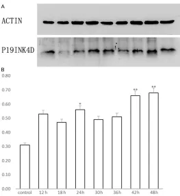
after the ip injection of gentamicin (P<0.01); a little peak value was observed at 24 h (P<0.05) (Figure 2).
Changes in P19 mRNA expression
A decrease in the P19 mRNA expression, which encodes the P19 protein, was reported in the cochlear lateral wall of rats 12-18 h after the ip injection of cisplatin. Also, P19 transcript levels
significantly increased 24 h after the cisplatin
injection (P<0.01) (Figure 3).
The change in P19 protein and mRNA expres-sion in cochlea basilar membrane with an irre-versible loss of a great extent of inner ear cells
Four days after the ip injection of cisplatin, ABR
threshold levels significantly decreased. It can
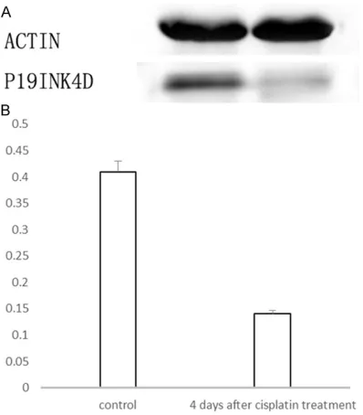
and mRNA level decreased significantly (Fig- ures 4 and 5).
Exons of CDKN2D sequence analysis
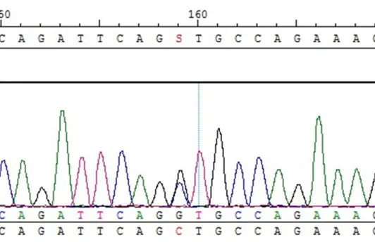
All 400 subjects could be analyzed successful-ly and showed a wild-type sequence (Figure 6).
However, no pathogenic mutation was detected in the coding exons of CDKN2D gene except a synony-mous mutation (449 C>G) in exon 2.
Discussion
Outer hair cells (OHCs) are one of the most vulnerable cellular tar-gets of cisplatin action. Other major targets in the cochlea are stria vascularis, supporting cells, inner hair cells (IHCs), and spiral ganglion cells [7]. In the control group, supporting cells exhibited moderate staining and outer hair cells were lightly immunoreactive; no positive reactant was detected in the inner hair cells. In the 48-h group, the expression of p19 increased, supporting cells exhib-ited intense staining, and moder-ate staining was seen in inner and
outer hair cells. The cell-specific
[image:5.612.92.524.73.220.2]distribution of p19 in the basilar membrane provides morphologi-cal evidence that it can play differ-ent roles in the developmdiffer-ent of drug-induced deafness.
Figure 1. Immunohistochemical staining of P19 (A, B) in rats cochlear basilar membrane. (A) In the control group, supporting cells (SC) exhibited moderate staining and inner hair cells (IHC) were lightly immunoreactive. P19 expres-sion in outer hair cells was negative. Original magnification: 400×. (B) In the 48-h group, the expresexpres-sion of P19ink4d increased, supporting cells (SC) exhibited intense staining, and inner and outer hair cells (IHC and OHC) exhibited moderate staining. Original magnification: 400×.
Figure 2. A. Western blotting of P19 protein in the basilar membrane of the rat cochlea. Lower panel: P19; upper panel: internal control (ACTIN). B. Semi-quantitative analysis shows the change in P19 after the admin-istration of a single dose of cisplatin (a significant increase, P<0.05 by the one-way ANOVA test, is marked by “*”; P<0.01 is marked by “**”).
Western blot also showed an increase in the p19 expression in 24 h; this increase was
sig-nificant 42 h after the ip injection of cisplatin.
Unexpectedly, the p19 protein levels were not consistent with mRNA levels. Though not
[image:5.612.90.353.291.577.2]cispla-tin, which became significant at 24 h (P<0.05).
In contrast, there was a tendency toward a decrease in the p19 mRNA expression, which encodes the p19 protein, 12-18 h after the ip
injection; no significant increase in mRNA was
observed until 24 h. A possible explanation for this discrepancy is that, in 12-24 h after a sin-gle-dose cisplatin treatment, the available p19 mRNA is rapidly translated, suggesting that cis-platin has a more rapid effect on the P19ink4d translation/processing than on the
transcrip-tion of the p19 gene. It indicates that the posttranscriptional mech-anism was responsible for the appearance of p19; this needs to be investigated further.
The chemotherapy drug cisplatin
is one of the doctors’ first lines of
defense against malignancies.
Cisplatin is cheap and efficacious,
but it has a number of side effects that can limit its use, of which nephrotoxicity and ototoxicity are
the most serious. Up to 75%-100%
[image:6.612.92.359.73.234.2]of patients receiving cisplatin che-motherapy develop progressive and irreversible SNHL [8]. There are some views on the mechanism of cisplatin ototoxicity, which still Figure 3. The change in P19 mRNA expression after the administration
of a single dose of cisplatin (a significant increase, P<0.01 by the one-way ANOVA test, is marked by “**”).
Figure 4. A. Western blotting of P19 protein expres-sion in cochlea basilar membrane. Lower panel: P19; upper panel: internal control (ACTIN). B. Semi-quan-titative analysis.
have no unified interpretation. The theory of
apoptosis is the most popular hypothesis. Cisplatin induces cell death by apoptosis but not necrotic cell death. However, the mecha-nisms that initiate cisplatin-induced apoptosis are not fully understood. The cell cycle check-points play an important role by sensing defects that occur during DNA replication and chromo-some segregation, and inducing a cell cycle arrest in response until the defects are repaired [9]. The cell cycle checkpoints are regulated by a family of protein kinases known as the cyclin-dependent kinases (CDKs).
P19 protein, like other INK4s, inhibits CDK4-cyclin D1 activity in vivo and induces G1 phase arrest. The abundance of the transcript of this gene was found to oscillate in a cell-cycle– dependent manner with the lowest expression at mid G1 and a maximal expression during the S phase [10]. Data obtained from sensory hair cells in Ink4d-/- mouse suggest a model in which the maintenance of the postmitotic state of hair cells is dependent on p19 alone [3]. A growing number of studies indicate that cell cycle reentry results in apoptosis by the re-acti-vation of the G1 cell cycle machinery [11-13]. The normal expression of P19 prevents the hair cells from entering an abnormal cell cycle,
[image:6.612.91.290.295.522.2]A few unique functions of p19 that distinguish it from its siblings have been reported. In addi-tion to its role in cell cycle regulaaddi-tion, p19 is also important for repairing DNA damage. It has been reported that UV light, cisplatin, and
β-amyloid peptide promote p19 transcriptional
induction and nuclear translocation [4]. How- ever, the mechanism(s) responsible for p19 upregulation in response to genotoxic stress are yet to be elucidated. Studies suggest that p19 protects cells from undergoing apoptosis
by allowing a more efficient DNA repair [14, 15].
More interestingly, p19-underexpressing cells were associated with impaired DNA repair activity, high frequency of chromosomal aberra-tions, increased apoptosis and, consequently, reduced cell survival when compared with their
p19-overexpressing counterparts [5, 6]. p19 activities in DNA repair and cell cycle arrest seem to be independent [14]. Thus, p19 acts as a chromatin accessibility factor that allows the access of the repair machinery to the DNA dam-age site [15].
In the course of eliminating the damaged cells by triggering
apop-tosis, a cell first tries to repair any
DNA damage and survive. When DNA damage overwhelms the re- pair capacity, apoptosis occurs.
The findings of this study demon -strated that p19 mRNA and
pro-tein levels both significantly
dec-reased in the rats with manifested hearing loss 4 days after cisplatin treatment. A previous research by Fang and Hongjun [18] found that the sequences of hair cells were destroyed and hair cell losses were induced 72 hours after treat-ment with 16 mg/kg (ip) of cispla-tin. The research indicated that at this point cisplatin treatment caused an obvious damage to the inner cells, which decreased the cells’ ability to maintain and man-ufacture the normal level of p19. Thus, it is presumed that if there is not yet an irreversible loss of a great extent of inner ear cells, the increased expression of p19 in rat inner ear cells might play an important role in repairing the tissue damage induced by cisplatin. It may participate in detecting DNA damage and directly relay it to the apoptotic machinery. A p19 mutation or a disturbance of its regulated expression can result in the loss of apoptotic control, which in turn may result in hearing loss.
The CDKN2D (cyclic-dependent kinase inhibitor 2D) gene is the human homolog of mouse gene p19. CDKN2D was not found in the inner ear
cells, and the level and cell-type specificity of
[image:7.612.92.360.67.234.2]the CKI expression in the human inner ear are not currently known. The gene CDKN2D was screened in two families that had autosomal recessive nonsyndromic hearing impairment (NSHI) from Pakistan but were negative for Figure 5. The change in P19 mRNA expression in cochlea basilar
mem-brane.
[image:7.612.93.359.283.455.2]functional sequence variants [16]. In this study, CDKN2D was analyzed in 400 samples without any common mutation of hearing loss, but no pathogenic mutation existed in the exons of this gene. The CDKN2D gene seems to be a rare causative gene for profound sensorineural deafness. One reason might be the higher availability of this short gene fragment. Also, animals derived from crosses of Ink4d-null with Kip1-null mice exhibited bradykinesia, proprio-ceptive abnormalities, and seizures, and died at about 18 days after birth [17]. Patients with a mutation of the CDKN2D gene usually die in utero or in early infancy or have a much more severe brain anomaly and malignancies, in which case it is impossible to carry on the genetic test. The Ping Chen’s report showed
that hearing is significantly compromised, start -ing between 7 and 15 weeks of age, and that hearing loss progresses into adulthood as a result of a mutation in the Ink4d gene. These data indicate that a mutation in the Ink4d gene can induce a progressive deafness, rather than a congenital severe hearing loss. The future research should be based on larger sample size or other target groups, such as acquired hearing loss in children.
Conclusion
The systematic administration of a single dose
of cisplatin can rapidly influence the expression
and distribution of P19 protein and mRNA. Cisplatin has a more rapid effect on the P19 translation/processing than on the transcrip-tion of the P19 gene, which suggests an unclear regulation at the posttranscriptional level that requires further investigation. P19 is one of the earliest markers appearing in response to cis-platin, before any toxic effect and cochlear dys-function. The P19 upregulation is probably one of the common responses of the inner ear cells to injury, such as acoustic trauma and early-stage drug ototoxicity. It may play an important role in the protection and repair of the dam-aged tissue. When DNA damage overwhelms the repair capacity and the irreversible inner ear cell loss induced by cisplatin occurs, the expression of P19 decreases.
No mutation existed within the coding exons of CDKN2D in 400 patients with profound senso-rineural deafness, which suggests only a minor
role of CDKN2D gene mutations in congenital severe hearing loss. Expression levels of
CDKN2D might influence acquired progressive
hearing loss, but the underlying mechanisms need to be explored in more detail.
Acknowledgements
This work was supported by the Cochlear Implant Program of Hunan, China, and National Basic Research Program of China (973) (grants 2012CB967900 and 2012CB967904) to D.H.X.
Disclosure of conflict of interest
None.
Address correspondence to: Dr. Dinghua Xie, De- partment of Otolaryngology Head and Neck Surgery, The Second Xiangya Hospital of Central South Uni- versity, Changsha, China. Tel: 86-13975-188898; E-mail: [email protected]
References
[1] Yan D, Liu XZ. Cochlear molecules and heredi-tary deafness. Front Biosci 2008; 13: 4972-83.
[2] Hereditary Hearing Loss homepage. In: http:// hereditaryhearingloss.org/[May 13 th, 2015 (aes)].
[3] Chen P, Zindy F, Abdala C, Liu F, Li XK, Roussel MF and Segil N. Progressive hearing loss in mice lacking the cycldependent kinase in-hibitor Ink4d. Nature Cell Biology 2005; 5: 422-426.
[4] Ceruti JM, Scassa ME, Marazita MC, Carcagno AC, Sirkin PF, Cánepa ET. Transcriptional up-regulation of p19INK4d upon diverse genotox-ic stress is critgenotox-ical for optimal DNA damage re-sponse. Int J Biochem Cell Biol 2009; 41: 1344-1353.
[5] Ceruti JM, Scassa ME, Flo JM, Varone CL, Canepa ET. Induction of p19INK4d in response to ultraviolet light improves DNA repair and confers resistance to apoptosis in neuroblas-toma cells. Oncogene 2005; 24: 4065-4080. [6] Scassa ME, Marazita MC, Ceruti JM, Carcagno
AL, Sirkin PF, Gonzalez-Cid M, Pignataro OP, Canepa ET. Cell cycle inhibitor, p19INK4d, pro-motes cell survival and decreases chromo-somal aberrations after genotoxic insult due to enhanced DNA repair. DNA Repair (Amst) 2007; 6: 626-638.
cisplatin-in-duced hearing loss. Hear Res 2013; 306: 46-53.
[8] Rybak LP, Whitworth CA, Mukherjea D, Ramku-mar V. Mechanisms of cisplatin-induced oto-toxicity and prevention. Hear Res 2007; 226: 157-167.
[9] Malumbres M, Barbacid M. Cell cycle, CDKs and cancer: a changing paradigm. Nat Rev Cancer 2009; 9: 153-166.
[10] Hirai H, Roussel MF, Kato JY, Ashmun RA, Sherr CJ. Novel INK4 proteins, p19 and p18 are spe-cific inhibitors of the cyclin D-dependent kinas-es CDK4 and CDK6. MoL Cell Biol 1995; 15: 2672-2681.
[11] Liu DX, Greene LA. Neuronal apoptosis at the G1/S cell cycle checkpoint. Cell Tissue Res 2001; 305: 217-228.
[12] Herrup K, Yang Y. Pictures in molecular medi-cine: contemplating Alzheimer’s disease as cancer: a loss of cell-cycle control. Trends Mol Med 2001; 7: 527.
[13] Nguyen MD, Mushynski WE and Julien JP. Cy-cling at the interface between neurodevelop-ment and neurodegeneration. Cell Death Dif-fer 2002; 9: 1294-1306.
[14] Marazita MC, Ogara MF, Sonzogni SV, Martí M, Dusetti NJ, Pignataro OP, Cánepa ET. CDK2 and PKA mediated-sequential phosphorylation is critical for p19INK4d function in the DNA damage response. PLoS One 2012; 7: e35638.
[15] Sonzogni SV, Ogara MF, Castillo DS, Sirkin PF, Radicella JP, Cánepa ET. Nuclear translocation of p19INK4d in response to oxidative DNA damage promotes chromatin relaxation. Mol Cell Biochem 2015; 398: 63-72.
[16] Santos RL, Hassan MJ, Sikandar S, Lee K, Ali G, Martin PE Jr, Wambangco MA, Ahmad W, Leal SM. DFNB68, a novel autosomal reces-sive non-syndromic hearing impairment locus at chromosomal region 19p13.2. Hum Genet 2006; 120: 85-92.
[17] Zindy F, Cunningham JJ, Sherr CJ, Jogal S, Sm-eyne RJ, Roussel MF. Postnatal neuronal prolif-eration in mice lacking Ink4d and Kip1 inhibi-tors of cyclin-dependent kinases. Proc Natl Acad Sci U S A 1999; 96: 13462-7.