Original Article
Involvement of spinal SIRT1 in development of chronic
constriction injury induced neuropathic pain in rats
Feng-Qin Luo, Qian Ma, Hui Zheng, Xiao-Wen Guo, Juan Zhang
Department of Anesthesiology, Zhejiang Hospital of Traditional Chinese Medicine, The First Affiliated Hospital of Zhejiang Chinese Medical University, Traditional Chinese Medical University, Hangzhou, Zhejiang Provincial, China Received December 12, 2016; Accepted December 27, 2016; Epub May 1, 2018; Published May 15, 2018
Abstract: It is known that the epigenetic process of histone acetylation is involved in the neuropathic pain. The aim of this study was to determine whether sirtuin type 1 (SIRT1), an NAD+ dependent deacetylase, affected allodynia and hyperalgesia in neuropathic pain. The neuropathic pain model was established by ligature of the right sciatic nerve to induce chronic constriction injury (CCI) in rats. Histone acetyltransferase (HAT) activity was increased and, and histone deacetylase (HDAC) activity was declined in tissue of the spinal dorsa horn in CCI rates by means of enzyme-linked immunosorbent assay (ELISA). The persistent hyperalgesia and allodynia caused by CCI were associ-ated with downregulation of SIRT1 and upregulation of acetylassoci-ated-H3 (Ac-H3) in tissue of the spinal cord by Western blot assay, which was reversed after intrathecal injection of SIRT1 agonist SRT1720. SRT1720 treatment achieved analgesic through inhibiting the acetylation of nuclear factor kappa B (NF-κB) and blocking the releases of the inflammatory factors including tumor necrosis factor-α (TNF-α) and interleukin (IL)-6 by means of Western blot and real-time quantitative PCR (RT-PCR), respectively. Taken together, these data suggest that SIRT1 in the spinal cord plays an important role in the neuropathic pain in the rat model.
Keywords: SIRT1, neuropathic pain, NF-κB, SRT1720, histone acetyltransferases, histone deacetylase
Introduction
As a result of a lesion or disease, neuropathic pain is characterized by hyperalgesia and allo-dynia affecting the somatosensory system at the peripheral or central level [1]. The underly-ing mechanisms of neuropathic pain are still poorly understood, therefore, it still remains as a challenge in the clinical treatment. Accumu- lative evidence indicates that histone acetyla-tion such as epigenetic modificaacetyla-tion greatly contributes to the development and mainte-nance of neuropathic pain [2, 3]. Epigenetics, an emerging field that is studying how environ-mental factors result in lasting changes in gene expression without altering DNA sequence. Covalent modification of the DNA-packaging histone that regulates the expression of noci-ceptive or analgetic genes is one of the charac-terized mechanisms of epigenetics [4, 5]. Controlling the dynamic interplay of HAT and HDAC enzymes is one of the processes. But, it is not clear how the balance of HAT/HDAC enzyme activity maintains the level of histone acetylation in the neuropathic pain.
The nicotinamide adenine dinucleotide (NAD+)
dependent deacetylase, SIRT1, has been sho- wn to regulate a wide variety of cellular pro-cesses, including aging and lifespan extension [6, 7]. Presently, SIRT1, as a member of the silent information regulator family, is gaining attention in the development of innovative treatment strategies for cancer, neurodegener-ative disorders, and metabolic disease [8, 9]. Many pain related transcription factors and the expression of their downstream targets are reg-ulated by SIRT1, such as nuclear factor kappa B (NF-κB), cAMP-response element binding pro-tein (CREB), and c-Jun [10-12]. We therefore speculated that SIRT1 deacetylase activity influenced the expression level of Ac-H3 and the transcriptional level of those possible pain-related genes may be involved in the neuro-pathic pain.
inflammation of nerve injury is related to allo-dynia and hyperalgesia of neuropathic pain [15]. NF-κB activity is increased in the dorsal root ganglia and the spinal cord of rats accom-panied by the neuropathic and inflammatory pain [16, 17]. Reports revealed that NF-κB plays a crucial role in the pathogenesis of neu-ropathic pain. The activation of NF-κB up-regu-lates the expression levels of inflammatory genes, such as tumor necrosis factor-α (TNF-α), and interleukin-6 (IL-6), and facilitates to pro-duce the hyperalgesia and allodynia [18]. Increasing evidences for an important role of NF-κB in the pain regulation, very few studies have addressed the mechanisms how to exert its effects on the neuropathic pain. Therefore, we hypothesized that SIRT1 was related to the neuropathic pain by a mechanism that might be involved in acetylization of NF-κB in the spi-nal cord of CCI rats.
Materials and methods
Animals
Adult male Sprague-Dawley rats (200-220 g) were purchased from Animal Experimental Center, Zhejiang Traditional Chinese Medical University, China. Animals were housed in groups of five per standard cage, on a 12 h light/dark cycle, in an air temperature and humidity controlled environment. In this study, experiments were carried out in accordance with local guidelines for the care of laboratory animals of Zhejiang Traditional Chinese Medical University, and were approved by the ethics committee for research on laboratory animal use of the institution.
CCI neuropathic pain model
CCI surgery was performed as previously described [19]. Rats were anesthetized with 4% pentobarbital sodium (Sh-haling testing cen-ters of biological reagents, Shanghai, China), and a 7-mm segment of the right common sci-atic nerve was exposed at the mid-thigh level. Four ligatures (4-0 chromic catgut) thread were loosely tied around the nerve, while the same procedure in the sham-operated control group was performed by exposing the right sciatic nerve without ligation. The muscle and skin were then closed with silk suture. Behavioral was assessed at 1, 3 and 5 day before surgery and at 7 day after surgery.
Implantation of intrathecal catheter
For spinal drug administration, the rats were chronically implanted with catheters as previ-ously described [20]. Intrathecal polyethylene catheter (polyethylene-10 tubing) was passed caudally from the cistern magna to the spinal cord level of lumbar enlargement. Proper loca-tion was determined by a temporary motor block of both hindlimbs after injection of 2% lidocaine 10 μL. Only rats with no evidence of neurologic deficit after the operation were stud-ied. All experiments were conducted 1 week after implantation of the intrathecal catheter. For intrathecal (i.t.) administration, the drugs were given using a microinjection syringe fol-lowed by a 10 μL flush.
Measurement of mechanical allodynia
Mechanical allodynia was assessed using an automated testing device (dynamic plantar anesthesiometer, UgoBasile, Italy) as described previously [21]. Rats were placed on a metal mesh floor to acclimate for at least 30 min. Then, von Frey filaments was pushed against the plantar surface of the right hind paw from below the floor of the test chamber with increas-ing pressure, which increased lineally from 0 to 50 g over a 20-s period. When rat removed its paw, the paw withdrawal threshold (PWT) was recorded automatically. The right hind paw was tested at 30-s intervals, and three responses were averaged to represent PWT.
Measurement of thermal hyperalgesia
Each rat was placed on a glass plate (Ugo Basile, Italy) with radiant heat equipment underneath. After a 30-min acclimation period, the radiant heat source under the glass floor was positioned directly under the right hind paw until the animal lifted its paw from the floor. The manipulator defined the thermal with-drawal latency (TWL) as the animal applied the radiant heat to withdraw its hind paw. TWL was measured three times at 5-min intervals, and its mean value was used as the latency of the response. The cut-off of heat application was automatically set at 20 s to avoid tissue damage.
Drugs
(DMSO; Sigma, USA) and administered (i.t. 40 µg/kg) once a day from day 0 to day 3 after the CCI operation. We have proved SRT1720 (i.t. 40 µg/kg) is enough to relieve CCI-induced ther-mal and mechanical pain behaviors, and data have been shown in the other paper of our labo-ratory (the article will be published). Control animals received the same volume of DMSO (DMSO group) intrathecal injections.
Enzyme-linked immunosorbent assay (ELISA)
The concentration of HDAC and HAT in ipsilat-eral L4-L5 spinal segments was measured with ELISA kits from BioVision (Mountain View, CA) following the manufacturer’s instructions. HDAC and HAT activity was expressed as ng/ mL protein. Each sample was run in triplicate, and the concentration was calculated from the kit’s standard curve.
Western blot analysis
The ipsilateral L4-L5 segments of the spinal cord from sham-operated, CCI, CCI+DMSO, and CCI+SRT1720 rats were quickly removed, and all tissues were rapidly frozen in liquid nitrogen and stored at 80°C until further processing. The frozen spinal cords were directly homoge-nized in a lysis buffer (12.5 KL/mg tissue) con-taining a cocktail of protease inhibitors and phenylmethylsulfonyl fluoride (Amresco, Solon, OH, USA). The supernatant, after centrifugation at 12,000 revolutions per minute for 15 min at 4°C, was used for Western blotting. The total protein level in the supernatants was measured using the Pierce bicinchoninic acid (BCA) Pro- tein Assay Kit (Thermo Scientific, Rockford, IL, USA). Samples were separated on 13% acryl-amide gels and then transferred onto polyvinyli-dene fluoride membranes for the detection of SIRT1, H3, Ac-H3, NF-κB and Ac-NF-κB. The membranes were blocked in 5% nonfat dry milk for 1 h at room temperature and then washed in Tris-buffered saline with tween (TBST) 3 times for 10 min each. The membranes were incubated with the primary antibody, rabbit anti-SIRT1 (1:2000; Abcam, Cambridge, UK), rabbit anti-H3 (1:2000), and acetyl-histone H3 (1:1000) (Cell Signaling Technology, Danvers, Massachusetts), anti-NF-κB and acetyl-acetyl (1:1000; Santa Cruz Biotechnology, Santa Cruz, California) primary antibody, horseradish per-oxidase (HRP)-rabbit-anti-tubulin and HRP-mouse-anti-glyceraldehyde 3-phosphate
dehy-drogenase (GAPDH) (Lianke, Hangzhou, China) at 4°C overnight. The blots were washed in TBST and incubated in the appropriate second-ary antibody, HRP-goat-anti-rabbit (1:3000, MultiSciences (Lianke) Biotech, Hangzhou, Chi- na) for 1 h at room temperature. All Western blot analyses were performed at least 3 times, and parallel results were obtained. The expres-sion level of proteins was calculated as the average of the densities in each band area from different rats. The expression of proteins was evaluated relative to GAPDH.
RT-PCR analysis
Total RNA was extracted from ipsilateral lumbar spinal cord tissues. Extracted RNA was pre-treated with DNase I at 37°C for 30 min. Before reverse transcription reaction, which was per-formed using a high capacity cDNA archived kit (TaKaRa, Japan), a Real-Time PCR Detection System (Roche, Switzerland) was used to con-tinuously monitor the intensity of fluorescence, which was directly proportional to the PCR products. The RT-PCR conditions were per-formed according to the manufacturer’s instruc-tions of TransStart Top Green qPCR SuperMix (TransGen Biotech, Beijing, China) included an initial denaturation for 30 s at 95°C, followed by 40 cycles of denaturation for 5 s at 95°C, annealing for 30 s at 60°C and extension at 72°C for 30 s using ABI 7300 Thermocycler (Applied Biosystems, Foster City, CA, USA) in triplicate. Primer sequences were listed as fol-lows: TNF-α, forward: 5’-CCAGGTTCTCTTCAAG- GGACAA-3’ and reverse: 5’-GGTATGAAATGGCA- AATCGGCT-3’; IL-6, forward: 5’-ACAAGGATTACG- AGCAGATGGT-3’ and reverse: 5’-GCAGGTTGTT- CTGGAAGTTGAG-3’; GAPDH, forward: 5’-GCCA- AAAGGGTCATCATCTC-3’ and reverse: 5’-GGCC- ATCCACAGTCTTCT-3’. GAPDH was used as con-trol of the input RNA level. The gene expression was calculated using the 2-ΔΔ Ct method.
Statistical analysis
Results
Changes in behavior induced by CCI
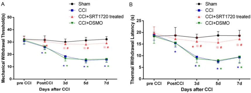
After CCI, the rats gradually showed the typical signs of thermal hyperalgesia and mechanical allodynia. Behaviors in sham-operated rats were not obviously changed. The changes in the right hind paw of PWT and TWL were dem-onstrated (Figure 1). There was no significant difference in PWT and TWL among the groups (P > 0.05) before CCI surgery. PWT and TWL of CCI rats decreased on day 1 after CCI, and
[image:4.612.91.520.72.232.2]were further reduced on day 3, 5 and 7 com-pared with sham-operated rats, indicating that the mechanical allodynia and thermal hyperal-gesia had been developed in CCI rats. In SRT1720-treated group, PWT and TWL went much higher on day1, 3, 5, and 7 compared to DMSO-treated group (Figure 1) (P < 0.05). We showed that the administration of SRT1720 (i.t. 40 µg/kg) was able to produce antinociception in rats with nerve injury. So, in our study, it is suggested that the activation of SIRT1 was involved and it produced analgesia in the neu-ropathic pain during CCI.
Figure 1. Changes in behavior induced by CCI. A. The paw withdrawal threshold was statistically decreased in CCI group by testing before surgery and at 1, 3, 5 and 7 days following the CCI operation in comparison with Sham group. B. Thermal withdrawal latency was statistically decreased in CCI group by testing before surgery and at 1, 3, 5 and 7 days following the CCI operation in comparison with Sham group. SRT1720 treated group reversed the change in the DMSO treated group. The data are expressed as the mean ± SEM (n = 6, *P < 0.05 vs Sham group, #P < 0.05 vs CCI group, ※P < 0.05 vs CCI+DSMO group).
[image:4.612.93.525.321.500.2]Changes in the activity of HDAC and HAT in neuropathic pain rats caused by CCI
To assess the involvement of epigenetics in the development of neuropathic pain, ELISA was used for detecting the dynamic interplay of HAT and HDAC. HAT activity was significantly higher in CCI group compared to that of Sham group and control group (Figure 2A; P < 0.05). For HDAC, both Sham group and CCI group had decreased levels of activity, but the difference was significant between CCI group and control group (Figure 2B; P < 0.05). Thus the balance of HAT/HDAC activity shifts towards the mainte-nance of histone acetylation in CCI-induced neuropathic pain. These findings might have showed that the activation of histone acetyl-transferases and inhibition of histone deacety-lase were participated in the development of neuropathic pain.
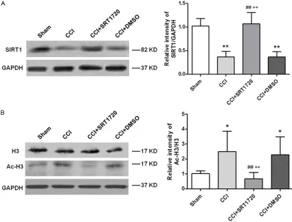
Changes in the expression levels of Ac-H3 and SIRT1 in neuropathic pain rats caused by CCI
The specific SIRT1 and Ac-H3 bands in the ipsi-lateral spinal cord that were derived using Western blot analysis (Figure 3). In contrast to sham-operated rats, remarkably decreased SIRT1 intensity and increased Ac-H3 intensity were observed in the ipsilateral spinal cord of rats in the CCI+DMSO group (P < 0.05). SRT1720 (i.t. 40 µg/kg) increased the CCI-induced SIRT1 depression and alleviated Ac-H3 changes in the spinal dorsal horn compared to the CCI+DMSO group (P < 0.01). These data suggested that the intrathecal SRT1720, as the SIRT1 agonist, reversed the downregulated SIRT1 expression and decreased the acetyla-tion level of histone H3 after CCI.
CCI-induced activation of NF-κB is related to SIRT1
In order to understand the underlying mecha-nism of SIRT1 effect in neuropathic pain, we detected NF-κB and acetylated-NF-κB (Ac-NF-κB) expression in the lumbar spinal cords after
nerve injury by Western blot analysis. As shown in Figure 4A, upregulation of NF-κB and Ac-NF-κB protein levels was detected in CCI rats com-pared with Sham-operated group (P < 0.01). NF-κB and Ac-NF-κB expression levels were sig-nificantly lower in SRT1720 rats than those in CCI/DMSO rats on day 7 after CCI operation (P
< 0.01 and P < 0.05). These results showed that CCI-induced pain increased the expression of the NF-κB and Ac-NF-κB, but the intrathecal SRT1720 demonstrated the inhibitory effect. The above data also suggests that the activa-tion of SIRT1, just as the effects of SRT1720, was related to a significant reduction of the acetylation level of NF-κB in the neuropathic pain.
Activation of TNF-α and IL-6 induced by CCI
NF-κB is a central regulator of the inflammatory process and plays a critical role in inflamma-tion. It regulates the expression of a group of proinflammatory mediators, such as the TNF-α and IL-6 [22]. As illustrated in Figure 4B, real-time PCR showed a significant upregulation of TNF-α and IL-6 mRNA expression 7 days after CCI compared to the Sham group (P < 0.01). As revealed by real-time PCR, there were signifi-cant differences in TNF-α and IL-6 between the CCI/DMSO group and SRT1720 group (P < 0.01). The above data showed that SIRT1 ago-nist, SRT1720 (i.t.) might inhibit activation of NF-κB and reduce the production of its down-stream proinflammatory mediators as TNF-α and IL-1β, indicating that the antinociceptive effect of SIRT1 might correlate with its anti-inflammatory action.
Discussion
In our study, we demonstrated first that CCI induced persistent hyperalgesia and allodynia that were associated with downregulation of SIRT1 and Ac-H3 expression in the spinal cords can be reversed by intrathecal injection of SIRT1 agonist SRT1720. The neuropathic pain model was established by ligature of the right
sciatic nerve to induce chronic constriction injury (CCI) in rats (Figure 1). We found that HAT activity was increased in the peripheral nerve injury and the HDAC activity was decreased in the spinal cord (Figure 2). The balance of HAT/ HDAC activity towards histone acetylation was broken in the CCI-inducing neuropathic pain. The last, a potential novel mechanism of SRT1720, a SIRT1 agonist, in analgesic action maybe can be achieved by the inhibition of acetylation of NF-κB and reduced the releases of the inflammatory factors TNF-α and IL-6. These data suggested that SIRT1 in the spinal cord plays an important role in the mainte-nance of neuropathic pain. In our study, SIRT1 expression in the spinal cord is down-regulated under neuropathic conditions (Figure 3); we suggested that the effect of SIRT1 is possibly related to the neuroinflammation in the pathic pain. SIRT1 regulates a variety of neuro-logical functions through the deacetylation of histones. The present study showed that CCI induced an increase in Ac-H3, just as the other studies have showed in the model of partial sci-atic nerve ligation and CCI [23, 24], and a decrease in SIRT1 expression in the spinal cord. The expression changes in Ac-H3 paral-leled the expression changes of SIRT1 in our study (Figure 4). This result further suggests that the activated acetylation is taken part in neuropathic pain processing. Specially, we found that the balance between HAT and HDAC was disrupted in the CCI pain model. It sup-ports the importance of epigenetic mecha-nisms in the development of neuropathic pain. SRT1720, as a small molecule activator of SIRT1, is structurally unrelated to, and 1,000-fold more potent than, resveratrol [25]. Resver- atrol, another SIRT1 agonist, has been proved its antinociceptive effects effect in a neuro-pathic pain model [24]. Study shows that the administration of SRT1720, once being sug-gested as a therapeutic for the treatment of type 2 diabetes [25], delayed and attenuated CCI-induced pain hypersensitivity, and decre- ased Ac-H3 and increased SIRT1 expression in the spinal dorsal horn. Thus, to our knowledge, SIRT1 activation is a promising new therapeutic approach for treating neuropathic pain. SIRT1 modifies multiple substrates through transcriptional regulators such as p53 and NF-κB. It is well reported that NF-κB is subject to deacetylation by SIRT1. Using a myeloid
spe-cific SIRT1 knockout mouse model, Schug et al. showed that ablation of SIRT1 in macrophages rendered NF-κB hyperacetylation, resulting in increased transcriptional activation of proin-flammatory target genes [26]. In addition, it has been shown that moderate overexpression of SIRT1 in mice led to the reduced NF-kB activity [27]. SRT1720 has been shown to suppress inflammatory cell infiltration and cytokine pro-duction in a mouse model of asthma [28]. Our results showed that SRT1720 may inhibit acti-vation of NF-κB and reduce the production of its downstream proinflammatory mediators as TNF-α and IL-6 (Figure 4). This result demon-strates that activation of SIRT1, with SRT1720, is possibly induced to decline behavioral hyper-sensitivity in CCI models through inhibiting deacetylation of NF-κB and decreasing tran-scriptional activation of proinflammatory target genes TNF-α and IL-6.
In conclusion, the present study indicates that the activation of acetylation plays a crucial role in persistent hyperalgesia and allodynia of the neuropathic pain. SIRT1 is involved in the neu-ropathic pain and the mechanism of SIRT1 may contribute to regulate acetylization of NF-κB in the spinal cord. Since SIRT1 agonist might be effective for the therapeutics in nerve injury of pain, it gives us a direction to find a new meth-od to therapy neuropathic pain.
Acknowledgements
Zhejiang Medical and Health Science and Technology Plan (Grant No. 2017RC023) and Natural Science Fund of Zhejiang province (Grant No. LY16H290003).
Disclosure of conflict of interest
None.
Address correspondence to: Dr. Juan Zhang, Depart- ment of Anesthesiology, Zhejiang Hospital of Tradi- tional Chinese Medicine, The First Affiliated Hospital of Zhejiang Chinese Medical University, 54 Youdian Road, Shangcheng District, Hangzhou, Zhejiang Provincial, China. Tel: 0086-571-87071760; Fax: 0086-571-87071760; E-mail: [email protected]
References
[2] Cherng CH, Lee KC, Chien CC, Chou KY, Cheng YC, Hsin ST, Lee SO, Shen CH, Tsai RY and Wong CS. Baicalin ameliorates neuropathic pain by suppressing HDAC1 expression in the spinal cord of spinal nerve ligation rats. J Formos Med Assoc 2014; 113: 513-520. [3] Kiguchi N, Kobayashi Y, Maeda T, Fukazawa Y,
Tohya K, Kimura M and Kishioka S. Epigenetic augmentation of the macrophage inflammato-ry protein 2/C-X-C chemokine receptor type 2 axis through histone H3 acetylation in injured peripheral nerves elicits neuropathic pain. J Pharmacol Exp Ther 2012; 340: 577-587. [4] Denk F, Huang W, Sidders B, Bithell A, Crow M,
Grist J, Sharma S, Ziemek D, Rice AS, Buckley NJ and McMahon SB. HDAC inhibitors attenu-ate the development of hypersensitivity in models of neuropathic pain. Pain 2013; 154: 1668-1679.
[5] Geranton SM. Targeting epigenetic mecha-nisms for pain relief. Curr Opin Pharmacol 2012; 12: 35-41.
[6] Baur JA, Ungvari Z, Minor RK, Le Couteur DG and de Cabo R. Are sirtuins viable targets for improving healthspan and lifespan? Nat Rev Drug Discov 2012; 11: 443-461.
[7] Wang F, Chen HZ, Lv X and Liu DP. SIRT1 as a novel potential treatment target for vascular aging and age-related vascular diseases. Curr Mol Med 2013; 13: 155-164.
[8] Herskovits AZ and Guarente L. SIRT1 in neuro-development and brain senescence. Neuron 2014; 81: 471-483.
[9] Lara E, Mai A, Calvanese V, Altucci L, Lopez-Nieva P, Martinez-Chantar ML, Varela-Rey M, Rotili D, Nebbioso A, Ropero S, Montoya G, Oyarzabal J, Velasco S, Serrano M, Witt M, Villar-Garea A, Imhof A, Mato JM, Esteller M and Fraga MF. Salermide, a Sirtuin inhibitor with a strong cancer-specific proapoptotic ef-fect. Oncogene 2009; 28: 781-791.
[10] Dey S, Bakthavatchalu V, Tseng MT, Wu P, Florence RL, Grulke EA, Yokel RA, Dhar SK, Yang HS, Chen Y and St Clair DK. Interactions between SIRT1 and AP-1 reveal a mechanistic insight into the growth promoting properties of alumina (Al2O3) nanoparticles in mouse skin epithelial cells. Carcinogenesis 2008; 29: 1920-1929.
[11] Monteserin-Garcia J, Al-Massadi O, Seoane LM, Alvarez CV, Shan B, Stalla J, Paez-Pereda M, Casanueva FF, Stalla GK and Theodoro- poulou M. Sirt1 inhibits the transcription factor CREB to regulate pituitary growth hormone synthesis. FASEB J 2013; 27: 1561-1571. [12] Yeung F, Hoberg JE, Ramsey CS, Keller MD,
Jones DR, Frye RA and Mayo MW. Modulation of NF-kappaB-dependent transcription and cell survival by the SIRT1 deacetylase. EMBO J 2004; 23: 2369-2380.
[13] Chen L, Fischle W, Verdin E and Greene WC. Duration of nuclear NF-kappaB action regulat-ed by reversible acetylation. Science 2001; 293: 1653-1657.
[14] Kauppinen A, Suuronen T, Ojala J, Kaarniranta K and Salminen A. Antagonistic crosstalk be-tween NF-kappaB and SIRT1 in the regulation of inflammation and metabolic disorders. Cell Signal 2013; 25: 1939-1948.
[15] Pathak NN, Balaganur V, Lingaraju MC, More AS, Kant V, Kumar D, Kumar D and Tandan SK. Antihyperalgesic and anti-inflammatory effects of atorvastatin in chronic constriction injury-induced neuropathic pain in rats. Inflammation 2013; 36: 1468-1478.
[16] Ma W and Bisby MA. Increased activation of nuclear factor kappa B in rat lumbar dorsal root ganglion neurons following partial sciatic nerve injuries. Brain Res 1998; 797: 243-254. [17] Chan CF, Sun WZ, Lin JK and Lin-Shiau SY.
Activation of transcription factors of nuclear factor kappa B, activator protein-1 and octam-er factors in hypoctam-eralgesia. Eur J Pharmacol 2000; 402: 61-68.
[18] Sun T, Luo J, Jia M, Li H, Li K and Fu Z. Small interfering RNA-mediated knockdown of NF-kappaBp65 attenuates neuropathic pain fol-lowing peripheral nerve injury in rats. Eur J Pharmacol 2012; 682: 79-85.
[19] Bennett GJ and Xie YK. A peripheral mononeu-ropathy in rat that produces disorders of pain sensation like those seen in man. Pain 1988; 33: 87-107.
[20] Yaksh TL and Rudy TA. Chronic catheterization of the spinal subarachnoid space. Physiol Behav 1976; 17: 1031-1036.
[21] Muthuraman A, Jaggi AS, Singh N and Singh D. Ameliorative effects of amiloride and pralidox-ime in chronic constriction injury and vincris-tine induced painful neuropathy in rats. Eur J Pharmacol 2008; 587: 104-111.
[22] Rahman MM, Halade GV, Williams PJ, Fer-nandes G. t10c12-CLA maintains higher bone mineral density during aging by modulating os-teoclastogenesis and bone marrow adiposity. J Cell Physiol 2011; 226: 2406-14.
[23] Uchida H, Matsushita Y and Ueda H. Epigenetic regulation of BDNF expression in the primary sensory neurons after peripheral nerve injury: Implications in the development of neuropath-ic pain. Neuroscience 2013; 240: 147-154. [24] Yin Q, Lu FF, Zhao Y, Cheng MY, Fan Q, Cui J, Liu
L, Cheng W and Yan CD. Resveratrol facilitates pain attenuation in a rat model of neuropathic pain through the activation of spinal Sirt1. Reg Anesth Pain Med 2013; 38: 93-99.
W, Iffland A, Lavu S, Medvedik O, Sinclair DA, Olefsky JM, Jirousek MR, Elliott PJ and Westphal CH. Small molecule activators of SIRT1 as therapeutics for the treatment of type 2 diabetes. Nature 2007; 450: 712-716. [26] Schug TT, Xu Q, Gao H, Peres-da-Silva A, Draper
DW, Fessler MB, Purushotham A and Li X. Myeloid deletion of SIRT1 induces inflamma-tory signaling in response to environmental stress. Mol Cell Biol 2010; 30: 4712-4721. [27] Chen J, Zhou Y, Mueller-Steiner S, Chen LF,
Kwon H, Yi S, Mucke L and Gan L. SIRT1 pro-tects against microglia-dependent amyloid-be-ta toxicity through inhibiting NF-kappaB signal-ing. J Biol Chem 2005; 280: 40364-40374.