Original Article
The RNA-binding protein Sam68 is critical for
non-small cell lung cancer cell proliferation
by regulating Wnt/β-catenin pathway
Xuebing Li1, Xuexia Zhou2, Feng Hua3, Yaguang Fan1, Lingling Zu1, Yuli Wang1, Wang Shen1, Hongli Pan1,
Qinghua Zhou1,4
1Tianjin Key Laboratory of Lung Cancer Metastasis and Tumor Microenvironment, Tianjin Lung Cancer Institute, Tianjin Medical University General Hospital, Tianjin, P. R. China; 2Department of Neuropathology, Tianjin Key Laboratory of Injuries, Variations and Regeneration of The Nervous System, Key Laboratory of Post-Trauma Neuro- Repair and Regeneration in Central Nervous System of Education Ministry, Tianjin Neurological Institute, Tianjin Medical University General Hospital, Tianjin, P. R. China; 3Department of Thoracic Surgery, Shandong Cancer Hos-pital and Institute, Jinan, P. R. China; 4Sichuan Lung Cancer Institute, Sichuan Lung Cancer Center, West China Hospital, Sichuan University, Chengdu, P. R. China
Received May 11, 2017; Accepted June 20, 2017; Epub August 1, 2017; Published August 15, 2017
Abstract: Src associated in mitosis, 68 kDa (Sam68) is a KH domain RNA-binding protein that regulates a broad scope of biological events, including RNA metabolism, transcription and signal transduction. Herein, we aimed to explore the expression, clinical significance and biological function of Sam68 in human non-small cell lung cancer (NSCLC). By applying quantitative real-time PCR (qRT-PCR), western blotting and immunohistochemistry (IHC) meth-ods, we found that nucleic localized Sam68 was markedly overexpressed in NSCLC tissues and cell lines. By X2 analysis and Kaplan-Meier survivial analysis between Sam68 expression and various clinicopathological features, Sam68 was found to be significantly associated with clinical T stage, advanced tumor grade, and short overall sur -vival. Finally, in vitro loss-of-function studies showed that knockdown of Sam68 inhibited cell proliferation, colony formation and cell cycle progression in NSCLC cells. Moreover, our results clarified that knockdown of Sam68 could suppress NSCLC cell proliferation via the inhibition of Wnt/β-catenin pathway. To conclude, our results demonstrat -ed that upregulation of Sam68 in NSCLC result-ed in poor prognosis, and it promot-ed cell proliferation via activating Wnt/β-catenin signaling pathway, which could serve as a novel biomarker for the prognosis and therapy of NSCLC.
Keywords: Sam68, NSCLC, cell proliferation, Wnt/β-catenin signaling
Introduction
Sam68 (SRC associated in mitosis of 68 kDa), also known as KHDRBS1 (KH domain-contain-ing, RNA-binddomain-contain-ing, signal-transduction associat-ed 1), belongs to the STAR (signal transduction and activation of RNA metabolism) family of RNA binding proteins (RBPs). It is initially iden-
tified by two groups as a protein that physically
interacts with and is phosphorylated by the tyrosine kinase c-SRC during mitosis [1, 2]. Sam68 contains an evolutionarily conserved hnRNP K homology domain (KH domain) for RNA binding, and it modulates several steps of RNA processing, including nuclear export, cytoplasmic RNA translation and alternative
splicing by binding to the AU-rich RNA struc-tures [3-5]. Besides, this protein has been described as an adaptor recruited in various signal transduction pathways via its proline- and tyrosine-rich sequence motifs, linking cel-lular signaling to RNA processing [6]. All the- se published reports demonstrate that Sam68 is a multifunctional factor that plays impor- tant roles in various cellular processes, includ-ing proliferation and differentiation.
line results in markedly growth retardation due to G2/M arrest [8]. Increasing studies suggest that Sam68 may act as an oncogene to promote cell proliferation, chemoresistance and metastasis [7, 9]. Moreover, Richard and colleagues have documented that haploinsuf-
ficiency of Sam68 delays mammary tumorigen -esis and metastasis in the PyMT transgenic mice model [10]. Nevertheless, whether the deregulation of Sam68 is a prevalent event in human cancer needs further investigation. NSCLC (non-small cell lung cancer) is one of the leading cause of cancer-related mortality in the world, with high incidence and an approxi-mately 16% of 5-year survival rate [11-13]. With air pollution becoming seriously and disgusting, NSCLC has become one of the most common newly diagnosed cancers and leading causes of cancer-related death in the developing coun-tries, especially in China [14-17]. The muta-tions, polymorphisms and overexpressions of p53, K-ras and EGFR (epidermal growth factor receptor) gene are all closely associated with NSCLC [18-21]. The elevated expression of Sam68 has also been observed in NSCLC, and high Sam68 expression predicts poor progno-sis of NSCLC patients [22]. To our best know- ledge, the role of Sam68 in NSCLC tumorigen-esis is still lacking from the present studies. Given that Sam68 is increased and correlated with lymph node metastasis and tumor TNM stage in NSCLC [22], and it drives mammary tumourigenesis [7, 10], we suppose that it may also exert proto-oncogenic functions in NSCLC. In an effort to investigate the role of Sam68 in NSCLC, we designed a series of experiments to clarify its expression pattern, clinical signi-
ficance, biological functions and underlined
mechanisms in NSCLC cells. Our study indicat-ed that knockdown of Sam68 suppressindicat-ed cell proliferation, colony formation and cell cycle
progression via inhibiting Wnt/β-catenin
pa-thway, and the upregulation of Sam68 could serve as a novel biomarker of prognosis and therapy target for NSCLC.
Materials and methods
Patients and tissues
Sixty fresh NSCLC tissues and their matched non-cancerous normal lung tissues were col-lected by Dr. Hua Feng (Department of Thora-
cic Surgery, Shandong Cancer Hospital and Institute, Jinan, P. R. China) from NSCLC pa- tients. All patients were diagnosed with
histo-logically confirmed NSCLC and clinicopathologi -cal data were available. All patients had pro-vided the written informed consent use of their tissues. For total RNA and total protein extrac-tion, tissues were immediately frozen by liquid nitrogen and stored at -80°C until used. Cell culture and reagents
Human bronchial epithelial Beas-2B cells and eight different lung cancer cells were cultured in 1640 or DMEM medium supplemented with 10% fetal bovine serum (Hyclone, Logan, UT,
USA), 100 units/ml penicillin and 100 μg/ml streptomycin at 37°C in a humidified 5% CO2
atmosphere. All these cell lines were from the cell bank of Tianjin Lung Cancer Institute (Tianjin, P. R. China). The antibody against Sam68, cyclin D1 and c-Myc was from Santa Cruz Biotechnology (Santa Cruz, CA, USA). The
antibody against β-actin was from Sigma (St.
Louis, MO, USA).
Short interference RNAs and transfections
For knocking down of endogenous Sam68 gene (NCBI Reference Sequence: NM_006559.2), two independent short interference RNA (si- RNA) oligos targeting Sam68 (siSam68-1 and siSam68-2) and control siRNA oligos (Control) were from Ribobio (Guangzhou, China). The sequences of these oligos are: Control: 5’- UUCUCCGAACGUGUCACGUdTdT-3’; siSam68-1: 5’-AGAGCUGUCCUACUUGAAUdTdT-3’; siSam- 68-2: 5’-AAGGCUACGAAGGCUAUUAdTdT-3’. Transient transfections of cells were perform- ed using Lipofectamine® 2000 (Invitrogen,
Carlsbad, CA, USA) as per the manufacture’s instructions.
Quantitative real-time PCR (qRT-PCR)
quantiza-tion method. SYBR Premix Ex Taq (Takara, Tokyo, Japan) was used to detect and quantify
the expression level of target gene. β-actin was
used as an internal control. The fold changes of mRNA levels were calculated by the 2-ΔΔCt
method. The primer sequences are as follows:
β-actin F: 5’-GATCATTGCTCCTCCTGAGC-3’;
β-actin R: 5’-ACTCCTGCTTGCTGATCCAC-3’; Sam- 68 F: 5’-GCGAGTGCTGATACCTGTCAAG-3’; Sam- 68 R: 5’-TCATTGAGCCCTTTCCCAAT-3’; cyclin D1 F: 5’-CTGGAGGTCTGCGAGGAACA-3’; cyclin D1 R: 5’-CTGCAGGCGGCTCTTTTTC-3’; c-Myc F: 5’-GCTCCTGGCAAAAGGTCAGA-3’; c-Myc R: 5’-CGCTGCGTAGTTGTGCTGAT-3’.
Western blotting
Relative protein expressions were determined by western blotting as described previously [23]. In brief, tissues or cells were lysed on ice for 30 min in RIPA buffer (Beyotime Bio- technology, Shanghai, China) supplemented
with 1 mM phenylmethylsulfonyl fluoride
(Be-yotime Biotechnology, Shanghai, China). The supernatant was collected after centrifugation at 4°C, 13000 rpm for 30 min. Equal amounts of protein were resolved on SDS-PAGE and transferred to a nitro-cellulose membrane. Pro- teins of interest were detected by western
blot-ting using specific antibodies.
Immunohistochemistry staining
Immunohistochemistry staining of tissues was performed as described [24]. Tissues were
formaldehyde-fixed and paraffin-embedded. The sections (5 μm thickness) were heat-immo
-bilized, deparaffinized and rehydrated.
Endo-genous peroxidases were blocked by 75% H2O2 in phosphate-buffered saline (PBS) for 30 min.
Antigen retrieval was finished by incubation in
10 mM citrate buffer (pH6.0) for 10 min, fol-lowed by incubation in 5% BSA blocking buffer for 1 h. The sections were incubated with pri-mary anti-Sam68 antibody (1:200) at 4°C over-night. After washes, the sections were incu- bated with secondary antibody for 1 h, and detected by incubation with streptavidin-horse-radish peroxidase complex. The sections we-
re finally visualized by 3,3-diaminobenzidine
(DAB) and subsequently photographed under a microscope.
Cell proliferation assays
A549 and H1299 cells (1.2×10³/well) were
seeded into 96-well plates after transfection
and incubated for another 4 d. At each day,
WST-8 (10 μl/well) from Cell Counting Kit-8
(Dojindo, Tokyo, Japan) was added and the absorbance at 450 nm was measured with a SpectraMax® M5 Multi-Mode Microplate Re-
ader (Molecular Devices, Sunnyvale, CA, USA). Colony formation assay
Colony formation assay was performed as described before [24]. After transfection, H1299 cells (2.0×10³/well) were seeded in
12-well plates with the medium containing 10% FBS. Medium was changed every other day. 12 days later, after removing the medium,
the cells were fixed with 4% formalin and
stained with 0.5% crystal violet solution for 0.5 h. After that, cells were washed twice with PBS, aspirated, allowed to air dry and photo-graphed. Cells were resolved with 1% SDS solution. The absorbance was measured using a SpectraMax® M5 Multi-Mode Microplate
Reader (Molecular Devices, Sunnyvale, CA, USA) at 600 nm.
Cell cycle analysis
Cell cycle analysis was determined by
propidi-um iodide (PI) staining and flow cytometry method as described previously [23]. Briefly, transfected A549 cells were fixed by 70%
ice-cold ethanol and stained with freshly prepared nuclei staining buffer (0.1% Triton X-100 in
PBS, 50 μg/ml of PI and 200 μg/ml of
RNa-seA) for 20 min at 37°C. Cell-cycle histograms were generated and analyzed with a FACS- CaliburTM Flow Cytometer (Becton Dickinson,
Franklin Lakes, NJ, USA). The percentage of cells in the G0/G1, S and G2/M phases were counted and compared.
Luciferase reporter assay
A549 and H1299 cells were seeded in tripli- cate in 24-well plates and allowed to settle
overnight. For basal level of Wnt/β-catenin
reporter assay: cells were co-transfected with
0.1 μg of the TOP flash or FOP flash reporter
plasmid (containing three copies of wild type or mutant TCF binding motif), 50 pmol siRNA
oli-gos and 0.05 μg of β-galactosidase
expres-sing plasmid as an internal control for trans-
fection efficiency by using Lipofectamine®
2000. For activated level of Wnt/β-catenin
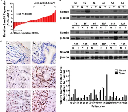
Figure 1. The expression of Sam68 is upregulated in NSCLC tissues. A. Determination of Sam68 mRNA level by qRT-PCR. ΔCtN: Ct value of β-actin was subtracted from Ct value of Sam68 of paired normal tissue. ΔCtT: Ct value of β-actin was subtracted from that of Sam68 of NSCLC tissue. Bar value (ΔCtN-ΔCtT) represented the difference between Sam68 mRNA level of NSCLC tissues and paired normal tissues. Bar value ≤-1 indicated that the ex -pression of Sam68 was decreased in NSCLC tissues. Bar value ≥1 indicated that the ex-pression of Sam68 was increased in NSCLC tissues. B. Determination of protein level of Sam68 by western blotting in 18 pairs of NSCLC tissues. The lower panel showed the quantitative results. N: normal; T: tumor. C. Representative images of Sam68 expression in alveolar epithelium (a), bronchial epithelial (b), adjacent lung adenocarcinoma (c), lung adenocar-cinoma (d), adjacent squamous cell lung caradenocar-cinoma (e) and squamous cell lung caradenocar-cinoma (f). Scale bar: 50 μm.
0.1 μg of the TOP flash, 50 pmol siRNA oligos, 0.05 μg of β-galactosidase expressing plas-mid and flag-β-catenin expression plasplas-mid by
using Lipofectamine® 2000. 72 h after
trans-fection, cells were harvested and analyzed for luciferase activities using Luciferase Repor- ter Assay Kit (Promega Corporation, Fitchburg,
WI, USA). β-galactosidase (β-gal) activity was
measured in assay buffer (100 mM pH7.5 phosphate, 2 mM MgCl2, 100 mM β-mer-captoethanol, 1.33 mg/mL o-nitrophenyl
β-D-galactopyranoside) at 420 nm. Relative Luc ac-
tivity was calculated as the ratio of Luc/β-gal
activity.
Statistical analyses
Statistical analyses were performed by two-tailed Student t-test using GraphPad Prism 5
software (La Jolla, CA, USA). Protein quantifica -tion was performed by ImageJ 1.46r software (NIH, Bethesda, MD, USA). Correlation between Sam68 expression and clinicopathological fea-tures was performed by X2 test using SAS 8.02
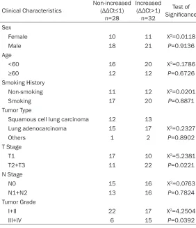
Table 1. Relationship between expression level of Sam68 and clinical and pathological features of the NSCLC individuals
Clinical Characteristics Non-increased (ΔΔCt≤1) n=28 Increased (ΔΔCt>1) n=32 Test of Significance Sex
Female 10 11 X2=0.0118
Male 18 21 P=0.9136
Age
<60 16 20 X2=0.1786
≥60 12 12 P=0.6726
Smoking History
Non-smoking 11 12 X2=0.0201
Smoking 17 20 P=0.8871
Tumor Type
Squamous cell lung carcinoma 12 13
Lung adenocarcinoma 15 17 X2=0.2327
Others 1 2 P=0.8902
T Stage
T1 17 10 X2=5.2381
T2+T3 11 22 P=0.0221
N Stage
N0 15 16 X2=0.0763
N1+N2 13 16 P=0.7824
Tumor Grade
I+II 22 17 X2=4.2504
III+IV 6 15 P=0.0392
Kaplan-Meier survival analysis was performed by logrank test using original data from KM plotter (http://www.kmplot.com/) or TCGA da- tabase (https://cancergenome.nih.gov/). Dif- ferences with P<0.05 were considered stati-
stically significant. Data were represented as
mean ± standard deviation (SD). Results
Upregulation of Sam68 in primary NSCLC tis-sues
We firstly applied qRT-PCR, western blotting
and IHC methods to determine the expression levels of Sam68 in 60 pairs of primary NSCLC tissues. The results displayed that both mRNA and protein levels of Sam68 were much high- er in NSCLC tissues than those in matched non-cancerous lung tissues (Figure 1A and 1B). Next, IHC analysis revealed that the ma- jor immunoreactivity of Sam68 located in the nucleus. In agreement with the above results, the immunoreactivity of Sam68 was more obvi-ously stained in the NSCLC tissues, compared
with the paired adjacent lung tissues. Moreover, the immu-noreactivity was even absent in the normal lung tissues (Figure 1C). Collectively, these
results confirmed that Sam68
is upregulated in NSCLC tis-sues at both transcriptional and translational levels, keep-ing in line with the previous report [22].
The prognostic significance of
Sam68 in NSCLC
Basing on the abovemention- ed results of Sam68 expres-sion in NSCLC tissues (Figure 1), we re-grouped these sam-ples according to mRNA level as non-increased Sam68 gro- up (n=28) and increased Sam- 68 group (n=32). Then statis- tic X2 analysis was conducted
to evaluate the association between Sam68 expression and each clinicopathological feature. As shown in Table 1, higher Sam68 expression was strongly associated with high- er T stage (P=0.0221) and advanced tumor grade (P=0.0392) in this cohort of 60 NSCLC patients. However, we found no difference between Sam68 expres-sion and other clinical variables, such as age, sex, smoking history, tumor type or N stage (P>0.05). In addition, Kaplan-Meier survival
analysis was further performed by logrank test using original data from KM plotter or TCGA database to elucidate the prognosis value of Sam68 in NSCLC. As shown in Figure 2A and 2B, patients with higher levels of Sam68
har-bored significantly shorter overall survival rates
than patients with lower Sam68 expression (P<0.0001), as calculated from two indepen-dent databases. Combining with the above results, we strengthened the conclusion that Sam68 is overexpressed in NSCLC tissues, and its higher expression predicts worse out- comes.
Upregulation of Sam68 in primary NSCLC cell lines
Figure 2. Higher Sam68 expression predicts worse overall survival. A. Kaplan-Meier analysis of the correlation be-tween Sam68 and overall survival of the LC patients using the data from KM plotter (n=1926, http://www.kmplot. com). B. Kaplan-Meier analysis of the correlation between Sam68 and overall survival of the LC patients using the data from TCGA database (n=230, https://cancergenome.nih.gov/). Patients were stratified into high and low ex -pression subgroups using the median of Sam68 mRNA level.
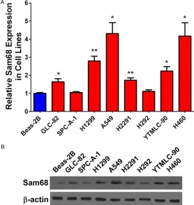
Figure 3. Expression of Sam68 is upregulated in lung cancer cells. A. Deter-mination of Sam68 mRNA by qRT-PCR in human bronchial epithelial Beas-2B cells and eight lung cancer cell lines (GLC-82, SPC-A-1, H1299, A549, H2291, H292, YTMLC-90 and H460). B. Determination of protein level of Sam68 by western blotting in human bronchial epithelial Beas-2B cells and eight lung cancer cell lines. *: P<0.05, **: P<0.01 versus Beas-2B cells.
epithelial cell line was also examined. As shown in Figure 3A, we observed that Sam68 was markedly overexpressed in most of the NSCLC cell lines. Among them, 4 cell lines exhibited
over 2-folds upregulation than the normal bronchial epithelial Beas-2B cells. Moreover, Sam-
68 protein was also confirmed
to be increased in six lung can-cer cell lines (Figure 3B). Taken
together, Sam68 is significant -ly upregulated in both NSCLC tissues and cell lines, indicat-ing its tumor promotindicat-ing roles in NSCLC.
Knockdown of Sam68 inhibits cell proliferation and cell cycle progression of NSCLC cells
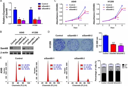
To investigate the biological function of Sam68 in NSCLC, Sam68-silenced A549 and H1- 299 cells were constructed us- ing two independent siRNAs
targeting Sam68. Efficient
[image:6.612.92.370.307.601.2]Figure 4. Knockdown of Sam68 inhibits cell proliferation of lung cancer cell. (A) A549 or H1299 cells were trans-fected with either control siRNA oligos or two Sam68 siRNA oligos. 72 h later, mRNA levels of Sam68 were detected by qRT-PCR. (B) A549 or H1299 cells were transfected as described in (A). 72 h later, protein levels of Sam68 were detected by western blotting. (C) A549 or H1299 cells were transfected as described in (A). In 24 h, cells were tryp-sinzed and seeded into 96-well plate. Cell proliferation assay was performed as described in Materials and Meth-ods. (D) A549 or H1299 cells were transfected as described in (A). In 24 h, the cells were trypsinzed and seeded into 12-well plate. Colony formation assay was performed as described in Materials and Methods. (E) A549 or H1299 cells were transfected as described in (A). In 72 h, the cells were harvested for cell cycle assay as described in Materials and Methods. The left panel depicted DNA content and the right panel showed the quantitative results. *: P<0.05, **: P<0.01, ***: P<0.001 versus control group.
was impaired by knockdown of Sam68, PI
staining followed by flow cytometry analysis
was performed in A549 cells. The result dem-onstrated that knockdown of Sam68 caused G0/G1 arrest (Figure 4E), indicating that Sam68 may promote NSCLC cell proliferation in a cell cycle-dependent manner.
Knockdown of Sam68 suppresses cell
prolif-eration via inhibiting Wnt/β-catenin signaling
Available data indicate that Wnt/β-catenin sig -naling substantially impacts on NSCLC tumori-genesis, prognosis, and resistance to therapy [25]. Meanwhile, we realized from one report that Sam68 could promote the nuclear
accu-mulation of β-catenin, facilitie Wnt/β-catenin
signaling activation and upregulate TCF/LEF
transcription activity in breast cancer cells [26]. To investigate whether knockdown of
Sam68 altered Wnt/β-catenin signaling activi -ty, luciferase reporter assay was utilized using its signaling reporter TOP Flash or FOP Flash which contains three copies of wild type or mutant TCF binding motif [27]. We found that knockdown of Sam68 suppressed the TOP
Flash activity, whereas no significant change
could be observed on TOP Flash activity (Figu- re 5A). Next, qRT-PCR and western blotting
were performed to examine the levels of
flag-Figure 5. Knockdown of Sam68 suppresses cell proliferation by inhibiting Wnt/β-catenin signaling pathway. A. A549 cells were co-transfected with 0.1 μg of the TOP flash or FOP flash reporter plasmid, 50 pmol siRNA oligos and 0.05 μg of β-galactosidase expressing plasmid. 72 h later, cells were lysed, luciferase and β-galactosidase activities were examined. Fold induction (Luc/β-gal) were calculated and expressed as means ± SD. B. Knockdown of Sam68 reduced the mRNA levels of downstream cyclin D1 and c-Myc genes as determined by qRT-PCR in A549 cells. C. Knockdown of Sam68 decreased the protein levels of downstream cyclin D1 and c-Myc as determined by western blotting in A549 and H1299 cells. D. H1299 cells were co-transfected with 0.1 μg of the TOP flash, 50 pmol siRNA oligos, 0.05 μg of β-galactosidase expressing plasmid and 0.25 μg flag-β-catenin expression plasmid. 72 h later, cells were lysed, luciferase and β-galactosidase activities were examined. Fold induction (Luc/β-gal) were calculated and expressed as means ± SD. E. H1299 cells were co-transfected with Sam68 siRNA oligos and flag-β-catenin expression plasmid. In 24 h, the cells were trypsinzed and seeded into 12-well plate. Colony formation assay was performed as described in Materials and Methods. **: P<0.01, ***: P<0.001, NS: no significance versus control group.
β-catenin in H1299 cells to investigate whether
loss of Sam68 could also alter the activated
level of Wnt/β-catenin signaling. We found that knockdown of Sam68 effectively inhibited flag-β-catenin-activated Wnt luciferase reporter
(P<0.001, Figure 5D). Finally, we investigated whether Sam68 knockdown could also inhibit
colony formation when Wnt/β-catenin signaling
was activated. The results showed that after
β-catenin was overexpressed, the clonogenic formation ability was significantly impaired by
loss of Sam68 (~70% inhibition), compared with that at basal level (~50% inhibition) (Figure 5E). Taken together, we demonstrated that knockdown of Sam68 suppressed NSCLC cell
proliferation via inhibiting Wnt/β-catenin
sig-naling. Discussion
[image:8.612.91.523.74.448.2]and NSCLC accounts for approximately 80% of all lung cancer subtypes, which contains ade-nocarcinoma, squamous cell carcinoma and large cell carcinoma [12, 28]. The 5-year sur-vival rate for lung cancer is only 16%, because a high proportion of patients are already with metastatic and advanced disease at diagnosis [13]. High mortalities of lung cancer patients can be majorly attributed to: (a) absence of accurate early diagnostic technologies, (b) re- sistance to currently available therapeutic me- thods. Short of understanding the precise molecular mechanism governing lung cancer carcinogenesis makes the actuality worse. Gene regulation in eukaryotes is a complex, multi-step process, including RNA transcrip-tion, alternative splicing, transport, localization and translation [29], in which RBPs partner the nascent RNA throughout its journey in the cell. The multi-functionality and the vast scope of targets regulated by RBPs make them impor-tant post transcriptional regulators. Now in- creasing evidence shows that the RBPs are dysregulated and make contributions to can- cer development [30].
In the present study, we sought to investigate
the expression, clinical significance and
bio-logical roles of Sam68, a well-studied RBP, in
human NSCLC cells. The first mentioned two
aspects have been disclosed by Zhang et al previously, that is, Sam68 is overexpressed in NSCLC tissues and high Sam68 expression predicts poor prognosis [22]. Through system-atic investigation, we also conclude that Sam- 68 is upregulated in both NSCLC tissues and cell lines, and its higher expression is signifi -cantly associated with clinical T stage, advan- ced tumor grade, and short overall survival. Besides, our cellular functional studies showed that knockdown of Sam68 inhibited cell prolif-eration, colony formation and cell cycle
pro-gression. Therefore, our findings add to the
un-derstanding of the mechanism of NSCLC tu- morigenesis in association with Sam68. Fur- thermore, our mechanistic studies revealed that knockdown of Sam68 suppresses cell
proliferation via inhibiting Wnt/β-catenin sig -naling, which is prominently activated in NSCLC and plays a critical role in lung tumorgenesis and metastasis [31]. However, the detailed mechanisms of Sam68 in promoting cell
prolif-eration through activating Wnt/β-catenin sig
-naling remain unclear. Its RNA binding and scaffolding features may be used as the cen- tral breakthroughs. In addition, whether other mechanisms exist which underlies Sam68-mediated growth promotion in NSCLC needs to be further explored.
In summary, our study provides the first com -prehensive view of aberrant upregulation of Sam68 in NSCLC cell proliferation in vitro via a
mechanism linked to activation of the
Wnt/β-catenin signaling pathway. We believe that our
findings provide new insights into the molecular
pathogenesis of NSCLC and implicate Sam68 as a potential prognostic biomarker and thera-peutic target for NSCLC.
Acknowledgements
This study was supported by grants from the National Natural Science Foundation of China (No. 81572288, to Qinghua Zhou; No. 81302002, to Xuebing Li; No. 81502166, to Xuexia Zhou), the Key Project of International Cooperation of Science and Technology Inno- vation between Governments, the National Key Research and Development Plan of China (No. 2016YEE0103400, to Qinghua Zhou), the Tianjin Natural Science Foundation (No. 14JC- QNJC12300, to Xuebing Li; No. 17JCYBJC2- 7100, to Xuexia Zhou; No. 17JCQNJC11700, to Hongli Pan; No. 17JCYBJC25400, to Yaguang Fan), and the “New Century” Talent Training Project of Tianjin Medical University General Hospital (2014, to Xuebing Li; 2016, to Xuexia Zhou).
Disclosure of conflict of interest
None.
Address correspondence to: Drs. Qinghua Zhou and Xuebing Li, Tianjin Key Laboratory of Lung Cancer Metastasis and Tumor Microenvironment, Tianjin Lung Cancer Institute, Tianjin Medical University General Hospital, No. 154 Anshan Street, Heping District, Tianjin 300052, P. R. China. Tel: +862260363020; Fax: +862260363013; E-mail: [email protected] (QHZ); [email protected] (XBL)
References
[2] Taylor SJ and Shalloway D. An RNA-binding pro-tein associated with Src through its SH2 and SH3 domains in mitosis. Nature 1994; 368: 867-871.
[3] Lin Q, Taylor SJ and Shalloway D. Specificity and determinants of Sam68 RNA binding. Im-plications for the biological function of K ho-mology domains. J Biol Chem 1997; 272: 27274-27280.
[4] Paronetto MP, Messina V, Bianchi E, Barchi M, Vogel G, Moretti C, Palombi F, Stefanini M, Ge-remia R, Richard S and Sette C. Sam68 regu-lates translation of target mRNAs in male germ cells, necessary for mouse spermatogenesis. J Cell Biol 2009; 185: 235-249.
[5] Stoss O, Olbrich M, Hartmann AM, Konig H, Memmott J, Andreadis A and Stamm S. The STAR/GSG family protein rSLM-2 regulates the selection of alternative splice sites. J Biol Chem 2001; 276: 8665-8673.
[6] Lukong KE and Richard S. Sam68, the KH do-main-containing superSTAR. Biochim Biophys Acta 2003; 1653: 73-86.
[7] Bielli P, Busa R, Paronetto MP and Sette C. The RNA-binding protein Sam68 is a multifunc-tional player in human cancer. Endocr Relat Cancer 2011; 18: R91-R102.
[8] Li QH, Haga I, Shimizu T, Itoh M, Kurosaki T and Fujisawa J. Retardation of the G2-M phase pro-gression on gene disruption of RNA binding protein Sam68 in the DT40 cell line. FEBS Lett 2002; 525: 145-150.
[9] Valacca C, Bonomi S, Buratti E, Pedrotti S, Bar-alle FE, Sette C, Ghigna C and Biamonti G. Sam68 regulates EMT through alternative splicing-activated nonsense-mediated mRNA decay of the SF2/ASF proto-oncogene. J Cell Biol 2010; 191: 87-99.
[10] Richard S, Vogel G, Huot ME, Guo T, Muller WJ and Lukong KE. Sam68 haploinsufficiency de -lays onset of mammary tumorigenesis and me-tastasis. Oncogene 2008; 27: 548-556. [11] Siegel RL, Miller KD and Jemal A. Cancer
sta-tistics, 2016. CA Cancer J Clin 2016; 66: 7-30. [12] Torre LA, Bray F, Siegel RL, Ferlay J, Lortet-Tieu-lent J and Jemal A. Global cancer statistics, 2012. CA Cancer J Clin 2015; 65: 87-108. [13] Torre LA, Siegel RL and Jemal A. Lung cancer
statistics. Adv Exp Med Biol 2016; 893: 1-19. [14] Chen W, Zheng R, Baade PD, Zhang S, Zeng H,
Bray F, Jemal A, Yu XQ and He J. Cancer statis-tics in China, 2015. CA Cancer J Clin 2016; 66: 115-132.
[15] Dehghani M, Keshtgar L, Javaheri MR, Derakh-shan Z, Oliveri Conti G, Zuccarello P and Fer-rante M. The effects of air pollutants on the mortality rate of lung cancer and leukemia. Mol Med Rep 2017; 15: 3390-3397.
[16] Downward GS, Hu W, Rothman N, Reiss B, Tromp P, Wu G, Wei F, Xu J, Seow WJ, Chapman RS, Lan Q and Vermeulen R. Quartz in ash, and air in a high lung cancer incidence area in Chi-na. Environ Pollut 2017; 221: 318-325. [17] Zheng R, Zeng H, Zuo T, Zhang S, Qiao Y, Zhou
Q and Chen W. Lung cancer incidence and mortality in China, 2011. Thorac Cancer 2016; 7: 94-99.
[18] Aviel-Ronen S, Blackhall FH, Shepherd FA and Tsao MS. K-ras mutations in non-small-cell lung carcinoma: a review. Clin Lung Cancer 2006; 8: 30-38.
[19] Liu C, Xu X and Zhou Y. Association between EGFR polymorphisms and the risk of lung can-cer. Int J Clin Exp Pathol 2015; 8: 15245-15249.
[20] Nicholson RI, Gee JM and Harper ME. EGFR and cancer prognosis. Eur J Cancer 2001; 37 Suppl 4: S9-15.
[21] Rodin SN and Rodin AS. Human lung cancer and p53: the interplay between mutagenesis and selection. Proc Natl Acad Sci U S A 2000; 97: 12244-12249.
[22] Zhang Z, Xu Y, Sun N, Zhang M, Xie J and Jiang Z. High Sam68 expression predicts poor prog-nosis in non-small cell lung cancer. Clin Transl Oncol 2014; 16: 886-891.
[23] Li X, Zhou X, Li Y, Zu L, Pan H, Liu B, Shen W, Fan Y and Zhou Q. Activating transcription fac-tor 3 promotes malignance of lung cancer cells in vitro. Thorac Cancer 2017; 8: 181-191. [24] Li X, Zhou X, Fan Y, Zhang Y, Zu L, Yao F and
Zhou Q. WW45, a Gli1 binding protein, nega-tively regulated hedgehog signaling in lung cancer. Oncotarget 2016; 7: 68966-68975. [25] Stewart DJ. Wnt signaling pathway in non-
small cell lung cancer. J Natl Cancer Inst 2014; 106: djt356.
[26] Wang L, Tian H, Yuan J, Wu H, Wu J and Zhu X. CONSORT: Sam68 is directly regulated by MiR-204 and promotes the self-renewal potential of breast cancer cells by activating the Wnt/ Beta-catenin signaling pathway. Medicine (Baltimore) 2015; 94: e2228.
[27] Ishitani T, Ninomiya-Tsuji J, Nagai S, Nishita M, Meneghini M, Barker N, Waterman M, Bower-man B, Clevers H, Shibuya H and Matsumoto K. The TAK1-NLK-MAPK-related pathway an-tagonizes signalling between beta-catenin and transcription factor TCF. Nature 1999; 399: 798-802.
[29] Aparicio LA, Abella V, Valladares M and Figueroa A. Posttranscriptional regulation by RNA-binding proteins during epithelial-to-mes-enchymal transition. Cell Mol Life Sci 2013; 70: 4463-4477.
[30] Wurth L and Gebauer F. RNA-binding proteins, multifaceted translational regulators in can- cer. Biochim Biophys Acta 2015; 1849: 881-886.