ORIGINAL RESEARCH
PERIPHERAL NERVOUS SYSTEM
Diffusivity Measurements Differentiate Benign from Malignant
Lesions in Patients with Peripheral Neuropathy or Plexopathy
E.L. Yuh, S. Jain Palrecha, G.M. Lagemann, M. Kliot, P.R. Weinstein, N.M. Barbaro, and C.T. Chin
ABSTRACT
BACKGROUND AND PURPOSE:Peripheral nerve disorders caused by benign and malignant primary nerve sheath tumors, infiltration or compression of nerves by metastatic disease, and postradiation neuritis demonstrate overlapping features on conventional MR imaging but require vastly different therapeutic approaches. We characterize and compare diffusivities of peripheral nerve lesions in patients undergoing MR neurography for peripheral neuropathy or brachial or lumbosacral plexopathy.
MATERIALS AND METHODS: Twenty-three patients, referred for MR neurography at our institution between 2003 and 2009 for a peripheral mononeuropathy or brachial or lumbosacral plexopathy and whose examinations included DWI, received a definitive diagnosis, based on biopsy results or clinical and imaging follow-up, for a masslike or infiltrative peripheral nerve or plexus lesion suspicious for tumor. Mean ADC values were determined within each lesion and compared across 3 groups (benign lesions, malignant lesions, and postradiation changes).
RESULTS:Both ANOVA and Kruskal-Wallis tests demonstrated a statistically significant difference in ADC values across the 3 groups (P⫽ .000023,P⫽.00056, respectively). Post hoc pair-wise comparisons showed that the ADC within malignant tumors differed significantly from that within benign tumors and postradiation changes. ADC within benign tumors and postradiation changes did not differ significantly from each other.
CONCLUSIONS: DWI may be highly effective for the differentiation of benign from malignant peripheral nerve masslike or infiltrative lesions.
P
eripheral neuropathies can be divided into mononeuropa-thies, polyneuropamononeuropa-thies, and plexopathies. Patients present with pain, sensory symptoms, and/or motor deficits in the distri-bution of a single peripheral nerve, multiple peripheral nerves, or a nerve plexus. Mononeuropathies affect a single peripheral nerve. Polyneuropathies affect multiple peripheral nerves. In plexopathies, symptoms are localized to the brachial or lumbosa-cral plexus.Polyneuropathies are generally attributable to systemic dis-eases (eg, diabetes and vitamin deficiencies), while mononeu-ropathies are most often due to trauma, nerve compression syndromes that occur at a few characteristic anatomic loca-tions, or mass lesions. History and physical examination, sup-plemented in a subset of cases by laboratory studies, electrodi-agnostic studies, and neuroimaging, are the main tools for diagnostic evaluation. In patients with a classic compression mononeuropathy, such as median nerve compression at the carpal tunnel, the diagnosis can often be made clinically and corroborated by needle electromyography, nerve conduction, and/or imaging studies.1-4 For mononeuropathies involving nerves not typically susceptible to compression syndromes, imaging can play an essential role in identifying the lesion and guiding management.
Plexopathies give rise to motor and/or sensory deficits in an extremity. Most brachial plexopathies (75%) are attributable to postradiation changes, primary and metastatic lung cancer, or metastatic breast cancer.5Common causes of lumbosacral plex-opathy are primary and metastatic tumor, including cervical, en-dometrial, ovarian, prostate, testicular, and colorectal cancer; Received February 22, 2014; accepted after revision June 23.
From the Departments of Radiology and Biomedical Imaging (E.L.Y., C.T.C.) and Neuro-logical Surgery (P.R.W.), University of California at San Francisco, San Francisco, Califor-nia; San Leandro Medical Center (S.J.P.), The Permanente Medical Group, San Leandro, California; Department of Radiology (G.M.L.), University of Pittsburgh, Pittsburgh, Penn-sylvania; Department of Neurosurgery (M.K.), Northwestern University Feinberg School of Medicine, Chicago, Illinois; and Goodman Campbell Brain and Spine (N.M.B.) and Department of Neurological Surgery (N.M.B.), Indiana University, Indianapolis, Indiana. E.L. Yuh and S. Jain Palrecha contributed equally to this work.
Please address correspondence to Cynthia T. Chin, MD, Department of Radiology and Biomedical Imaging, University of California at San Francisco, 505 Parnassus Ave, Box 0628, San Francisco, CA 94143-0628; e-mail: [email protected]
Indicates article with supplemental on-line table.
postradiation changes; and diabetes.6For patients with a history of radiation for malignancy, recurrent tumor with nerve invasion must be distinguished from radiation plexopathy; both can de-velop months to years following therapy and can have similar clinical presentations.6
Although benign and malignant primary nerve sheath tumors, infiltration of nerves by metastatic disease, and postradiation neuritis require different therapeutic approaches, they also dem-onstrate overlapping features on MR imaging, including T2 hy-perintensity, focal enlargement, and enhancement.7,8Diffusivity measurements from DWI may be helpful in differentiating dis-tinct pathologic entities. In prior studies, DWI was useful in dif-ferentiating malignant and benign peripheral nerve sheath tu-mors,9retroperitoneal masses,10head and neck tumors,11,12and lymph nodes.13,14Other studies have demonstrated differences in the diffusivities of adult15or pediatric brain tumors16that corre-late with tumor grade and/or histologic type. In this study, we focus on masslike or infiltrative lesions of the peripheral nerves detected by MR imaging in patients presenting clinically with a peripheral mononeuropathy or plexopathy. We characterize and compare the diffusivities of these lesions and demonstrate signif-icant differences among benign and malignant peripheral nerve tumors and postradiation changes.
MATERIALS AND METHODS
Study PopulationTwenty-three patients referred for MR neurography at our insti-tution between 2003 and 2009 by neurologists, neurosurgeons, and oncologists for a clinical indication of a peripheral monon-europathy or brachial or lumbosacral plexopathy and who re-ceived a definitive diagnosis of a masslike or infiltrative nerve lesion based on biopsy results, long-term clinical and imaging follow-up, or intermediate-term follow-up supplemented by PET, neurologic examination, and/or nerve conduction studies were included in the study population. We included patients with noncystic “mass”-like lesions, defined as noncystic lesions with a diameter at least 50% larger than that of the apparent nerve of origin as well as patients with more infiltrative lesions consisting of more subtle nerve thickening and/or effacement of normally visualized interfascicular and surrounding fat planes without the presence of a well-defined “mass.” Study participants were di-vided into 3 groups according to the final most likely pathologic diagnosis. Group 1 (n⫽10) consisted of benign lesions; Group 2 (n⫽7), malignant tumors; and Group 3 (n⫽6), postradiation changes, without evidence of residual tumor.
MR Neurography
MR imaging sequences performed at 1.5T (Gyroscan Intera 1.5T; Philips Healthcare, Best, the Netherlands) included axial and cor-onal STIR (TR⫽2200 ms, TE⫽20 ms, TI⫽160 ms, NEX⫽4, FOV⫽22, matrix⫽256⫻192, slice thickness/gap⫽3/0.3 mm), T1 and fat-saturated postgadolinium T1-weighted spin-echo (TR⫽500, TE⫽14, NEX⫽3), and DWI (single-shot echo-planar imaging, 6 directions, TR⫽2 ms⫻pulse-pulse interval, TE⫽15 ms, FOV⫽22, matrix⫽256⫻144, slice thickness/ gap⫽5.0/0.5 mm, b-value⫽400 s/mm2). ADC maps were
cal-culated using the Philips vendor software on the MR imaging scanner console immediately after acquisition of DWI data.
Mean and SD of ADC values within approximately 1-cm ROIs drawn within the lesions were determined independently by 2 board-certified radiologists. Each radiologist drew multiple ROIs for lesions that exceeded 3 cm and averaged the means within these ROIs, to avoid limited regional sampling of spatially heter-ogeneous lesions. Nonenhancing T2 hyperintense areas suspi-cious for cystic or necrotic areas were not included within any ROI. Volumes of masslike lesions were approximated by using the volume formula for an ellipsoid; volume⫽(4/3)⫻⫻a⫻b⫻c, where a, b, and c were orthogonal linear dimensions of the lesion measured by one radiologist. (Volume measurements were not performed on infiltrative lesions that consisted of more subtle nerve thickening without a well-defined mass with a diameter at least 50% larger than that of the nerve of origin.)
Statistical Analysis
[image:2.594.303.531.46.287.2]FIG 2. Biopsy-proven schwannoma of the left median nerve in a 48-year-old woman presenting with left upper extremity pain and paresthesias. Axial fat-suppressed T2 (A), axial fat-saturated postgadolinium T1 (B), maximum-intensity-projection DWI (C), and axial ADC (D) demonstrate a vividly enhancing, heterogeneously T2 hyperintense mass (arrows) along the median nerve. The ADC value within the mass was 2.1⫾0.36⫻10⫺3 mm2/s. The lesion was resected due to progressive symptoms.
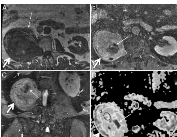
[image:3.594.141.447.46.326.2] [image:3.594.140.446.377.696.2]FIG 4. Metastatic renal cell carcinoma in a 46-year-old woman with left arm weakness. Axial STIR (A), axial fat-saturated postgadolinium T1 (B), axial DWI (C), and axial ADC (D) demonstrate a T2 hyperintense, enhancing mass (arrows) along the left C6 nerve just outside the left C5–C6 neural foramen. ADC within the mass was 1.08⫾0.09⫻10⫺3mm2/s. The mass was subtotally resected and irradiated.
[image:4.594.140.447.48.371.2] [image:4.594.140.446.417.655.2]tween separate measurements performed for each lesion by a sin-gle radiologist. Interrater reliability was determined through the Pearson correlation coefficient between measurements per-formed on each lesion by the 2 different radiologists.
One-way between-groups ANOVA was performed to assess for a statistically significant difference in ADC values among the 3 groups (benign lesions, malignant lesions, and postradi-ation changes) atP⬍.05. Post hoc pair-wise comparisons were FIG 6. Biopsy-proven non-Hodgkin lymphoma infiltrating the right sciatic nerve. Axial STIR (A), axial fat-saturated postgadolinium T1 (B), and axial ADC map (C) demonstrate a T2 hyperintense minimally enhancing soft-tissue mass (arrows) along the right sciatic nerve at the greater sciatic foramen. ADC within the mass was 0.69⫾0.33⫻10⫺3mm2/s. CT-guided biopsy was nondiagnostic.D, MR imaging– guided biopsy was performed, yielding a pathologic diagnosis of diffuse large B-cell lymphoma. The patient underwent chemotherapy and radiation to the mass.
[image:5.594.134.453.46.353.2] [image:5.594.134.453.422.614.2]then performed using the Tukey test to assess for statistically significant pair-wise differences between pairs of groups at P⬍.05.
Although ADC values within each group satisfied the Shapiro-Wilk test for normality, the sensitivity of this test for nonnormal-ity may be reduced for small sample sizes.17The nonparametric Kruskal-Wallis test was also performed on the ADC values to assess for a statistically significant difference across groups atP⬍ .05. Post hoc nonparametric pair-wise comparisons were per-formed using Mann-WhitneyUtests to assess for statistically sig-nificant differences between pairs of groups atP⬍.05. All statis-tical analyses were performed using SPSS, Version 19 (IBM, Armonk, New York).
This study was approved by the insti-tutional committee on human subjects research. All patient data stored for the purpose of this research study were ano-nymized by using accession numbers to identify each case.
RESULTS
The On-line Table shows diagnoses in 23 patients divided into 3 groups on the basis of biopsy results (n⫽10); long-term clin-ical and imaging follow-up (n⫽10; mean follow-up, 40⫾31 months); or interme-diate-term follow-up supplemented by PET, neurologic examination, and/or nerve conduction studies (n⫽3, mean follow-up, 4⫾1 months). Group 1 (n⫽ 10) consisted of benign masses, including 4 proven schwannomas, 1 biopsy-proven neurofibroma, and 5 peripheral nerve masses that demonstrated both long-term stability in size and conven-tional MR imaging features characteristic of a benign primary nerve sheath tumor. Group 2 (n⫽7), malignant tumors, con-sisted of 1 biopsy-proven malignant pe-ripheral nerve sheath tumor, 1 biopsy-proven rhabdomyosarcoma, 2 cases of metastatic breast cancer involving the brachial plexus (biopsy-proven) or the sacral nerves, 1 case of biopsy-proven metastatic renal cell carcinoma involving a cervical nerve, 1 case of biopsy-proven non-Hodgkin lymphoma involving the sciatic nerve, and 1 case of acute lym-phoblastic leukemia involving the bra-chial plexus. Group 3 (n⫽6) consisted of postradiation changes without evi-dence of residual tumor in 6 cases of previously irradiated malignancies, in-cluding 3 cases of breast cancer, 1 high-grade sarcoma, 1 case of Hodgkin lym-phoma, and 1 case of oral squamous cell carcinoma. Lesion volumes ranged from 1 to 5530 cm3(median, 37 cm3; 25th percentile, 14 cm3; 75th percentile, 122 cm3).
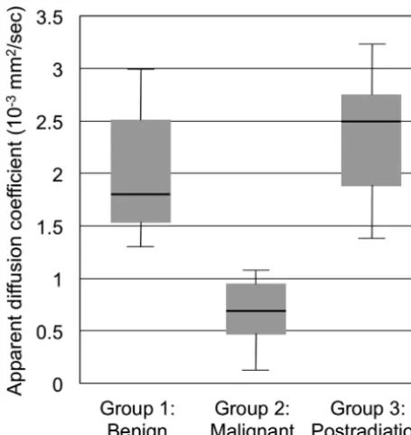
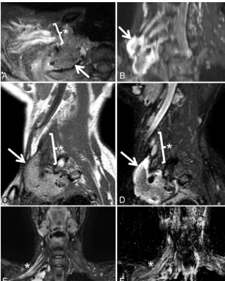
Fig 1 demonstrates ADC values within Groups 1–3. Postradi-ation changes without evidence of residual tumor (Group 3) dem-onstrated the highest median ADC value of 2.50⫻10⫺3mm2/s (interquartile range⫽0.87⫻10⫺3mm2/s, minimum⫽1.39⫻ 10⫺3mm2/s, maximum⫽3.22⫻10⫺3mm2/s, mean ADC⫽ 2.37⫻10⫺3mm2/s, SD⫽0.61⫻10⫺3mm2/s), followed closely by benign lesions (Group 1) with a median ADC value of 1.81⫻ 10⫺3mm2/s (interquartile range⫽0.98⫻10⫺3mm2/s, mini-mum⫽1.30⫻10⫺3mm2/s, maximum⫽2.97⫻10⫺3mm2/s, mean ADC⫽1.99⫻10⫺3mm2/s, SD⫽0.60⫻10⫺3mm2/s). Malignant lesions (Group 2) recorded the lowest median ADC FIG 8. Postradiation brachial plexopathy in a 42-year-old woman with progressive right upper
[image:6.594.53.373.46.445.2]value of 0.69⫻10⫺3mm2/s (interquartile range⫽0.49⫻10⫺3 mm2/s, minimum⫽0.26⫻10⫺3mm2/s, maximum⫽1.08⫻ 10⫺3mm2/s, mean ADC⫽0.69⫻10⫺3mm2/s, SD⫽0.28⫻ 10⫺3mm2/s). Intrarater and interrater reliability for ADC mea-surements, characterized by the Pearson correlation coefficientr, werer⫽0.98 (P⫽7.0⫻10⫺7) for intrarater reliability andr⫽ 0.88 (P⫽.0011) for interrater reliability.
One-way between-groups ANOVA demonstrated a statisti-cally significant difference in ADC values across the 3 groups:F (df1⫽2,df2⫽20)⫽19.0,P⫽.000023. Post hoc pair-wise comparisons using the Tukey test showed that the mean ADC values for Group 1 (1.99⫻10⫺3mm2/s) and Group 2 (0.69⫻ 10⫺3 mm2/s) were significantly different at P ⫽ .0002 and mean ADC values for Group 2 (0.69 ⫻ 10⫺3 mm2/s) and Group 3 (2.37⫻10⫺3mm2/s) were significantly different at P⫽.000039. The ADC for Groups 1 and 3 did not differ sig-nificantly from each other (P⫽.36).
The nonparametric Kruskal-Wallis test also showed a statisti-cally significant difference in ADC across the 3 groups, with2 (df⫽2, 23 subjects)⫽15.0 andP⫽.00056. Finally, post hoc nonparametric pair-wise comparisons using the Mann-Whitney Utest showed a statistically significant difference in ADC between Groups 1 and 2 (P⫽.00010) and between Groups 2 and 3 (P⫽ .0012), but no statistically significant difference between Groups 1 and 3 (P⫽.97).
Figs 2–9 demonstrate MR images of representative cases from each of the 3 groups (benign lesions, malignant lesions, and post-radiation changes).
DISCUSSION
Peripheral polyneuropathies affect multiple peripheral nerves and are generally caused by systemic diseases, most commonly diabetes, and vitamin deficiency related to alcohol use or perni-cious anemia, in the United States and Europe. Polyneuropathies
are usually diagnosed through clinical history, physical examination, laboratory studies, nerve conduction studies, and nee-dle electromyography.6,18Unlike polyneu-ropathies, mononeuropathies and plex-opathies are most often due to trauma, nerve compression syndromes at charac-teristic anatomic locations, mass lesions, and postradiation changes; and imaging can play a decisive role in diagnosis. In a study of MR imaging of symptomatic nontraumatic brachial plexopathy, post-radiation change was the most common cause, accounting for 31% of cases, with metastatic breast cancer and primary or metastatic lung cancer accounting for 24% and 19% of cases, respectively. The remaining 26% of cases were caused by a wide variety of benign and malignant tu-mors.5Unlike many cases of diffuse poly-neuropathy, patients with brachial or lumbosacral plexopathy frequently un-dergo MR imaging due to the localized distribution of symptoms and presumed localized extent of the pathologic findings.19,20
We characterized the diffusivity of masslike or infiltrative le-sions of the peripheral nerves discovered on MR imaging per-formed for a clinical indication of peripheral mononeuropathy or brachial or lumbosacral plexopathy. We demonstrated a statisti-cally significant difference among the diffusivities of benign and malignant tumors and postradiation changes. This result is com-patible with other studies that have demonstrated diffusivity to be generally inversely correlated with tumor cellularity and tumor grade in several contexts, including adult and pediatric brain tu-mors, head and neck masses, lymph nodes, retroperitoneal soft-tissue masses,10-16,21and MR neurography.9Our results are also consistent with a prior study9in which a statistically significant difference in ADC values between benign (1.85⫾0.40⫻10⫺3 mm2/s) and malignant (0.90⫾0.25⫻10⫺3mm2/s) peripheral nerve tumors and tumorlike masses was reported (P⬍.001). In the current study, we extended the analysis to include infiltrative postradiation changes of the peripheral nerves that were not in-cluded in that previous study. We report a complete separation of ADC values between benign and malignant lesions, with malig-nant lesions demonstrating ADCⱕ1.08⫻10⫺3mm2/s, and be-nign lesions demonstrating ADCⱖ1.30⫻10⫺3mm2/s.
The apparent diffusion coefficient is a measure of the diffusiv-ity, or microscopic mobildiffusiv-ity, of water protons in tissue. The dif-ference in ADC values of the lesions in our study was likely due to factors such as tumor cellularity, integrity of cell membranes, nu-clear-to-cytoplasmic ratio, and the water content of the extracel-lular matrix. These have been postulated to account for low dif-fusivity within malignant solid tumors in prior studies.10-16,21A study of benign and malignant extracranial soft-tissue tumors in children showed an inverse relationship between ADC measure-ments and cellular density derived from histologic analysis, but correlation was only moderate (R2⫽0.54), and it was postulated FIG 9. Marked increase in diffusivity following radiation therapy for metastatic breast cancer in
[image:7.594.56.371.48.227.2]that additional factors such as extracellular water content also influence the ADC.22Myxoid matrix is known to be abundant in both schwannomas and neurofibromas. Schwannomas, particu-larly Antoni B tissue within schwannomas, contain a high water content, due to low cellularity and high mucin and low collagen content,23and this likely accounts for their high diffusivity.
Limitations of this study include the lack of biopsy-proven diagnoses for all patients, the likelihood that diffusion character-istics of postradiation changes evolve with time, and the need for validation in a larger study population. Some malignant periph-eral nerve sheath tumors, which may arise either de novo or from malignant degeneration of plexiform neurofibromas, have been shown to have a significant myxoid content; one prior study of soft-tissue masses reported that ADC failed to differentiate benign from malignant myxoid soft-tissue tumors, though only one ma-lignant peripheral nerve sheath tumor was included in that study.21ADC values in postradiation change are likely to be af-fected by features such as edema, fibrous-inflammatory reaction, and vascular permeability that are temporally dynamic, particu-larly in the first few months after treatment. Long-term follow-up imaging rather than biopsy was more often performed for lesions with benign features on conventional imaging, introducing a bias, because biopsy was performed on all lesions that ultimately ceived a malignant diagnosis but on only a subset of lesions re-ceiving a benign diagnosis. Finally, rare entities such as perineu-roma24and posttraumatic neuroma appear as mass lesions of the peripheral nerves, but were not included within our study population.
CONCLUSIONS
We demonstrate a statistically significant difference in diffusivity between biopsy-proven malignant lesions, and lesions with either biopsy-proven benign histology or stable size and benign features on follow-up imaging. Although our results need to be validated in a larger study population, the pattern of diffusivity values within benign and malignant lesions described here may be help-ful in selecting patients for percutaneous tissue sampling, debulk-ing versus en bloc total resection, and/or short-term clinical and imaging follow-up.
Disclosures: Nicholas M. Barbaro—UNRELATED:Grants/Grants Pending: National Institute of Neurological Disorders and Stroke,* Elekta.* *Money paid to the institution.
REFERENCES
1. Spinner RJ.Outcomes for peripheral nerve entrapment syndromes.
Clin Neurosurg2006;53:285–94
2. Britz GW, Haynor DR, Kuntz C, et al.Carpal tunnel syndrome: cor-relation of magnetic resonance imaging, clinical, electrodiagnostic, and intraoperative findings.Neurosurgery1995;37:1097–103 3. Guggenberger R, Markovic D, Eppenberger P, et al.Assessment of
median nerve with MR neurography by using diffusion-tensor
imaging: normative and pathologic diffusion values.Radiology
2012;265:194 –203
4. Keen NN, Chin CT, Engstrom JW, et al.Diagnosing ulnar neuropa-thy at the elbow using magnetic resonance neurography.Skeletal Radiol2012;41:401– 07
5. Wittenberg KH, Adkins MC.MR imaging of nontraumatic brachial plexopathies: frequency and spectrum of findings.Radiographics
2000;20:1023–32
6. Amato AA, Barohn RJ.Peripheral neuropathy.In: Longo DL, Fauci AS, Kasper DL, et al, eds.Harrison’s Principles of Internal Medicine.
18th ed. New York: McGraw-Hill; 2011:3448 –72
7. Stoll G, Bendszus M, Perez J, et al.Magnetic resonance imaging of the peripheral nerves.J Neurol2009;256:1043–51
8. Li CS, Huang GS, Wu HD, et al.Differentiation of soft tissue benign and malignant peripheral nerve sheath tumors with magnetic res-onance imaging.Clin Imaging2008;32:121–27
9. Chhabra A, Thakkar RS, Andreisek G, et al.Anatomic MR imaging and functional diffusion tensor imaging of peripheral nerve tumors and tumorlike conditions.AJNR Am J Neuroradiol2013;34:802– 07 10. Nakayama T, Yoshimitsu K, Irie H, et al.Usefulness of the calculated apparent diffusion coefficient value in the differential diagnosis of retroperitoneal masses.J Magn Reson Imaging2004;20:735– 42 11. Thoeny HC, De Keyzer F, King AD.Diffusion-weighted MR imaging
in the head and neck.Radiology2012;263:19 –32
12. Abdel Razek AA, Kandeel AY, Soliman N, et al.Role of diffusion-weighted echo-planar MR imaging in differentiation of residual or recurrent head and neck tumors and posttreatment changes.AJNR Am J Neuroradiol2007;28:1146 –52
13. Fornasa F, Nesoti MV, Bovo C, et al.Diffusion-weighted magnetic resonance imaging in the characterization of axillary lymph nodes in patients with breast cancer. J Magn Reson Imaging 2012;36: 858 – 64
14. Kos¸ucu P, Tekinbas C, Erol M, et al.Mediastinal lymph nodes: as-sessment with diffusion-weighted MR imaging.J Magn Reson Imag-ing2009;30:292–97
15. Yamasaki F, Kurisu K, Satoh K, et al.Apparent diffusion coefficient of human brain tumors at MR imaging.Radiology2005;235:985–91 16. Kan P, Liu JK, Hedlund G, et al.The role of diffusion-weighted mag-netic resonance imaging in pediatric brain tumors.Childs Nerv Syst
2006;22:1435–39
17. Shapiro SS, Wilk MB, Chen HJ.A comparative study of various tests for normality.J Am Statist Assoc1968;63:1343–72
18. England JD, Asbury AK.Peripheral neuropathy.Lancet2004;363: 2151– 61
19. Grant GA, Britz GW, Goodkin R, et al.The utility of magnetic reso-nance imaging in evaluating peripheral nerve disorders. Muscle Nerve2002;25:314 –31
20. Thyagarajan D, Cascino T, Harms G.Magnetic resonance imaging in brachial plexopathy of cancer.Neurology1995;45:421–27 21. Nagata S, Nishimura H, Uchida M, et al.Diffusion-weighted
imag-ing of soft tissue tumors: usefulness of the apparent diffusion coef-ficient for differential diagnosis.Radiat Med2008;26:287–95 22. Humphries PD, Sebire NJ, Siegel MJ, et al.Tumors in pediatric
pa-tients at diffusion-weighted MR imaging. Radiology 2007;245: 848 –54
23. Wippold FJ, Lubner M, Perrin RJ, et al.Neuropathology for the neuroradiologist: Antoni A and Antoni B tissue patterns.AJNR Am J Neuroradiol2007;28:1633–38