Characterization of serum complement activity in serum of
the Komodo dragon (Varanus komodoensis)
Mark Merchant1*, Danyell Henry1, Rodolfo Falconi1, Becky Muscher2, Judith Bryja3
1Department of Chemistry, McNeese State University, Lake Charles, USA 2Department of Herpetology, San Antonio Zoo, San Antonio, USA 3Department of Herpetology, Houston Zoo, Houston, USA
Email: *[email protected]
Received 27 July 2012; revised 3 September 2012; accepted 13 September 2012
ABSTRACT
Incubation of different volumes of serum from the Komodo dragon (Varanus komodoensis) with sheep red blood cells (SRBCs) resulted in volume-depend- ent hemolysis, as measured spectrophotometrically at 540 nm. The hemolysis occurred rapidly, with almost 90% of the hemolytic activity occurring within 20 min of incubation. A thermal profile showed that Komodo dragon serum exhibited low activity from 5˚C-20˚C, but exerted maximum activity at 35˚C, which was substantially reduced at 40˚C. The maxi-mum activity was observed near optimal tempera-tures to which Komodo dragons thermoregulate. Mild heat treatment of Komodo dragon serum (56˚C, 30 min) depleted the ability to hemolyze SRBCs. In addition, preincubation of Komodo dragon serum with only 5 mM EDTA or phosphate, both chelators of divalent metal ions, reduced the hemolytic activity sharply. These results indicate that the hemolytic ac-tivity was due to the presence of a potent serum com-plement system. Incubation of Komodo dragon serum with 5 mM EDTA and 15 mM Ca2+ or Mg2+, but not Ba2+, Zn2+, or Fe2+, completely restored activity. These results indicate that Komodo dragon serum complement activity requires the presence of Mg2+ or Ca2+. This is the first assessment of innate immune activity of a Varanid.
Keywords:Innate Immunity; SRBC Hemolysis Varanid
1. INTRODUCTION
Innate immunity comprises that part of microbial defense which responds in a non-specific manner to infiltration by microbes. Serum complement is one of the first lines of defense against infection, and is made up of approxi-mately 11 different proteins that can be activated by
three different mechanisms, the classical (antibody-de- pendent) pathway [1], the alternative pathway [2], and the lectin-dependent pathway [3]. Once activated, all three mechanisms work in a proteolytic cascade fashion, resulting in the eventual formation of a multi-protein “membrane attack complex” [4] in the outer membrane of the microbe, causing leakage of cellular contents and lysis. Some of the proteolytic complement protein frag-ments also act to opsonize microbes for phagocytosis [5], serve as attractants for the chemotaxis of macrophages and neutrophils to the site of infection [6], and function as anaphylactic factors [7], causing mast cell degranula-tion and vascular permeability [8]. Deficiencies in com-plement proteins have been associated with susceptibility to bacterial infection [9], Leiner’s disease [10], fulminant hepatic failure [11], and many other clinical conditions [12]. Serum complement is among the most important components of innate immunity, and has been identified and characterized across a broad spectrum of diverse taxa, including mammals [13] and ancient invertebrates [14,15].
The Komodo dragon (Varanus komodoensis) is the world’s largest lizard. Its range is restricted to several small islands in the Indonesian archipelago. These ani-mals are carnivorous reptilians, and are known to feed on prey items much larger than themselves. Komodo drag-ons typically lie at ambush points and bite their prey, delivering a heavy load of infectious bacteria from their saliva [16,17], and then often follow their prey for days, or even weeks, before they succumb to systemic infec-tion. In addition, Komodo dragons have been reported to deliver potent toxins in their bites that prevent coagula-tion and cause prey items to succumb to systemic shock [18]. Although these animals exhibit aggressive behav-iors toward members of their own species during territo-rial disputes, and often bite each other during feeding frenzies [19], Komodo dragons do not seem to experi-ence the same fate as their prey. This seems to indicate that these animals have developed an immune system
that allows them to combat potentially high loads of pathogenic bacteria. However, because of their remote location and endangered status, little is known concern-ing the physiology, biochemistry, and immunology of these apex predators. Recent studies in our laboratory have shown that the serum from Komodo dragons exhib-its potent and broad-acting antibacterial properties [20]. This study was undertaken to examine the serum com-plement system of the Komodo dragon, and is the first characterization of an innate immune component in a Varanid.
2. MATERIALS AND METHODS
Chemicals and biochemicals: Sheep red blood cells (SRBCs) were purchased from Rockland Immunoche- micals (Gilbertsville, PA, USA). Ethylene diamine tetra- acetate (EDTA), CaCl2, MgCl2, BaSO4, FeCl2, and ZnCl2
were purchased from Sigma Chemical Company (St. Louis, MO, USA).
Collection of samples: Blood samples were collected from Komodo dragons at the Houston and San Antonio zoos. Blood was drawn from the tail vein, transferred to Vaccutainer™ tubes, and allowed to clot for at least five h before the serum was collected. The amount of blood collected from each individual depended on the size of the animal, and was at the discretion of the attending veterinarian at each institution. Blood was collected from the tail caudal veins three adults (20 - 81.5 kg) and five juveniles (1.5 - 6.2 kg), transferred to Vaccutainer™ tubes, and allowed to clot for at least five hr before se-rum was collected by centrifugation. The sese-rum was pooled so that average antibacterial values for this spe-cies could be generated. The collection of blood from these animals was conducted in accordance with the Animal Care and Use institutional policies of the Hous-ton and San AnHous-tonio Zoos.
Serum volume-dependent SRBC hemolysis: The functionality of the Komodo dragon serum complement system of proteins was examined using a SRBC lysis assay modified from the method of Mayer [21], and pre-viously described for crocodilians [22]. To measure the volume dependence of SRBC activity on Komodo dra-gon serum, different volumes of serum (0 - 15 µL) were diluted to 150 µL total volume with a 0.9% saline solu-tion and then 150 µL of 2% SRBCs was added to the solution. The samples were allowed to incubate for 60 min, followed by centrifugation at 16,000 × g for 5 min at am- bient temperature. The optical densities of the resulting supernatants were measured using the Bio-Rad Bench- mark Plus™ microplate spectrophotometer at 540 nm.
Kinetic analysis of SRBC hemolysis: For the deter-mination of the hemolytic kinetic profile, a solution composed of 250 µL of Komodo dragon serum, 5.15 mL
of a 0.9% saline solution, and 5.40 mL of a 1% SRBC was prepared. In quadruplicate, 290 µL of solution was dispensed into separate vials. At different time intervals (0 - 120 min), the samples were centrifuged and trans-ferred to microtiter plates to measure optical density (540 nm) as described above.
Temperature-dependent SRBC hemolysis: Komodo dragon-mediated serum complement SRBC hemolysis activity was also assayed at different temperatures. Ali-quots of Komodo dragon serum (5 μL) and 145 μL of saline were incubated at different temperatures (5˚C - 40˚C in increments of 5˚C) for 10 min. The reaction was initiated by the addition of 150 μL of 2% SRBCs, and allowed to continue for 30 min. The samples were then centrifuged (16,000 × g) and transferred to microtiter plates to measure optical density (540 nm) as described above.
Effects of divalent metal ions and heat on Komodo dragon complement: To examine the effects chelators of divalent metal ions on the Komodo dragon comple-ment protein system, 100 μL of serum solution with SRBC and saline solution was spiked with either 1 μL of 500 mM EDTA or 500 mM Na3PO4. The resulting serum
samples (5 μL), which contained 5 mM EDTA or Na3PO4, were incubated (30 min) with 145 μL of saline
and 150 μL of 1% SRBCs. Another aliquot of serum (100 μL) was heated to 56˚C for 30 min. The serum was allowed to cool to ambient temperature, and then 5 μL was incubated (30 min) with 145 μL of saline and 150
μL of 1% SRBCs. These samples were centrifuged at 16,000 × g for 5 min, and the optical density of each sample was measured using the Bio-Rad Benchmark Plus™ microplate spectrophotometer at 540 nm.
To determine the specific requirement of Komodo dragon serum complement activity for divalent metal ions, serum was treated with 5 mM EDTA in the absence, and in the presence, of 15 µM CaCl2, MgCl2, ZnCl2,
BaSO4, and FeCl2 (Table 1). The samples (150 μL) were
then incubated with 150 μL of 2% SRBCs for 30 min at ambient temperature. The samples were centrifuged, and the optical densities of the supernatants were measured at 540 nm as described above.
Table 1. Effects of heat treatment and EDTA on hemolytic activity of serum from the Komodo dragon. Komodo dragon serum was treated with EDTA, or subjected to mild heat treat-ment, and then exposed to 1% SRBCs for 30 min at ambient temperature. The results are expressed as the percentage of maximum lysis, and represent the means ± standard deviation for four determinations.
SRBC Treatment % Maximum Activity
None 0.0%
Komodo dragon serum 97.3 ± 2.9%
Serum, 56˚C, 30 min 6.3 ± 0.1%
Serum + 5 mM EDTA 4.2 ± 0.2%
Serum + 5 mM EDTA + 15 mM Ca2+ 93.8 ± 3.4%
Serum + 5 mM EDTA + 15 mM Mg2+ 94.7 ± 2.6%
Serum + 5 mM EDTA + 15 mM Zn2+ 6.7 ± 0.9%
Serum + 5 mM EDTA + 15 mM Ba2+ 7.8 ± 1.1%
Serum + 5 mM EDTA + 15 mM Fe2+ 3.7 ± 0.4%
3. RESULTS
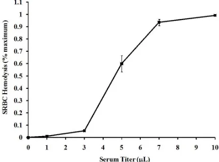
[image:3.595.59.286.164.323.2]Exposure of different dilutions of serum from Varanus komodoensis to SRBCs resulted in volume-dependent hemolysis (Figure 1). Treatment of a 500 μL suspension of 1% SRBCs (v/v) with only 3 μL of serum resulted in measureable (p < 0.5) hemolysis, as 5.5% ± 0.9% of the SRBCs were hemolyzed. Significant hemolysis of SRBCs occurs with infusion of a small quantity of Komodo dragon serum. Increased volumes of 5 and 7 μL of serum produced 60.0% ± 6.5% and 93.6% ± 2.6% hemolysis, respectively. However, a further increase to 10 μL of serum produced 99.3% ± 0.6% hemolysis, which indi-cated that a near-maximal response could be obtained by the use of only 7 μL of serum.
Figure 2 shows the kinetic response of the hemolytic activity of serum from Varanus komodoensis, at two dif-ferent volumes, toward SRBCs. At the lower volume (3 mL), an increase in SRBC hemolysis was not observed until 30 min of incubation with Varanus komodoensis serum. The activity increased to only 15.9% ± 0.3% of maximum activity at 60 min. However, the activity in-creased sharply to 91.2% ± 0.8% at 120 min. In contrast, the higher serum volume (7 mL) produced a sharp in-crease (26.0% ± 2.7%) at only 5 min of incubation with 1% SRBCs. The activity increased rapidly, in a linear fashion, to 88.5% ± 2.8% at 20 min. The linear relation of the increase (y = 0.0464x – 0.011) exhibited a correla-tion coefficient of 0.985.
[image:3.595.315.527.353.536.2]Incubation of 1% SRBCs with serum from Varanus komodoensis at different temperartures (5˚C - 40˚C) re-sulted in temperature-dependent hemolysis (Figure 3). Hemolysis at 5˚C - 20˚C was low (7.4% - 9.5% of maxi-mum), and was not statistically different between these
Figure 1. Titer-dependent hemolysis of SRBCs by Komodo dragon serum. Incubation of 1% (v/v) SRBCs with different volumes of Komodo dragon serum for 30 min resulted in a volume-dependent hemolysis, as measured spectrophotometri-cally as described in the Materials and Methods. The results are expressed as % of maximum hemolysis, relative to a positive control, and represent the means ± standard deviations, and represent the results of four independent determinations.
Figure 2. Kinetic analysis of SRBC hemolysis by Komodo dragon serum. Incubation of 1% SRBCs (v/v) with Komodo dragon serum, diluted in normal saline, for different amounts of time resulted in a time-dependent hemolysis. The results are expressed as the % of maximum hemolysis, relative to a posi-tive control, and represent the means ± standard deviations, and represent the results of four independent determinations.
temperatures (p > 0.05). The hemolytic activity increased (p < 0.05) at 25˚C (28.7% ± 2.8%), 30˚C (53.3% ± 2.3%), and 35˚C (68.7% ± 5.1%). However, the activity at 40˚C resulted in decreased (p < 0.05) activity, to 52.0% ± 2.6% of maximum activity.
Figure 3. Effects of temperature on hemolysis of SRBCs by Komodo dragon serum. Incubation of 1% SRBCs (v/v) with 0.5% Komodo dragon serum, diluted with normal saline, for 30 min resulted in a temperature-dependent hemolysis. The results are expressed as % of maximum hemolysis, relative to a posi-tive control, and represent the results of four independent de-terminations.
ability to hemolyze SRBCs to only 6.3% ± 2.8% of maximal activity. Likewise, pretreatment of Komodo dragon serum with 5 mM EDTA resulted in only 4.2% ± 0.2% hemolytic activity. However, incubation of serum with 5 mM EDTA and 15 mM Ca2+, Mg2+, Ba2+, Zn2+, or
Fe2+ resulted in 93.8% ± 3.4%, 94.7% ± 2.6%, 7.8% ±
1.1%, 6.7% ± 0.9%, or 3.7% ± 0.4% of maximal hemo-lytic activity, respectively.
4. DISCUSSION
The components of the innate immune systems of eu-karyotic organisms constantly survey their surroundings, primarily through molecular pattern recognition, distin-guishing self from non-self tissues, and respond to inva-sion of microbes in a nonspecific manner. The response is rapid, and acts as a first line of defense against infec-tion. In higher vertebrates, such as mammals, both the innate and acquired immune components are well-de- veloped [23]. However, the immune systems of reptilians are believed to have a less developed acquired immune system [24], but the innate immunity of some, particu-larly crocodilians, have been shown to have a highly developed innate immunity [25-29]. In addition, it has been suggested that more ancient poikilothermic verte-brates, such as teleost fish [30-32] and crocodilians [33], have followed the evolutionary path to develop a more efficient innate immune system, while mammals have developed more effective adaptive immunity. Previous studies have demonstrated that serum complement activ-ity in several different crocodilian species is far more potent than that of other phyla [29,33-35]. The results presented in this study show that the complement activity in the serum of the Komodo dragon is much more active than that of crocodilians.
The SRBC hemolysis assay has been used as a clinical tool to assess human complement activity for many years [36]. In addition, this assay has been modified to assess crocodilian complement activity [37]. The fact that the SRBC hemolysis by Komodo dragon serum is extremely heat-labile, sensitive to proteases (data not shown), and requires either Mg2+ or Ca2+ are all indications that this
activity is due to the presence of serum complement. The alternate serum complement pathway is activated, in the absence of antibody:antigen interaction (classical path-way) or protein:carbohydrate interaction (lectin pathpath-way), by the detection of non-self molecular pattern recogni-tion, which results in the formation of a membrane attack complex (MAC) in the outer membrane of microbes. However, in the presence of SRBCs, the MAC causes rapid hemolysis, which provides an easy way to measure complement activity in vitro by the measurement of he-moglobin spectrophotometrically at 540 nm [37].
The relation of Komodo dragon serum volume with hemolytic activity showed a typical sigmoidal curve, with a CH50 of approximately 9 µL/1000 µL incubation
(Figure 1). These data show the potent capacity with which the Komodo dragon serum disrupts the integrity of SRBCs. By way of comparison, the CH50 for the
Ameri-can alligator (Alligator mississippiensis) was found to be 539 µL [33], or 60 times higher than for the Komodo dragon. These data support the fact that Komodo dragon serum exhibits potent antibacterial activities [20], and might provide a mechanism for these observed activities.
The kinetic curve, depicted in Figure 2, shows that different volumes of Komodo dragon serum exhibit dif-ferent rates of SRBC hemolysis. The shape of the curve with high Komodo dragon serum volume is very similar to that of human complement [38], and also to the sev-eral crocodilian species such as the American alligator (Alligator mississippiensis) [33], American crocodile (Crocodylus acutus) [27], and the differences in the shapes of the kinetic curves strongly suggest that the serum complement enzymes exhibit positive cooperativ-ity. At the lower concentrations, the cooperativity is not observed due to reduced contact between the enzymes, and thus the onset of activity occurs much later. Both of the kinetic curves arrive at the same maximum activities, given enough time, due to the higher degree of coopera-tivity in samples with the higher plasma protein concen-trations.
35˚C (Figure 3), very near the 37˚C - 38˚C preferred temperature range to which Komodo dragons thermo-regulate [35]. These results are consistent with those ob-tained for the American alligator [33] and the saltwater (Crocodylus porosus) and freshwater (Crocodylus johns- toni) crocodiles in Australia [34], and the American crocodile (Crocodylus acutus) [27], and the broad- snouted caiman (Caiman latirostris) [29].
The results shown in Table 1 reveal similar character-istics of Komodo dragon serum with the ability of croco-dilian serum to hemolyze SRBCs [34,41]. Mammalian serum complement activity is known to be thermally unstable [12], and the results displayed in Table 1 pro-vide supporting epro-vidence that Komodo dragon serum complement exhibits the same thermal lability. Mild heat treatment of Komodo dragon serum at 56˚C for 30 min, classical serum complement thermal inactivation condi-tions [39], resulted in a substantial decrease in activity. In addition, mammalian serum complement function is known to require both Mg2+ and Ca2+ [38], while croco-
dilian complement function requires either Mg2+ or Ca2+
[41]. The results from the present study (Table 1) shows that Komodo dragon serum complement, like that of crocodilians, requires the presence of either Mg2+ or Ca2+.
Treatment of Komodo dragon serum with only 5 mM EDTA produced a large decrease in hemolytic activity. However, this activity can be restored by the inclusion of 15 mM Mg2+ or Ca2+, indicating the requirement for only
one of these metals. In addition, the results clearly illus-trate that not all divalent metal ions will restore EDTA- depleted Komodo dragon serum complement. The addi-tion of 15 mM Ba2+, Zn2+, or Fe2+ ions resulted in
com-plement activity which was not statistically different (p > 0.05) than treatment with EDTA alone. The inabilities of these divalent cations to restore EDTA-depleted Komodo dragon serum complement activity demonstrate the spe- cificity of the complement proteins for Mg2+ and Ca2+.
This same specificity has been noted in several croco-dileian species [27,34,41].
The results presented in this study show that the serum from the Komodo dragon exhibits potent and rapid se-rum complement activity. The activity occurs in a vol-ume-, time-, and temperature-dependent manner, and requires either Mg2+ or Ca2+ for activity. These activities
are consistent with an animal that lives an aggressive lifestyle and harbors potentially infectious microbes in their saliva for the purpose of feeding. This study repre-sents the first investigation of innate immunity of a Va-ranid lizard.
5. ACKNOWLEDGEMENTS
The authors wish to acknowledge the help of San Antonio Zoo em-ployees: J. Stephen McCusker (Director), Alan Kardon (Curator of
Herpetology and Aquarium), and Rob Coke and Jenny Nollman (Vet-erinarians), and Houston Zoo employees Rick Barongi (Director), Stan Mays (Curator of Herpetology), and Maryanne Tocidlowski (Veteri-narian), for permission to work with zoo animals and collection of samples. In addition, we also thank technicians Courtney Threadgill, Candice Robinson, and Hepetology/Aquarium staff (San Antonio Zoo) for help in restraining animals during blood collection. This research was funded by a McNeese State University Alumni Association Fac-ulty Development Award. In addition, support was provided by a NSF Research Commercialization/Educational Enhancement Plan grant administered by the Louisiana Board of Regents.
REFERENCES
[1] Cooper, N.R. (1985) The classical complement pathway: Activation and regulation of the first complement com-ponent. Advances in Immunology, 37, 151-216.
doi:10.1016/S0065-2776(08)60340-5
[2] Pillemer, L., Blum, L., Lepow, I., et al. (1954) The prop-erdin system and immunity: I. Demonstration and isola-tion of a new serum protein, properdin, and its role in immune phenomena. Science, 120, 279-285.
doi:10.1126/science.120.3112.279
[3] Fujita, T., Matsushita, M. and Endo, Y. (2004)The lectin- complement pathway: Its role in innate immunity and evolution. Immunological Reviews, 198, 185-202. doi:10.1111/j.0105-2896.2004.0123.x
[4] Muller-Eberhard, H.J. (1986) The membrane attack com-plex of complement. Annual Reviews in Immunology, 4, 503-528. doi:10.1146/annurev.iy.04.040186.002443 [5] Tofte, R.W., Petersen, P.K., Kim, Y., et al. (1980)
Opsonization of four Bacteroides species: Role of the classical complement pathway and immunoglobulin.
In-fection and Immunity, 2, 693-696.
[6] Ward, P.A., Chocrane, C.G. and Mueller-Eberhard, H.J. (1965) The role of serum complement in chemotaxis of leukocytes in vitro. Journal of Experimental Medicine, 122, 327-346. doi:10.1084/jem.122.2.327
[7] Rhyne, M.B. and Germuth F.G. (1961) The relationships between serum complement activity and the development allergic lesions in rabbits. Journal of Experimental
Medi-cine, 114, 633-646. doi:10.1084/jem.114.5.633
[8] Muller-Eberhard, H.J. (1988) Molecular organization and function of the complement system. Annual Reviews in
Biochemistry, 57, 321-347.
doi:10.1146/annurev.bi.57.070188.001541
[9] Miller, M.E. and Nillson, U.R. (1970) A familial defi-ciency of the phagocytosis-enhancing activity of serum related to a dysfunction of the fifth component of com-plement (C5). New England Journal of Medicine, 282, 354-358. doi:10.1056/NEJM197002122820702
[10] Jacobs, J. and Miller, M. (1972) Fatal familial Leiner’s disease: A deficiency of the opsonic activity of serum complement. Pediatrics, 49, 225-232.
21, 643-649. doi:10.1136/gut.21.8.643
[12] Morgan, B.P. and Walport, M.J. (1991) Complement deficiency and disease. Immunolology Today, 12, 301- 306. doi:10.1016/0167-5699(91)90003-C
[13] Ruddy, S., Gigli, I. and Austen, K.F. (1971) The com-plement system of man. New England Journal of
Medi-cine, 287, 489-495. doi:10.1056/NEJM197209072871005
[14] Day, N., Gewurz, H., Johannsen, R., et al. (1970) Com- plement and complement-like activity in lower verte-brates and inverteverte-brates. Journal of Experimental
Medi-cine, 132, 941-950. doi:10.1084/jem.132.5.941
[15] Gigli, I. and Austen, K.F. (1971) Phylogeny and function of the complement system. Microbiology, 25, 309-332. doi:10.1146/annurev.mi.25.100171.001521
[16] Pianka, E.R. and King, D.R. (2004) Introduction. In: Pianka, E.R., King, D.R. and King, R.A., Eds., Varanoid Lizards of the World, Indiana University Press, Bloom- ington, 3-9.
[17] Montgomery, J.M., Gillespie, D., Sastrawan, P., Freeking et al. (2002) Aerobic salivary bacteria in wild and captive Komodo dragons. Journal of Wildlife Diseases, 38, 545- 551.
[18] Fry, B.G., Wroe, S., Teeuwisse, W., et al. (2009) A cen-tral role for venom in predation by Varanus komodoensis (Komodo Dragon) and the extinct giant Varanus (Mega-
lania) priscus. Proceedings of the National Academy of
Science, 106, 8969-8974. doi:10.1073/pnas.0810883106
[19] Earley, R.L., Attum, O. and Eason, P. (2002) Varanid combat: Perspectives from game theory. Amphibia-Rep-
tilia, 23, 469-485. doi:10.1163/15685380260462374
[20] Merchant, M., Henry, D, Falconi, R., et al. (2012) Anti- bacterial activities of serum from the Komodo dragon (Varanus komodoensis). Microbiology Research, in Press. [21] Mayer, M.M. (1967) Complement and complement fixa-
tion. In: Kabat, E.A. and Mayer, M.M., Eds., Experimen- tal Immunochemistry, 2nd Edition, Thomas, Springfiled, 133-240.
[22] Merchant, M., Hammack, T., Dronette, J., et al. (2006) Assessment of innate immune activity of crocodilians using a spectroscopic assay based on the hemolysis of sheep red blood cells. Spectroscopy Letters, 39, 337-343. doi:10.1080/00387010600781290
[23] St. Georgiev, V. (2009) Mammalian host defenses: Innate and adaptive immunity. National Institute of Allergy and Infectious Diseases, NIH Infectious Disease, Part III, 577-626.
[24] Cuchens, M.A. and Clem, L.W. (1979) Phylogeny of lymphocyte heterogeneity. IV. Evidence for T-like and B-like cells in reptiles. Developmental and Comparative
Immunolology, 3, 465-475.
doi:10.1016/S0145-305X(79)80042-7
[25] Merchant, M., Williams, S. and Hardy, R. (2008) Super- oxide production by leukocytes in the American alligator (Alligator mississippiensis). Comparative Biochemistry
and Physiolology B, 152, 67-71.
doi:10.1016/j.cbpb.2008.09.089
[26] Merchant, M., Heard, R. and Monroe, C. (2009)
Charac-terization of phospholipase A2 activity in serum of the
American alligator (Alligator mississippiensis). Journal
of Experimental Zoology A, 311, 662-666.
doi:10.1002/jez.553
[27] Merchant, M., McFatter, J., Mead, S., et al. (2009)Iden- tification and characterization of serum complement ac- tivity in the American crocodile (Crocodylus acutus).
Vetereinary Immunolology and Immunopathology, 133,
165-169. doi:10.1016/j.vetimm.2009.07.016
[28] Merchant, M., Monroe, C. and Falconi, R. (2009) Char-acterization of dipeptidyl peptidase IV enzyme activity in the blood of the American alligator (Alligator mississip-
piensis). Comparative Biochemistry and Physiolology B,
154, 341-345. doi:10.1016/j.cbpb.2009.07.010
[29] Siroski, P., Merchant, M., Parachú Marcó, V., et al. (2010) Characterization of serum complement activity of the broad snouted caiman (Caiman latirostris, Crocodilia: Alligatoridae). Zoological Studies, 49, 64-70.
[30] Boshra, H., Gelman, A.E. and Sunyer, J.O. (2004) Struc-tural and functional characterization of complement C4 and C1s-like molecules in teleost fish: Insights into the evolution of classical and alternative pathways. Journal
of Immunology, 173, 349-359.
[31] Sunyer, J.O. and Lambris, J.D. (1998) Evolution and diversity of the complement system of poikilothermic vertebrates. Immunological Reviews, 166, 39-57.
doi:10.1111/j.1600-065X.1998.tb01251.x
[32] Sunyer, J.O., Zarkadis, I.K. and Lambris, J.D. (1998) Complement diversity: A mechanism for generating im-mune diversity. Immunolology Today, 19, 519-523. doi:10.1016/S0167-5699(98)01341-3
[33] Merchant, M., Roche, C., Sweeney, A., et al. (2005) Identification of serum complement activity in the Amer- ican alligator (Alligator mississippiensis). Comparative
Biochemistry and Physiology B, 141, 281-288.
doi:10.1016/j.cbpc.2005.03.009
[34] Merchant, M. and Britton, A. (2006) Characterization of serum complement activity of the saltwater (Crocodylus porosus) and freshwater (Crocodylus johnstoni) croco-diles. Comparative Biochemistry and Physiology A, 143, 488-493. doi:10.1016/j.cbpa.2006.01.009
[35] Major, S., Fontenot Jr., C.L., Pojman Sr., J.A., et al. (2011) Serum complement activity in the three-toed am-phiuma (Amam-phiuma tridactylum). Comparative
Immu-nolology, Microbiology, and Infectious Disease, 34, 115-
121. doi:10.1016/j.cimid.2010.04.001
[36] Lutz, D. and Lutz, J.M. (1997) Komodo: The living dragon. Revised Edition, DIMI Press, Salem.
[37] Mayer, M.M. (1967) Complement and complement fixa-tion. In: Kabat, E.A. and Mayer, M.M., Eds., Experimen-tal Immunochemistry, 2nd Edition, R. Thomas, Spring-field, 133-240.
[38] Merchant, T., Hammack, T., Sanders, P., et al. (2006) Rapid and inexpensive method for the spectroscopic de-termination of innate immunity of crocodilians.
Spec-troscopy Letters, 39, 337-343.
doi:10.1080/00387010600781290
kills E. coli: I. Location of the lethal lesion. Journal of
Immunology, 127, 1146-1151.
[40] Soltis, R.D., Hasz, D., Morris, M.J., et al. (1979) The effect of heat inactivation of serum on aggregation of immunoglobulins. Immunology, 36, 37-45.
[41] Levine, L., Osler, A.G., et al. (1953) The role of calcium and magnesium ions in complement fixation and immune
hemolysis: III. The respective roles of calcium and mag-nesium ions in immunehemolysis. Journal of
Immunol-ogy, 71, 374-379.
[42] Merchant, M., Verret, B. and Elsey, R.M. (2005) Diva-lent metal requirements for serum complement activity in the American alligator (Alligator mississippiensis). Com-