RESEARCH ARTICLE
PATHOPHYSIOLOGICAL MECHANISM BEHIND DIABETIC CARDIOVASCULAR DISORDERS
*Dr. Pratima Tripathi
Department of Biosciences, Sri Sathya Sai Institute of Higher Learning, Anantapur-515001, Andhra Pradesh
ARTICLE INFO ABSTRACT
Fatal and non-fatal Coronary Artery Diseases (CAD) are increased 2-4 fold in patients with diabetes and autopsy as compared to non-diabetic patients with Coronary Heart Disease (CHD). Immediate and long-term post- Myocardial Infarction (MI) mortality is increased 1.5-2 fold among diabetic patients. Despite a comparably small infarct size, diabetic patients have a far greater risk of developing highly fatal post-MI complications when compared to non diabetic patients. Following MI, the surviving myocardium of diabetic patients becomes hyperkinetic to compensate for non-viable infarcted myocardium in an attempt to maintain cardiac output. However, in diabetic patients, these areas of myocardium cannot achieve this compensatory enhancement in function due to a complex set of intra- and extra-myocardial factors superimposed on an already reduced coronary artery flow reserve. Endomyocardial samples from diabetic patients show enhanced thickening of capillary basement membrane, myocellular atrophy and hypertrophy with myocardial and, interstitial fibrosis, which further reduces the function of the myocardium. In this review the author have looked into various cardiovascular complications in diabetic condition and the pathophysiological mechanisms lying behind each.
Copyright © Dr. Pratima Tripathi. This is an open access article distributed under the Creative Commons Attribution License, which permits unrestricted use, distribution, and reproduction in any medium, provided the original work is properly cited.
INTRODUCTION
Diabetes mellitus is a metabolic disorder of multiple etiologies characterized by chronic hyperglycemia with disturbances in carbohydrate, fat and protein metabolism resulting from defects of insulin secretion, insulin action, or a combination of both and is associated with the development of specific long term organ damage due to microvascular diseases (Williamson
et al., 1993). Type 1diabetes is due to virtually complete lack
of endogenous pancreatic insulin production, whereas in type 2 diabetes, the rising blood glucose results from a combination of genetic predisposition, unhealthy diet, physical inactivity and increasing weight with a central disturbance resulting in complex pathophysiological processes (Nishikawa et al., 2000). Patients with diabetes are at higher risk of cardiovascular, cerebrovascular and peripheral artery disease. Among the several risk factors associated with CVD such as hypertension, cigarette smoking and dyslipidemia, diabetes is an important contributor to CVD risk and mortality. The reverse association is equally strong (Thomas et al., 1994). Today atherosclerosis accounts for about 70-80% deaths in diabetic individuals as compared to 30% in general populations. The increased risk of CVD in diabetic individuals can be explained in part by the clustering of traditional risk factors such as dyslipidemia, hypertension, hyperglycemia, and an increased tendency to thrombosis. Recent evidences show that increased oxidative stress and excess production of
*Corresponding author:Dr. Pratima Tripathi
Department of Biosciences, Sri Sathya Sai Institute of Higher Learning, Anantapur-515001, Andhra Pradesh
advanced glycation end products contribute to the development of diabetic complications, which are mediated at least by endothelial dysfunction (Graier et al., 1995). Since the worldwide prevalence of diabetes is increasing in ever-alarming proportions, CVD is becoming the most frequent cause of death. The various types of CVD associated with diabetes are acute myocardial infarction, diabetic cardiomyopathy, Cardiac Autonomic Nephropathy (CAN) and Cardiac Allograft Vasculopathy (CAV). The complexity and pandemic proportions of heart diseases and diabetes call for a comprehensive understanding of the pathophysiological mechanisms involved and their integration into a broader picture.
Hyperglycemia and Coronary Heart Diseases
Hyperglycemia may accelerate atherogenesis through various mechanisms, such as glycation of collagen and other vessel wall proteins, excess production of atherogenic lipoproteins and, reactive oxygen species, increased oxidative stress, glycation of several lipids and lipoproteins and also through endothelial damage, and hemorrheological abnormalities. In hyperglycemic conditions, glucose is reduced by Aldolase reductase to sorbitol which in some tissues is further oxidized to fructose upon sorbitol-dehydrogenase-catalyzed oxidation. The conversion of glucose to fructose (the polyol pathway) results not only in the utilization of NADPH but also in the accumulation of reduced NAD. This shift in the redox state of pyridine coenzymes induces a state of pseudohypoxia resulting into hypoxia like responses (Williamson et al., 1993). Polyol
ISSN: 0975-833X
International Journal of Current Research
Vol. 6, Issue, 01, pp.4426-4436, January,2014
INTERNATIONAL JOURNAL OF CURRENT RESEARCH
Article History:
Received 26th September, 2013
Received in revised form
11th October, 2013
Accepted 23rd December, 2013
Published online 26th January,2014
Key words:
Diabetic vascular complications, Inflammation,
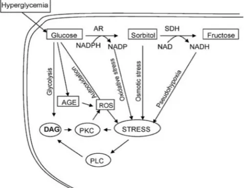
pathway mediated alterations in pyridine nucleotides have been linked to diverse metabolic changes such as block of Nitric Oxide (NO) and activation of protein kinases (Nishikawa et al., 2000). The increase in NADH due to elevated polyol pathway activity could increase the synthesis of diacylglycerol (DAG) from dihydroxy acetone phosphate (Thomas et al., 1994). DAG level activates phospholipase C and members of protein kinase C family that plays a key role in mesangial expansion and smooth muscle cells proliferation induced by high glucose (Graier et al., 1995). In the presence of normal glucose (5.5mM) Aldolase Reductase (AR) catalysed reduction of glucose represents less than 3% of total glucose utilization, whereas in presence of high glucose (20mM), more than 30% of glucose is used by AR (Srivastava et al., 2005). This reduction pathway imposes a significant strain on NADPH supply. Because NADPH is used for several critical reductive metabolic steps, such as detoxification of reactive oxygen species and hydroperoxides by the glutathione reductase/ glutathione peroxidase system, a large drain on the NADPH pool could compromise the ability of the cell to protect itself from oxidative stress (Dvornik et al., 1992). Hyperglycemia leads to the formation of advanced glycation end products (AGES), which are proteins or lipids that become glycated after exposure to sugars (Goldin et al. 2006). The presence and accumulation of AGES in many different cell types affects extracellular and intracellular structure and function. AGES cause cross-linkage of vessel wall proteins, leading to thickening and leakage of the vasculature and the formation of irreversible and abnormal deposits of plasma-derived proteins in the sub intimal layers of arteries (Goldin et al., 2006). In addition they engage the receptors of nuclear factor kappa B (NF-kB), a transcription factor that coordinates the inflammatory response. Furthermore soluble AGES activate monocytes and block Nitric Oxide (NO) activity in the endothelium (Yan et al., 2006), (Wautier and Schmidt, 2004). Higher cellular glucose causes increased production of ROS by several mechanisms (Fig 1). ROS mediates direct endothelial cell damage, oxidizes AGES and low density lipo-proteins (LDLs) thereby increasing the chances of atherogenesis.
Fig. 1. Involvement of polyol pathway in diabetic complications (Source: Srivastava et al., 2005)
During hyperglycemia, reduction of glucose to sorbitol by AR constitutes the first and the rate-limiting step of the polyol pathway that converts glucose to fructose via sorbitol dehydrogenase (SDH). In this pathway both NADPH and NAD+ are consumed as cofactors for the enzymes AR and SDH. Osmotic stress due to accumulation of sorbitol and oxidative stress due to changes in the ratio of NADPH/NADP+ and reduced NAD (NADH)/NAD+ are the major cause of various complications of secondary diabetes (Srivastava et al., 2005).
Insulin resistance and CVD
Insulin resistance is associated with both T1 and T2 diabetes mellitus (Daneman, 2006). In addition it may cluster with several classical CVD risk factors, such as visceral obesity, lipid abnormalities, hypertension and impaired glucose
tolerance to form a condition known as metabolic syndrome (Eckel et al., 2005). Controversy remains regarding
involvement of insulin resistance in the pathogenesis of atherosclerosis (Ferrannini and Iozzo, 2006). At the cellular level, there are two major insulin-regulated pathways, known as the metabolic and mitogenic pathways (Van Gaal et al., 2006). Normally, insulin binds to Its Receptor (IR), and activates Insulin Receptor Substrate (IRS-1) by phosphorylation of tyrosine residues. This, in turn activates phosphotidylinositol 3-kinase (PI-3K). In people with obesity, impaired glucose tolerance (IGT), type 2 diabetes mellitus (T2DM) and other insulin resistant states, there is a severe defect in the activation of IRS-1 which results into impaired glucose transport into the cell. This leads to a rise in blood glucose level and the resultant hyperglycemia, stimulates insulin release and development of hyperinsulinemia (Van Gaal et al., 2006). Although the metabolic pathway (IRS-1/PI-3K pathway) is severely impaired in any condition of insulin resistance, the mitogenic pathway (proceeding through mitogen-activated protein; MAP-kinase) (Fig.2) seems to retain its sensitivity to insulin, and any prevailing hyperinsulinemia would therefore, lead to an excessive stimulation of this pathway (Cersosimo and DeFronzo, 2006). This may result in the release of a number of growth and inflammatory factors and, promote proliferation and migration of vascular smooth muscle cells (Eckel et al., 2005).
Oxidative stress plays a casual role in insulin resistance and might be linked with visceral adiposity (Houstis et al., 2006). Mature adipocytes functions as an endocrine/paracrine organ that secretes numerous adipokines, cytokines and growth factors, in insulin resistance. A significant number of these proanthrogenic factors can be released from components of visceral fat, such as infiltrating macrophages that are not adipocytes (Weisberg et al., 2003). Several mechanisms link obesity to CAD such as the increased levels of Nonesterified Fatty Acids (NEFA) that results from increased lipolysis, and disturbances in adipokine and cytokine secretions. These processes in turn are linked with insulin resistance (Van Gaal
et al., 2006). Several adipokines and cytokines, such as
adiponectin, interleukin-6 (IL-6), retinol binding protein-4 (RBP-4), reisistin, and tomor-necrosis factor-α are associated with insulin resistance. RBP-4 is an adipocyte derived molecule that is elevated prior to the onset of diabetes (Graham
[image:2.595.39.289.516.708.2]and promote insulin resistance (Yang et al., 2005). Visceral fat releases IL-6, which can contribute to local and systemic imflammation, and elevation of C-reactive protein levels (Van Gaal et al., 2006). Thus, insulin resistance is intricately linked with visceral adipocity, and oxidative stress, and it may promote endothelial dysfunction and CAD.
Fig.2. Schematic representation of the overproduction of oxidants from NADPH oxidase and, mitochondrial sources in mammalian skeletal muscle, with subsequent enhanced engagement of p38 MAPK and other stress-activated kinases, including JNK, GSK-3, IKKβ, and others, associated with diminished insulin-stimulated insulin signaling and reduced glucose transport activity. (Source: Van Gaal et al., 2006).
Abnormalities in fatty acid metabolism and CVD
Lipid abnormalities are often found in diabetic patients (Chahil and Ginsberg, 2006). In T1 Diabetes Mellitus (T1DM), sustained hyperglycemia is associated with increased concentrations of total cholesterol, LDL and triglycerides and low concentration of HDL. In T2DM, dyslipidemia is associated with insulin resistance and is characterized by raised triglyceride, and low HDL concentration. LDL cholesterol is present in the form of highly atherogenic small, dense LDL particles (Chahil and Ginsberg, 2006). These LDL particles bind to the receptors on monocytes or macrophages and lead to the formation of foam cells. They can undergo oxidation through several mechanisms thereby contributing to atherogenesis at an early age (Mehta et al., 2006). Small dense LDL particles oxidize more readily than their counterparts and due to their small size they are likely to penetrate the arterial wall more easily. These particles possess high affinity for arterial wall proteoglycans thus prolonging their residence time in the subendothelial space (Packard, 2006). Elevated triglyceride promotes the increase in triglyceride-rich lipoprotein that is found to be involved in the development of atherosclerosis. Chylomicrons and large VLDLs are not capable of entering the arterial wall whereas small VLDLs and intermediate density lipoproteins (LDLs) can easily enter the arterial intima (Steiner, 1998). As a result certain triglyceride rich lipoproteins are atherogenic while others are not (Steiner, 1998). α, β unsaturated aldehydes which are produced during β-oxidation of alkoxy radicals derived from ω-6 polyunsaturated fatty acids are highly reactive (Esterbauer et
al., 1991). They are generated in high concentrations by
peroxidative reactions of lipids and lipoproteins and have been found to mediate and amplify the cellular effects of the free radical precursors (Esterbauer et al., 1991). HDL particles have a role in reverse cholesterol transport (from non-hepatic cells to
the liver), have direct effects on the endothelial cells and possess antioxidant properties like inhibition of LDL oxidation by transition metal ions, prevention of lipid hydroperoxide formation, reduction of apoptosis by inhibiting caspases and activating protein kinase Akt, activating eNOS to produce NO. In addition to lipid peroxidation, high concentrations of α, β unsaturated aldehydes are also generated by other biochemical processes, like myeloperoxidase catalysed oxidation of amino acids and oxidative modification of nucleosides (Assmann and Gotto, 2004). Accumulation of these unsaturated aldehydes and their products are found to be associated with increased formation of ROS leading to atherosclerosis, ischemia-reperfusion etc.
Hypertension and Diabetic CVD
Hypertension is at least twice as common in people with diabetes as in general population and is also more frequent in people with impaired glucose tolerance. It affects upto 70% of patients with diabetes mellitus and has some unique characteristics such as increased salt sensitivity, volume expansion, loss of nocturnal dipping of pulse, and isolated systolic hypertension (Yudkin, 2002). Sustained hypertension is associated with structural and functional alterations of both large arteries and arterioles. Hypertensive arteries experience remodeling of arterial walls due to increased vascular stiffness probably caused by accumulation of collagen and smooth muscle cell proliferation (Lehoux, 2006). The co-existence of hypertension and, diabetes increases the risk of developing macrovascular complications (myocardial infarction, stroke) and also microvascular complications (nephropathy, retinopathy etc) (Wahren et al., 2000). The vigorous treatment of hypertension may reduce the progression of these complications. In T1D, hypertension develops years after diagnosis usually already reflecting the development of diabetic nephropathy (Piero et al., 2008). Blood pressure tends to increase three years after the onset of micro albuminuria (Wahren, 2004). In patients with T2D, hypertension may be present at diagnosis and even before the elevation of blood glucose levels as reported by Kannel and collegues (Kannel et
al., 1974). The association between hypertension and, obesity
is well established leading to a higher rate of cardiovascular morbidity and mortality in patients with these two conditions (Kannel et al., 1974). The treatment of hypertension in diabetic patients aim at the prevention of CVD, minimizing the progression of renal disease and diabetic retinopathy. T2D patients may benefit more from tight control of blood pressure than with strict control of blood glucose levels. Initial treatments should include non-pharmacological measures such as weight reduction (in overweight and obesity), regular excercising, reducing salt intake (<1500mg/day), avoiding excessive alcohol consumption and smoking cessation. Pharmacological therapy should be initiated in all diabetics who persist with BP>130/80mmHg, when a change in lifestyle has already been implemented for 3 months or when the maximum BP levels are already higher than 140/90mmHg at diagnosis.
Endothelial dysfunction and CVD
adhesion and trafficking, prevention of platelet adhesion and regulation of blood flow via modulation of vessel patency (Feletou and Vanhoutte, 2006). These functions of the endothelium are mediated by various molecules such as nitric oxide, prostacyclin, C-type natriuretic peptide, Angiotensin II, plasminogen activator and so on (Feletou and Vanhoutte, 2006). In diabetics, hyperglycemia promotes the formation of ROS/AGEs, which stimulates the endothelial expression of pro-inflammatory cytokines such as interleukin-1 and monocyte chemoattractant protein-1, as well as leucocyte adhesion molecules (Lum and Rockbock, 2001). Oxidative stress due to excess production of ROS leads to reduction in the level of NO, and degradation of eNOS cofactor tetrahydrobiopterin (BH4) (Quagliaro et al., 2007). Insulin resistance also, causes alterations of PI-3K/Akt pathway which in turn lead to a marked decrease in eNOS activation. All these alterations confer a strong proatherogenic profile, which plays a major role in the development of CAD (Joshua et al., 2005). Endothelial dysfunction in T1D is an important determinant of inflammatory activity regardless of the presence or absence of complications, and hence can be considered as early marker of CVD (Schram et al., 2003). The disturbances in vascular responses can be seen by impaired flow mediated dilation responses associated with increased carotid artery intima-media thickness. Several markers of endothelial function in T1D such as, von Willebrand factor, thrombomodulin, selectin, plasminogen activator inhibitor-1(PAI-1), Type-IV collagen and tissue plasminogen activator (tPA) are increased. It has been shown that the cellular adhesion molecule E-selectin may enhance CAD prediction beyond traditional risk factors in T1D (Khan et al., 2000). Other markers of low grade inflammation levels such as oxidized LDL, monocyte IL-6, superoxide anion, plasma C-Reactive Proteins (CRP), Scd40L and nitrityrosine levels are found to be elevated in T1D patients (Hadi and AlSuwaidi, 2007). Thus endothelial dysfunction in T1D represents a high risk for micro and, macro angiopathy and hyperglycemia, appears to be one of the main causes along with other factors like environment, and genes (Hadi and AlSuwaidi, 2007). T2D is independently associated with impaired flow mediated dilatation. Endothelial dysfunction is considered to be the determinant factor for the vascular complications that is aggravated, rather than caused by hyperglycemia, because of the presence of many other risk factors such as obesity, hypertension, dislipidemia and ageing (Kolluru et al., 2012). This may be due to increased calpain dependent decrease in the association with eNOS. Inhibition of calpain activity decreases endothelial cell surface expression of the pro-inflammatory adhesion molecules ICAM-1 and, VCAM-1 during hyperglycemia (Stalker et al., 2003). T2D is associated with insulin resistance that causes eNOS inhibition by alterations in PI-3K/Akt pathway. Abnormalities in vasculature along with insulin resistance are also found in first degree relative to T2D patients even without presence of classic cardiovascular risk factors (Basha et al., 2012).
Oxidative stress and diabetic vascular complications
Increased intracellular glucose concentration results in the activation of alternative pathways of metabolism such as hexosamine and aldolase reductase pathways, both involved in the pathophysiology of chronic complications of diabetes. These pathways trigger an increased production of ROS, leads
to the formation of advanced AGEs and the activation of protein kinase C (PKC) resulting into increased oxidative stress. NADPH oxidase, nitric oxide synthase (NOS), xanthine oxidase, mitochondrial chain electron transport, lipooxygenase, cycloxygenase and cytochrome P450 are the main sources of ROS in the body. The ROS produced by these enzymes act as second messengers regulating the expression of redox signal sensitive gene (NF-kB) and also play vital role in production of inflammatory mediators (Griedling, 2004). ROS produced in the vascular wall are involved in various cellular events such as mitosis, apoptosis, hypertrophy, gene transcription and protein synthesis (Heerebeek et al., 2002). In diabetics, mitochondrial O2- anion act as a factor initiating a cascade of events that
results in increased production of ROS and RNS through activation of NF-kB. In addition, NOS can divert the production of nitric oxide (NO) to generate O2
-
in deficiency of L-arginine or tetrahydrobiopterin (BH4) in the endothelium of diabetic patients. When both L-arginine and BH4 are produced and, antioxidant enzymes are absent, the formation of peroxynitrite (NOO-) occurs, causing damage to cellular structures such as DNA, lipids, proteins etc. (Ballinger, 2000).
Under normal conditions the presence of ROS induces the expression of antioxidant enzymes as a defense mechanism. But even a short duration of diabetes without any chronic complications, less plasma antioxidants and uric acid levels suggests oxidative stress early in the disease (Marra et al., 2002). Non-enzymic extracellular antioxidants like α-tocopherol, vit. A, β-carotene, ascorbic acid, albumin and uric acid are particularly important to protect against lipid, protein and DNA damage. Another important component of antioxidant defense in diabetes is hepatoglobin that binds free hemoglobin resulting in the inhibition of iron-induced oxidative damage, since hemoglobin released in the blood after hemolysis of senescent erythrocytes is a potent oxidant (Levy
et al., 2002). The failure in the removal of ROS in the absence
Inflammation Cascade, Diabetes and Atherosclerosis
Diabetes, obesity and insulin resistance are associated with subclinical inflammation characterized by over expression of cytokines produced by adipose tissue, activated macrophages and other cells. Inflammatory mediators, such as TNF-α, interleukin-1 (IL-1), IL-6, leptin, resistin, monocyte chemoattractant protein-1(MCP-1), plasminogen activator inhibitor-1 (PAI-1), C-reactive protein (CRP), fibrinogen, angiotensin, visfatin, retinol binding protein-4 and adiponectin are involved in signaling pathways, in insulin action and perpetuation of inflammatory response (Shoelson et al., 2006). These cytokines are involved in the chronic inflammatory process of the vessel wall, promoting lipid accumulation with consequent development of atherosclerosis and CVD (Vicenova et al., 2009) (Fig. 3) Atherosclerosis is a complex multifactorial disease, and its acceleration in diabetes may be explained by several conditions including hyperglycemia, increased oxidative stress, advanced glycation end products (AGE), dyslipidemia, autonomic imbalance, hyperinsulenemia, excess inflammatory markers and genetic variables (Ait Oufella et al., 2011). It is assumed that the adipose tissue initiates obesity-induced inflammation and leads to the recruitment of immune cells which contributes to the maintenance of inflammatory response, besides leading to endothelial dysfunction with increased expression of adhesion molecules (ICAM-1, V-CAM-1, P-selectin and E-selectin), migration of monocytes, neutrophiles and T-lymphocytes (Wellen and Hotamisligil, 2005). Insulin resistance induces chronic elevation in free fatty acids (FFA) plasma concentration leading to increased storage of triglycerides in muscle, promoting reduction of muscle glucose uptake and increased hepatic glucose production that have been shown to
impair insulin action and promote hyperinsulenemia (De Fronzo, 2004).
Hyperinsulenemia can induce cardiomyocyte hypertrophy through myocyte growth induced by the activation of P13K/Akt-1 pathway and also by enhancing FFA levels. FFA are also implicated in the development of myocardial contractile dysfunction (Poornima, 2006). Several cytokines described to be related with insulin resistance are also involved with the development of atherosclerosis and CVD. TNF-α and other cytokines, FFA and ROS, activate inflammatory pathways and promote the expression of numerous genes involved in insulin resistance (Wellen and Hotamisligil, 2005). IL-1is another cytokine produced as a consequence of stress and cell injury mainly by macrophages that modulate key events in the process of atherosclerosis such as vessel wall inflammation, leukocyte chemotaxis and adhesion by increasing expression of VCAM-1 and MCP-1, angiogenesis through vascular endothelial growth factor (VEFG) induction, upregulation of matrix metalloproteinases (MMP), and destabilization of atheromatous plaques, that can lead to plaque rupture and thrombosis (Vicenova et al., 2009). CRP is an acute phase protein and is primarily derived from IL-6 hepatic biosynthesis. Atherogenic mechanisms of CRP include impaired production of endothelial NO and prostacyclin; increased production of endothelin-1 and other cell adhesion molecules, MCP-1, IL-8 and PAI-1; ROS and proinflammatory macrophage production; monocyte adhesion and chemotaxis; uptake of oxidized LDL; CRP also stimulates the expression of
metalloproteinases, activates NF-kB and promotes cell proliferation in vascular smooth muscle cells due to upregulation of the angiotensin type 1-receptor (Chait, 2005). Adiponectin has many protective actions in the atherosclerosis process due to the inhibition of LDL oxidation, activation of macrophages, reduction of adhesion molecule (VCAM and ICAM), inhibition of proliferation, and migration of smooth cells, and an increased production of NO in endothelial cells (Ferrarezi, 2007). Adiponectin is markedly reduced with increased obesity, and in diabetes and, hypoadiponectinemia is associated with an increase in CVD rates (Yamauchi, 2002). Leptin is a hormone secreted by adipose tissue and primarily involved in the regulation of energy expenditure and, food intake. Plasma leptin concentrations are increased in obese and diabetic patients (Yang and Barouch, 2007). Leptin has been shown to participate in the development of atherosclerosis in several ways: inducing oxidative stress; increasing the production of MCP-1, endothelin-1 (ET-1) which leads to cardiomyocyte hypertrophy; promoting migration, proliferation, hypertrophy of vascular smooth muscle cells (VSMC) and vascular cell wall calcification; stimulating platelet aggregation, attenuating cardiomyocyte contractility through increased nitric oxide production, reduction of intracellular calcium and decreased β-adrenergic response (Yang and Barouch, 2007).
Fig.3. Role of inflammation in plaque disruption leading to Atherosclerosis. (Source: Vicenova et al., 2009).
Diabetic Cardiomyopathy
patients. Early pathological features of DCM include non-specific interstitial changes within preserved monocytes which progress to more marked interstitial and perivascular fibrosis, with deposition of periodic acid Sciff-positive material, myocyte hypertrophy, microvascular alterations (thickening of basement membrane and micro-aneurysms), ventricular dilatation and dystolic dysfunction with either decreased or maintained systolic function (Fonarow and Srikanthan, 2006), (Marwick, 2006). Metabolic and hormonal factors, autonomic neuropathy and, microangiopathic alterations are the main ethiopathologic factors. The most important mechanisms of diabetic cardiomyopathy are metabolic disturbances (depletion of glucose transporter 4, increased free fatty acids, carnitine deficiency, changes in calcium homeostasis), myocardial fibrosis (association with increases in angiotensin II, IGF-I, and inflammatory cytokines), small vessel disease (microangiopathy, impaired coronary flow reserve, and endothelial dysfunction), cardiac autonomic neuropathy (denervation and alterations in myocardial catecholamine levels), and insulin resistance (hyperinsulinemia and reduced insulin sensitivity) (Fang et al., 2004). It is shown that diabetes is associated with a cardiomyopathy, independent of comorbid conditions, and that metabolic disturbances, myocardial fibrosis, small vessel disease, cardiac autonomic neuropathy, and insulin resistance may all contribute to the development of diabetic heart disease. In summary, various pathogenic factors of diabetes most likely induce ROS that in turn causes the endoplasmic reticulum stress and associated cell death. Cardiac cell death will initiate the cardiac inflammation and remodeling and eventually cardiac dysfunction, that is, diabetic cardiomyopathy. The pathogenic factors of diabetes include hyperglycemia, hyperlipidemia, and Angiotensin II and so on.
Molecular basis for diabetic cardiomyopathy
Hyperglycemia, hyperlipidemia and increased ROS induce alterations in downstream transcription factors which result in changes in gene expression, myocardial substrate utilization, myocyte growth, endothelial function, and myocardial compliance. Hyperglycemia may mediate its damaging effects through a series of secondary transducers. One of the principle abnormalities is the excess generation of AGEs, which deactivate NO and impair coronary vasodilation. Sustained hyperglycaemia causes excess formation of mitochondrial ROS, which affects transcription, leading to contractile dysfunction (Rosen et al., 1998). An increase in ROS decreases NO levels, which leads to myocardial inflammation and endothelial dysfunction via poly ADP-ribose polymerase (PARP), inhibition of which has been shown to reverse diabetic endothelial dysfunction (Soriano et al., 2001). The severity of diastolic dysfunction correlates with HbA1c (glycated haemoglobin) levels and the likely cause is AGE induced formation of ROS, resulting in myocardial collagen deposition and fibrosis. Enhanced expression of metallothionein, a potent antioxidant, limits the development of diabetic cardiomyopathy and, by breaking collagen cross-links improves diastolic function (Candido et al., 2003). Recently, the process of advanced glycation has been related directly to alterations in myocardial calcium handling and hence contractility (Lee and East, 2001). Sarcoplasmic/ endoplasmic-reticulum Ca2+ -ATPase 2a (SERCA2a) is responsible for replenishing intracellular calcium stores following release; this results in the
termination of contraction thus playing an integral part in cardiac relaxation. SERCA2a is a P-type ATPase that utilizes energy from the hydrolysis of the terminal phosphate bond of ATP to pump calcium against its electrochemical gradient (Lee and East, 2001). The turnover rate of SERC2a is low, which makes it susceptible to post-translational modification, especially in a chronic condition like diabetes. Thus advanced glycation of SERCA2a has been shown to lead to a decrease in its activity and a prolongation of cardiac relaxation (Bidasee
et al., 2004). Independent of the effects of hyperlipidaemia on
coronary artery endothelial function, the increased dependence of diabetic myocardium on fatty acid supply results in several major cellular metabolic perturbations. Thus there is increased
β-oxidation and mitochondrial accumulation of long-chain acyl
carnitines, leading to uncoupling of oxidative phosphorylation (Stanley et al., 1997). Enhanced fatty acid oxidation decreases glucose and pyruvate utilization by inhibiting pyruvate dehydrogenase. The net result is an excess of glycolytic intermediates and increased synthesis of ceramide (a toxic lipid compound) leading to apoptosis, which can be prevented by the peroxisome-proliferator-activated receptor (PPAR- α and γ) agonist, troglitazone (Zhou et al., 2000). Thus impaired glycolysis, pyruvate oxidation, lactate uptake and a greater dependence on fatty acids as a source of acetyl CoA, leads to a perturbation of myocardial bioenergetics and contraction/relaxation coupling (Rodrigues et al., 1998). Classic pharmacotherapy is aimed at restoring the balance between ATP synthesis and breakdown by increasing oxygen delivery (i.e. long-acting nitrates or Ca2+ channel antagonist) or by decreasing cardiac power by reducing blood pressure and heart rate (Ca2+ channel antagonist or β-blocker). An alternative agent which may readdress the altered bioenergetics of diabetic cardiomyopathy is trimetazidine [1-(2,3,4-trimethoxibenzyl)-piperazine], as it partially inhibits myocardial fatty acid oxidation, increases carbohydrate oxidation and reduces lactate production (Stanley and Marzilli, 2003). It has been found that in Type II diabetic patients with ischaemic cardiomyopathy randomized to receive either trimetazidine or placebo for 6 months, a beneficial effect was
observed on LV volumes and LV ejection fraction (Rosano
et al., 2003).
Role of Metabolic and Hormonal factors
decreased myocardial function (Cai and Kang, 2003). In addition, hyperglycemia increases the oxidative stress and glycosylation processes. Glycosylation of p53 protein leads to activation of angiotensin II synthesis and, necrosis (Farhangkhoee et al., 2006). Hyperglycemia also activates the protein kinase Cβ11 pathway again promoting myocardial necrosis and fibrosis (Farhangkhoee et al., 2006).
Cardiac autonomic neuropathy (CAN)
Cardiac autonomic neuropathy (CAN) have been associated with myocardial dysfunction and the development of CAN reduces the survival rate in DM patients by 44% to 85% (Vinik
et al., 2003). Other studies found that in coronary resistance
vessels, CAN was associated with an impaired vasodilator response to increased sympathetic stimulation so that 20% of DM patients with no ischemic heart disease suffers from abnormal diastolic filling related to severity of CAN (Vinik et
al., 2003). Ventricular filling abnormalities are also prominent
in patients with CAN. Catecholamines regulate the contractility of cardiac myocytes by acting on sarcoplasmic reticulum calcium uptake and, under conditions of β- adrenergic receptor stimulation cardiac performance is enhanced (Kogler and Ruegg, 1997). These effects are mediated by cAMP-dependent phosphorylation of proteins located in the sarcolemma, the membrane of the sarcoplasmic reticulum, and in the myofibrils of the cardiomyocytes. In DM, the cardiac-β adrenergic system may be enhanced, which can induce myocyte hypertrophy, interstitial fibrosis and reduced contractile function,
accompanied by myocyte apoptosis (Marwick, 2006). α- adrenergic stimulation, which has anti-apoptotic effects,
seems to be decreased in DM (Lee and East, 2001). CAN may contribute to impaired diastolic function and is associated with an increased cardiovascular risk in diabetic patients. Diabetic autonomic neuropathy is associated with an impaired vasodilator response of coronary resistance vessels to increased sympathetic stimulation (DiCarli et al., 1999).
Twenty one percent of patients with Type I diabetes without ischemic heart disease have abnormal diastolic filling which is associated with the severity of CAN (Kahn et al.,. 1986). Similarly ventricular filling abnormalities are most prominent in patients with autonomic neuropathy (Airaksinen et al., 1989). Studies have shown a correlation between myocardial sympathetic innervation derived from scintigraphy and the E/A ratio (ratio of early to late peak mitral filling wave velocities) in Doppler echocardiography, providing evidence that an abnormal sympathetic innervation of the heart may contribute to a disturbance in LV filling. Sympathetic dysfunction has been related to both systolic and diastolic dysfunction in Type II diabetes (Annonu et al., 2001). An abnormal systolic blood pressure response to standing was correlated significantly with a reduced mitral E/A ratio (Fig. 4). Studies also reflect an association between parasympathetic and cardiac dysfunction as evidenced by the association between significantly lower mean heart rate variation during deep breathing, and abnormal diastolic peak filling rate in diabetic patients (Uusitupa et al., 1988). The mitral E/A ratio have been shown to be significantly reduced in patients with autonomic neuropathy and a significant correlation was observed between the E/A ratio and autonomic neuropathy (Monteagudo et al. 2000).
Cardiac Allograft Vasculopathy (CAV)
CAV is a CVD that occurs in heart transplant recipients (Valantine, 2004). It is a rapidly progressive form of atherosclerosis characterized by intimal proliferation, and luminal stenosis of epicardial branches, occlusion of small arteries, and MI with the progression of the disease. Both immunologic and non-immunologic mechanisms are involved in the pathogenesis of CAV. The initiating event is subclinical endothelial cell injury in the coronary graft, caused by
ischemia-reperfusion damage or host versus graft immune response. This leads to a cascade of destructive events involving cytokines, inflammatory mediators, complement activation and leukocyte adhesion molecules (Ramzy et al., 2005). These changes produce inflammation, thrombosis, SMC proliferation and vasoconstriction. The non-immunologic factors leading to CAV are DM, both pre existing and new onset after transplantation (caused by immunosuppression), and metabolic abnormalities associated with insulin resistance. These factors play a vital role in accelerating vascular damage through various mechanisms as involved in CAD in non-transplant cases (Valantine, 2004). The histological changes that characterize CAV are not uniform. Pathological examination of coronary arteries from human cardiac allografts has shown a broad spectrum of abnormalities, ranging from concentric fibrous intimal thickening to complicated atherosclerotic plaques that bear a close resemblance to spontaneous atherosclerosis (Pucci et al., 1990). It has been demonstrated that early intimal proliferation progresses with time and with subsequent increases in lipid deposits and, calcification of the coronary vessel (Billingham, 1992). Atheromas formation and diffusion of intracellular and, extracellular, accumulation of lipids in both intimal and medial walls are frequent occurrences (McManus et al., 1995).
The internal elastic lamina remains almost intact except for small breaks (Billingham, 1995). Early after transplantation, diffused fibrous intimal thickening or a vasculitis predominates. Late after transplantation, focal atherosclerotic plaques, intimal thickening, or a mixture of both is found. The smaller branches are often occluded before the larger epicardial arteries, resulting in small, stellate infarcts (Neish et al., 1992). Despite exuberant intimal proliferation, the media of the vessel is rarely thickened and sometimes becomes narrower than in normal conditions (Billingham, 1995). The cellular infiltrate of intimal proliferative lesions consists of modified smooth muscle cells, macrophages/monocytes, and T lymphocytes (Billingham, 1995). Although the exact pathogenesis of CAV remains to be established, several lines of data suggest that it is primarily an immune-mediated disease (Treasure and Alexander, 1995). Limitation of the proliferative vascular disease to the allograft arterial and venous tree, the often diffuse nature of allograft vascular involvement, the development of CAV in cardiac allografts of animal models with some histocompatibility mismatch, and the lack of development in isografts support the immunologic hypothesis of CAV development. Studies suggest that immunologic mechanisms operating in a milieu of nonimmunologic risk factors constitute the principal stimuli that result in progressive myointimal hyperplasia (Treasure and Alexander, 1995). The initial event of CAV is probably a subclinical graft coronary endothelial injury. The endothelial cell is the major determinant of vessel wall function. It normally inhibits thrombus formation and leukocyte adhesion, regulates vasomotor function, and inhibits vascular smooth muscle cell proliferation. Damage to the endothelium could alter any or all of these functions, predisposing the artery to inflammation, thrombosis, vasoconstriction, and vascular smooth muscle cell growth (Treasure and Alexander, 1995). After human cardiac transplantation, humoral or more important cellular responses to HLA antigens and vascular endothelial cell antigens are potential sources of endothelial damage. CD4 lymphocyte–
induced upregulation of MHC class II antigens on endothelial cells (subsequent to MHC-I antigen detection by CD8 lymphocytes) elicits a cellular immune response (Libby et al., 1992).
The role of MHC donor-recipient differences in the pathogenesis of CAV has not yet been completely elucidated. The ability to produce CAV in animals transplanted with an MHC-identical graft and, more recently, to document the occurrence of allograft rejection in genetically engineered animals lacking MHC genes should spur further investigations of other allograft-specific antigens, distinct from those of the MHC, which may play an important role in the development of CAV. Irrespective of the initial specific immune-mediated injury, the cascade of events that follows appears to be a physiologically nonspecific inflammatory response (Duquesnoy and Demetris, 1995). It is important to note that activated lymphocytes secrete interferon-γ, which stimulates production of ICAM-1 (Hayry et al., 1989). The involvement of adhesion molecules plays a crucial role in regulating the interaction of inflammatory cells with cells in the vascular wall because the adherence of leukocytes to vascular endothelium is a prerequisite for transmigration. Expression of vascular adhesion molecules (VCAM-1, ICAM-1, and ELAM-1) on endothelial cells and medial smooth muscle cells in cardiac transplant patients has been observed and, early ICAM-1 expression could be correlated to early development of angiographically visible CAV (Labarrere et al., 1995). The intercellular network, via macrophages, T lymphocytes, endothelial cells, and smooth muscle cells, generates a variety of stimulatory cytokines (IL-1, IL-2, IL-6, and tumor necrosis factor-α) and growth factors (PDGF, FGF, EGF, TGF-β etc) that promote the development of the chronic allograft lesion (Duquesnoy and Demetris, 1995).
Conclusion
Diabetic heart disease is caused by complex interactions that result from various mechanisms; associated with hyperglycemia, dyslipidemia, hypertension and possibly insulin resistance. All these lead to increased oxidative stress and enhanced glycosylation of several humoral and vessel wall proteins, which cause endothelial damage, and structural changes in coronary arteries. The damaged endothelial cells become a source of ROS and RNS which along with several other factors leads to proatherosclerotic processes. Some of these mechanisms progresses into the development of diabetic cardiomyopathy, in which myocyte substrate utilization and neural influences play a major role. Further investigations are needed to elucidate the beneficial role of oral anti-diabetic agents in case of diabetic heart disease. Also, early initiation of therapies aimed at reducing inflammation and oxidative stress may be beneficial in reducing CVD in diabetes. Casual anti-oxidant therapies with compounds that acts as intracellular scavengers or as anti-oxidant enzyme can modulate ROS-sensitive signaling pathways may represent a potential avenue of therapy in diabetics to prevent the onset and progression of cardiovascular complications.
REFERENCES
Airaksinen K, Kostinen J, Akaheimo M, Huikuri H. 1989. Augmentation of atrial contraction to LV filling in IDDM subjects as assessed by Doppler echocardiograph. Diabetes
Care. 12: 159–61.
Ait-Oufella H, Taleb S, Mallat Z et al. 2011. Recent advances on the role of cytokines in atherosclerosis. Arteriosc
Thromb Vasc Biol. 31 (5): 969–79.
Annonu AK, Fattah AA, Mokhtar MS, Ghareeb S, Elhendy A. 2001. LV systolic and diastolic functional abnormalities in asymptomatic patients with non-insulin-dependent diabetes mellitus. J Am Soc Echocardiogr. 14: 885–91.
Assmann G, Gotto AM Jr. 2004. HDL cholesterol and protective factors in atherosclerosis. Circulation. 109 (23): III8-14.
Ballinger SW, Patterson C, Yan CN et al. 2000. Hydrogen peroxide- and peroxynitrite induced mitochondrial DNA damage and dysfunction in vascular endothelial and smooth muscle cells. Circ Res. 86 (9): 960–66.
Basha B, Samuel SM, Triggle CR et al. 2012. Endothelial dysfunction in diabetes mellitus: possible involvement of endoplasmic reticulum stress. Experiment Diabetes Res. 2012, Article ID 481840, 14 pages.
Berry C, Brosnan MJ, Fennell J, Hamilton CA, Dominiczak AF. 2001. Oxidative stress and vascular damage in hypertension. Curr Opinion in Nephrol and Hypert.10 (2): 247–55.
Bidasee KR., Zhang Y, Shao C H et al. 2004. Diabetes increases formation of advanced glycation end products on sarco(endo)plasmic reticulum Ca2+-ATPase. Diabetes. 53: 463–73.
Billingham ME. 1992. Histopathology of graft coronary disease. J Heart Lung Transplant . 11: S38-44.
Billingham ME. 1995. Pathology of graft vascular disease after heart and heart-lung transplantation and its relationship to obliterative bronchiolitis. Transplant Proc. 27: 2013-2016.
Cai L, Kang YJ. 2003. Cell death and diabetic cardiomyopathy.
Cardiovasc Toxicol. 3 (3): 219-28.
Candido R, Forbes JM, Thomas MC et al. 2003. A breaker of advanced glycation end products attenuates diabetes induced myocardial structural changes. Circ Res. 92: 785– 92.
Cersosimo E, DeFronzo RA. 2006. Insulin resistance and endothelial dysfunction: the road map to cardiovascular diseases. Diabetes Metab Res Rev. 22: 423-36.
Chahil TJ, Ginsberg HN. 2006. Diabetic dyslipidemia.
Endocrinol Metab Clin North Am. 35 (3): 491-510.
Chait A, Chang YH, Oram JF, Heinecke JW. 2005. Lipoprotein-associated inflammatory proteins: markers or mediators of cardiovascular disease. J of Lipid Res. 46 (3): 389–403.
Daneman D. Type 1 diabetes. 2006. Lancet. 367 (9513): 847-58.
DeFronzo RA. 2004. Pathogenesis of type 2 diabetes mellitus.
Medic Clin of North America. 88 (4): 787–835.
Di Carli M F, Bianco-Batlles D, Landa M E et al. 1999. Effects of autonomic neuropathy on coronary blood flow in patients with diabetes mellitus. Circulation. 100: 813–19. Dong C, Wilson JE, Winters GL, McManus BM. 1996. Human
transplant coronary artery disease: pathological evidence for Fas-mediated apoptotic cytotoxicity in allograft arteriopathy. Lab Invest. 74 (5): 921-31.
Duquesnoy RJ, Demetris AJ. 1995. Immunopathology of cardiac transplant rejection. Curr Opin Cardiol . 10 (2): 193-206.
Dvornik D. 1992. Aldose reductase inhibitors as pathobiochemical probes. J Diabetes Compl. 6: 25–34. Eckel RH, Grundy SM, Zimmet PZ. 2005. The metabolic
syndrome. Lancet. 365: 1415-28.
Esterbauer H, Schaur R J, Zollner H. 1991. Chemistry and biochemistry of 4-hydroxynonenal, malonaldehyde and related aldehydes. Free Radic Biol Med. 11(1): 81–128. Fang ZY, Prins JB, Marwick TH. 2004. Diabetic
cardiomyopathy: evidence, mechanisms, and therapeutic implications. Endocr Rev . 25 (4): 543-67.
Farhangkhoee H, Khan ZA, Kaur H, Xin X, Chen S, Chakrabarti S. 2006. Vascular endothelial dysfunction in diabetic cardiomyopathy: pathogenesis and potential treatment targets. Pharmacol Ther. 111 (2): 384-99. Feletou M, Vanhoutte PM. 2006. Endothelial dysfunction: a
multifaceted disorder. Am J Physiol Heart Circ Physiol. 291(3): H985-1002.
Ferrannini E, Iozzo P. 2006. Is insulin resistance atherogenic? A review of the evidence. Atheroscler Suppl. 7(4): 5-10. Ferrarezi DAF, Cheurfa N, Reis AF, Fumeron F, Velho G.
2007. Adiponectin gene and cardiovascular risk in type 2 diabetic patients: a review of evidences. Arquivos
Brasileiros de Endocrinologia e Metabologia. 51(2):153–
59.
Fonarow GC, Srikanthan P. 2006. Diabetic cardiomyopathy.
Endocrinol Metab Clin North Am. 35: 575-599.
Gibbons GH. 1995. The pathogenesis of graft vascular disease: implications of vascular remodeling. J Heart Lung
Transplant. 14: S149-58.
Goldin A, Beckman JA, Schmidt AM, Creager MA. 2006. Advanced glycation end products: sparking the development of diabetic vascular injury. Circulation. 114 (6): 597-605.
Graham TE, Yang Q, Bluher M et al. 2006. Retinol-binding protein 4 and insulin resistance in lean, obese, and diabetic subjects. N Engl J Med. 354 (24): 2552–63.
Graier WF, Grubenthal I, Dittrich P, Wascher TC, Kostner GM. 1995. Intracellular mechanism of high D-glucose-induced modulation of vascular cell proliferation. Eur J
Pharmacol. 294: 221–29.
Griendling KK. 2004. Novel NADPH oxidases in the cardiovascular system. Heart. 90 (5): 491–93.
Hadi HAR, Al Suwaidi JA. 2007. Endothelial dysfunction in diabetes mellitus. Vasc Health and Risk Mgmt. 3 (6): 853–-76.
Hayry P, Renkonen R, Leszczynski D et al. 1989. Local events in graft rejection. Transplant Proc . 21: 3716-20.
Heerebeek LV, Meischl C, Stooker W, Meijer CJLM, Niessen HWM, Roos D. 2002. NADPH oxidase: new source of reactive oxygen species in the vascular system. Jof Clinl
Pathol. 55 (8): 561-68.
Houstis N, Rosen ED, and Lander ES. 2006. Reactive oxygen species have a causal role in multiple forms of insulin resistance Nature. 440 (7086): 944–48.
Isner JM, Kearney M, Bortman S, Passeri J. 1995. Apoptosis in human atherosclerosis and restenosis. Circulation . 91 (11): 2703-11.
Joshua IG, Zhang Q, Falcone JC, Bratcher AP, Rodriguez WE, Tyagi SC. 2005. Mechanisms of endothelial dysfunction with development of type 1 diabetes mellitus: role of insulin and C-peptide. J Cell Biochem. 96: 1149-56. Kahn K, Zola B, Juni J E, Vinik A I. 1986. Radionuclide
assessment of LV diastolic filling pressures in diabetes mellitus with and without cardiac autonomic neuropathy. J
Am Coll Cardiol. 7: 1303–09.
Kannel WB, Hjortland M, Castelli WP. 1974. Role of diabetes in congestive heart failure: the Framingham study. Am J
Cardiol. 34: 29-34.
Khan F, Elhadd TA, Greene SA, Belch JJF. 2000. Impaired skin microvascular function in children, adolescents, and young adults with type I diabetes. Diabetes Care. 23 (2): 215–20.
Kogler H, Ruegg JC. (1997) Cardiac contractility: modulation of myofibrillar calcium sensitivity by beta-adrenergic stimulation. Isr J Med Sci. 33:1-7.
Kolluru GK, Bir SC, Kevil CG. 2012. Endothelial dysfunction and diabetes: effects on angiogenesis,vascular remodeling and wound healing. International Journal of Vasc Med. 2012: Article ID918267, 30 pages.
Labarrere CA, Pitts D, Nelson DR, Faulk WP. 1995. Coronary artery disease in cardiac allografts: association with arteriolar endothelial HLA-DR and ICAM-1 antigens. Transplant Proc. 27 (3): 1939-40.
Lee AG, East JM. 2001. What the structure of a calcium pump tells us about its mechanism. Biochem J. 356 (Pt 3): 665– 83.
Lehoux S, Castier Y, Tedgui A. 2006. Molecular mechanisms of the vascular responses to haemodynamic forces. J Intern
Med 259: 381-92.
Levy AP, Hochberg I, Jablonski K et al. 2002. Haptoglobin phenotype is an independent risk factor for cardiovascular
disease in individuals with diabetes: the strong heart study.
J Amer College Cardiol. 40 (11):1984–90.
Libby P, Swanson SJ, Tanaka H. 1992. Immunopathology of coronary arteriosclerosis in transplanted hearts. J Heart
Lung Transplant. 11:S5-6.
Lum H, Roebuck KA. 2001. Oxidant stress and endothelial cell dysfunction. Am J Physiol Cell Physiol . 280 (4): C719-41. Marra G, Cotroneo P, Pitocco D et al. 2002. Early increase of
oxidative stress and reduced antioxidant defenses in patients with uncomplicated type 1 diabetes: a case for gender difference. Diabetes Care.25 (2): 370-75.
Marwick TH. (2006) Diabetic heart disease. Heart. 92 (3): 296-300.
McManus BM, Horley KJ, Wilson JE et al. 1995. Prominence of coronary arterial wall lipids in human heart allografts. Am J Pathol . 147 (2): 293-308.
Mehta JL, Rasouli N, Sinha AK, Molavi B. 2006. Oxidative stress in diabetes: a mechanistic overview of its effects on atherogenesis and myocardial dysfunction. Int J Biochem
Cell Biol. 38 (5-6): 794-803.
Monteagudo PT, Moises VA, Kohlmann Jr O et al. 2000. Influence of autonomic neuropathy upon LV dysfunction in insulin-dependent diabetic patients. Clin Cardiol. 23: 371– 75.
Neish AS, Loh E, Schoen FJ. 1992. Myocardial changes in cardiac transplant-associated coronary arteriosclerosis: potential for timely diagnosis. J Am Coll Cardiol. 19(3): 586-92.
Nishikawa T, Edelstein D, Du XL et al. 2000. Normalizing mitochondrial superoxide production blocks three pathways of hyperglycaemic damage. Nature.404 (6779):787–90. Packard CJ. 2006. Small dense low-density lipoprotein and its
role as an independent predictor of cardiovascular disease.
Curr Opin Lipidol. 17: 412-17.
Piero M, Francesco D, Davide L, Francesco P. 2008. An overview of pancreatic beta-cell defects in human type 2 diabetes: Implications for treatment. Regulatory Peptides.146: 4–11.
Poornima IG, Parikh P, Shannon RP. 2006. Diabetic cardiomyopathy: the search for a unifying hypothesis. Circ
Res. 98 (5): 596–605.
Pucci AM, Forbes RDC, Billingham ME. 1990. Pathologic features in long-term cardiac allograft. J Heart Transplant . 9: 339-45.
Quagliaro L, Piconi L, Assaloni R, Da Ros R, Szabo C, Ceriello A: 2007. Primary role of superoxide anion generation in the cascade of events leading to endothelial dysfunction and damage in high glucose treated HUVEC.
Nutr Metab Cardiovasc Dis. 17: 257–67.
Rabinovitch M, Molossi S, Clausell N. 1995. Cytokine-mediated fibronectin production and transendothelial migration of lymphocytes in the mechanism of cardiac allograft vascular disease: efficacy of novel therapeutic approaches. J Heart Lung Transplant. 14: S116-23.
Ramzy D, Rao V, Brahm J, Miriuka S, Delgado D, Ross HJ. (2005) Cardiac allograft vasculopathy: a review. Can J
Surg. 48 (4): 319-27.
Rodrigues B, Cam MC, McNeill JH. 1998. Metabolic disturbances in diabetic cardiomyopathy. Mol Cell
Biochem. 180 (1-2): 53–7.
patients with coronary artery disease: a double-blind placebo-controlled study. Cardiovasc Diabetol. 2: 16–25. Rosen P, Du X, Tschope D. 1998. Role of oxygen derived free
radicals for vascular dysfunction in the diabetic heart: prevention by α-tocopherol. Mol Cell Biochem. 188: 103– 11.
Russell ME, Wallace AF, Wyner LR, Newell JB, Karnoswky MJ. 1995. Upregulation and modulation of inducible nitric oxide synthase in rat cardiac allografts with chronic rejection and transplant arterioscleroses. Circulation. 92 (3): 457-64.
Schram M T, Chaturvedi N, Schalkwijk C et al. 2003. Vascular risk factors andmarkers of endothelial function as determinants of inflammatory markers in type 1 diabetes: the EURODIAB prospective complications study. Diabetes
Care. 26 (7): 2165–73.
Shoelson SE, Lee J, Goldfine AB. 2006. Inflammation and insulin resistance. J Clin Invest. 116 (7): 1793–1801. Soriano FG, Pacher P, Mabley J, Liaudet L, Szabo C. 2001.
Rapid reversal of the diabetic endothelial dysfunction by pharmacological inhibition of poly(ADP-ribose) polymerase. Circ Res. 89 (8): 684–91.
Srivastava S K, Ramana K V, Bhatnagar A. 2005. Role of Aldose Reductase and Oxidative Damage in Diabetes and the Consequent Potential for Therapeutic Options. Endoc
Rev. 26 (3) 380-92.
Stalker TJ, Skvarka CB, Scalia R. 2003. A novel role for calpains in the endothelial dysfunction of hyperglycemia.
The FASEB Journal. 17 (11), 1511–13.
Stanley WC, Lopaschuk GD, McCormack JG. 1997. Regulation of energy substrate metabolism in the diabetic heart. Cardiovasc Res. 34 (1): 25–33.
Stanley WC, Marzilli M. 2003. Metabolic therapy in the treatment of ischaemic heart disease: the pharmacology of trimetazidine. Fundam Clin Pharmacol. 17 (2): 133–45. Steiner G. 1998. Intermediate-density lipoproteins, diabetes
and coronary artery disease. Diabetes Res Clin Pract. 40: S29-S33.
Thomas TP, Porcellati F, Kato K, Stevens MJ, Sherman WR, Greene DA. 1994. Effects of glucose on sorbitol pathway activation, cellular redox, and metabolism of myo-inositol, phosphoinositide, and diacylglycerol in cultured human retinal pigment epithelial cells. J Clin Invest.93 (6): 2718–-24.
Treasure CB, Alexander RW. 1995. Relevance of vascular biology to the ischemic syndromes of coronary atherosclerosis. Cardiovasc Drugs Ther. 9: 13-19.
Uusitupa M, Mustonen J, Laakso M et al. 1988 Impairment of diastolic function in middle aged type 1 and type 2 diabetic
patients free of cardiovascular disease. Diabetologia. 31 (11): 783–91.
Valantine H. 2004. Cardiac allograft vasculopathy after heart transplantation: risk factors and management. J Heart Lung
Transplant. 23(5): S187-93.
Van Gaal LF, Martens IL, De Block CE. 2006. Mechanisms linking obesity with cardiovascular disease. Nature. 444: 875-80.
Vicenova B, Vopalensky V, Burysek L et al. 2009. Emerging role of interleukin-1 in cardiovascular disease. Physiol Res. 58: 481–98.
Vinik AI, Maser RE, Mitchell BD, Freeman R. 2003. Diabetic autonomic neuropathy. Diabetes Care. 26 (5): 1553-79. Wahren J, Ekberg K, Johansson J et al. 2000. Role of
C-peptide in human physiology. Am J Physiol Endocrinol
Metab. 278 (5): E759-68.
Wahren J. 2004. C-peptide: new findings and therapeutic implications in diabetes. Clin Physiol Funct Imaging. 24: 180-189.
Wautier JL, Schmidt AM. 2004. Protein glycation: a firm link to endothelial cell dysfunction. Circ Res.95: 233-38. Weisberg SP, McCann D, Desai M, Rosenbaum M, Leibel RL,
Ferrante AW, Jr. 2003. Obesity is associated with macrophage accumulation in adipose tissue. J Clin Invest. 112 (12): 1796–1808.
Wellen KE, Hotamisligil GS. 2005. Inflammation, stress, and diabetes. J of Clin Invest. 115 (5): 1111–19.
Williamson JR, Chang K, Frangos M et al. 1993. Hyperglycemic pseudohypoxia and diabetic complications.
Diabetes. 42 (6): 801–13.
Yamauchi T, Kamon J, Minokoshi Y et al. 2002. Adiponectin stimulates glucose utilization and fatty-acid oxidation by activating AMP-activated protein kinase. Nature Med. 8 (11): 1288–95.
Yan SF, Yan SD, Herold K, Ramsamy R, Schmidt AM. 2006. Receptor for advanced glycation end products and the cardiovascular complications of diabetes and beyond: lessons from Ageing. Endocrinol Metab Clin North Am. 35: 511-24.
Yang Q, Graham TE, Mody N et al. 2005. Serum retinol binding protein 4 contributes to insulin resistance in obesity and type 2 diabetes. Nature. 436 (7049): 356–62.
Yang R, Barouch LA. 2007. Leptin signaling and obesity: cardiovascular consequences. Circ Res. 101(6): 545–59. Yudkin JS. 2002. Mechanisms of vascular disease in
hypertension with special reference to diabetes. Semin Vasc
Med. 2: 139-48.
Zhou Y, Grayburn P, Karim A et al. 2000. Lipotoxic heart disease in obese rats: implications for human obesity. Proc
Natl Acad Sci U.S.A. 97: 1784–89.