R E S E A R C H
Open Access
Comparative proteomic analysis of
Ulva
prolifera
response to high temperature
stress
Meihua Fan
1*, Xue Sun
2, Zhi Liao
1, Jianxin Wang
1, Yahe Li
2and Nianjun Xu
2*Abstract
Background:Ulva proliferabelongs to green macroalgae and is the dominant species of green tide. It is distributed worldwide and is therefore subject to high-temperature stress during the growth process. However, the adaptation mechanisms of the response ofU. proliferato high temperatures have not been clearly investigated yet.
Methods:In this study, isobaric tags for relative and absolute quantitation (iTRAQ) labelling was applied in combination with the liquid chromatography-tandem mass spectrometry (LC-MS/MS) to conduct comparative proteomic analysis of the response ofU. proliferato high-temperature stress and to elucidate the involvement of this response in adaptation mechanisms. Differentially expressed proteins (DEPs) ofU. proliferaunder high temperature (denote UpHT) compared with the control (UpC) were identified. Bioinformatic analyses including GO analysis, pathway analysis, and pathway enrichment analysis was performed to analyse the key metabolic pathways that underlie the thermal tolerance
mechanism through protein networks. Quantitative real-time PCR and western blot were performed to validate selected proteins.
Results:In the present study, 1223 DEPs were identified under high temperature compared with the control, which included 790 up-regulated and 433 down-regulated proteins. The high-temperature stimulus mainly induced the expression of glutathione S-transferase, heat shock protein, ascorbate peroxidase, manganese superoxide dismutase, ubiquitin-related protein, lhcSR, rubisco activase, serine/threonine protein kinase 2, adenylate kinase, Ca2+-dependent protein kinase (CDPK), disease resistance protein EDS1, metacaspase type II, NDPK2a, 26S proteasome regulatory subunit, ubiquinone oxidoreductase, ATP synthase subunit, SnRK2s, and cytochrome P450. The down-regulated proteins were photosynthesis-related proteins, glutathione reductase, catalase-peroxidase, thioredoxin, thioredoxin peroxidase, PP2C, and carbon fixation-related proteins. Furthermore, biological index analysis indicated that protein content and SOD activity decreased; the value of Fv/Fm dropped to the lowest point after culture for 96 h. However, APX activity and MDA content increased under high temperature.
Conclusion:The present study implied an increase in proteins that were associated with the stress response, oxidative phosphorylation, the cytokinin signal transduction pathway, the abscisic acid signal transduction pathway, and the glutathione metabolism pathway. Proteins that were associated with photosynthesis, carbon fixation in photosynthesis organisms, and the photosynthesis antenna protein pathway were decreased. These pathways played a pivotal role in high temperature regulation. These novel proteins provide a good starting point for further research into their functions using genetic or other approaches. These findings significantly improve the understanding of the molecular mechanisms involved in the tolerance of algae to high-temperature stress.
Keywords:Ulva prolifera, Proteome analysis, High temperature, iTRAQ
* Correspondence:[email protected];[email protected]
1Marine Sciences and Technology College, Zhejiang Ocean University,
Zhoushan, Zhejiang 316000, China
2Key Laboratory of Marine Biotechnology of Zhejiang Province, School of
Marine Sciences, Ningbo University, Ningbo, Zhejiang 315211, China
Background
Ulva prolifera (Chlorophyta) belongs to Chlorophyta, Ulvales, Ulvaceae and is a type of adaptable large algae. It is rich in protein, carbohydrates, crude fibre, vitamins, amino acids, unsaturated fatty acids and other bioactive substances.U. proliferawas developed as a food mater-ial, for refining raw materials, and as aquaculture feed. The natural reproductive ability ofU. proliferais strong, and it is the dominant species in the Yellow Sea and East China Sea. The breakout ofU. proliferain Qingdao since June 2008 has destroyed the natural environment and has a great influence on tourism and the aquatic indus-try. In addition, U. prolifera had been abundant in Xiangshan of the East China Sea from January to March. However, it disappeared in April. Therefore, change in the environment, especially temperature, had a great in-fluence on the growth of U. prolifera.To adapt to these environmental changes, U. prolifera has developed mechanisms to adapt to different types of stresses in-cluding high temperatures, cold, hypersalinity and ultra-violet radiation [1, 2]. High temperature is a major environmental factor that limitsU. proliferayield. It can affect photosynthesis, respiration, water balance, mem-brane stability, hormone levels, and primary and second-ary metabolites [3]. Direct injuries that are associated with high temperature include protein denaturation, ag-gregation, and increased fluidity of membrane lipids. In-direct or slower heat injuries include inactivation of enzymes in chloroplasts and mitochondria, inhibition of protein synthesis, protein degradation and loss of mem-brane integrity [4–6]. Photosynthesis is a process that is very sensitive to heat stress. The inhibition of photo-system II (PSII) leads to an invariable change in chloro-phyll fluorescence [7]. To counter the effects of heat stress on cellular metabolism, plants respond to temperature changes by reprogramming their transcrip-tome, proteome, metabolome and lipidome. Such changes are aimed at establishing a new steady-state bal-ance of metabolic processes that can enable the organ-ism to function, survive and even reproduce at a higher temperature [5]. Previous studies have focused on physiological changes including photosynthesis, respir-ation, cell-membrane stability, hormone changes and in-duced antioxidant systems and heat-shock-protein expression in U. prolifera at high temperatures [8–10]. The effect of heat stress onU. proliferaappears to be as-sociated with multiple processes and mechanisms in-cluding stress-related genes, transcription factors, and metabolism [11]. However, the transcription patterns are not always directly consistent with protein expression levels. To the best of our knowledge, the transcriptome expression analysis of the responses of U. prolifera to abiotic stresses such as low light or low temperature have been reported [1]. However, little attention has
been given to the proteomic changed analysis of the re-sponse ofU. proliferato high temperatures.
Recent advances in the use of iTRAQ labelling of pep-tides and proteins for the relative quantification of an entire organism or cell proteome under different experi-mental conditions offer promising tools for the discovery of biomarkers at the protein level. Proteomic responses to heat stress have been widely studied in many species, such asAspergillus flavus[12],Phaeodactylum tricornu-tum[13], andPyropia yezoensis[14]. In addition, prote-omic analysis also has been used to identify the metabolic responses of Synechocystis PCC 6803 to bio-fuel butanol, ethanol and hexane treatments [15–17]. However, it has been rarely reported inU. prolifera.
We describe here a procedure for proteomic analysis of theU. proliferausing iTRAQ labelling to identify dif-ferentially expressed proteins upon exposure to high temperature. The goal of the study is to use the molecu-lar information in U. prolifera and provide a better un-derstanding of the thermotolerance mechanism in U. proliferaand other macroalgae of the same habitats.
Methods
Macroalgae collection and preparation
Macroalgae used for this study were collected from the intertidal zone of the Xiangshan port of the East China Sea, Ningbo, China (121.82424 E, 29.552086 N) during low tide. U. prolifera samples were extensively rinsed with filtered (0.2 μm) seawater and placed into an en-amel tray to further remove debris and epiphytes. Fur-thermore, the U. proliferasamples were disinfected with 0.2% KI for 10 min and flushed with sterile seawater. Next,U. proliferasamples that were inoculated in Prova-soli medium [18] with 200μg/mL ampicillin were placed in a biochemical incubator that was set to the appropri-ate temperature (25 °C) and light intensity (100μmol s−
1
m−2on a 12 h: 12 h light dark cycle). To obtain rela-tively sterile materials, theU. proliferasamples were dis-infected and transferred to fresh Provasoli medium every 7 days and continuously cultured for 10 days at 25 °C.
Experimental design
U. prolifera samples were transferred to a high-temperature 35 °C growth chamber and subjected to the same irradiance and photoperiod (denoted UpHT) as the treatment group, with three biological repeats. In addition, theU. proliferasamples were cultured at 25 °C as the control group (denoted UpC), with three bio-logical repeats. The gametophyte thalli were harvested at 3 h, snap frozen using liquid nitrogen and maintained at −70 °C until protein extraction. In addition,U. prolifera
Protein extraction
Proteins were extracted using the following method: the
U. prolifera samples were disrupted in lysis buffer (in-cluding 0.1 M/L Tris-HCl, 1.4 M/L NaCl, 0.02 M/L Na2EDTA, 2% CTAB, 0.1% DIECA, 2% PVP K-30, 0.2%
β-mercaptoethanol with pH adjusted to 8.0) with enzyme inhibitors (phenylmethyl sulphonyl fluoride, PMSF) using a tissue grinder and were sonicated on ice. The expected proteins were extracted after centrifuga-tion at 25,000 g for 20 mins. The supernatants were carefully removed and mixed with an appropriate vol-ume of cold acetone and stored at −20 °C overnight. The mixture was centrifuged again. The pellets were dis-solved using lysis buffer. Next, 10 mM DTT (dithiothrei-tol) was added to the solution, and the solution was maintained at 56 °C for 1 h to reduce the disulphide bonds of peptides. Next, 55 mM IAM (iodacetamide) was added to the solution, and the solution was main-tained in a darkroom for 45 min. An appropriate volume of chilled acetone was added into the solution, which was maintained at −20 °C for 2 h. The pellet was dis-solved with lysis buffer to obtain the protein solution. Final protein concentrations were determined using BCA kits according to the manufacturer’s instructions.
Protein digestion and iTRAQ labelling
Each 100 μg of protein was digested in trypsin solution (1:10) and incubated at 37 °C for 4 h. The digested pep-tides were labelled using iTRAQ reagents according to the manufacturer’s instructions. iTRAQ Reagent 6-Plex kit (Applied Biosystems, Foster City, CA, USA) was used for iTRAQ labelling according to the manufacturer’s protocol with some modifications. The iTRAQ labelling reagents 113, 114 and 115 were used to label three bio-logical replicates from the control group; similarly, 116, 117 and 118 were used to label three biological repli-cates from UpHT.
Peptide fractionation via reverse phase chromatography
Prominence high-performance liquid chromatography (HPLC) system (LC-20AB; Shimadzu, Kyoto, Japan) that was connected to a reversed-phase column was used to perform the first dimension of peptide separation. The iTRAQ-labelled sample was dried using a speedvac and resuspended in 2 mL of 5% acetonitrile, and the pH was adjusted to approximately pH 9.8. The resuspended sam-ple solution was injected into a Gemini C18 column (4.6 × 250 mm, 5-μm particles). The following separation gradient with buffer B (98% ACN, 0.1% FA) was used at a flow rate of 1000 μL/min: 5% of mobile phase B in 10 min, 5 to 35% of mobile phase B in 40 min, and 35 to 95% of mobile phase B in 1 min. The system was then maintained in 95% of mobile phase B in 3 min, which was decreased to 5% within 1 min before equilibrating with 5%
solvent B for 10 min. Elution was monitored by measuring the absorbance at 214 nm, and fractions are collected every 1 min. The peptides were subjected to nanoelectros-pray ionization followed by tandem mass spectrometry (MS/MS) in a Q EXACTIVE (Thermo Fisher Scientific, San Jose, CA) that was coupled online to the HPLC.
Identification and quantification of peptides and proteins
The MS/MS data were searched against a protein se-quence database that was derived from the transcrip-tome database of U. prolifera and partion for other green algae proteins using the ProteinPilot™software 4.2 (SCIEX) for peptide identification and quantification. The MS/MS spectra obtained were searched using the following user-defined search parameters. The MS/MS spectra were searched against a decoy database to esti-mate the false discovery rate (FDR) for peptide identifi-cation. For iTRAQ studies, 95% protein confidence level was used as the identification criterion.
Proteomics data processing
The raw MS/MS data were converted into “.mgf” files using ProteinPilot software (AB Sciex). Mascot version 2.3.0 (Matrix Sciences, London, UK) was used to search against the transcriptome database ofU. proliferaand par-tion for other green algae proteins. To identify false posi-tives, raw spectra from the actual database were compared with a generated database of random sequences. Only peptides with significant scores at the 95% confidence level were considered reliable and used for protein identi-fication. Protein quantitative ratios were weighted and normalized relative to the median ratio in Mascot. Only proteins with significant quantitative ratios between the two treatments (p< 0.05) and with fold changes > 1.2 or < 0.83 were considered differentially expressed. Functional annotation and classification of proteins for the DEPs was conducted using the Blast2GO program (https:// www.blast2go.com/) [19]. KEGG pathway analysis was performed using ( http://kobas.cbi.pku.edu.cn/anno_i-den.php). The clustering of the heat map was conducted using Mev4.9. Enrichment analysis was conducted using the singular enrichment analysis (SEA) tool in the agriGO toolkit [20]. Subcellular localizations of proteins were de-termined using TargetP [21].
RNA isolation and qRT-PCR
the manufacturer’s protocol. The purity and integrity of the RNA was verified via absorbance measurements at 260 nm using a Nanodrop ND-1000 spectrophotometer (NanoDrop Technologies Inc., USA) and via electro-phoretic separation using a Bioanalyzer and the RNA Nano kit (Agilent Technologies). From the DEGs, 8 re-lated genes were selected to verify the reliability of the transcriptome by real-time fluorescence quantitative PCR. Quantitative real-time PCR was performed on an ABI Quant Studio6 Flex (USA) using SYBR Green PCR kits (Trans, China) according to the manufacturers’ in-structions. Each cycle of the thermal amplification followed the universal protocol according to the manu-facturer’s instructions: 94 °C for 30 s, followed by 40 cy-cles with 15 s at 94 °C and 1 min at 55 °C. For all qRT-PCR results, β-actin was used as the housekeeping gene, for which sequences of the specific primers have been previously published. The gene-specific primers used to amplify each gene were as follows: β-actin for-ward primer was 5’-AGGATGCATACGTTGGTGAA-3’,
and β-Actin reverse primer was 5’-TTGTGGTGC
CAAATCTTCTC-3’[8].
Western blot
The protein samples were electrophoresed via 12% SDS-PAGE and transferred onto nitrocellulose mem-branes (Millipore, Germany). After blocking with 5% skim milk, the membranes were incubated with the rabbit polyclonal anti-HSP70 (Agrisera AS0837, Sweden,1: 3000), Rabbit polyclonal Rubisco large subu-nit,form I and form II antibody (Agrisera AS03037, Sweden 1:10000) at 4 °C overnight, respectively. Subse-quently, the membranes were washed with PBS five times and incubated with goat anti-rabbit IgG(H + L) secondary antibody (Thermo Fisher Scientific, USA) at a ratio of 1:5000 at room temperature for 2 h. Finally, the membranes were developed using an enhanced chemilu-minescence kit (Santa Cruz, USA)[22].
PPI network analysis
The PPI data ofU. prolifera were downloaded from the STRING database. Each interaction has a combined score, which represents the reliability of the interaction between the proteins. The PPI interactions with a com-bined score (0: lowest confidence; 1: highest confidence) larger than 0.4 were used for further network analysis. All differentially expressed proteins were mapped onto the PPI network, and the Cytoscape tool was used to visualize the network.
Physiological and biochemical indicators
It was proposed that physiological adaptations ofU. pro-lifera may enable it to survive the harsh intertidal environment and contribute to subsequent blooms. To
investigate the effects of high temperature on antioxi-dant activities and photosynthesis indexes, theU. prolif-era samples were quickly frozen using liquid nitrogen and stored at −80 °C. For antioxidative enzyme extrac-tion, 1.0 g ofU. proliferawas homogenized in 5.0 mL of extraction buffer containing 1 mM EDTA, 0.05% Triton-X-100, 2% PVP, and 1 mM ascorbate in 50 mM phosphate buffer, pH 7.8. This mixture was centrifuged at 12,000 g for 20 min at 4 °C [11, 23]. The resulting supernatant was stored at −20 °C for the assay of the following antioxidant enzymes.
Superoxide dismutase activity determination
Total superoxide dismutase activity was determined via the inhibition of the photochemical reduction of the chloride nitroblue tetrazolium (NBT) at 560 nm. The enzyme activity was expressed as unit U/g fresh weight, and one SOD unit was defined as the quantity required to inhibit the photoreduction rate of NBT by 50% [24].
Ascorbate peroxidase (APX) activity determination
Ascorbate peroxidase (APX) activity was determined by measuring the decrease in absorbance of ascorbic acid (AsA) at 290 nm. The reaction mixture contained 2.6 mL of PBS (pH 7.5, containing 0.1 mmol/L EDTA and 0.5 mmol/L AsA), 0.1 mL of sample, and 0.3 mL of 2 mmol/L H2O2[24,25].
Estimation of lipid peroxidation (MDA content)
Fresh samples (500 mg each) were homogenized in 10 mL of 0.1% trichloroacetic acid (TCA). The hom-ogenate was centrifuged at 15,000 g for 5 min. Next, 2 mL aliquot of supernatant was taken and 4 mL of 0.5% thiobarbituric acid (TBA) in 20% TCA was added to it. The mixture was heated at 95 °C for 30 min and quickly cooled in an ice bath. After centrifugation at 10,000 g for 10 min to remove the suspended turbidity, the absorbance of the supernatant was recorded at 532 nm absorbance on a UV-visible spectrophotometer (Che-mito Spectrascan, UV 2600). The value of non-specific absorption at 600 nm was subtracted. The MDA con-tent was calculated using its absorption coefficient of 155 mmol/ cm [26,27].
Chlorophyll fluorescence parameters measurements
FV/Fm; the photochemical quenching coefficient (qP) and nonphotochemical quenching coefficient (NPQ) were also determined. The rapid light curves (RLCs) consisted of the fluorescence response to eight different and increasing actinic irradiance levels over the range of 0∼820 μmol photons m−2 s−1. The parameters of the RLCs were calculated following the formula described by Jassby and Platt: rETR (relative electron transport rate) = rETRmax × tanh(α×I/ rETRmax), where rETRmax is the saturated maximum rETR, tanh is the hyperbolic tangent function,αis the initial slope of the RLC (the ef-ficiency of the electron transport), and I is the incident irradiance [28].
Results and discussion Annotation of proteome data
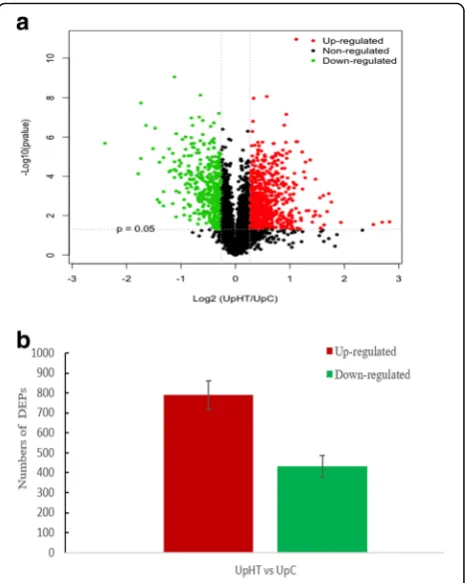
Protein response to heat stress in U. prolifera were re-vealed by iTRAQ analysis. A total of 283,344 spectra were generated; 15,546 peptides and 4449 proteins were identified with the cut-off of Mascot Percolator Q value<= 0.01 [29]. Differentially expressed proteins (DEPs) were identified upon an expression ratio > 1.20 or < 0.83 and p < 0.05 under UpHT compared to UpC [30–32]. During high-temperature stress, a total of 1223 proteins were found to be significantly changed, of which, 790 DEPs were up-regulated and 433 DEPs were down-regulated (Fig.1, Table1), which indicated that al-most 27.50% of the identified proteins changed their abundance significantly in response to the heat stress.
GO function analysis of DEPs
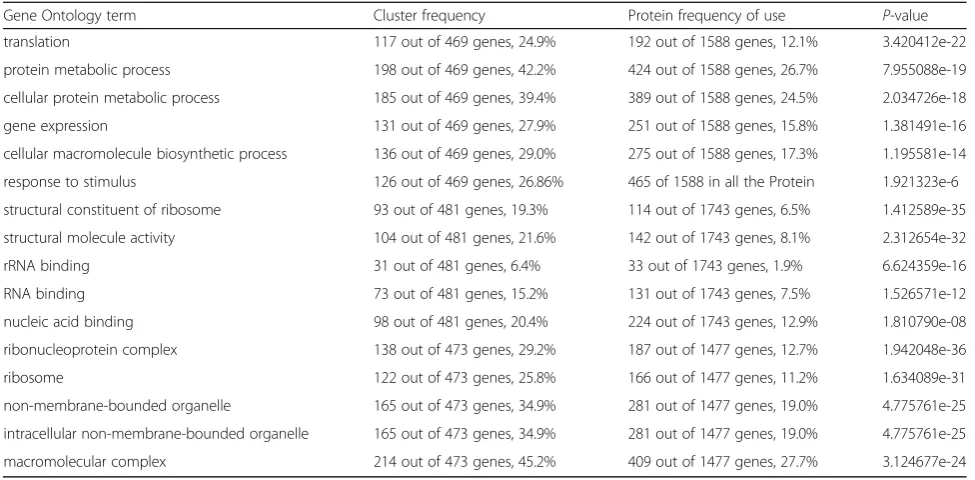
GO analysis was conducted on DEPs using the Blast2GO program; 469, 481 and 473 DEPs were successfully assigned to the biological processes, molecular function, and cellular components GO categories, respectively. The distribution of GO terms (the second level) for the three categories is shown in Table 2. For the biological process category, the most abundant groups were meta-bolic process (42.20%), cellular process (39.40%), and translation (24.90%). In addition, 126 DEPs belonged to response to stimulus (26.86%), of which 86 DEPs were up-regulated and 40 DEPs were down-regulated. For the cellular component category, cells (63.59%), cell parts (63.59%), and organelles (51.79%) were the most abun-dant groups. In the molecular function category, most of the DEPs were classified into catalytic activity (58.07%) and binding functions (45.70%); antioxidant activity only accounted for 0.72% of the DEPs (Table2). Furthermore, subcellular localization of the 790 up-regulated charac-terized proteins showed that 54 proteins (6.84%) were located in the chloroplast, 139 proteins (17.60%) were assigned to the mitochondria, 42 proteins (5.32%) belonged to the secretory pathway, and 512 proteins (12%) were classified as belonging to other locations.
Forty-three of the DEPs had unknown locations (Fig.2). Furthermore, subcellular localization analysis of the 433 down-regulated proteins showed that quite many chloroplast proteins and mitochondria proteins are re-lated to the thermotolerance ofU. prolifera.
COG function class of DEPs
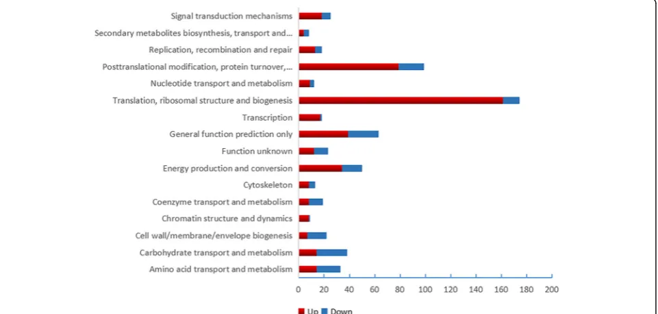
In the COG function analysis, the largest category was
“translation, ribosomal structure and biogenesis”(161 DEPs up-regulated, 13 DEPs down-regulated), followed by“ post-translational modification, protein turnover, molecular chaperone”(79DEPs up-regulated and 20 DEPs down-regu-lated) and “general function prediction only” (39 DEPs up-regulated and 24 DEPs down-regulated) (Fig.3).
KEGG pathway analysis of DEPs
KEGG pathway analysis of DEPs were conducted online at
http://www.genome.jp/kegg/kegg2.html; 557 up-regulated proteins were mapped to 97 KEGG pathways, and the top 15 pathways are listed in Table3. The pathway enrichment
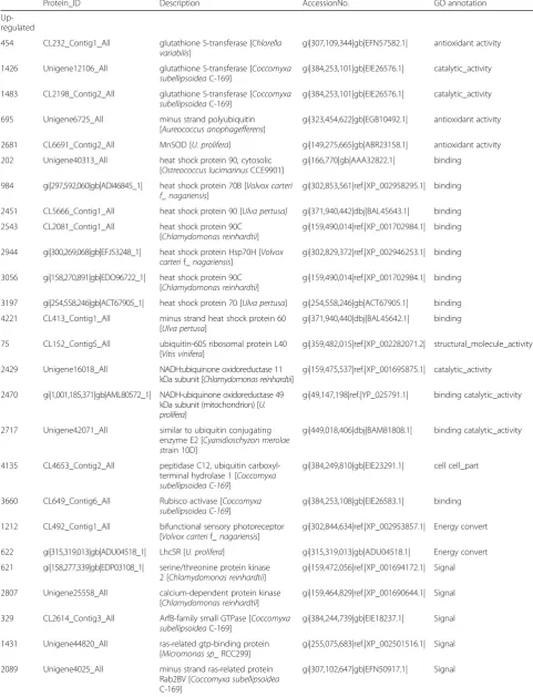
Table 1Partion DEPs under UpHT vs UpC conditions
Protein_ID Description AccessionNo. GO annotation
Up-regulated
454 CL232_Contig1_All glutathione S-transferase [Chlorella
variabilis]
gi|307,109,344|gb|EFN57582.1| antioxidant activity
1426 Unigene12106_All glutathione S-transferase [Coccomyxa
subellipsoideaC-169]
gi|384,253,101|gb|EIE26576.1| catalytic_activity
1483 CL2198_Contig2_All glutathione S-transferase [Coccomyxa
subellipsoideaC-169]
gi|384,253,101|gb|EIE26576.1| catalytic_activity
695 Unigene6725_All minus strand polyubiquitin
[Aureococcus anophagefferens]
gi|323,454,622|gb|EGB10492.1| antioxidant activity
2681 CL6691_Contig2_All MnSOD [U. prolifera] gi|149,275,665|gb|ABR23158.1| antioxidant activity
202 Unigene40313_All heat shock protein 90, cytosolic
[Ostreococcus lucimarinusCCE9901]
gi|166,770|gb|AAA32822.1| binding
984 gi|297,592,060|gb|ADI46845_1| heat shock protein 70B [Volvox carteri f_ nagariensis]
gi|302,853,561|ref.|XP_002958295.1| binding
2451 CL5666_Contig1_All heat shock protein 90 [Ulva pertusa] gi|371,940,442|dbj|BAL45643.1| binding
2543 CL2081_Contig1_All heat shock protein 90C
[Chlamydomonas reinhardtii]
gi|159,490,014|ref.|XP_001702984.1| binding
2944 gi|300,269,068|gb|EFJ53248_1| heat shock protein Hsp70H [Volvox carterif_nagariensis]
gi|302,829,372|ref.|XP_002946253.1| binding
3056 gi|158,270,891|gb|EDO96722_1| heat shock protein 90C [Chlamydomonas reinhardtii]
gi|159,490,014|ref.|XP_001702984.1| binding
3197 gi|254,558,246|gb|ACT67905_1| heat shock protein 70 [Ulva pertusa] gi|254,558,246|gb|ACT67905.1| binding
4221 CL413_Contig1_All minus strand heat shock protein 60
[Ulva pertusa]
gi|371,940,440|dbj|BAL45642.1| binding
75 CL152_Contig5_All ubiquitin-60S ribosomal protein L40
[Vitis vinifera]
gi|359,482,015|ref.|XP_002282071.2| structural_molecule_activity
2429 Unigene16018_All NADH:ubiquinone oxidoreductase 11
kDa subunit [Chlamydomonas reinhardtii]
gi|159,475,537|ref.|XP_001695875.1| catalytic_activity
2470 gi|1,001,185,371|gb|AML80572_1| NADH-ubiquinone oxidoreductase 49 kDa subunit (mitochondrion) [U. prolifera]
gi|49,147,198|ref.|YP_025791.1| binding catalytic_activity
2717 Unigene42071_All similar to ubiquitin conjugating
enzyme E2 [Cyanidioschyzon merolae strain 10D]
gi|449,018,406|dbj|BAM81808.1| binding catalytic_activity
4135 CL4653_Contig2_All peptidase C12, ubiquitin
carboxyl-terminal hydrolase 1 [Coccomyxa subellipsoidea C-169]
gi|384,249,810|gb|EIE23291.1| cell cell_part
3660 CL649_Contig6_All Rubisco activase [Coccomyxa
subellipsoidea C-169]
gi|384,253,108|gb|EIE26583.1| binding
1212 CL492_Contig1_All bifunctional sensory photoreceptor
[Volvox carterif_ nagariensis]
gi|302,844,634|ref.|XP_002953857.1| Energy convert
622 gi|315,319,013|gb|ADU04518_1| LhcSR [U. prolifera] gi|315,319,013|gb|ADU04518.1| Energy convert
621 gi|158,277,339|gb|EDP03108_1| serine/threonine protein kinase 2 [Chlamydomonas reinhardtii]
gi|159,472,056|ref.|XP_001694172.1| Signal
2807 Unigene25558_All calcium-dependent protein kinase
[Chlamydomonas reinhardtii]
gi|159,464,829|ref.|XP_001690644.1| Signal
329 CL2614_Contig3_All ArfB-family small GTPase [Coccomyxa
subellipsoideaC-169]
gi|384,244,739|gb|EIE18237.1| Signal
1431 Unigene44820_All ras-related gtp-binding protein
[Micromonas sp_ RCC299]
gi|255,075,683|ref.|XP_002501516.1| Signal
2089 Unigene4025_All minus strand ras-related protein
Rab2BV [Coccomyxa subellipsoidea
C-169]
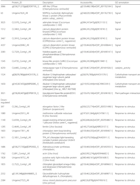
Table 1Partion DEPs under UpHT vs UpC conditions(Continued)
Protein_ID Description AccessionNo. GO annotation
3066 gi|158,271,373|gb|EDO97193_1| ARF-like GTPase
[Chlamydomonas reinhardtii]
gi|159,488,198|ref.|XP_001702104.1| Signal
63 Unigene7624_All NDPK2a nucleotide diphosphate
kinase 2 protein [Physcomitrella patenssubsp_ patens]
gi|168,020,398|ref.|XP_001762730.1| Signal
3025 CL5370_Contig1_All adenylate kinase [Coccomyxa
subellipsoideaC-169]
gi|384,247,647|gb|EIE21133.1| Signal
1512 CL3463_Contig1_All calcium-dependent protein
kinase(CDPK)[Coccomyxa subellipsoideaC-169]
gi|384,245,250|gb|EIE18745.1| Signal
3467 CL3910_Contig4_All calcium-dependent protein kinase
(CDPK)[Volvox carterif_ nagariensis]
gi|384,245,250|gb|EIE18745.1| Signal
4337 Unigene26962_All calcium-dependent protein kinase
(CDPK) [Chlamydomonas reinhardtii]
gi|159,464,829|ref.|XP_001690644.1| Signal
3383 CL7329_Contig1_All histidine-aspartic acid
phosphotransferase [Chlamydomonas reinhardtii]
gi|159,463,834|ref.|XP_001690147.1| Signal
1545 CL4735_Contig1_All kinase-like protein-SnRK2 [Coccomyxa
subellipsoideaC-169]
gi|384,245,989|gb|EIE19481.1| Signal
4259 CL6485_Contig1_All metacaspase type II [Chlamydomonas
reinhardtii]
gi|159,467,293|ref.|XP_001691826.1| catalytic_activity
1524 gi|28,076,780|gb|AAO31578_1| ribulose-1,5-bisphosphate carboxylase/ oxygenase large subunit, partial (chloroplast) [Ulva lactuca]
gi|28,076,780|gb|AAO31578.1| Carbohydrate transport and metabolism
1650 gi|145,567,455|gb|ABP82085_1| ribulose-1,5-bisphosphate carboxylase/ oxygenase large subunit, partial (chloroplast) [Ulva sp_ WELT A027500]
gi|219,932,444|emb|CAR65339.1| Carbohydrate transport and metabolism
3021 gi|158,282,407|gb|EDP08159_1| triacylglycerol lipase-like protein(EDS1) [Chlamydomonas reinhardtii]
gi|159,476,166|ref.|XP_001696182.1| Plant-pathogen interaction
Down-regulated
397 CL206_Contig2_All elongation factor 2-like
[Solanum lycopersicum]
gi|356,525,774|ref.|XP_003531498.1| Response to stimulus
1080 Unigene25013_All methionine sulfoxide reductase
B [Ulva fasciata]
gi|197,631,364|gb|ACH70611.1| Response to stimulus
1140 CL6594_Contig3_All oxygen-evolving enhancer protein
1 precursor [Volvox carterif_nagariensis]
gi|302,846,662|ref.|XP_002954867.1| Response to stimulus
1295 CL4649_Contig1_All ferritin [Ulva fasciata] gi|149,275,663|gb|ABR23157.1| Response to stimulus
1603 Unigene17811_All chloroplast stem-loop-binding
protein [Chlamydomonas reinhardtii]
gi|159,464,355|ref.|XP_001690407.1| Response to stimulus
1611 CL1851_Contig5_All TPA_inf: chloroplast light-harvesting
complex II protein precursor Lhcbm5 [Acetabularia acetabulum]
gi|145,079,356|tpg|DAA05911.1| Response to stimulus
1686 gi|158,277,735|gb|EDP03502_1| Adenylosuccinate synthetase, chloroplastic;
gi|159,469,564|ref.|XP_001692933.1| Response to stimulus
1762 CL891_Contig1_All actin [Ulva linza] gi|363,992,274|gb|AEW46682.1| Response to stimulus
1828 Unigene18752_All putative early light-inducible protein
2 [Ulva linza]
gi|380,468,161|gb|AFD61608.1| Response to stimulus
1855 CL7525_Contig1_All chloroplast glycerolipid omega-3-fatty
acid desaturase [Chlamydomonas reinhardtii]
gi|159,462,866|ref.|XP_001689663.1| Response to stimulus
2532 gi|1,181,548|gb|AAA86855_1| Glyceraldehyde-3-phosphate
dehydrogenase A, chloroplastic;
gi|159,463,282|ref.|XP_001689871.1| Response to stimulus
2584 Unigene27523_All minus strand plastocyanin precursor
[Ulva pertusa]
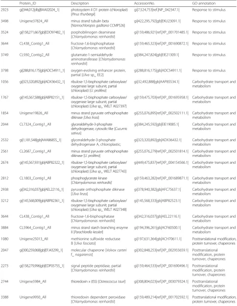
Table 1Partion DEPs under UpHT vs UpC conditions(Continued)
Protein_ID Description AccessionNo. GO annotation
2923 gi|344,012|dbj|BAA02024_1| photosystem II D1 protein (chloroplast) [Pinus thunbergii]
gi|7,524,753|ref.|NP_042347.1| Response to stimulus
3498 Unigene37824_All minus strand tubulin beta
[Nannochloropsis gaditanaCCMP526]
gi|422,295,792|gb|EKU23091.1| Response to stimulus
3524 gi|158,271,667|gb|EDO97482_1| porphobilinogen deaminase [Chlamydomonas reinhardtii]
gi|159,486,921|ref.|XP_001701485.1| Response to stimulus
3644 CL438_Contig1_All fructose-1,6-bisphosphatase
[Chlamydomonas reinhardtii]
gi|159,465,323|ref.|XP_001690872.1| Response to stimulus
3749 CL930_Contig2_All glutamate-1-semialdehyde
aminotransferase [Chlamydomonas reinhardtii]
gi|384,247,824|gb|EIE21309.1| Response to stimulus
3798 gi|288,816,175|gb|ADC54911_1| oxygen-evolving enhancer protein, partial [Ulvasp_ EE2]
gi|288,816,175|gb|ADC54911.1| Response to stimulus
1056 gi|323,320,892|gb|ADX36432_1| ribulose-1,5-bisphosphate carboxylase/ oxygenase large subunit, partial (chloroplast) [U. prolifera]
gi|32,492,888|gb|AAP85534.1| Carbohydrate transport and metabolism
1767 gi|145,567,588|gb|ABP82151_1| ribulose-1,5-bisphosphate carboxylase/ oxygenase large subunit, partial (chloroplast) [Ulvasp_WELT A027397]
gi|159,475,703|ref.|XP_001695958.1| Carbohydrate transport and metabolism
1854 Unigene19826_All minus strand pyruvate orthophosphate
dikinase[Ulva linza]
gi|255,076,895|ref.|XP_002502111.1| Carbohydrate transport and metabolism
2044 CL7324_Contig1_All glyceraldehyde-3-phosphate
dehydrogenase, cytosolic-like [Cucumis sativus]
gi|384,245,592|gb|EIE19085.1| Carbohydrate transport and metabolism
2532 gi|1,181,548|gb|AAA86855_1| glyceraldehyde-3-phosphate dehydrogenase A, chloroplastic;
gi|323,320,892|gb|ADX36432.1| Carbohydrate transport and metabolism
2561 CL2687_Contig1_All minus strand pyruvate orthophosphate
dikinase [U. prolifera]
gi|255,076,279|ref.|XP_002501814.1| Carbohydrate transport and metabolism
2674 gi|145,567,931|gb|ABP82322_1| ribulose-1,5-bisphosphate carboxylase/ oxygenase large subunit, partial (chloroplast) [Ulva sp_ WELT A027743]
gi|449,475,837|ref.|XP_004154566.1| Carbohydrate transport and metabolism
2812 CL1803_Contig1_All phosphoglycerate kinase
[Chlamydomonas reinhardtii]
gi|159,463,282|ref.|XP_001689871.1| Carbohydrate transport and metabolism
2938 gi|342,316,037|gb|AEL22116_1| pyruvate orthophosphate dikinase [Ulva linza]
gi|378,940,382|gb|AFC75637.1| Carbohydrate transport and metabolism
3212 gi|145,568,009|gb|ABP82361_1| ribulose-1,5-bisphosphate carboxylase/ oxygenase large subunit, partial (chloroplast) [Ulvasp_ WELT A027348]
gi|145,568,333|gb|ABP82523.1| Carbohydrate transport and metabolism
3644 CL438_Contig1_All fructose-1,6-bisphosphatase
[Chlamydomonas reinhardtii]
gi|342,316,037|gb|AEL22116.1| Carbohydrate transport and metabolism
3884 CL5964_Contig1_All minus strand starch branching enzyme
II [Parachlorella kessleri]
gi|194,396,261|gb|ACF60500.1| Carbohydrate transport and metabolism
1080 Unigene25013_All methionine sulfoxide reductase
B [Ulva fasciata]
gi|197,631,364|gb|ACH70611.1| Posttranslational modification, protein turnover, chaperones
2047 gi|300,259,068|gb|EFJ43299_1| molecular chaperone [Volvox carteri
f_nagariensis]
gi|302,848,253|ref.|XP_002955659.1| Posttranslational modification, protein turnover, chaperones
2273 gi|158,279,996|gb|EDP05755_1| signal peptide peptidase, partial [Chlamydomonas reinhardtii]
gi|159,464,533|ref.|XP_001690496.1| Posttranslational modification, protein turnover, chaperones
2744 Unigene5984_All thioredoxin x (ISS) [Ostreococcus tauri] gi|308,804,023|ref.|XP_003079324.1| Posttranslational modification, protein turnover, chaperones
3388 Unigene9950_All thioredoxin dependent peroxidase
[Chlamydomonas reinhardtii]
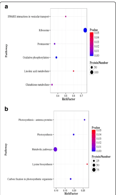
analysis showed that the most significantly enriched path-ways among the 557 up-regulated proteins were ribosome (134, 24.05%), oxidative phosphorylation (32, 5.75%), prote-asome (14, 2.51%), glutathione metabolism (15, 2.69%), SNARE interactions in vesicular transport (5, 0.89%) and linoleic acid metabolism (3, 0.53%) (Fig.4a). Two hundred and thirty-three down-regulated proteins were enriched in photosynthesis (15, 6.44%), carbon fixation in photosyn-thesis organisms (15, 6.44%), photosynphotosyn-thesis antenna pro-teins (8, 8.43%), metabolic pathways (91, 39.06%) and lysine biosynthesis (3, 1.2%) (Fig. 4b). At present, glutathione S-transferase, heat shock proteins, MnSOD and ubiquitin-related proteins were up-regulated. The results
indicated that upon high-temperature stimulus,U. prolifera
induces defence mechanisms, and the expression of heat shock proteins and antioxidant-associated proteins is increased.
Expression analysis of DEPs in response to stimulus
Abiotic stresses such as high temperature, low temperature, and drought induced plant defence mecha-nisms, including the expression of antioxidant enzymes to regulate their adaptation [33]. In higher plants, the signal response to high-temperature stress involves a re-duction in the synthesis of normal proteins and is ac-companied by accelerated expression of heat-responsive Table 1Partion DEPs under UpHT vs UpC conditions(Continued)
Protein_ID Description AccessionNo. GO annotation
4415 CL3087_Contig4_All molecular chaperone [Volvox carteri
f_nagariensis]
gi|302,839,709|ref.|XP_002951411.1| Posttranslational modification, protein turnover, chaperones
141 CL451_Contig4_All diadenosine tetraphosphatase and
related serine/threonine protein phosphatases[Chlorella variabilis]
gi|307,103,797|gb|EFN52054.1| Signal transduction
mechanisms
242 CL5191_Contig2_All serine/threonine protein phosphatase
[Guillardia thetaCCMP2712]
gi|348,671,120|gb|EGZ10941.1| Signal transduction
mechanisms
250 Unigene7524_All protein serine/threonine phosphatase
2C [Coccomyxa subellipsoideaC-169]
gi|307,104,872|gb|EFN53124.1| Signal transduction
mechanisms
3622 CL6423_Contig1_All minus strand protein tyrosine
phosphatase [Ectocarpus siliculosus]
gi|299,116,693|emb|CBN74838.1| Signal transduction
mechanisms
4212 CL4299_Contig1_All serine/threonine protein phosphatase
[Volvox carterif_ nagariensis]
gi|302,851,958|ref.|XP_002957501.1| Signal transduction
mechanisms
2947 Unigene205_All phytoene dehydrogenase and related
proteins[Chlorella variabilis]
gi|384,249,750|gb|EIE23231.1| Secondary metabolites biosynthesis, transport
3019 gi|158,273,526|gb|EDO99315_1| cytochrome P450, CYP85 clan, partial [Chlamydomonas reinhardtii]
gi|159,481,496|ref.|XP_001698815.1| Secondary metabolites biosynthesis
3998 CL7806_Contig1_All carotenoid cleavage dioxygenase
[Chlamydomonas reinhardtii]
gi|159,474,908|ref.|XP_001695565.1| Secondary metabolites biosynthesis
4156 CL6885_Contig2_All phytoene dehydrogenase and related
proteins [Volvox carterif_nagariensis]
gi|302,835,624|ref.|XP_002949373.1| Secondary metabolites biosynthesis
397 CL206_Contig2_All elongation factor 2-like [Solanum
lycopersicum]
gi|356,525,774|ref.|XP_003531498.1| Translation
4324 gi|452,119,419|gb|AGG09538_1| elongation factor Tu, partial (chloroplast) [U. prolifera]
gi|452,119,405|gb|AGG09531.1| Translation
756 Unigene5495_All oxidoreductase [Coccomyxa
subellipsoideaC-169]
gi|384,249,901|gb|EIE23381.1| Energy production and conversion
1323 CL514_Contig1_All glutathione reductase [Ulva fasciata] gi|82,658,844|gb|ABB88584.1| Energy production and
conversion
1554 CL3939_Contig2_All glutathione reductase [Ulva fasciata] gi|82,658,844|gb|ABB88584.1| Energy production and
conversion
1808 Unigene43735_All photosystem I subunit VII (chloroplast)
[Trebouxiophyceaesp_ MX-AZ01]
gi|357,467,753|ref.|XP_003604161.1| Energy production and conversion
2584 Unigene27523_All plastocyanin precursor [Ulva pertusa] gi|48,526,878|gb|AAT45616.1| Energy production and
genes and HSPs. Reactive oxygen species (ROS) refer to free radicals, including hydrogen peroxide (H2O2),
sing-let oxygen (O2−), and hydroxyl radicals (OH−). Excessive
amounts of ROS can damage macromolecules and cell membranes. To remove toxic ROS, algae have developed a number of antioxidant systems that serve as protective mechanisms, namely, superoxide dismutases (SOD), cat-alases, peroxidases, thioredoxin, and glutathione [34]. In the present study, high-temperature stress can lead to protein misfolding and inactivation and desiccation of plant cells. Heat shock proteins are important molecular chaperones of the Hsp family, which prevents the aggre-gation of misfolded proteins. Significant up-regulation was observed for the heat shock protein family, which
included 90 (CL5666_Contig1_All, 1.5-fold), 90C (Uni-gene40313_All, 1.78-fold), 70 (gi|219764948|emb|-CAW63927_1|, 1.4-fold), ClpB chaperone, Hsp100 family (CL517_Contig2_All, 1.64-fold). Therefore, it could play a role in protecting the protein structure early in the stress response. DNA repair protein Rad50 (CL6208_Contig1_All, 1.22-fold) were identified and sig-nificantly up-regulated under high temperature. It means that the DNA repair protein Rad50 played an important role in DNA damage induced by high temperature. We also determined that antioxidant proteins exhibited highly dynamic changes in response to the high-temperature stimulus. Reactive oxygen or nitrogen species are gener-ated in the plant cell during the extreme stress condition, Table 2GO analysis of DEPs (UpHT vs UpC) (top15)
Gene Ontology term Cluster frequency Protein frequency of use P-value
translation 117 out of 469 genes, 24.9% 192 out of 1588 genes, 12.1% 3.420412e-22
protein metabolic process 198 out of 469 genes, 42.2% 424 out of 1588 genes, 26.7% 7.955088e-19
cellular protein metabolic process 185 out of 469 genes, 39.4% 389 out of 1588 genes, 24.5% 2.034726e-18
gene expression 131 out of 469 genes, 27.9% 251 out of 1588 genes, 15.8% 1.381491e-16
cellular macromolecule biosynthetic process 136 out of 469 genes, 29.0% 275 out of 1588 genes, 17.3% 1.195581e-14
response to stimulus 126 out of 469 genes, 26.86% 465 of 1588 in all the Protein 1.921323e-6
structural constituent of ribosome 93 out of 481 genes, 19.3% 114 out of 1743 genes, 6.5% 1.412589e-35
structural molecule activity 104 out of 481 genes, 21.6% 142 out of 1743 genes, 8.1% 2.312654e-32
rRNA binding 31 out of 481 genes, 6.4% 33 out of 1743 genes, 1.9% 6.624359e-16
RNA binding 73 out of 481 genes, 15.2% 131 out of 1743 genes, 7.5% 1.526571e-12
nucleic acid binding 98 out of 481 genes, 20.4% 224 out of 1743 genes, 12.9% 1.810790e-08
ribonucleoprotein complex 138 out of 473 genes, 29.2% 187 out of 1477 genes, 12.7% 1.942048e-36
ribosome 122 out of 473 genes, 25.8% 166 out of 1477 genes, 11.2% 1.634089e-31
non-membrane-bounded organelle 165 out of 473 genes, 34.9% 281 out of 1477 genes, 19.0% 4.775761e-25
intracellular non-membrane-bounded organelle 165 out of 473 genes, 34.9% 281 out of 1477 genes, 19.0% 4.775761e-25
macromolecular complex 214 out of 473 genes, 45.2% 409 out of 1477 genes, 27.7% 3.124677e-24
which produces toxic compounds after reacting with the organic molecules. The glutathione-S-transferase (GST) enzymes play a significant role in detoxifying these toxins and help in their excretion or sequestration; the enzymes catalyse reduction of glutathione (GSH; a tripeptide Glu-Cys-Gly) by electrophilic and hydrophobic toxic mol-ecules that are generated under stress to convert them to non-toxic and soluble conjugates [35]. The expression of glutathione S-transferase (Unigene15839_All,1.45-fold), MnSOD (CL6691_Contig2_All, 1.42-fold), ascorbate peroxidase (CL1454_Contig2_All,1.28-fold) and polyubi-quitin (Unigene6725_All, 1.54-fold) were up-regulated. In addition, the expression of catalase-peroxidase (CL5101_ Contig1_All, 0.66-fold), glutathione reductase (CL514_ Contig1_All, 0.77-fold), and thioredoxin-dependent peroxidase (Unigene9950_All, 0.77-fold) were down-regulated.
In addition, the 26S proteasome regulatory subunit is a component of the hexameric ring of AAA-ATPases that forms the base of the 19S regulatory particle (RP). This subunit plays specific roles in plant proteasomes by helping to promote the assembly of the RP with the 20S core protease (CP) and gate the CP to prevent indiscrim-inate degradation of cytosolic and nuclear proteins. In plants, this subunit plays an important role in diverse processes that include shoot and root apical meristem maintenance, cell size regulation, and stress responses [36]. In the present study, the 26S proteasome regulatory subunit (CL1786_Contig3_All, CL2484_Contig2_All, 1.3 or 1.32-fold) was induced under high-temperature stress. The result is consistent with the observation that the ex-pression of the 26S proteasome subunit RPN10 is
upregulated by salt stress in Dunaliella viridis [37]. The result indicated that the 26S proteasome regulatory sub-unit played a key role in stress tolerance. Programmed cell death (PCD) is an important biological phenomenon. Algae can activate PCD when exposed to external patho-gens and other stresses. Metacaspases often play an im-portant role in plant PCD, and a large number of experiments have shown that metacaspases are involved in plant PCD. In PCD, type II metacaspase was found to be transported from the cytoplasm to the nucleus, which promoted chromosome degradation. In the present study, mov34-domain-containing protein (Unigene14048_All, 1.33-fold) and metacaspase type II (CL6485_Contig1_All, 1.25-fold) were up-regulated. The results indicated that the high-temperature stimulus accelerated the accumula-tion of ROS, inhibited the growth ofU. prolifera, induced the expression of partial stress related proteins, and allevi-ated the stimulation. This process is consistent with the results that are reported for the brown alga Ectocarpus siliculosus[38].
Expression analysis of DEPs that are involved in the oxidative phosphorylation pathway
The oxidative phosphorylation (OXPHOS) process com-prises an electron-transfer chain (ETC) that is driven by substrate oxidation and is coupled to the synthesis of ATP through an electrochemical transmembrane gradi-ent. Oxidative phosphorylation is a vital part of metabol-ism, it also produces reactive oxygen species such as superoxide and hydrogen peroxide, which leads to propagation of free radicals, damages cells and contrib-utes to disease, and possibly ageing. The previous study
showed that low temperature, UVB radiation and phoxim-induced increase in oxidative phosphorylation gene [39–41]. In the present study, 34DEPs were mapped to the oxidative phosphorylation pathway (Figs.
5 and 6). The expression of 32 DEPs were found to be increased, and 2 DEPs were down-regulated. NADH:ubi-quinone oxidoreductase (CL6587_Contig2_All), succin-ate dehydrogenase (CL2133_Contig2_All), cytochrome C oxidase subunit (CL1977_Contig2_All), and ATP synthe-sis subunit (gi|807046094|gb|AKC35152_1) were all sig-nificantly up-regulated by 1.36 fold, 1.59 fold, 1.23 fold, and 1.83 fold, respectively. The results are consistent with previous results. These results indicated that high-temperature induction enhanced the activity of the
ETC complex in the mitochondria. The imbalance be-tween the activity changes of ETC may lead to the accu-mulation of reactive oxygen species.
Expression analysis of DEPs involved in phytohormone and signal transduction pathways
Studies have investigated the expression of the hormone-related proteins that were changed under abiotic stress conditions [42]. In the present study, expression pattern analysis showed that the high-temperature stimulus chan-ged the expression of key enzymes involved in phytohor-mone signal transduction and horphytohor-mone biosynthesis. Approximately 12 DEPs were identified in phytohormone signal transduction (Fig. 7). In carotenoid biosynthesis, Table 3KEGG pathway of DEPs(top15)(UpHT vs UpC)
Pathway DEPs with pathway
annotation
All Proteins with pathway annotation (3046)
P value Pathway ID
Up-regulated
Ribosome 134 179 2.73E-66 ko03010
Oxidative phosphorylation 32 84 1.18E-05 ko00190
Proteasome 14 39 0.006672349 ko03050
Glutathione metabolism 15 46 0.01352303 ko00480
SNARE interactions in vesicular transport 5 10 0.0225611 ko04130
Linoleic acid metabolism 3 5 0.04546205 ko00591
Protein processing in endoplasmic reticulum 23 90 0.05135553 ko04141
Phagosome 13 45 0.05413349 ko04145
Spliceosome 25 101 0.06097134 ko03040
Non-homologous end-joining 2 3 0.08799354 ko03450
RNA transport 22 95 0.1338129 ko03013
Porphyrin and chlorophyll metabolism 12 47 0.1356993 ko00860
Taurine and hypotaurine metabolism 1 1 0.1828628 ko00430
Vancomycin resistance 1 1 0.1828628 ko01502
Plant-pathogen interaction 11 48 0.2512615 ko04626
Down-regulated
Photosynthesis 15 76 0.00047354 ko00195
Carbon fixation in photosynthetic organisms 15 83 0.001247104 ko00710
Photosynthesis - antenna proteins 8 31 0.001746649 ko00196
Metabolic pathways 91 952 0.005208609 ko01100
Lysine biosynthesis 3 11 0.04605148 ko00300
Betalain biosynthesis 1 1 0.07649376 ko00965
Carotenoid biosynthesis 4 23 0.09360185 ko00906
Selenocompound metabolism 3 17 0.1357731 ko00450
Monobactam biosynthesis 2 9 0.146953 ko00261
Degradation of aromatic compounds 1 2 0.1471594 ko01220
Tyrosine metabolism 3 18 0.1542241 ko00350
Tryptophan metabolism 3 19 0.1734048 ko00380
One carbon pool by folate 3 19 0.1734048 ko00670
cytochrome P450 (Unigene18531_All) was significantly up-regulated by 1.23 fold. The expression of violaxanthin de-epoxidase-related protein (CL1646_Contig1_Alland CL1850_Contig11_All), prolycopene isomerase (Unige-ne205_All) and zeaxanthin epoxidase (CL4385_Conti-g1_All) were down-regulated by 0.83 fold, 0.74 fold, 0.81 fold, and 0.82 fold compared to the control. In brassinos-teroid (BR) biosynthesis, molecular and biochemical stud-ies revealed that two different cytochrome P450s, CYP90B2 and CYP724B1/D11, are the rate-limiting step of brassinosteroid biosynthesis. Three significant DEPs
that were mapped to the BR synthesis pathway the
expression of cytochrome P450s 90B/724B
(gi|158273526|gb|EDO99315_1|, 0.69-fold) were de-creased. The expression of 90C1D1 (CL3922_Contig1_All, 1.29-fold) and CYP735A (Unigene18531_All, 1.23-fold) were up-regulated. CYP735A encodes cytokinin hydroxy-lases that catalyse iP-nucleotide converts to trans-Zeatin. The signalling transduction of the cytokinin pathway and abscisic acid (ABA) pathway played an important role in the growth and development of plants and the regulation of stress resistance. AHP (Histidine-containing phospho-transfer protein) mainly functions as two-component phosphorelay mediators between cytokinin sensor histi-dine kinases and response regulators (B-type ARRs) and plays an important role in promoting cytokinin signal transduction via the multistep His-to-Asp phosphorelay. Biochemical and molecular genetic studies inArabidopsis
have identified protein phosphatase 2C (PP2C) enzymes as negative regulators in plant signal transduction pro-cesses [43]. In the absence of ABA, PP2C mainly inhibited the active state of SnRK2s protein. The inhibition of the active state of SnRK2s protein result in a decrease in down-stream transcriptional regulation. ABA relieved the inhib-ition effect of PP2C on SnRK2s, activated the SnRK2s protein, and initiated downstream signal transduction. In the present study, the expression of the AHP protein (CL7329_Contig1_All, 1.25-fold) and SnRK2s (CL4735_ Contig1_All, 1.27-fold) protein was up-regulated. However, the expression of PP2C (CL5191_Contig2_All, 0.8-fold) was down-regulated, which indicated that the signal transduc-tion pathway of cytokinin and ABA was induced at a high temperature. The result is consistent with previous rze-search [42, 43]. Plant growth is co-ordinately regulated by environmental and hormonal signals. Brassinosteroid (BR), cytokinin and ABA played essential roles in growth regula-tion via light and temperature; however, the interacregula-tions be-tween hormone signal transduction and environmental signals remain poorly understood at the molecular level. In summary, the results indicated that phytohormone signal regulation played a key role in the high-temperature stimu-lus and enhanced cytokinin and ABA signal transduction pathway. However, the regulation mechanism should be further considered.
Expression analysis of DEPs involved in photosynthesis-related proteins
Photosynthesis is very sensitive to high-temperature stress. Changes in environmental temperature are pri-marily reflected by photosynthesis, which triggers a response aimed at attaining the best possible perform-ance under the new conditions. Several studies have shown that high-temperature stress can significantly inhibit photosynthesis [44, 45]. In green algae, light-harvesting chlorophyll protein complexes are the
major light-harvesting complexes. These complexes in-clude photosystem II (light-harvesting complex II and photosynthetic reaction centre) and photosystem I (LHCI and photosynthetic reaction centre). The light-harvesting complex is a complex of subunit proteins that may be part of a larger supercomplex of the photosystem, and the function is to collect more of the incoming light. In the previous study, LHCI of photosystem I Chlamydomonas reinhardtii is composed of nine genes. Three product complexes (Lhca1, 3 and 7) showed emission peaks in the
range of 680–690 nm, thus accounting for the blue contri-bution observed in the native preparations. Three other complexes (Lhca5, 6 and 8) exhibit emission in the 695– 700 nm range. This emission component is not clearly re-solved in the fluorescence spectra of the native LHCI preparations, possibly due to energy transfer to the red forms of a neighbouring complex or due to a change in the absorption properties of these complexes, because of protein–protein interactions, when embedded in the PSI– LHCI supercomplex, as previously proposed [46]. Three more complexes, namely, Lhca2, 4 and 9, exhibited emission above 707 nm with the red-most form (715 nm) associated with Lhca2, which indicates that Lhca2 is re-sponsible for the red-most emission in vivo. Up-regulation of Lhca4 and Lhca9 was observed under iron-deficiency conditions, and it correlates with a shift to the blue end of the emission spectrum (from 710 to 704 nm) and with a relative increase in intensity of the emission above 700 nm, compared to that for the emission below 700 nm [47]. This indicates an increase in the content of red forms that emit approximately 705 nm in the antenna, in agree-ment with the fluorescence characteristics of Lhca4 and Lhca9. Under the same conditions, down-regulation of Lhca5 was also detected, leadin to the suggestion that Lhca4 can substitute for Lhca5, which possibly provides a better sink for energy dissipation. The finding that these antennas contain the lowest energy forms supports the hypothesis. A similar effect was recently observed in higher plants, where Lhca5 could substitute for Lhca4 [48, 49]. In the present study, the LHCI sub-units lhca1 (CL4203_Contig1_All, 0.72-fold) and lhca3 (CL654_Contig2_All, 0.8-fold) were down-regulated, and lhca2 (LhcSR, gi|315319013|gb|ADU04518_1|, 1.3-fold) was up-regulated. It may be possible that the decrease in the emission of blue light results in en-ergy transfer to the red forms. Lhca2 is responsible for the red-most emission. The up-regulation of lhca2 indicated that the lowest energy forms were native or-ganized. Lhca1 and lhca3 were unstable and under-went decomposition under high temperature. The
Fig. 5The oxidative phosphorylation pathway
results were consistent with that obtained by Naumann et al. [47]. In addition, the two most abun-dant light-harvesting complex II are Lhcb1 and Lhcb2, which make up light-harvesting complex (LHC) II trimers. They are also involved in facilitating state transitions, a process during which energy bal-ance between photosystem (PS) II and I is achieved. In the present study, the LHCII subunits lhcb1 and lhcb2 were also down-regulated. Down-regulation of LHCII would significantly influence light absorbance and energy transfer in U. prolifera upon a high– temperature stimulus (Fig. 8). The results were con-sistent with the observation that salt stress induces a decrease in excitation energy transfer from phycobili-somes to photosystem II but an increase to photo-system I in the cyanobacterium Spirulina platensis
[50]. Eighteen DEPs were mapped to the photosyn-thesis pathway, and the expression of 14 DEPs were down-regulated. Ferredoxin-NADP+ reductase (FNR) is one of the important enzymes involving in many biochemical and physiological metabolism processes,
such as electron transfer, carbon dioxide fixation, nitrogen assimilation and antioxidation. Ferredoxin-NAD-P+reductase (Unigene9738_All, 0.82-fold and CL7087_Contig1_All, 0.81-fold) and photosystem I and photosystem II subunit proteins were identified and found to be decreased.
However, the expression of the chloroplast ATP synthase subunit that was encoded by 5 unigenes (gi|807,046,094|g-b|AKC35152_1|, 1.82-fold) was up-regulated.
This result indicated that the high temperature inhibited photosynthesis of U. prolifera and induced stress-related LhcSR protein expression, which was con-sistent with the expression of LhcSR in U. linza in a study that was conducted by Dong et al. [51]. The electrons from PSI and PSII might transfer to oxygen, and thereby lead to substantial generation of reactive oxygen species. Enrichment of the oxidative phos-phorylation pathway under cold treatment suggested that high–temperature induced ROS production might occur. However, this hypothesis still lacks experimen-tal evidence.
Fig. 7The cluster of plant hormone biosynthesis and signal transduction related proteins(UpHT vs UpC). Red indicated the expression of protein up-regulated, green indicated the expression of protein down-regulated
Expression analysis of DEPs involved in the carbon-fixation pathway
According to CO2assimilation mechanisms, plants can be
classified into three types: C3, C4, and CAM. C4plants have
acquired a series of high-performance photosynthetic genes during evolution, which confer more efficient photosyn-thesis than that in C3plants under adverse conditions such
as high light intensity, temperature, and low CO2
concen-tration. In the present study, 19 DEPs were mapped to car-bon fixation in photosynthetic organisms (Fig. 9). The expression of 14 DEPs was down-regulated, and that of 5 DEPs was up-regulated. In C4-dicarboxylic acid cycle, the
key regulated enzymes aspartate aminotransferase (Unige-ne42677_All, 0.71-fold), pyruvate orthophosphate dikinase (Unigene19826_All, 0.73-fold), and NADP malic enzyme (CL5095_Contig3_All, 0.81-fold) were down-regulated. At the same time, pyruvate orthophosphate dikinase (Unigene19826_All, 0.73-fold) and NADP malic enzyme were also found to be down-regulated in CAM. Moreover, the expressions of plastid transketolase (Unigene20747_All, 0.43-fold), fructose-1,6-bisphosphatase (CL438_Conti-g1_All, 0.81-fold), glyceraldehyde-3-phosphate dehydrogen-ase (CL7324_Contig1_All, 0.72-fold) and phosphoglycerate kinase (CL1803_Contig1_All, 0.81-fold) were down-regu-lated. Ribulose-1,5-bisphosphatecarboxylase/oxygenase large subunit was found to be encoded by 7 DEPs, of which, 4 DEPs were up-regulated, and 3 DEPs were down-regu-lated. The result demonstrated that the expression of the key enzyme in the carbon-fixation pathway was down-regulated.
The activation of rubisco in vivo requires the presence of the regulatory protein rubisco activase. This enzyme facilitates the release of sugar phosphate inhibitors from rubisco catalytic sites thereby influencing carbamylation. Rubisco activase was used to explore the role of the
enzyme in C4photosynthesis at a high temperature. It is
concluded that during short-term treatment at 40 °C, ru-bisco activase content is not the only factor that modulates rubisco carbamylation during C4
photosyn-thesis [52]. In the present study, rubisco activase (CL649_Contig6_All, 1.32-fold) was up-regulated; how-ever, it is not the only factor. At the same time, soluble carbonic anhydrase precursor encoded by CL7120_Con-tig2_All was down-regulated, and CL6328_Contig1_All was up-regulated. In other words, to protect the energy balance, U. prolifera decreased photosynthesis and carbon fixation of metabolic activity under high-temperature conditions. However, increase in the ex-pression of carbonic anhydrase precursor and rubisco activase play an important role in the acceleration to photosynthesis balance adjustment.
Expression analysis of DEPs involved in the glutathione metabolism pathway
A total of 18 DEPs were mapped to the glutathione me-tabolism pathway. The expression of key enzymes in glutathione metabolism pathway were observed to sig-nificantly change. Glutathione S-transferase (GST) catal-yses the nucleophilic fusion of reduced glutathione (GSH; a tripeptide Glu-Cys-Gly) with electrophilic and hydrophobic toxic molecules, which were generated under stress, to convert them to non-toxic and soluble conjugates. GSTs play an important role in various metabolic pathways, and they are involved in detoxifica-tion of oxidative lipid peroxide metabolites, hormone metabolism, stress resistance, protection of cell oxidative stress injury, cell signal transduction and disease resist-ance [53–55]. Ascorbate peroxidase (APXs) catalyses the H2O2-dependent oxidation of ascorbate in plants, algae
and certain cyanobacteria. Glutathione reductase (GR)
catalyses the reduction of glutathione disulphide (GSSG) to the sulphydryl form glutathione (GSH), which is a critical molecule in resisting oxidative stress and maintaining the reducing environment inside the cell [56–58]. In the present study, APX (CL1454_Contig2_All, 1.28-fold), glutathione S-transferase (CL2243_Contig1_All, 1.53-fold), glucose-6-phosphate 1-dehydrogenase 2(CL2243_Conti-g1_All, 1.53-fold) and 6-phosphogluconate dehydrogenase (gi|158274793|gb|EDP00573_1|, 1.32-fold), decarboxyla-ting(gi|158274793|gb|EDP00573_1|, 1.32-fold) were found to significantly increase. However, the expression of GR(CL514_Contig1_All, 0.77-fold) was down-regulated (Fig. 10). It is indicated that high temperature stimulus accumulated more ROS, induced the expression of resistance-related protein glutathione S-transferase and APX.
Expression analysis of Ca2+-binding protein
Ca2+-dependent signalling processes are beneficial to plant perception, and they respond to diverse environ-mental stressors, such as osmotic stress and high temperature [59–61]. In our study, the expression of Hsp90 (Unigene40313_All, 1.78-fold), CDPK (Unige-ne26962_All, 1.35-fold) and EDS1 (gi|158,282,407|g-b|EDP08159_1|, 1.4-fold) was up-regulated in U. prolifera under the high-temperature stimulus. In addition, CDPK can activate H2O2 accumulation
through regulating NADPH oxidase. The results indi-cated that the high-temperature stimulus accelerated ROS signalling, CDPK and EDS1 expression. In conclu-sion, the Ca2+-signal also played an important role in high-temperature stress inU. prolifera.
Expression analysis of DEPs involved in other types of metabolisms
Linoleic acid (LA), a carboxylic acid, belongs to one of the two families ofessential fatty acids. It is converted by vari-ouslipoxygenasesandcyclooxygenasesand the cytochrome P450 enzyme. All of these LA products exhibit bioactivity, and they are implicated in human physiology and pathology as indicated in the cited linkages. In the present study, 4 DEPs were mapped to linoleic acid metabolism. The expression of the alpha-beta hydrolase superfamily (gi|158279131|gb|EDP04893_1|, 2.05-fold), cytochrome P450 (Unigene18531_All, 1.23-fold), lipoxygenase (Unige-ne17329_All, 3.21-fold), and 12-oxophytodienoic acid re-ductase (CL2323_Contig1_All, 1.47-fold) was up-regulated. In addition, glycan, taurine, and hypotaurine played a key role in plant response to stress. The expression of glycan-biosynthesis-related proteins changed significantly under high-temperature stimulus. The expression of dolichyl-phosphate beta-glucosyl transferase (ALG5,
Unigene5913_All, 1.29-fold) was up-regulated. However, alpha-1,2-mannosyltransferase (ALG9,
CL1555_Conti-g1_All, 0.78-fold) was down-regulated. Glycosyl transferase is encoded by two DEPs; one is down-regulated, and the other is up-regulated. Moreover, it has been suggested that taurine, hypotaurine and their metabolic precursors (cysteic acid, cysteamine and cysteine sulphinic acid) might act as antioxidants through scavenging ·OH, HOCl and H2O2. It
is concluded that cysteamine and hypotaurine are far more likely to act as antioxidants in vivo than taurine, provided that they are present in a sufficient concentration at sites of oxidant generation [62]. In our study, the expression of cys-tathionine beta-synthase (Unigene42486_All, 1.47-fold) was observed to be up-regulated. This result indicates that the
high-temperature stimulus induced cysteamine biosynthesis and increased the antioxidant levels.
qPCR to verify the reliability of the proteome
The expression of 8 unigenes were investigated via qPCR. The results were consistent with the available proteome data (Additional file 1: Table S1). Quantitative real time polymerase chain reaction results indicated that the uni-genes that encode heat shock protein 90, APX, MnSOD, glutathione reductase, glutathione S-transferase, RbcL, heat shock protein 70 and ATP synthase were all signifi-cantly up-regulated by 3.152, 2.502, 2.106, 0.420, 2.320, 2.522, 2.242 and 3.252-fold, respectively.
Validation of differentially expressed proteins identified by western blot
Two proteins, Hsp70 and RbcL identified DEPs with marked differences in expression determined by iTRAQ based quantitative analysis were selected to be verified by western blot analysis (Fig. 11). Hsp70 and RbcL protein was significantly up-regulated in UpHT groups as com-pared with control group (p< 0.05). The results which were found by western blot is consistent with the findings in iTRAQ analysis. It is well established that Hsp70 and RbcL could play a role in protecting the protein structure and photosynthesis early in the stress response.
PPI network
In plants, proteins do not function in isolation within cells but as part of a network. In this study, a PPI net-work was generated to highlight the interactions and relationships between different proteins. To obtain inter-action between proteins, we constructed a PPI network that was based on data downloaded from the STRING
database. For the UpHT vs UpC protein, we chose 102 proteins that were involved in response to stimulus, photosynthesis, carbon fixation in photosynthetic organ-isms, oxidative phosphorylation, glutathione metabolism, plant signal transduction and plant-pathogen interaction to construct the PPI. Based on the differential expression patterns at the protein levels in UpHT compared to UpC (Fig. 12). In the present study, there are 37 unigenes nodes and 194 interactions in the PPI network. The PPI network indicated that heat shock protein 90C (CL2081_Contig1_All, 1.49-fold), heat shock protein 90C (gi|158270891|gb|EDO96722_1|, 1.44-fold), heat shock protein 70 (gi|219764948|emb|CAW63927_1|, 1.4-fold), ClpB chaperone, Hsp100 family (CL517_Conti-g2_All and Unigene15739_All, 1.64-fold and 1.46-fold)
and heat shock protein 70B
(gi|297592060|g-b|ADI46845_1|, 1.82-fold) are involved in interactions with proteins from other functional groups and played important signal regulated. In addition, soluble inorganic pyrophosphatase 2 (CL408_Contig1_All, 1.51-fold), plastid transketolase (Unigene20747_All, 0.43-fold), F1F0 ATP synthase gamma subunit (Unigene20819_All, 1.69-fold), ribulose-1,5-bisphosphate carboxylase/oxy-genase large subunit (gi|323,320,892|gb|ADX36432_1|, 0.8-fold), serine/threonine-protein kinase CTR1 (gi|300257667|gb|EFJ41912_1|, 2.66-fold), component of
cytosolic 80S ribosome and 40S small subunit
(CL2825_Contig3_All, 1.3-fold), flagellar associated pro-tein (CL4919_Contig2_All, 1.53) as the key regulation factor and construct PPI under high temperature.
A pathway model of high temperature stress responses in U. prolifera
Using our results and previous studies, we propose a pu-tative synergistic response network for U. prolifera that responds to high-temperature stress. As shown in Fig.13, the reaction of U. prolifera to high-temperature stress rapidly accumulates ROS, which leads to a series of metabolic changes. First, high-temperature stress in-duces stress signals to generate differential accumulation of signal transduction components. High temperature causes the cell ROS concentration to rapidly increase, and high concentrations of ROS result in an increasing release of Ca2+into the cytoplasm. When the concentra-tion of Ca2+ reaches a particular threshold, CDPK is ac-tivated. As a result, plants are able to produce appropriate responses to the stress conditions. In an emergency response, HSP, as the key molecule, is signifi-cantly up-regulated to protect proteins from misfolding. To remove the accumulated ROS, the antioxidant sys-tem is activated, and proteins that regulate the redox balance, including MnSOD, GSTs, APX and other re-sponse factors, are up-regulated. These proteins can break down excessive intracellular ROS and restore the
normal intracellular redox environment. Proteins with important functions in energy metabolism are continu-ously upregulated to generate ATP, which allows plants to perform vital metabolic functions and to combat stress. Oxidative phosphorylation is the major approach to generate ATP, accompanied by the accumulation of ROS. The high-temperature stimulus induced upregulation of electron transport chain complex protein: NADH:ubiqui-none oxidoreductase, succinate dehydrogenase, cyto-chrome C oxidase subunit, and ATP synthesis subunit in mitochondria. The imbalance between the activity changes of ETC may lead to accumulation of reactive oxygen spe-cies. The possible reason was that changes in the quinone pool redox state are responsible for the apparent inorganic phosphate activation of complex III. Complex III is respon-sible for higher ROS production during physiological work-ing conditions relative to complex I [63]. At the same time, protein synthesis increased, and cytosolic 80S ribosome and 40S small subunit, nucleolar protein, and component of C/D snoRNPs were all up-regulated. However, the effect of high temperature on proteins involved in photosynthesis and carbon fixation are downregulated, and the result was
consistent with the proteome response of Brachypodium distachyon to drought stress [64].
Biological index and photosynthesis index
Antioxidant enzymes played an important role in pre-venting the oxidation of biological molecules and pro-tecting plant tissues from damage caused by ROS that was induced by multiple environmental stresses. SODs catalysed O2−+ 2H+→O2+ H2O2. In addition, APX and
CAT catalysed H2O2→O2+ H2O. In the present study,
the effects of high temperature were investigated on the activity of antioxidant enzymes, the chlorophyll content, and the chlorophyll fluorescence parameter of U. prolif-era. The result indicated that under a high-temperature stimulus, the protein content decreased during the all process compared to the control group (Additional file
1: Figure S1). SOD activity was decreased under high temperature compared to the control group. However, the APX activity increased in the later period (Add-itional file 1: Figure S2, Fig. 4). The MDA content in-creased over the time and reached the highest value after 96 h, which was significantly different from the
Fig. 12PPI network ofU. proliferaresponse to high temperature stimuls. Red indicated the expression of protein up-regulated,blue indicated the expression of protein down-regulated. CL2081_Contig1_All: heat shock protein 90C; gi|158270891|gb|EDO96722_1|:heat shock protein 90C; gi|219764948|emb|CAW63927_1|:heat-shock inducible Hsp70;Unigene20819_All:F1F0 ATP synthase gamma subunit; CL517_Contig2_All:ClpB chaperone, Hsp100 family; gi|1001185371|gb|AML80572_1|:NADH-ubiquinone oxidoreductase 49 kDa subunit; CL4445_Contig2_All:prohibitin; gi|315319013|gb|ADU04518_1|:LhcSR; gi|300264644|gb|EFJ48839_1|:Stress-induced-phosphoprotein; CL408_Contig1_All:soluble inorganic pyrophosphatase 2; gi|300257667|gb|EFJ41912_1|:Serine/threonine-protein kinase CTR1;CL2825_Contig3_All:component of cytosolic 80S ribosome and 40S small subunit;Unigene15739_All:ClpB chaperone, Hsp100 family; CL4169_Contig1_All:nucleolar protein, component of C/D
control group (Additional file 1: Figure S3). Further-more, the value of Fv/Fm dropped to the lowest point after 96 h of culture (Additional file 1: Figure S5). Our results provide important information for the selection of heat-resistantU. prolifera.
Conclusions
A total of 1223 differentially expressed proteins were iden-tified in response to heat stress. The up-regulated proteins were enriched in ribosome, oxidative phosphorylation, proteasome, glutathione metabolism, SNARE interactions in vesicular transport and linoleic acid metabolism path-way. The down-regulated proteins were in photosynthesis, carbon fixation, photosynthesis antenna proteins, and metabolic pathways. The up-regulated proteins were mainly glutathione S-transferases, heat shock proteins, manganese superoxide dismutase, ubiquitin-related pro-tein, lhcSR, rubisco activase, serine/threonine protein kinase 2, adenylate kinase, Ca2+-dependent protein kinase (CDPK), disease resistance protein EDS1, mov34-domain-containing protein, metacaspase type II, NDPK2a, 26S proteasome regulatory subunit, ubiquinone oxidoreduc-tase, ATP synthase subunit and cytochrome C oxidase subunit AHP, SnRK2s, and cytochrome P450. The down-regulated proteins were photosynthesis-related pro-teins, glutathione reductase, thioredoxin, thioredoxin peroxidase and carbon-fixation-related proteins. Overall, stress response, oxidative phosphorylation, cytokinin
signal transduction pathway, abscisic acid signal transduc-tion pathway, and Ca2+ signal transduction played key roles in high-temperature regulation. Moreover, high temperature might lead to photosynthesis and inhibition of carbon fixation in U. prolifera. These novel proteins provide a good starting point for further research into their functions using genetic or other approaches. These findings significantly improve the understanding of the molecular mechanisms that are involved in the tolerance of algae to high-temperature stress.
Additional files
Additional file 1: Figure S1.Effects of high temperature on protein content ofU. prolifera.Figure S2.Effects of high temperature on superoxide dismutase activity ofU. prolifera.Figure S3.Effects of high temperature on MDA contents ofU. prolifera.Figure S4.Effects of high temperature on APX activity ofU. prolifera.Figure S5.Effects of high temperature on the maximum quantum yield ofU. prolifera.Table S1. qPCR validation of the proteome data. (DOCX 402 kb)
Acknowledgments Not applicable.
Funding
This work was sponsored by Zhejiang ocean university doctoral research startup funding project (Q1701), University student innovation training program(xj2018011), and the National Natural Science Foundation of China (41606129, 31671009, 41276122). This research was also sponsored by the K.C.Wong Magna Fund in Ningbo University.