R E S E A R C H
Open Access
CD300f immunoreceptor contributes to
peripheral nerve regeneration by the
modulation of macrophage inflammatory
phenotype
Hugo Peluffo
1,2†, Patricia Solari-Saquieres
1†, Maria Luciana Negro-Demontel
1, Isaac Francos-Quijorna
3,
Xavier Navarro
3, Ruben López-Vales
3, Joan Sayós
4and Natalia Lago
1,5*Abstract
Background:It has recently become evident that activating/inhibitory cell surface immune receptors play a critical role in regulating immune and inflammatory processes in the central nervous system (CNS). The immunoreceptor CD300f expressed on monocytes, neutrophils, and mast cells modulates inflammation, phagocytosis, and outcome in models of autoimmune demyelination, allergy, and systemic lupus erythematosus. On the other hand, a finely regulated inflammatory response is essential to induce regeneration after injury to peripheral nerves since hematogenous macrophages, together with resident macrophages and de-differentiated Schwann cells, phagocyte distal axonal and myelin debris in a well-orchestrated inflammatory response. The possible roles and expression of CD300f and its ligands have not been reported under these conditions.
Methods: By using quantitative PCR (QPCR) and CD300f-IgG2a fusion protein, we show the expression of CD300f and its ligands in the normal and crush injured sciatic nerve. The putative role of CD300f in peripheral nerve regeneration was analyzed by blocking receptor-ligand interaction with the same CD300f-IgG2a soluble receptor fusion protein in sciatic nerves of Thy1-YFP-H mice injected at the time of injury. Macrophage M1/M2 polarization phenotype was also analyzed by CD206 and iNOS expression.
Results:We found an upregulation of CD300f mRNA and protein expression after injury. Moreover, the ligands are present in restricted membrane patches of Schwann cells, which remain stable after the lesion. The lesioned sciatic nerves of Thy1-YFP-H mice injected with a single dose of CD300f-IgG2a show long lasting effects on nerve regeneration characterized by a lower number of YFP-positive fibres growing into the tibial nerve after 10 days post lesion (dpl) and a delayed functional recovery when compared to PBS- or IgG2a-administered control groups. Animals treated with CD300f-IgG2a show at 10 dpl higher numbers of macrophages and CD206-positive cells and lower levels of iNOS expression than both control groups. At later time points (28 dpl), increased numbers of macrophages and iNOS expression occur.
Conclusions: Taken together, these results show that the pair CD300f ligand is implicated in Wallerian degeneration and nerve regeneration by modulating both the influx and phenotype of macrophages.
Keywords: Regeneration, Immunoreceptors, CD300, Macrophage M1/M2 phenotype, Schwann cell, Wallerian degeneration, Phagocytosis
* Correspondence:[email protected]
†Equal contributors
1
Neuroinflammation and Gene Therapy Laboratory, Institut Pasteur Montevideo, Mataojo 2020, CP 11400 Montevideo, Uruguay
5
Neurodegeneration Laboratory, Institut Pasteur Montevideo, Montevideo, Uruguay
Full list of author information is available at the end of the article
Introduction
Although axons in the peripheral nervous system (PNS) have the capacity to regenerate and reach distal targets after a mechanical injury, functional recovery is usually not complete [1]. Successful axonal regeneration and functional reinnervation depends on different factors such as severity and site of nerve injury, age of the sub-ject, and the distance that axons have to grow until they reach distal targets, among others [2, 3].
After a peripheral nerve injury, the distal portion of the nerve undergoes progressive degeneration in a process called Wallerian degeneration (WD) [4]. While WD in the PNS is fast, taking 14–21 days to clear axonal and myelin debris, it is dramatically slow in the central nervous sys-tem (CNS) [5]. This fact has suggested that slow or defi-cient myelin and debris clearance from the injury site could create an inhibitory environment for axonal regen-eration. Accordingly, Wlds mutant mouse with a delayed WD shows impairment of axonal regeneration [6, 7]. Thus, endogenous or therapeutic compounds increasing the speed of WD might enhance axonal regeneration and target reinnervation. WD begins with axonal degener-ation, followed by myelin ovoid breakdown and myelin clearance by Schwann cells and resident and infiltrating macrophages [8, 9]. The recruitment of resident macro-phages to the injury site starts within hours while the infil-tration of macrophages from blood begins 2–3 days after injury and peaks between 7 and 14 days [10, 11]. Finally, myelin clearance is complete from 8 to 14 days after nerve injury [12]. Some authors have classified WD in a two-stage process: the first one, an inflammatory process when pro-inflammatory cytokines such as IL-1βand TNFαare produced mainly by resident macrophages and Schwann cells, and a second stage of WD which aims at resolution of inflammation with secretion of anti-inflammatory cyto-kines such as IL-10 by infiltrated macrophages and Schwann cells [13–16]. New insights into macrophage activation related to macrophage polarization and their pro-inflammatory or anti-inflammatory responses have been reported [17, 18], and recently macrophages have been classified as M1 or “classically” activated macro-phages and M2 or “alternatively activated” macro-phages, depending on the profile of cytokines required for their activation [19, 20]. Taken together, these data suggest that macrophages involved in WD might be po-larized to the M1 phenotype on the first stage and to the M2 phenotype for the resolution of inflammation. Different markers have been suggested to be representa-tive of the different phenotypes, such as CD206 (man-nose receptor) or arginase I for M2 and iNOS or IL-1β for M1 phenotype [20, 21]. Despite this important breakthrough in activated macrophage classification, only few studies have been published describing the M1/M2 macrophage phenotype after a peripheral nerve
injury [22, 23]. Overall, the differences seen between macrophage phenotype in the PNS and CNS could con-tribute to explain the differences between the effective WD process in the PNS in comparison with the CNS where WD is very slow and inhibitory factors for nerve re-generation remain in the damaged tissue. Thus, modula-tion of inflammamodula-tion and macrophage polarizamodula-tion to a M1-M2 phenotype may represent a strategy to pro-mote regeneration in the PNS and the CNS. However, additional studies are needed to firmly establish this hypothesis.
regulated the clearance of apoptotic cells, being macro-phages derived from CD300f knockout mice deficient for phagocytosis of apoptotic cells. Other recent reports suggest the existence of other main ligands for mouse CD300f, as phosphatidylcholine or ceramide [40, 41], and for human CD300f (IREM1), as sphingomyelin [42]. Despite the importance of CD300f in the regulation of inflammation and clearance of cell debris and apoptotic cells, no data is available regarding the expression of CD300f or its ligands in the normal and lesioned nerve and its role in regeneration.
In the present work, we characterize peripheral nerve expression of CD300f after a crush injury and the pres-ence of its ligands. Moreover, by using soluble receptor fusion protein CD300f-IgG2a, we show that the blockade of the interaction between the immunoreceptor and its ligands impairs axonal regeneration and modulates macrophage M1/M2 phenotype.
Materials and methods Animal surgery and treatment
Both male and female adult (8–12 weeks old) Thy1-yellow fluorescent protein mice, line H (Thy1-YFP-H; Jackson Laboratories, Bar Harbor, USA) [43], were used in these studies. All experimental procedures were ap-proved by the IPMon Animal Care Committee and con-ducted according to international FELASA guidelines, national law, and ethical guidelines (Uruguayan Animal Care Committee).
The surgical procedure was carried out following ster-ile precautions. Mice were anesthetized with ketamine-xylazine (90–10 mg/kg) and the right sciatic nerve at the mid thigh level was exposed. Treatments were per-formed by direct injections at 45 mm from the tip of the third digit in 2μl of sterile PBS, using a fine glass micro-pipette connected to a Hamilton syringe. Immediately after the injection and at the same position, the nerve was crushed in two different directions 30 s each time with fine forceps. The crush site was labeled with lamp black powder. The wound was closed with 5–0 monony-lon Ethimonony-lon sutures (Ethicon) and disinfected. The sciatic and tibial nerves and the plantar skin were harvested at 24 h, 3, 10, and 28 days post lesion (dpl).
All nerve injections were performed in 2μl PBS and at 10 μg/ml concentration of the following products: rCD300f-IgG2a (rat extracellular domain of CD300f fused to mouse IgG2a protein) or purified mouse mye-loma IgG2a (Invitrogen, Cat. N° 026200).
Histological and immunohistochemical procedures At 10 and 28 days post lesion (dpl), mice were deeply anesthetized with pentobarbitone and intracardially perfused with saline followed by 4 % paraformalde-hyde in 0.1 M phosphate buffer solution. The sciatic
nerve, including its main tibial branch, was dissected to the ankle level and harvested. A sciatic nerve seg-ment 3 mm distal to the injury site was postfixed in 4 % paraformaldehyde for 3 h, transferred to 30 % su-crose, and frozen for further immunohistochemistry procedures. The tibial nerve was sampled in two, one segment of approximately 14 mm was washed with PBS 0.01 M and whole mounted on slides in Mowiol mounting medium, whereas another segment of 2 mm at the ankle level was dissected out, postfixed in 2 % glutaraldehyde in 0.1 M phosphate buffer, and processed for embedding in Epon resin for semithin section preparations.
The sciatic nerve was cut longitudinally in the cryo-stat (8 μm thickness) and stored at −20 °C until used. Non-specific antibody binding was blocked with PBS 0.01 M + 1 % Triton + 10 % fetal bovine serum for 1 h at room temperature. Sections were then incubated over-night at room temperature with the following primary antibodies: rabbit anti-Iba-1 (1:3000; Wako 019-19741), rat anti-mouse CD206 (1:500; Serotec MCA2235), rabbit anti-iNOS (1:500; Calbiochem 482728), and rat anti-mouse F4/80 (1:150; Serotec MCA497). Macro-phages were also demonstrated using biotinylated
Licoper-sicon esculentum(tomato) lectin (6 μg/ml; L9389; Sigma-Aldrich). After washes with PBS-Triton 1 %, sections were incubated for detection with appropriate secondary anti-bodies (Invitrogen) and DAPI. Controls were made to rule out nonspecific staining by incubation without the pri-mary antibody.
For the recognition of mouse CD300f ligand, immuno-histochemical stainings using a soluble fusion protein containing the extracellular domain of rCD300f fused to the Fc region of the IgG2a mouse heavy chain or control mouse IgG2a were performed (both at 10 μg/ml). The studies were done in teased fibres and cryostat sections.
For immunohistochemistry of teased fibres, sciatic nerves were freshly dissected out and immediately immersed in 4 % paraformaldehyde in 0.1 M phosphate buffer for 3 h. After washing with PBS, the perineural sheath was removed and nerve bundles were separated using a pair of fine needles. Teased fibres were blocked with PBS 0.01 M + 1 % Triton + 10 % fetal bovine serum for 1 h at room temperature and then incubated with the following primary antibodies: rabbit anti-MBP (1:100; Aldrich M3821), rat anti-S100 (1:200; Sigma-Aldrich HPA006462), and rCD300f-IgG2a (10 μg/ml), overnight at room temperature. After washes with PBS-Triton 1 %, sections were incubated for detection with ap-propriate secondary antibodies (Invitrogen) and DAPI.
were obtained. Non-specific antibody binding was blocked with PBS 0.01 M + 0.3 % Triton + 1 % normal goat serum for 1 h at room temperature. Sections were then incu-bated in primary rabbit antiserum against protein gene product 9.5 (PGP9.5, 1:1000; Ultraclone) for 48 h at 4 °C. After several washes, sections were incubated for detec-tion with appropriate secondary antibodies for 24 h at 4 °C and mounted on gelatin-coated slides. Five sections from each sample were used to quantify the number and density of nerve fibres present in the epidermis of the paw pads.
Tissue sections were examined using an Olympus IX81 microscope and images of the longitudinal sections were acquired at 20× with an AxioCam MRm Zeiss camera attached to a computer for further counts and imaging processing by using ImageJ software. Confocal images of teased fibres were acquired using a Leica TCS SP5 II confocal microscope.
Semithin sections (1μm) were obtained from the tibial nerve blocks. Images of whole tibial nerve cross section were acquired at 10× with an AxioCam MRm Zeiss camera attached to a computer, while sets of images chosen by systematic random sampling of squares repre-senting at least 30 % of the nerve cross-sectional area were acquired at 100×. Measurements of the cross-sectional area of the whole nerve as well as counts of the number of myelinated fibres were carried out by using ImageJ software.
Flow cytometry
Cell surface expression of the CD300f (CLM-1) was tested by indirect immunofluorescence following stand-ard techniques using a monoclonal anti-CLM-1 from hamster and the corresponding isotypic control [37, 39]. Cells from uninjured and crushed sciatic nerve were analyzed by flow cytometry at 3, 10, and 28 dpl as described previously [45] with some modifications. Briefly, animals were perfused with PBS to eliminate blood. Crushed sciatic nerves were harvested, cut in little pieces, and passed through a cell strainer of 70μm and the cell suspension centrifuged. Samples were incu-bated with anti-mouse CD16/CD32 (1:100; Biolegend, Cat N°101319) for 15 min at 4 °C to block the nonspecific binding of immunoglobulins to the Fc receptors. Cells were incubated with CD45-PerCP (Biolegend), CD11b-PE-Cy7 (Biolegend), F4/80-APC (eBioscience), and mono-clonal hamster anti-CLM-1 antibody (5 ug/mL) which was a generous gift from Genentech (San Francisco, CA) or an isotypic control (armenian IgG hamster from Serotec, Cat N° MCA2356), in PBS for 30 min at 4 °C. After washing in PBS, cells were incubated with an anti-armenian hamster IgG-FITC secondary antibody (Biolegend, Cat N°405502) in PBS for 30 min at 4 °C (dilution 1:100). Samples were
analyzed with BD FACSCanto II Flow Cytometer and FlowJo Software (BD Biosciences).
Evaluation of axonal regeneration
Tibial nerves from crushed sciatic nerves at 10 dpl were whole mounted onto microscope slides and coverslipped in Mowiol mounting medium. The number of YFP-positive fibres was visualized using an Olympus IX81 microscope. Regenerating axons were counted at 1-mm increments along the length of the tibial nerve beginning at 8–9 mm from the crush injury site. All evaluations were conducted by a researcher blinded to the treatment groups as described [46].
Functional evaluation
The walking track sciatic functional index (SFI) test was also carried out to assess recovery of locomotor func-tion. The plantar surface of the mouse hindpaws was painted with black ink prior to crossing a runway. Foot-prints corresponding to the operated and intact paws were easily identified. The print length (PL) and the dis-tance between the first and fifth toes (toe spread, TS) and between the second and fourth toes (intermediate toe spread, IT) were measured. The three parameters were combined in the SFI [47] to quantify changes in walking patterns. The SFI varies between 0 (for unin-jured) and −100 (for maximal impaired gait). The walk-ing track test was carried out prior to surgery to obtain baseline scores and then on days 4, 7, 10, 14, 17, and 28 dpl to assess the recovery of locomotor function. A re-searcher blinded to the treatment groups conducted all evaluations.
Isolation of RNA and QPCR
endogenous control (FAM-MGB 4333760) was included in the model to standardize each reaction run with respect to RNA integrity and sample loading. QPCR was per-formed using the Corbett Rotorgene 6000 apparatus and software. Cycling conditions were 50 °C for 2 min, 95 °C for 10 min, followed by 45 cycles at 95 °C for 15 s and 60 °C for 1 min. [48].
Production of rCD300f-IgG2a
Chinese hamster ovary (CHO-K1) cells were stably transfected with pSecTag/mIgG2a constructs [26] and positive cells were selected with 250 mg/mL of Zeocin (Invivogen, San Diego, CA, USA). The chimerical pro-tein was purified from the supernatant using a propro-tein A-sepharose column (GE Healthcare, Pittsburgh, PA, USA) as described before [26].
Phagocytosis assay
Ten days after the crush injury and the different treat-ments, mice were anesthetized and approximately 8 mm of the lesioned nerve distal to the injury site was obtained, epineurium dissected and discarded, and in-cubated in PBS + collagenase (2 mg/ml) for 30 min at 37 °C. After homogenization, single-cell suspensions from each nerve in separate wells were plated for 2 h in DMEM supplemented with 10 % fetal bovine serum and penicillin 100 U/ml, streptomycin 100μg/ml and in the presence of fluorescent beads (1:1000; Life Technolo-gies, F-8762). After several washes, cells were fixed in 4 % PFA and the number of beads per cell were quanti-fied under epifluorescence microscope observation by a treatment-blinded researcher.
Data processing and statistical analysis
All data are shown as mean ± standard error of the mean (SEM). Statistical analysis of behavioral data (SFI) was determined using two-way repeated measures ANOVA followed by Bonferroni post hoc analysis. One-way analysis of variance (ANOVA) followed by Tukey’s post hoc analysis was used for experimental data with more than two experimental groups. A value of p≤0.05 was considered to be statistically significant.
Results
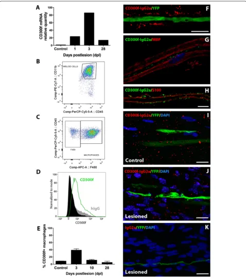
CD300f expression in the sciatic nerve after injury Expression of the immune receptor CD300f in the sci-atic nerve was assessed by QPCR and flow cytometry in control uninjured and crush lesioned samples. Low basal CD300f mRNA expression was detected; its levels increased at 1 day post lesion (dpl), peaking at 3 dpl and declining thereafter, showing at 28 dpl slightly lower levels than at 1 dpl but still remaining high in comparison with uninjured nerves (Fig. 1a). Protein ex-pression of CD300f and several macrophage markers
(F4/80, CD45, and CD11b) were analyzed by flow cy-tometry in uninjured and crushed sciatic nerves at 3, 10, and 28 dpl (Fig. 1b–e). A small proportion of resi-dent CD45+/CD11b+/F4/80+ macrophages expressed CD300f in the uninjured nerve (Fig. 1e). After crush sciatic nerve lesion, there was an increase in the total number of CD45+/CD11b+/F4/80+ macrophages present in the injured nerve in comparison with a non-injured nerve. Nearly 40 % of these macrophages expressed CD300f at 3 dpl, and the expression declined pro-gressively to 28 dpl. Moreover, few F4/80 negative and CD11b+/CD45+ positive cells at 3 and 10 dpl also expressed CD300f, most probably representing neutro-phils and maybe mast cells (not shown).
CD300f ligand is expressed in the PNS
In order to characterize the presence of the ligand of CD300f in the PNS, we performed immunohistochemi-cal studies using rCD300f-IgG2a fusion protein as de-scribed [50] on both frozen sections and teased fibres. Uninjured teased fibres from Thy1-YFP-H transgenic mice showed a punctate staining pattern with rCD300f-IgG2a, not co-localizing with the axonal cytoplasm (Fig. 1f ). While no co-localization was observed at the MBP-positive myelin domain of Schwann cells (Fig. 1g), partial co-localization with the external limit of S100-positive Schwann cell domain was apparent (Fig. 1h). Frozen sections of crushed sciatic nerve were used in order to assess the expression of the ligand after the in-jury. At 10 dpl, no apparent difference was observed in the staining for the ligand in comparison with an unin-jured nerve (Fig. 1i, j). No staining was detected using IgG2a negative control (Fig. 1k).
CD300f participates in axonal regeneration
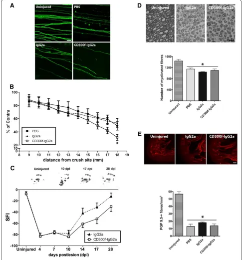
PBS control groups (Fig. 2a, b). We further evaluated the recovery of function after crush injury by analyzing the hindpaw prints to obtain the SFI. After crush sciatic nerve injury, the SFI drops to−80 % whereas it recovers by day 28 up to −20 % with no significant differences when compared to pre-injury values. After CD300f-IgG2a treatment, a strong tendency (p= 0.07) towards delayed functional recovery was observed compared to
IgG2a control animals (Fig. 2c). A detailed study of re-generated fibres was performed at 28 dpl by analyzing the number of regenerated myelinated fibres counted in semithin sections in the tibial nerve at the ankle level, and no differences between groups were observed (Fig. 2d). In addition, distal re-innervation was assessed by counting the number of PGP9.5-positive fibres in the epidermis. The number of PGP9.5-positive fibres was
significantly lower in injured groups than in the contra-lateral skin at 28 dpl but no differences were seen be-tween treatments (Fig. 2e). Taken together, these data show that a single injection of the soluble CD300f-IgG2a transiently delayed the regeneration, followed by en-dogenous compensations that achieve normal regener-ation at longer time points.
Blocking CD300f/ligand interaction induced macrophage accumulation and changes in phenotype
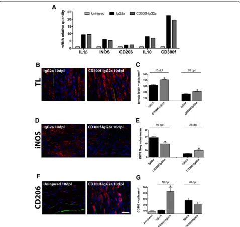
In order to understand the mechanism of impaired re-generation by blocking CD300f function, we assessed the level of inflammatory markers and macrophage infiltra-tion and phenotype. QPCR from nerve samples at 1 dpl showed increased mRNA for IL-1β, iNOS, CD206, and IL-10 when compared to uninjured nerves (Fig. 3a). However, no significant differences were observed after the injection of CD300f-IgG2a or IgG2a. Moreover, the blockade of CD300f/ligand interaction did not alter CD300f mRNA at 1 dpl. Interestingly, later on at 10 dpl, immunohistochemistry against the general macrophage marker tomato lectin in the sciatic nerve distally to the crush showed increased numbers of positive cells with the typical morphology of phagocytic macrophages, which were further increased by treatment with CD300f-IgG2a (Fig. 3b, c). Similar results were obtained with anti-Iba-1 general macrophage marker (not shown). At 28 dpl, phagocytic macrophages could still be seen in the sciatic nerve although at lower numbers than at 10 dpl (Fig. 3c). However, in the group injected with CD300f-IgG2a, macrophages remained at significantly higher numbers than in the control group.
We further evaluated whether the blockade of the interaction between CD300f and its ligand had an effect in the phenotype of the macrophages that infiltrated into the nerve. We performed immunohistochemistry against the mannose receptor (CD206) as a marker of alternative M2 macrophages and against iNOS as a marker of clas-sical proinflammatory M1 macrophages. iNOS labeling
was significantly reduced in animals treated with rCD300f-IgG2a at 10 dpl but increased at 28 dpl in com-parison with the injured control group (Fig. 3d, e).
CD206 labeling of the uninjured and IgG2a-treated nerves showed a low number of CD206-positive cells scattered through the nerve without differences between uninjured and injured nerves at 10 dpl. However, in the group of animals injected with rCD300-IgG2a, there was a significantly higher number of CD206-positive cells at 10 dpl (Fig. 3f, g). At 28 dpl, the injured control group showed higher numbers of CD206-positive cells in com-parison with uninjured nerves and control injured nerves at 10 dpl but without significant differences be-tween the injured control group and the rCD300f-IgG2a-treated group. Interestingly, considering all the experimental groups together, the number of regenerat-ing YFP-positive fibres negatively correlated with the number of CD206-positive cells at 10 dpl (Spearman’s rank test, p= 0.02, r=−0.64). This correlation was not observed at 28 dpl.
Tomato lectin has been widely used as a marker of microglia/macrophages and endothelial cells in the CNS [55, 56]. Here, we show that tomato lectin is also a good marker for macrophages in the normal and le-sioned nerve when compared to Iba-1 and F4/80 (Figs. 4 and 5). Tomato lectin labeling increased from 3 dpl peaking at 10–14 days post lesion (Fig. 4) as pub-lished by other authors using other macrophages markers such as F4/80 and CD11b [10, 11]. Tomato lec-tin and Iba-1 labeled the very few resident macrophages of the normal nerve (Fig. 4a–c). Interestingly, an important heterogeneity of macrophages was observed after the le-sion. Most infiltrated large foamy macrophages at all time points were intensely stained with tomato lectin, Iba-1, and F4/80 markers (Figs. 4d–l and 5). However, smaller cells showed different combinations of stainings, in-cluding tomato lectin-positive and Iba-1 and F4/80-negative staining. This latter category would also include the endothelial cells. Moreover, both Iba-1 and F4/80
(See figure on previous page.)
stained small macrophages that remained unstained with tomato lectin (Fig. 4d–i red arrows, Fig. 5 arrows). This macrophage phenotype heterogeneity could also be ob-served with M1/M2 markers like iNOS and CD206. Al-though most iNOS and CD206 staining co-localized with the big foamy tomato lectin and F4/80-positive macro-phages (Fig. 5 arrowheads), round and smaller tomato lectin-negative and F4/80-positive macrophages also expressed iNOS or CD206 (Fig. 5 arrows). Taken together,
these data show that all iNOS and CD206 staining was de-tected inside tomato lectin, Iba-1, or F4/80-positive cells.
Phagocytosis after blockade of CD300f
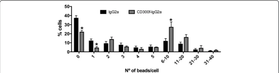
the number of beads/cell was quantified. The treat-ment of the nerve with CD300f-IgG2a at the time of the lesion, induced 10 days later, increased phagocytic activity of nerve cells when compared to IgG2a treat-ment. A reduction in the percentage of cells with 0–1 phagocyted beads and an increase in the percentage of cells with 6–10 phagocyted beads were observed after CD300f-IgG2a treatment (Fig. 6).
Discussion
regeneration. This delayed regeneration is associated to a modulation in the number and phenotype of M1/M2 macrophages in the lesioned nerve.
Recent reports have shown that phospholipids as phos-phatidylcholine or phosphatidylserine are ligands for CLM-1, the mouse orthologue of CD300f [35, 36, 41]. Phosphati-dylserine/CLM-1 interaction contributes to apoptotic cell clearance and hence to dampen inflammation [35, 36]. Cer-amide has also been described as a putative ligand for this receptor, contributing to dampening inflammatory reac-tions of mast cells in several allergy models by the activa-tion of CLM-1 negative signaling [40]. The interacactiva-tion of activating/inhibitory immunoreceptors with lipids appears to be a more general phenomenon [41]. For instance, some gangliosides and 3-O-sulfo-β-d-galactosylceramide (C24:1) are potential ligands for CD300b/CLM-7 [57]. In the
central nervous system, staining of the brain and spinal cord with rat or human CD300f-IgG2a showed a distinctive punctuate pattern, mainly in oligodendrocytes of the white matter [50]. Accordingly, we show here a similar punctuate staining pattern with CD300f-IgG2a in peripheral nerves. Interestingly, by using teased nerve fibres and Thy1-YFP-H mice, we evidence the specific subcellular localization of the CD300f ligands to what appears to be the outer cell membrane of the non-myelinating S100-positive domain of myelinating Schwann cells previously described [58] and not to the MBP-positive myelin sheath or the axonal com-partment. However, we cannot discard the possibility that the ligand may also be present in non-myelinating Schwann cells or some component of the extracellular matrix. Although electron microscope procedures are necessary to determine its precise location, our confocal Fig. 5iNOS and CD206 stain a heterogeneous population of macrophages. Single focal plane confocal microscopy for iNOS or CD206 combined with tomato lectin and F4/80 macrophage staining evidenced that most cells co-stain with tomato lectin and F4/80, and some of them are in addition iNOS or CD206 positive (arrowheads). Moreover, there is a population of round and smaller F4/80-positive and tomato lectin negative macrophages that also shows iNOS or CD206 staining (arrows). Scale bar: 20μm
images suggest the localization of the ligand in the outer non-myelin Schwann cell membrane [58]. The normal presence of the ligands in Schwann cells and oli-godendrocytes point to supplementary roles in addition to the phosphatidylserine “eat me” signal or the ceramide-induced signaling previously described. Other authors have shown that CD200, the ligand for the inhibitory im-mune receptor CD200R, is expressed in Schwann cells in the intact nerve [59]. In the CNS, this receptor induces a tonic anti-inflammatory signal contributing to set the threshold and magnitude of proinflammatory signaling [24, 60]. Whether the ligand of CD300f expressed on Schwann cells and oligodendrocytes also contributes to the maintenance of this tonic anti-inflammatory state is an open question. Chang and co-workers showed that CD200 is downregulated after crush injury in the site of lesion [59]. They hypothesized that, after nerve injury, CD200 is downregulated in order to decrease immuno-suppression and enhance influx of macrophages and the inflammatory response to eliminate myelin and axonal debris. The ligands for CD300f do not decrease at least at 10 dpl, suggesting different signaling mechanisms for these two receptors. Interestingly, despite intense inva-sion of macrophages into the nerve, a very similar staining pattern was observed for the CD300f ligands, suggesting that macrophages do not bear the ligand. In accordance, microglial cells in vitro did not stain with CD300f-IgG2a [50].
Recent findings suggest that the anti-inflammatory physiological state of most tissues is not only a passive state resulting from absence of inflammatory stimuli but an active condition that requires participation of several molecules responsible for the suppression of potentially inflammatory stimuli. Under this paradigm, a physio-logical function for the ligands of inhibitory receptors like CD200R, CD300a, or CD300f could be to contrib-ute to the so-called “On” and “Off” signals [61]. “Off” signals are constitutively present in the brain paren-chyma and are expressed mainly by healthy neurons, whereas “On” signals are expressed by endangered or impaired neurons [62]. The integration of all the inhibi-tory and activating inputs shapes the phenotype and re-sponse of microglial cells or macrophages accordingly. In fact, several activating and inhibitory receptors simi-lar to CD300f like CD200R, TREM2, or SIGLECs have been reported to be key regulators of microglial and macrophage activation [24, 25]. Thus, these mecha-nisms, including CD300f and its ligands, could also be in place for the interaction of macrophages and Schwann cells in the normal and injured PNS to regu-late the inflammatory status.
CD300f has been reported to deliver both activating and inhibiting signals [28, 35–37]. However, only inhibi-tory signals have been found on monocytic cell lines. For
determining the altered inflammatory response and de-layed regeneration observed. In addition to the modula-tion of the proinflammatory phenotype, CD300f may also contribute to dampen inflammatory reactions promoting phagocytosis of apoptotic cells [36]. Phagocytosis of mye-lin and cell debris is a critical component of WD and suc-cessful regeneration [5]. The impaired regeneration after the single injection of CD300f-IgG2a might be related to inhibition of phagocytosis and thus delayed debris clear-ance. Accordingly, at 10 dpl, when no CD300f-IgG2a re-mains, the accumulation of debris may trigger the increased phagocytosis of nerve cells observed here that may be responsible for the delayed but successful regener-ation at 28 dpl.
Despite the importance of macrophage phenotype in WD and axonal regeneration, only a few reports [22, 23] have described the expression of M1/M2 phenotypic cell markers after nerve injury and repair. In a recent paper, Ydens and colleagues made a description of the different markers of M1 and M2 macrophages after nerve tran-section and repair in mice, showing a rapid M2 polarization of macrophages after axotomy [22]. They evaluated a high number of markers of inflammation in-cluding iNOS, CD206, and IL-1βat different time points after nerve injury and mainly by QPCR. In accordance with our results, they observed a rapid fast induction of mRNA for IL-1βand IL-10 at 1 dpl. Moreover, they ob-served the upregulation of other M2 markers like Arg1, Ym1, or TREM2. They also reported that the mRNA for CD206 did not show changes at the different time points evaluated (until 14 dpl). In accordance, we did not ob-serve notable changes in CD206 mRNA at 1 dpl or in protein level at 10 dpl in comparison with uninjured nerves. However, we also analyzed longer time points (28 dpl) to sample processes of resolution of the neuro-inflammation and found an increase in CD206 staining in comparison with both uninjured and 10 dpl control injured sciatic nerves. This late increase might be a con-sequence of signals aimed to resolve inflammation by adjusting macrophage polarization towards a healing phenotype. In relation to M1 macrophage polarization after nerve injury, Ydens and colleagues did not show a significant change in iNOS, IL-12p40, or INFγ mRNA levels at the different time points post lesion studied. However, in the present work, we have seen a significant increase in the iNOS mRNA at 24 h after lesion and in the iNOS protein at 10 and 28 dpl. These differences in the results could be due to the type of nerve lesion used between the two studies, i.e., nerve section or nerve crush. Further experiments are needed to es-tablish the effect of the different M1/M2 markers on nerve neuroinflammation and regeneration. In this line, we show that the manipulation of the CD300f/ ligand interaction induces impairment of regeneration
associated to important changes in M1/M2 markers. After a sciatic nerve crush injury, a single injection of CD300f-IgG2a significantly increased the number of to-mato lectin-positive macrophages and CD206-positive cells and decreased iNOS immunoreactivity at 10 dpl, whereas an opposite effect was found at 28 dpl. These data suggest that blocking CD300f-ligand interactions not only contributes to an enhanced recruitment of macrophages but also to a change in the phenotype of normally recruited macrophages towards an early M2 phenotype, followed by a switch to a M1 phenotype of some macrophages later on. Interestingly, Mokarram and colleagues induced a nerve section followed by tubulization repair and IL-4 or INFγ treatment to polarize macrophages towards a M2 or M1 phenotype, respectively. Only IL-4 but not INFγtreatment induced increased Schwann cell migration, macrophage recruit-ment, macrophage polarization towards M2 phenotype, and regeneration [23]. In the absence of any treatment, they reported a main macrophage polarization towards an M1 phenotype at 21 dpl, while we show mainly a polarization towards an M2 phenotype at 28 dpl. More-over, Mokarram and colleagues reported that the in-crease in CD206-positive cells at 21 dpl positively correlated with regeneration, while we observe in fact a negative correlation at 10 dpl and no correlation at 28 dpl. This apparent contradiction between both studies may be explained by the different nerve injury models, i.e., nerve crush versus section and tubulization, where the neuroinflammatory conditions are different and where the structural maintenance of the epi-, peri-, and endoneurium has strong effects. Moreover, the treat-ment with IL-4 may change fundatreat-mental endogenous neuroinflammatory mechanisms influencing the final outcome of regeneration observed.
Conclusions
macrophages that take part in the first stage of WD (1–10 dpl) would be polarized mainly towards a pro-inflammatory M1 phenotype whereas the resolution of inflammation at later stages (15–30 dpl) would be driven predominantly by M2 macrophages.
Competing interests
The authors declare that they have no competing interests.
Authors’contributions
NL and HP conceived the study, designed and carried out most of the experiments, and wrote the manuscript. PS carried out the functional evaluation and part of the immunohistochemistry assays. MLN and JS produced and purified the CD300f-IgG2a fusion protein. NL, IFQ, and RLV performed the FACS analysis. XN, RLV, and JS contributed to the interpretation of the results and discussion. All the authors read and approved the final manuscript.
Acknowledgements
This research was supported by Fundació Marató TV3 (110533), Catalunya, Spain, Comisión Sectorial de Investigación Científica (CSIC-UDELAR), Uruguay, PEDECIBA, Uruguay, Agencia Nacional de Investigación e Innovación (ANII), Uruguay, and FOCEM (MERCOSUR Structural Convergence Fund), COF 03/ 1111, CIBERNED, and TERCEL funds from the Instituto de Salud Carlos III of Spain. The authors thank the technical help of Jessica Jaramillo, Marta Valeri, and Natalia Puig.
Author details
1
Neuroinflammation and Gene Therapy Laboratory, Institut Pasteur Montevideo, Mataojo 2020, CP 11400 Montevideo, Uruguay.2Department of
Histology and Embryology, Faculty of Medicine, UDELAR, Montevideo, Uruguay.3Institute of Neurosciences and Department of Cell Biology,
Physiology and Immunology, Universitat Autònoma de Barcelona, and Centro de Investigación Biomédica en Red sobre Enfermedades Neurodegenerativas (CIBERNED), Bellaterra, Spain.4Immunobiology Group, CIBBIM-Nanomedicine Program, Hospital Universitari Vall d’Hebron, Institut de Recerca (VHIR), Universitat Autonoma de Barcelona, Barcelona, Spain.
5Neurodegeneration Laboratory, Institut Pasteur Montevideo, Montevideo,
Uruguay.
Received: 10 February 2015 Accepted: 21 July 2015
References
1. Lago N, Navarro X. Correlation between target reinnervation and distribution of motor axons in the injured rat sciatic nerve. J Neurotrauma. 2006;23:227–40. 2. Deumens R, Bozkurt A, Meek MF, Marcus MA, Joosten EA, Weis J, et al.
Repairing injured peripheral nerves: bridging the gap. Prog Neurobiol. 2010;92:245–76.
3. Allodi I, Udina E, Navarro X. Specificity of peripheral nerve regeneration: interactions at the axon level. Prog Neurobiol. 2012;98:16–37. 4. Waller A. Experiments on the section of the glossopharyngeal and
hypoglossal nerves of the frog, observations of the alterations produced thereby in the structure of their primitive fibers. Phil Transact Royal Soc London. 1850;140:423–9.
5. Vargas ME, Barres BA. Why is Wallerian degeneration in the CNS so slow? Annu Rev Neurosci. 2007;30:153–79.
6. Schafer M, Fruttiger M, Montag D, Schachner M, Martini R. Disruption of the gene for the myelin-associated glycoprotein improves axonal regrowth along myelin in C57BL/Wlds mice. Neuron. 1996;16:1107–13. 7. Fruttiger M, Montag D, Schachner M, Martini R. Crucial role for the
myelin-associated glycoprotein in the maintenance of axon-myelin integrity. Eur J Neurosci. 1995;7:511–5.
8. Gaudet AD, Popovich PG, Ramer MS. Wallerian degeneration: gaining perspective on inflammatory events after peripheral nerve injury. J Neuroinflammation. 2011;8:110.
9. Rotshenker S. Wallerian degeneration: the innate-immune response to traumatic nerve injury. J Neuroinflammation. 2011;8:109.
10. Avellino AM, Hart D, Dailey AT, MacKinnon M, Ellegala D, Kliot M. Differential macrophage responses in the peripheral and central nervous system during Wallerian degeneration of axons. Exp Neurol. 1995;136:183–98.
11. Omura T, Omura K, Sano M, Sawada T, Hasegawa T, Nagano A. Spatiotemporal quantification of recruit and resident macrophages after crush nerve injury utilizing immunohistochemistry. Brain Res. 2005;1057:29–36.
12. Be’eri H, Reichert F, Saada A, Rotshenker S. The cytokine network of Wallerian degeneration: IL-10 and GM-CSF. Eur J Neurosci. 1998;10:2707–13. 13. Shamash S, Reichert F, Rotshenker S. The cytokine network of Wallerian
degeneration: tumor necrosis factor-alpha, interleukin-1alpha, and interleukin-1beta. J Neurosci. 2002;22:3052–60.
14. Sawada T, Sano M, Omura T, Omura K, Hasegawa T, Funahashi S, et al. Spatiotemporal quantification of tumor necrosis factor-alpha and interleukin-10 after crush injury in rat sciatic nerve utilizing immunohistochemistry. Neurosci Lett. 2007;417:55–60.
15. Liefner M, Siebert H, Sachse T, Michel U, Kollias G, Bruck W. The role of TNF-alpha during Wallerian degeneration. J Neuroimmunol. 2000;108:147–52.
16. George A, Kleinschnitz C, Zelenka M, Brinkhoff J, Stoll G, Sommer C. Wallerian degeneration after crush or chronic constriction injury of rodent sciatic nerve is associated with a depletion of endoneurial interleukin-10 protein. Exp Neurol. 2004;188:187–91.
17. Gordon S. Alternative activation of macrophages. Nat Rev Immunol. 2003;3:23–35.
18. Geissmann F, Manz MG, Jung S, Sieweke MH, Merad M, Ley K. Development of monocytes, macrophages, and dendritic cells. Science. 2010;327:656–61. 19. Mantovani A, Sica A, Sozzani S, Allavena P, Vecchi A, Locati M. The
chemokine system in diverse forms of macrophage activation and polarization. Trends Immunol. 2004;25:677–86.
20. David S, Kroner A. Repertoire of microglial and macrophage responses after spinal cord injury. Nat Rev Neurosci. 2011;12:388–99.
21. Kigerl KA, Gensel JC, Ankeny DP, Alexander JK, Donnelly DJ, Popovich PG. Identification of two distinct macrophage subsets with divergent effects causing either neurotoxicity or regeneration in the injured mouse spinal cord. J Neurosci. 2009;29:13435–44.
22. Ydens E, Cauwels A, Asselbergh B, Goethals S, Peeraer L, Lornet G, et al. Acute injury in the peripheral nervous system triggers an alternative macrophage response. J Neuroinflammation. 2012;9:176.
23. Mokarram N, Merchant A, Mukhatyar V, Patel G, Bellamkonda RV. Effect of modulating macrophage phenotype on peripheral nerve repair. Biomaterials. 2012;33:8793–801.
24. Hoek RM, Ruuls SR, Murphy CA, Wright GJ, Goddard R, Zurawski SM, et al. Down-regulation of the macrophage lineage through interaction with OX2 (CD200). Science. 2000;290:1768–71.
25. Neumann H, Takahashi K. Essential role of the microglial triggering receptor expressed on myeloid cells-2 (TREM2) for central nervous tissue immune homeostasis. J Neuroimmunol. 2007;184:92–9.
26. Alvarez-Errico D, Aguilar H, Kitzig F, Brckalo T, Sayos J, Lopez-Botet M. IREM-1 is a novel inhibitory receptor expressed by myeloid cells. Eur J Immunol. 2004;34:3690–701.
27. Aguilar H, Alvarez-Errico D, Garcia-Montero AC, Orfao A, Sayos J, Lopez-Botet M. Molecular characterization of a novel immune receptor restricted to the monocytic lineage. J Immunol. 2004;173:6703–11.
28. Alvarez-Errico D, Sayos J, Lopez-Botet M. The IREM-1 (CD300f) inhibitory receptor associates with the p85alpha subunit of phosphoinositide 3-kinase. J Immunol. 2007;178:808–16.
29. Comas-Casellas E, Martinez-Barriocanal A, Miro F, Ejarque-Ortiz A, Schwartz Jr S, Martin M, et al. Cloning and characterization of CD300d, a novel member of the human CD300 family of immune receptors. J Biol Chem. 2012;287:9682–93.
30. Martinez-Barriocanal A, Sayos J. Molecular and functional characterization of CD300b, a new activating immunoglobulin receptor able to transduce signals through two different pathways. J Immunol. 2006;177:2819–30. 31. Borrego F. The CD300 molecules: an emerging family of regulators of the
immune system. Blood. 2013;121(11):1951–60.
32. Martinez-Barriocanal A, Comas-Casellas E, Schwartz Jr S, Martin M, Sayos J. CD300 heterocomplexes, a new and family-restricted mechanism for myeloid cell signaling regulation. J Biol Chem. 2010;285:41781–94. 33. Torres-Espin A, Hernandez J, Navarro X. Gene expression changes in the
34. Nielsen R, Bustamante C, Clark AG, Glanowski S, Sackton TB, Hubisz MJ, et al. A scan for positively selected genes in the genomes of humans and chimpanzees. PLoS Biol. 2005;3, e170.
35. Choi SC, Simhadri VR, Tian L, Gil-Krzewska A, Krzewski K, Borrego F, et al. Cutting edge: mouse CD300f (CMRF-35-like molecule-1) recognizes outer membrane-exposed phosphatidylserine and can promote phagocytosis. J Immunol. 2011;187:3483–7.
36. Tian L, Choi SC, Murakami Y, Allen J, Morse 3rd HC, Qi CF, et al. p85alpha recruitment by the CD300f phosphatidylserine receptor mediates apoptotic cell clearance required for autoimmunity suppression. Nat Commun. 2014;5:3146.
37. Ejarque-Ortiz A, Sola C, Martinez-Barriocanal A, Schwartz Jr S, Martin M, Peluffo H, et al. The receptor CMRF35-like molecule-1 (CLM-1) enhances the production of LPS-induced pro-inflammatory mediators during microglial activation. PLoS ONE. 2015;10, e0123928.
38. Moshkovits I, Karo-Atar D, Itan M, Reichman H, Rozenberg P, Morgenstern-Ben-Baruch N, et al. CD300f associates with IL-4 receptorαand amplifies IL-4–induced immune cell responses. Proc Natl Acad Sci U S A. 2015;112(28):8708–13.
39. Xi H, Katschke Jr KJ, Helmy KY, Wark PA, Kljavin N, Clark H, et al. Negative regulation of autoimmune demyelination by the inhibitory receptor CLM-1. J Exp Med. 2010;207:7–16.
40. Izawa K, Yamanishi Y, Maehara A, Takahashi M, Isobe M, Ito S, et al. The receptor LMIR3 negatively regulates mast cell activation and allergic responses by binding to extracellular ceramide. Immunity. 2012;37:827–39. 41. Cannon JP, O’Driscoll M, Litman GW. Specific lipid recognition is a general feature of CD300 and TREM molecules. Immunogenetics. 2012;64:39–47. 42. Izawa K, Isobe M, Matsukawa T, Ito S, Maehara A, Takahashi M, et al.
Sphingomyelin and ceramide are physiological ligands for human LMIR3/ CD300f, inhibiting FcepsilonRI-mediated mast cell activation. J Allergy Clin Immunol. 2014;133:270–3. e271-277.
43. Feng G, Mellor RH, Bernstein M, Keller-Peck C, Nguyen QT, Wallace M, et al. Imaging neuronal subsets in transgenic mice expressing multiple spectral variants of GFP. Neuron. 2000;28:41–51.
44. Ale A, Bruna J, Morell M, van de Velde H, Monbaliu J, Navarro X, et al. Treatment with anti-TNF alpha protects against the neuropathy induced by the proteasome inhibitor bortezomib in a mouse model. Exp Neurol. 2014;253:165–73.
45. Klopstein A, Santos-Nogueira E, Francos-Quijorna I, Redensek A, David S, Navarro X, Lopez-Vales R: Beneficial effects of alphaB-crystallin in spinal cord contusion injury. J Neurosci 2012, 32:14478–14488.
46. Fricker FR, Lago N, Balarajah S, Tsantoulas C, Tanna S, Zhu N, et al. Axonally derived neuregulin-1 is required for remyelination and regeneration after nerve injury in adulthood. J Neurosci. 2011;31:3225–33.
47. Bain JR, Mackinnon SE, Hunter DA. Functional evaluation of complete sciatic, peroneal, and posterior tibial nerve lesions in the rat. Plast Reconstr Surg. 1989;83:129–38.
48. Lopez-Vales R, Navarro X, Shimizu T, Baskakis C, Kokotos G, Constantinou-Kokotou V, et al. Intracellular phospholipase A(2) group IVA and group VIA play important roles in Wallerian degeneration and axon regeneration after peripheral nerve injury. Brain. 2008;131:2620–31.
49. Pfaffl MW. A new mathematical model for relative quantification in real-time RT-PCR. Nucleic Acids Res. 2001;29, e45.
50. Peluffo H, Ali-Ruiz D, Ejarque-Ortiz A, Heras-Alvarez V, Comas-Casellas E, Martinez-Barriocanal A, et al. Overexpression of the immunoreceptor CD300f has a neuroprotective role in a model of acute brain injury. Brain Pathol. 2011;22:318–28.
51. Fricker FR, Zhu N, Tsantoulas C, Abrahamsen B, Nassar MA, Thakur M, et al. Sensory axon-derived neuregulin-1 is required for axoglial signaling and normal sensory function but not for long-term axon maintenance. J Neurosci. 2009;29:7667–78.
52. Groves ML, McKeon R, Werner E, Nagarsheth M, Meador W, English AW. Axon regeneration in peripheral nerves is enhanced by proteoglycan degradation. Exp Neurol. 2005;195:278–92.
53. Beirowski B, Berek L, Adalbert R, Wagner D, Grumme DS, Addicks K, et al. Quantitative and qualitative analysis of Wallerian degeneration using restricted axonal labelling in YFP-H mice. J Neurosci Methods. 2004;134:23–35. 54. English AW, Meador W, Carrasco DI. Neurotrophin-4/5 is required for the
early growth of regenerating axons in peripheral nerves. Eur J Neurosci. 2005;21:2624–34.
55. Acarin L, Vela JM, Gonzalez B, Castellano B. Demonstration of poly-N-acetyl lactosamine residues in ameboid and ramified microglial cells in rat brain by tomato lectin binding. J Histochem Cytochem. 1994;42:1033–41. 56. Peluffo H, Acarin L, Faiz M, Castellano B, Gonzalez B. Cu/Zn superoxide
dismutase expression in the postnatal rat brain following an excitotoxic injury. J Neuroinflammation. 2005;2:12.
57. Phongsisay V, Iizasa E, Hara H, Yamasaki S. 3-O-sulfo-beta-d-galactose moiety of endogenous sulfoglycolipids is a potential ligand for immunoglobulin-like receptor LMIR5. Mol Immunol. 2015;63:595–9. 58. Court FA, Zambroni D, Pavoni E, Colombelli C, Baragli C, Figlia G, et al.
MMP2-9 cleavage of dystroglycan alters the size and molecular composition of Schwann cell domains. J Neurosci. 2011;31:12208–17.
59. Chang CY, Lee YH, Jiang-Shieh YF, Chien HF, Pai MH, Chen HM, et al. Novel distribution of cluster of differentiation 200 adhesion molecule in glial cells of the peripheral nervous system of rats and its modulation after nerve injury. Neuroscience. 2011;183:32–46.
60. Linnartz B, Wang Y, Neumann H. Microglial immunoreceptor tyrosine-based activation and inhibition motif signaling in neuroinflammation. Int J Alzheimers Dis. 2010;2010.
61. Biber K, Neumann H, Inoue K, Boddeke HW. Neuronal‘On’and‘Off’signals control microglia. Trends Neurosci. 2007;30:596–602.
62. Ransohoff RM, Cardona AE. The myeloid cells of the central nervous system parenchyma. Nature. 2010;468:253–62.
63. Kim EJ, Lee SM, Suk K, Lee WH. CD300a and CD300f differentially regulate the MyD88 and TRIF-mediated TLR signalling pathways through activation of SHP-1 and/or SHP-2 in human monocytic cell lines. Immunology. 2012;135:226–35.
64. Mueller M, Leonhard C, Wacker K, Ringelstein EB, Okabe M, Hickey WF, et al. Macrophage response to peripheral nerve injury: the quantitative contribution of resident and hematogenous macrophages. Lab Invest. 2003;83:175–85.
65. Nadeau S, Filali M, Zhang J, Kerr BJ, Rivest S, Soulet D, et al. Functional recovery after peripheral nerve injury is dependent on the pro-inflammatory cytokines IL-1beta and TNF: implications for neuropathic pain. J Neurosci. 2011;31:12533–42.
Submit your next manuscript to BioMed Central and take full advantage of:
• Convenient online submission
• Thorough peer review
• No space constraints or color figure charges
• Immediate publication on acceptance
• Inclusion in PubMed, CAS, Scopus and Google Scholar
• Research which is freely available for redistribution