Open Access
Research
Proteomic study on gender differences in aging kidney of mice
Hanna Amelina and Susana Cristobal*
Address: Department of Biochemistry and Biophysics, Stockholm University, SE-106 91, Stockholm, Sweden
Email: Hanna Amelina - [email protected]; Susana Cristobal* - [email protected] * Corresponding author
Abstract
Background: This study aims to analyze sex differences in mice aging kidney. We applied a proteomic technique based on subfractionation, and liquid chromatography coupled with 2-DE. Samples from male and female CD1-Swiss outbred mice from 28 weeks, 52 weeks, and 76 weeks were analysed by 2-DE, and selected proteins were identified by matrix assisted laser desorption ionisation time-of-flight mass spectrometry (MALDI-TOF MS).
Results: This proteomic analysis detected age-related changes in protein expression in 55 protein-spots, corresponding to 22 spots in males and 33 spots in females. We found a protein expression signature (PES) of aging composed by 8 spots, common for both genders. The identified proteins indicated increases in oxidative and proteolytic proteins and decreases in glycolytic proteins, and antioxidant enzymes.
Conclusion: Our results provide insights into the gender differences associated to the decline of kidney function in aging. Thus, we show that proteomics can provide valuable information on age-related changes in expression levels of proteins and age-related modifications. This pilot study is still far from providing candidates for aging-biomarkers. However, we suggest that the analysis of these proteins could suggest mechanisms of cellular aging in kidney, and improve the kidney selection for transplantation.
Background
Aging studies in tissues such as brain have attracted a lot of attention, however the kidney has been neglected [1]. Very recently, differential expression of proteins involved in metabolism, transport, and stress response in kidney has been reported from aging male mouse [2]. Although this organ shows a quantifiable decline of function with age, the gender differences have not been analyzed in pre-vious proteomics studies [3]. There is an approximately 25% decline in the glomerular filtration rate starting at age 40 for humans and the ability of the medulla to con-centrate urine declines progressively with age. Therefore, any disease affecting the organ, including hypertension
and diabetes mellitus, accelerates the age-related changes in kidney. Moreover, impaired kidneys are targets for transplantation. Therefore, novel aging kidney biomark-ers could also improve the selection of older donor organs for transplantation.
Aging is among the most complex biological phenomena. It is a complex process resulting from changes in the expression and regulation of numerous genes over time. Most physiological functions decline with age because cells accumulate damage over time. This slow incremental damage results in the gradual loss of differentiated func-tions and growth rate. This process is accompanied by an
Published: 9 April 2009
Proteome Science 2009, 7:16 doi:10.1186/1477-5956-7-16
Received: 30 June 2008 Accepted: 9 April 2009
This article is available from: http://www.proteomesci.com/content/7/1/16
© 2009 Amelina and Cristobal; licensee BioMed Central Ltd.
increased probability for the development of cancer [4]. Mounting evidences indicate that a specific gene could be connected to the extended longevity. However, the uni-versal explanation for these life-extending effects has not yet been found. Alterations in the expression of individual proteins have reported this effect. These mechanisms include: (i) telomere repair [5]; (ii) stress response [6]; (iii) anti-oxidant defense [7]; (iv) nicotinamide deamina-tion [8]; (v) insulin/insulin-like growth factor-1 signaling [9]; and (vi) histone deacetylation [10]. However, the glo-bal view of aging has become more complex with the understanding that some of these pathways can be con-nected. The ability to survey the entire proteome or a sub-set of the proteomes offers new opportunities to study the complex biological phenomenon of aging in an unbiased manner.
Studies in model organisms such as Saccharomyces cerevi-siae, Caenorhabditis elegans, Mus musculus, and Drosophila melanogaster have provided much of our insight into the underlying biological pathways associated with aging. However, a key question is still whether the mechanisms of aging are conserved between species with different lifespan. Murine models have been used to investigate the expression of proteins and their oxidation in the brains of the senescence-accelerated mouse (SAM) as a potential animal model of Alzheimer's disease [11]; the differential expression of the liver proteome [12]; and the differential gene expression profiles in the hippocampus to reveal the mechanisms involved in age-related learning and mem-ory deficits [13]. The CD1-Swiss outbred mouse has been utilized to study brain mitochondrial dysfunction in aging [14]. Among others, proteomic techniques have been applied to examine the effect of anti-aging agents on human endothelial cells [15], to study differential protein expression and glycosylation of membrane proteins using Hutchinson-Gilford progeria syndrome fibroblasts [16], and to investigate age-related changes in the glycation of human aortic elastin [17]. These studies clearly indicate the value of additional proteomic studies of aging.
Tissue-specific quantitative assessment of protein expres-sion could reveal preferential biochemical pathways affected by aging. Different mammalian tissues have dis-tinct energy needs, primary functions, and regeneration capacities. The first quantitative proteomic study of rat mitochondria from various tissues has been recently pub-lished [18]. We have applied proteomics to characterise the mouse peroxisomes from liver and kidney [19]. Com-parative proteomics has been utilised to examine the effect of aging on the cellular proteome from rat skeletal muscle [20], mice brain [11], and on specific organelles such as the Golgi apparatus and endoplasmic reticulum [21] or mitochondrial proteins in mice [22], in rat [3], in bovine heart [23], and rat brain [24]. Our group has
per-formed a peroxisomal proteomic analysis of liver and kid-ney in young and old mice [25].
In this study, we present a subproteomic analysis of mice kidney during the aging process focusing on the gender differences. Here, we show that although age-associated changes are widespread among different functional classes of proteins, the gender effect should not be under-estimated as a differential factor in aging studies. Finally, we discuss the possible role of these age-related protein modifications in the functional lifespan of the mouse kid-ney.
Results
Proteins differentially expressed with age and gender The principal aim of this study was to characterize age-dependent changes in the subproteome of mice kidney of both genders. The tissues were first subjected to a simple fractionation in order to obtain organelle-enriched frac-tions [26]. The reproducibility of fractionation procedure as well as enrichment of specific organellar fractions dur-ing purification process was controlled by enzymatic anal-yses and Western immunoblots [see additional files 1 and 2]. Although cell fractionation from fresh tissues could provide higher recovery, in our experiment we utilized rapidly fresh frozen kidneys in order to be able to process all the samples simultaneously. Obtained fractions were then processed by liquid chromatography using Q-sepha-rose as an anion exchanger. The elution fractions were applied onto 2-DE, followed by colloidal Coomassie Blue staining. On average, 300 spots were detected on each 2-DE map. Statistical analysis was utilized to compare the average spot ratio of expression between the 2-DE maps from different genders and ages.
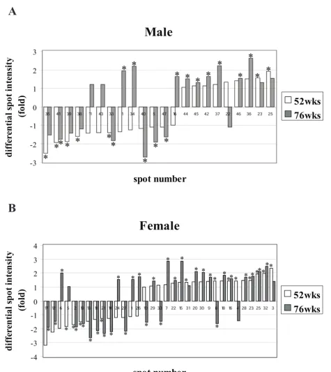
For the convenience of time-point comparisons, we divided the mice into the following groups: young mice (28 weeks old), adult mice (52 weeks old), and old mice (76 weeks old). Summarising the results, eight spots were found that compose a protein expression signature (PES) of aging, common for both genders [see additional file 3]. Studying the variation in protein expression by gender, in males, 22 protein spots showed significant changes in protein expression between young and the two groups of older mouse tissues. In the 52-week old group, 13 spots were down-regulated up to 2.5 fold, and 9 spots were up-regulated up to 1.9-fold. In the 76-week old group, 8 spots were down-regulated up to 2.7-fold and 13 spots were up-regulated up to 2.6-fold. These differentially expressed proteins are illustrated in Fig. 1A, 1B, 1C, 2A and addi-tional file 4.
were down-regulated up to 3.2-fold, and 19 spots were up-regulated up to 2.4-fold. In the 76 week-old group, 13 spots were down-regulated up to 2.6-fold, and 20 spots were up-regulated up to 2.8-fold (Fig. 1D, 1E, 1F and 2B). Detailed protein expression data can be found in addi-tional file 4.
Multivariate analysis
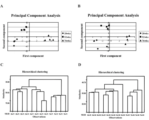
To validate whether the obtained PES could constitute a robust set of biomarkers, principal component analysis (PCA) and hierarchical clustering were performed. PCA is a useful tool for data categorization, since it separates the dominating features in a data set. It is remarkable that the first principal component clearly discriminates the oldest group from the two younger groups in both genders. Both sets of gels, corresponding to males and females, are situ-ated in the positive side of the x-axis (Fig. 3A and 3B). The second principal component separated adult groups from
the young ones. The adult group is clustered in the posi-tive side of the y-axis, opposite from the young group.
Hierarchical clustering supported the results obtained with PCA. All of the gels were clustered together with a 39% similarity for female samples and 48% similarity for the male samples. For both genders, the similarity between young and adults was over 50% and separated those two groups from the old samples. The group with the highest similarity among all groups was the oldest female specimens, which showed 70% similarity level (Fig. 3C and 3D).
Identification of differentially expressed proteins
The proteins that showed significant age-dependent varia-tion in expression from male and female mouse kidney were trypsin-digested and analysed by MALDI-TOF MS. For males, 12 proteins, and 23 proteins for females, were
2-DE reference gel images of male and female mouse kidney specimens from different ages showing protein expression profiles
Figure 1
2-DE reference gel images of male and female mouse kidney specimens from different ages showing protein expression profiles. The equal amounts of solubilised proteins (300 μg) were separated on 11 cm pI 5-8 linear gradient strips in the first dimension and on 12.5% SDS-PAGE in the second dimension. Gels were calibrated for molecular mass (in kDa) and pI (in pI units) with external pI and mass standards and stained with CBB G-250. Proteins with significantly different expression levels between ages are marked with circles and numbered. Numbers refer to protein spot numbers in other figures and
addi-tional file 5. (A) gender: male, age: 28 weeks; (B) gender: male, age: 52 weeks; (C) gender: male, age: 76 weeks, (D) gender:
female, age: 28 weeks; (E) gender: female, age: 52 weeks; (F) gender: female, age: 76 weeks.
A B C
D E F
5 pI 8 5 pI 8 5 pI 8
5 pI 8 5 pI 8 5 pI 8
76700
56300 35900
24600 16800
11500 76700
56300 35900
24600 16800
11500
76700
56300 35900
24600 16800 11500 76700
56300 35900
24600 16800 11500
Mr (Da)
Proteins differently expressed at the age of 52 and 76 weeks versus 28 weeks
Figure 2
Proteins differently expressed at the age of 52 and 76 weeks versus 28 weeks. (A) male, (B) female. The vertical axis corresponds to differential spot intensity, above the 0 value for the up-regulated protein-spots and below the 0 value for the down-regulated ones. According to 28 weeks old group, in the horizontal axis the down-regulated proteins are organised with the lowest values on the left side and the up-regulated ones show the highest values on the right side. Statistically
signifi-cant folds (one-way ANOVA, p < 0.05) are indicated with asterixes.
Female
spot number
d
if
feren
ti
a
l s
p
o
t i
n
te
n
si
ty
(fo
ld
)
52wks
76wks
Male
spot number
d
if
feren
ti
a
l s
p
o
t i
n
te
n
si
ty
(fo
ld
)
52wks
76wks
A
successfully identified [see additional file 5] These spots composing an aging-PES were identified in this study (Fig. 1 and 4). These proteins are involved in energetic path-ways and transport, and one of them (heat shock protein 9A) functions as molecular chaperone. We can organise the aging-related response in the following patterns of expression: a group of proteins that showed the same ten-dency of response in both genders, and a group of pro-teins that responded in a different manner depending on the gender. A group of proteins that were up-regulated in both genders is composed of transferrin, 3- hydroxyisobu-tyrate dehydrogenase, isocytrate dehydrogenase 1, and the hypothetical protein LOC70984 (spot 23); a group of
pro-teins that were down-regulated in both genders includes spot-protein number 33 that we were unable to identify. A group of proteins with gender-specific response consists of ATP synthase and another isoform of the hypothetical protein LOC70984 (spot 22), that were up-regulated in males and down-regulated in females; and a group of pro-teins that were down-regulated in males and up-regulated in females represented by heat shock protein 9A.
Functional classification of the differentially expressed proteins
The proteins identified in this study were functional clas-sified by their biological association with the aging
proc-Organization of data by multivariate analysis
Figure 3
Organization of data by multivariate analysis. (A) and (C) correspond to female; (B) and (D) correspond to male. (A)
and (B) – principle component analysis score plots. PCA has been performed on a correlation matrix, each spot represents a
gel. (C) and (D) – hierarchical clustering dendrogram with single linkage and Euclidean distance. The spots that participated in
both types of multivariate analysis were present in 100% of the 2-DE maps and passed the filter of the one-way ANOVA (p < 0.05) statistical test.
A B
C D
Principal Component Analysis
-6 -4 -2 0 2 4 6
-6 -4 -2 0 2 4 6 8
First component
S
eco
n
d
co
m
p
o
n
en
t
28wks 52wks 76wks
Principal Component Analysis
-4 -3 -2 -1 0 1 2 3 4
-6 -4 -2 0 2 4 6
First component
Se
c
o
nd c
o
m
p
o
n
e
n
t
28wks 52wks 76wks
76w M3 76w M4 76w M2 76w M1 28w M3 52w M2 52w M4 52w M3 52w M1 28w M4 28w M2 28w M1 48.79
65.86
82.93
100.00
Observations
Similar
ity
Hierarchicalclustering
76w F3 76w F2 76w F4 76w F1 52w F4 52w F3 52w F2 52w F1 28w F4 28w F3 28w F2 28w F1 39.88
59.92
79.96
100.00
Observations
Similar
ity
ess. Using AmiGO browser http:// amigo.geneontology.org/cgi-bin/amigo/go.cgi and gene onthology (GO) mapping data serviced from QickGO http://www.ebi.ac.uk/GOA/index.html, the identified proteins were classified into the following groups accord-ing to their biological process: bindaccord-ing, transport, metab-olism, response to stimulus, development, and biological process unknown. In most cases, we used GO mapping of the proteins from Mus musculus, and from Rattus norvegicus
when there was no information available for the mouse proteins. We categorized 12 identified proteins from male mouse kidney and 23 proteins from female mouse kidney according to the biological process described in GO terms (Fig. 5). The functional classification showed that metab-olism-associated proteins played an equally important role in the age-associated changes in protein expression
for both genders, whereas the expression level of proteins involved in the developmental processes changed signifi-cantly with age only in the case of females.
Discussion
Proteins differentially expressed with age and gender This first aim of this subproteomic analysis was to study sex differences in mice aging kidney and to discover pos-sible novel biomarkers. The understanding of kidney age-related changes that compromise function of the organ during aging could improve the organ selection for kidney transplantation. In order to reduce proteome complexity and gain resolution, we utilised simple fractionation with subsequent anion-exchange chromatography coupled with 2-DE. This method was initially developed in our laboratory and successfully applied for the marine
pollu-Protein expression signature common for male and female aged mouse kidney
Figure 4
Protein expression signature common for male and female aged mouse kidney. (A) Differential protein expression
in fold from the 8 proteins composing common age-related protein expression profiles in both genders. (B) Venn diagram
rep-resenting differently expressed proteins in common for male and female mouse kidney.
8
14
25
female male
8
14
25
female male
A
B
tion assessment [26]. Using this technique, we obtained an organelle-enriched fraction and some cytosolic pro-teins. The reproducibility of this method and the fraction characterization was well-defined in our previous studies [26]. The fraction mainly contained mitochondria, perox-isomes, and microsomes, the organelles known to be affected by aging [21,25,27].
The aging-specific proteomic changes observed in this study highlight the importance of gender differences. Two pieces of evidences support this idea: i) the variation in protein expression with age was higher in females than in male samples; and ii) the functional classification of iden-tified proteins revealed an important role of functions such as metabolism and response to stimulus in males and development in females. The gender difference has emerged as an important factor influencing protein expression. In aging studies, the longer female life span has been reported for humans, but gender differences have also been observed in non-human mammals [28], other vertebrates [29], and invertebrates [30]. In studies of telomere length in middle-aged populations, a signifi-cantly faster rate of age-dependent telomere-length decrease has been reported in men compared to women [31]. Oxidative damage is also an important factor in reducing life span [32] and males and females suffer dif-ferent levels of oxidative damage. Both, human and mice males express lower level of protective enzymes against oxidative damage [33,34]. However, a similar age-related expression pattern was observed in both genders,
consist-ing on an increase in the number of up-regulated proteins and a decrease in the number of down-regulated ones.
We observed that in CD1-Swiss outbred mice, a mouse model for accelerated aging [14], the ratio of aging-related proteomic changes in the studied subproteome was mod-erate, slightly higher for females than for male samples. However, we have previously detected stronger age-related variation from proteins involved in mitochondrial
β-oxidation from kidney samples of aged male C57bl/6J mice [25], and Kim et al. [3] have also reported a decrease over 6-fold in proteins from the cytoskeleton in the rat proteome. The differences between these three studies could be attributed to utilising various species and focus-ing on different subproteomes. Hence, the battery of pro-tein expression changes reported here could be explained by theories of aging. Age-specific functional decline is not a result of the complete failure of a small number of cellu-lar processes, it is rather a slight weakening of many path-ways that cumulatively causes a significant decrease in cell function [35].
The statistical analysis confirmed the robustness of the proteomic analysis method using biological replicates. The gels from young males are the only ones that showed certain interindividual variability, and the gels from the older group were the most similar, independent of gen-der. The proteome variations could clearly divide young and adults from the group of the old ones. In the PCA, the gels from the young and adult groups, and the gels from
Classification of identified age-related male (A) and female (B) mouse kidney proteins according to biological process charac-terization of the Gene Ontology system
Figure 5
Classification of identified age-related male (A) and female (B) mouse kidney proteins according to biological process characterization of the Gene Ontology system.
A B
Male mouse kidney
14%
14% 14% 14%
44%
binding
transport
me tabolism
re sponse to slimulus
unknown
Female mouse kidney
4% 17% 21%
13%
8% 37%
binding
transport
me tabolism
re sponse to stimulus
de ve lopme nt
the old group occupied opposite sides of the first compo-nent, and the young samples were also clustered together and differentiated from the adults in the second compo-nent. Moreover, the differences between the adults and the oldest group arise from the hierarchical analysis. In agreement with this observation, a decline in protein expression at the age of 18 months and a recovery in a 24-month-old group has been reported [25].
Identification of differentially expressed proteins
The identification of seven out of the eight proteins that composed the common PES associated with aging in this study arose some points of discussion. Among these pro-teins, transferrin (spot 1) was significantly down-regu-lated in the adult group and up-regudown-regu-lated in the older group in both genders. Although the main role of transfer-rin is to work as an iron carrier, it plays additional func-tions in the maintenance of metabolic activities in mammalian cells as growth, survival, and differentiation factor [36]. Therefore, changes in the expression of trans-ferrin could be correlated with aging-associated kidney impairment. On one hand, binding of iron, which can be toxic, promotes free radical formation via the Fenton and Haber-Weiss reactions, thus resulting in oxidative damage to tissues [37]. On the other hand, decrease of transferrin levels may lead to pathological free-serum iron levels ren-dering individuals more susceptible to infection by iron-dependent microorganisms [38]. In addition, an age-related increase in transferrin in urine as an indirect effect of alterations of renal tubular proximal receptors with age has been reported [39].
Another up-regulated enzyme of this group was isocitrate dehydrogenase 1 (IDH). This protein provides NADPH in cytosolic and peroxisomal compartments of mammalian cells and contains a type I peroxisomal targeting sequence (an Ala-Lys-Leu tripeptide at the carboxyl terminus). The mammalian IDH fulfills many cellular functions, particu-larly in peroxisomes, where NADPH is required for the β -oxidation of very long-chain fatty acids and for the bio-synthesis of isoprenoids, ether phospholipids, and poten-tially cholesterol [40]. In cell culture systems, the overexpression of IDH was correlated with protection against damage from oxidants or radiation [41,42]. More-over, in another 2-DE-based study, this enzyme was also up-regulated in the adult rat brain [43]. The up-regulation of this protein to improve the cell protection against oxi-dation could partially explain the increases detected in our study. In IMR-90 cell cultures, the cytosolic IDH grad-ually increased with age up to the 46–48 population dou-bling level and then gradually decreased [44].
Age-dependent up-regulation of 3-hydroxyisobutyrate dehydrogenase, a mitochondrial enzyme involved in valine catabolism, has been previously reported. It has
been shown that higher levels of 3-hydroxyisobutyrate dehydrogenase may reflect the increased need for energy because of the reduced glucose metabolism in the neural retinas of old rat [45]. On the other hand, in young tis-sues, tumour necrosis factor α and the peroxisome prolif-erator, nafenopin, induce the up-regulation of this protein in rat hepatocytes [46].
Among the proteins that showed various age-related expression pattern in different genders, the heat shock protein (HSP) 9A (spot 5) was identified. HSP 9A was down-regulated with age in males whereas it was regu-lated in females. On the contrary, ATP synthase was up-regulated in males and down-up-regulated in females. It has been reported that the improved efficiency of the mito-chondrial respiratory chain could directly decrease the generation of ROS [47]. Two enzymes involved in the ammonia detoxication pathway showed opposite response with age in females, with up-regulation of orni-thine aminotransferase and down-regulation of glutamate dehydrogenase. However, the levels of these enzymes also increased in the aged group of a senescence-accelerated mouse but not in the aged control group [12]. The possi-ble increase in the accumulation of toxic free ammonia with age could be a consequence of this cellular response. On the other hand, the down-regulation of the glycolytic enzyme, phosphoglycerate mutase, that we observed in this study has been extensively reported for Alzheimer's disease brain samples [48]. This reduced protein expres-sion in combination with impairment of protein function caused by its oxidation would increase the aging associ-ated oxidative damage caused by ROS. It has also been proposed that high glycolytic rate can partially protect the cells against oxidative damage, however, oxidative stress eventually inhibits glycolysis [49].
Conclusion
Aging kidney is still a controversial issue, several studies pointed out that certain structural dysfunctions impaired kidney with age and others suggested that the molecular age of a kidney was not directly correlated with the indi-viduals age [52]. There is still a need to better understand the process of renal senescence, and to include the gender factor in those studies. Those are the key factors to find biomarkers of aging kidney. The outcome from our pilot study in mice is still far from providing novel candidates for biomarkers. However, the biological functions of the identified proteins revealed indirect evidence of age-related physiological changes. Therefore, we emphasize that an extensive proteomic analysis is needed to gain a better understanding of the mechanisms involved in the aging kidney, and to interpret the biological significance of the protein profiles. This approach could accelerate and lead to a high-throughput screening that could result in a better identification of organs for transplantation.
Methods
Animals and tissueThe mice used in this study were CD1-Swiss outbred strain of 28- (young), 52- (adult), and 76-(old) week-old, both male and female specimens [14]. The animals were grown at the Department of Experimental Animals of the University of Cadiz, housed in small groups, and kept at 22 ± 2°C with 12:12-h light-dark cycles and with full access to water and food. Experiments were carried out in accordance with the Guiding Principles for Research Involv-ing Animals and Human BeInvolv-ings of the American Physiolog-ical Society, the Guidelines of the European Union Council (86/609/CEE), and the Spanish regulations (BOE 67/8509-12, 1988) for laboratory animals and were approved by the Scientific Committee of the University of Cadiz. The animals were euthanized by cervical disloca-tion, kidneys were collected, and frozen immediately and stored at -80°C until further use. The dissected frozen kid-ney tissues were kindly provided by Dr. A. Navarro at Uni-versity of Cádiz (Spain). All mice were sacrificed the same day; four kidney samples per each age and gender group (24 kidney samples in total) were used in this study.
Sample preparation and 2-DE PAGE
Mice kidney homogenates were prepared by previously described methods [26]. Briefly, frozen tissues (approxi-mately 200 mg per sample) were homogenized in ice-cold homogenization buffer containing 250 mM sucrose, 5 mM MOPS, 1 mM EDTA-Na2, 0.1% ethanol (v/v), 0.2 mM DTT, and the following protease inhibitors: 0.2 mM PMSF, 2 μM leupeptin, 2 μM pepstatin, 1 mM ε -aminoc-aproic acid. The homogenates were centrifuged at 100 × g
for 10 min at 4°C to pellet the cell debris and nuclei. The pellet was resuspended in homogenization buffer and centrifuged again at 100 × g for 10 min. The post-nuclear
supernatants were collected and centrifuged at 1950 × g
for 10 min, and the final supernatant was subjected to anion-exchange chromatography with Q-Sepharose Fast Flow (Amersham Biosciences, Uppsala, Sweden) as a matrix. This step reveals a larger number of low-abun-dance proteins and to remove proteins that otherwise would interfere with the resolution. All the steps of chro-matography procedure were performed as previously described [26]. The Q-Sepharose Fast Flow matrix was equilibrated three times with 40 mM Tris-HCl buffer, pH 8.0. One volume of the organelle-enriched fraction was combined with another volume of dilution buffer, 40 mM Tris-HCl, pH 9.0, to obtain a final pH of 8.0. This fraction was loaded onto a tube containing equilibrated Q-Sepha-rose beads and incubated on ice for 15 min. The flow-through fractions were discarded, and Q-Sepharose beads were washed three times with 40 mM Tris-HCl, pH 8.0, and finally, it was eluted twice with elution buffer con-taining 40 mM Tris-HCl and 1 M KCl, pH 8.0. The eluted fractions were precipitated by 20% TCA in 100% cold ace-tone with 0.07% β-mercaptoethanol and washed with 1 ml acetone and 0.07% (v/v) β-mercaptoethanol. Proteins extracted by this method were solubilized in a solubiliza-tion buffer (7 M urea, 2 M thiourea, 2% CHAPS (w/v), 0.5% Triton X-100, 1% β-mercaptoethanol, 1% (v/v) Pharmalyte (3–10), 1% DTT (w/v)), modified from Rab-illoud [53]. Afterwards, samples were alkylated with 30 mM IAA for 15 min in darkness and then mixed with a rehydration solution containing 8 M urea, 2% CHAPS (w/ v), 15 mM DTT, 1% β-mercaptoethanol (v/v) and 0.2% Pharmalyte (v/v) (3–10). Solubilized samples were applied onto 11 cm IPG strips, pH 4–7 (Bio-Rad, Her-cules, CA). Protein concentrations were measured accord-ing to Bradford [54] usaccord-ing bovine serum albumin as a standard. The total protein applied per gel was 300 μg. Isoelectric focusing was performed on a Protean IEF Cell (Bio-Rad) at 20°C using the following program: passive rehydration for 12 h, rapid voltage slope at all the steps, step 1: 250 V for 15 min, step 2: 8000 V for 2.5 h and step 3: at 8000 V until it reached 35000 Vh. After this, the IPG strips were reduced (1% DTT (w/v)) and then were alkylated (4% IAA (w/v)) in equilibration buffer (6 M urea, 50 mM Tris pH 8.8, 30% glycerol (v/v), 2% SDS (w/ v) and 0.002% CBB (w/v)). The second dimension was carried out on homogeneous 12.5% T Criterion precast gels (Bio-Rad, Hercules, CA), at 120 V for 2 h using a Cri-terion Dodeca Cell (Bio-Rad).
Image acquisition and analysis
and membranes to compare the level of protein expres-sion between kidney from young, adult and old mice. Each spot intensity volume was processed by background substraction and total spot volume normalization, giving the spot volume percentage (Vol%). For the matching, two match sets were created grouping gels from different genders, four gels in each match set were organized into three match sets according to age. After completion of spot matching, the normalized spot intensity of each pro-tein spot from individual gels was compared between groups using statistical analysis. Statistical significance was assessed by one-way ANOVA using MINITAB 14 soft-ware, with probability value p < 0.05 considered signifi-cant. For spots that showed significant differences, multiple comparisons were performed using Tukey test.
Multivariate analysis
Using Image Master 2D Platinum 6.0 and MINITAB 14 statistical software, the data were processed with two dif-ferent kinds of multivariate analysis: PCA and hierarchical clustering, including protein spots present in 100% of the 2-DE maps and significantly expressed according to statis-tical analysis.
Trypsin digestion and MALDI-TOF MS analysis
The protein spots, excised with blade and transferred into a microcentrifuge tube, were washed 3 times with 25 mM ammonium bicarbonate (NH4HCO3) in 50% acetonitrile (ACN), followed by dehydration with 100% ACN. The solvent was then removed, and the gel pieces were dried in a flow hood. The gel pieces were then rehydrated with 12.5 ng/μl modified trypsin (Promega, Madison, WI, USA) in 50 mM NH4HCO3 with the minimal volume to cover the gel pieces and incubated overnight at 37°C. Pep-tides were extracted with 50% ACN/0.1% TFA, desalted, and concentrated using ZipTipC18 (Millipore, Billerica, MA) in accordance with manufacturer instructions. The peptide extracts (1 μl) were mixed with an equal volume of saturated matrix solution (10 mg/ml of α -cyano-4-hydroxycinnamic acid (Sigma) in 50% ACN/0.1% TFA) directly on the target and allowed to dry at room temper-ature. MALDI-TOF analysis was performed in reflector mode on a Voyager-DE STR MALDI-TOF mass spectrome-ter from Applied Biosystems (Fosspectrome-ter City, CA). The exspectrome-ter- exter-nal calibration was carried out using Sequazyme Peptide Mass Standard Kit (Applied Biosystems). Trypsin autodi-gestion products (842.51, 1045.56, and 2211.10 Da) were used for internal calibration. The MALDI spectra used for protein identification were searched against the National Center for Biotechnology Information (NCBI) protein databases using the MASCOT search engine http:// www.matrixscience.com. Peptide mass fingerprinting used the assumption that peptides are monoisotopic, oxi-dized at methionine residues, and carbamidomethylated at cysteine residues. Up to one missed trypsin cleavage
was allowed. A mass tolerance of 100 ppm was the win-dow of error allowed for matching the peptide mass val-ues. Probability-based MOWSE scores were estimated by comparison of search results against estimated random match population and were reported as -10 × log10(p) where p is the absolute probability. MOWSE scores greater than 63 were considered significant. All the protein iden-tifications were in the expected size ranges based on posi-tion on the gel. All spectra were averaged over 300 shots.
List of abbreviations
2-DE: two-dimensional gel electrophoresis; MALDI-TOF MS: matrix assisted laser desorption ionisation time-of-flight mass spectrometry; DNPH: 2, 4-dinitrophenylhy-drazine; PES: protein expression signature; HSP: heat shock protein; IDH: isocitrate dehydrogenase; GSH: glut-hathione reduced; CBB G-250: Coomassie Brilliant Blue G250.
Competing interests
The authors declare that they have no competing interests.
Authors' contributions
HA carried out the 2-DE and MALDI-MS analysis, per-formed the statistical analysis, and drafted the manu-script. SC was responsible for designing the experimental strategy and writing the manuscript. HA and SC read and approved the final manuscript.
Additional material
Additional file 1
Enrichment of some organellar marker enzymes during the subcellu-lar fractionation procedure. A – Graph illustrating the purification of acid phosphatase (lysosomal marker); B – graph illustrating the purifica-tion of catalase (peroxisomal marker); C – supplementary table with information on the absolute values of specific acid phosphatase activity at different fractionation steps; D – supplementary table with information on the absolute values of specific catalase activity at different fractionation steps. A – total homogenate; B – postnuclear fraction; C – heavy mito-chondrial fraction; D – organelle – enriched fraction; E – eluate. Purifi-cation of an enzyme was calculated as SEA(fr.X)/SEA(fr.A), where SEA is specific enzymatic activity, and fr. X is fraction B – E.
Click here for file
[http://www.biomedcentral.com/content/supplementary/1477-5956-7-16-S1.pdf]
Additional file 2
Representative Western blots illustrating the enrichment of specific organelles during the fractionation procedure. A – total homogenate; B – postnuclear fraction; C – heavy mitochondrial fraction; D – organelle – enriched fraction; E – eluate from LC.
Click here for file
Acknowledgements
This project was supported by grants from the Swedish Research Council, VR, Magnus Bergvalls Stiftelse, Stiftelsen Längmanska kulturfonden, Lars Hiertas Minne, Helge Ax:son Johnsons Stifelse, Carl Trygger Stiftelse, Stif-telse Oscar and Lilli Lamms Minne, the CBR funding, SSF, VINNOVA and Wallenberg foundation. We also thank to Prof. Ana Navarro and her team from Cadiz University, Spain for providing the tissues from CD1-mice and Stockholm University Proteomic Facility (SUPF).
References
1. Yang S, Liu T, Li S, Zhang X, Ding Q, Que H, Yan X, Wei K, Liu S:
Comparative proteomic analysis of brains of naturally aging mice. Neuroscience 2008, 154:1107-1120.
2. Chakravarti B, Seshi B, Ratanaprayul W, Dalal N, Lin L, Raval A, Chakravarti DN: Proteome profiling of aging in mouse models: differential expression of proteins involved in metabolism, transport, and stress response in kidney. Proteomics 2009,
9:580-597.
3. Kim CH, Park DU, Chung AS, Zou Y, Jung KJ, Sung BK, Yu BP, Chung HY: Proteomic analysis of post-mitochondrial fractions of young and old rat kidney. Exp Gerontol 2004, 39:1155-1168. 4. Rubin H: Cell aging in vivo and in vitro. Mech Ageing Dev 1997,
98:1-35.
5. Hemann MT, Strong MA, Hao LY, Greider CW: The shortest tel-omere, not average telomere length, is critical for cell viabil-ity and chromosome stabilviabil-ity. Cell 2001, 107:67-77.
6. Song J, Takeda M, Morimoto RI: Bag1-Hsp70 mediates a physio-logical stress signalling pathway that regulates Raf-1/ERK and cell growth. Nat Cell Biol 2001, 3:276-282.
7. Melov S: Mitochondrial oxidative stress. Physiologic conse-quences and potential for a role in aging. Ann N Y Acad Sci 2000,
908:219-225.
8. Anderson RM, Bitterman KJ, Wood JG, Medvedik O, Sinclair DA:
Nicotinamide and PNC1 govern lifespan extension by calorie restriction in Saccharomyces cerevisiae. Nature 2003,
423:181-185.
9. Arantes-Oliveira N, Berman JR, Kenyon C: Healthy animals with extreme longevity. Science 2003, 302:611.
10. Tissenbaum HA, Guarente L: Increased dosage of a sir-2 gene extends lifespan in Caenorhabditis elegans. Nature 2001,
410:227-230.
11. Poon HF, Vaishnav RA, Getchell TV, Getchell ML, Butterfield DA:
Quantitative proteomics analysis of differential protein
expression and oxidative modification of specific proteins in the brains of old mice. Neurobiol Aging 2006, 27:1010-1019. 12. Cho YM, Bae SH, Choi BK, Cho SY, Song CW, Yoo JK, Paik YK:
Dif-ferential expression of the liver proteome in senescence accelerated mice. Proteomics 2003, 3:1883-1894.
13. Cheng XR, Zhou WX, Zhang YX, Zhou DS, Yang RF, Chen LF: Dif-ferential gene expression profiles in the hippocampus of senescence-accelerated mouse. Neurobiol Aging 2007,
28:497-506.
14. Navarro A, Boveris A: Brain mitochondrial dysfunction in aging: conditions that improve survival, neurological per-formance and mitochondrial function. Front Biosci 2007,
12:1154-1163.
15. Lee JH, Chung KY, Bang D, Lee KH: Searching for aging-related proteins in human dermal microvascular endothelial cells treated with anti-aging agents. Proteomics 2006, 6:1351-1361. 16. Robinson LJ, Karlsson NG, Weiss AS, Packer NH: Proteomic
anal-ysis of the genetic premature aging disease Hutchinson Gil-ford progeria syndrome reveals differential protein expression and glycosylation. J Proteome Res 2003, 2:556-557. 17. Konova E, Baydanoff S, Atanasova M, Velkova A: Age-related
changes in the glycation of human aortic elastin. Exp Gerontol
2004, 39:249-254.
18. Forner F, Foster LJ, Campanaro S, Valle G, Mann M: Quantitative proteomic comparison of rat mitochondria from muscle, heart, and liver. Mol Cell Proteomics 2006, 5:608-619.
19. Mi J, Kirchner E, Cristobal S: Quantitative proteomic compari-son of mouse peroxisomes from liver and kidney. Proteomics
2007, 7:1916-1928.
20. Piec I, Listrat A, Alliot J, Chambon C, Taylor RG, Bechet D: Differ-ential proteome analysis of aging in rat skeletal muscle. Faseb J 2005, 19:1143-1145.
21. Drahos KL, Tran HC, Kiri AN, Lan W, McRorie DK, Horn MJ: Com-parison of Golgi apparatus and endoplasmic reticulum pro-teins from livers of juvenile and aged rats using a novel technique for separation and enrichment of organelles. J Bio-mol Tech 2005, 16:347-355.
22. Chang J, Van Remmen H, Cornell J, Richardson A, Ward WF: Com-parative proteomics: characterization of a two-dimensional gel electrophoresis system to study the effect of aging on mitochondrial proteins. Mech Ageing Dev 2003, 124:33-41. 23. Kiri AN, Tran HC, Drahos KL, Lan W, McRorie DK, Horn MJ:
Pro-teomic changes in bovine heart mitochondria with age: using a novel technique for organelle separation and enrichment.
J Biomol Tech 2005, 16:371-379.
24. Poon HF, Shepherd HM, Reed TT, Calabrese V, Stella AM, Pennisi G, Cai J, Pierce WM, Klein JB, Butterfield DA: Proteomics analysis provides insight into caloric restriction mediated oxidation and expression of brain proteins associated with age-related impaired cellular processes: Mitochondrial dysfunction, glutamate dysregulation and impaired protein synthesis.
Neurobiol Aging 2006, 27:1020-1034.
25. Mi J, Garcia-Arcos I, Alvarez R, Cristobal S: Age-related subpro-teomic analysis of mouse liver and kidney peroxisomes. Pro-teome Sci 2007, 5:19.
26. Amelina H, Apraiz I, Sun W, Cristobal S: Proteomics-based method for the assessment of marine pollution using liquid chromatography coupled with two-dimensional electro-phoresis. J Proteome Res 2007, 6:2094-2104.
27. Navarro A, Lopez-Cepero JM, Bandez MJ, Sanchez-Pino MJ, Gomez C, Cadenas E, Boveris A: Hippocampal mitochondrial dysfunc-tion in rat aging. Am J Physiol Regul Integr Comp Physiol 2008,
294:R501-509.
28. Moore SL, Wilson K: Parasites as a viability cost of sexual selec-tion in natural populaselec-tions of mammals. Science 2002,
297:2015-2018.
29. Ottinger MA, Mobarak M, Abdelnabi M, Roth G, Proudman J, Ingram DK: Effects of calorie restriction on reproductive and adrenal systems in Japanese quail: are responses similar to mam-mals, particularly primates? Mech Ageing Dev 2005, 126:967-975. 30. Das M: Age determination and longevity in fishes. Gerontology
1994, 40:70-96.
31. Bekaert S, De Meyer T, Rietzschel ER, De Buyzere ML, De Bacquer D, Langlois M, Segers P, Cooman L, Van Damme P, Cassiman P, et al.:
Telomere length and cardiovascular risk factors in a
middle-Additional file 3
3-D views of differentially expressed proteins common for both gen-ders.
Click here for file
[http://www.biomedcentral.com/content/supplementary/1477-5956-7-16-S3.pdf]
Additional file 4
Protein expression data. A- Female mouse kidney, B- Male mouse kid-ney.
Click here for file
[http://www.biomedcentral.com/content/supplementary/1477-5956-7-16-S4.pdf]
Additional file 5
List of age-related mouse kidney proteins identified by MALDI-TOF-MS. (A) Female; (B) Male. Proteins' numbers correspond to the numbers from 2-DE gels shown in Figs. 1 and 2. Obs., observed.
Click here for file
Publish with BioMed Central and every scientist can read your work free of charge
"BioMed Central will be the most significant development for disseminating the results of biomedical researc h in our lifetime."
Sir Paul Nurse, Cancer Research UK
Your research papers will be:
available free of charge to the entire biomedical community
peer reviewed and published immediately upon acceptance
cited in PubMed and archived on PubMed Central
yours — you keep the copyright
Submit your manuscript here:
http://www.biomedcentral.com/info/publishing_adv.asp
BioMedcentral aged population free of overt cardiovascular disease. Aging
Cell 2007, 6:639-647.
32. Johnson FB, Sinclair DA, Guarente L: Molecular biology of aging.
Cell 1999, 96:291-302.
33. Ide T, Tsutsui H, Ohashi N, Hayashidani S, Suematsu N, Tsuchihashi M, Tamai H, Takeshita A: Greater oxidative stress in healthy young men compared with premenopausal women. Arterio-scler Thromb Vasc Biol 2002, 22:438-442.
34. Tomas-Zapico C, Alvarez-Garcia O, Sierra V, Vega-Naredo I, Cabal-lero B, Joaquin Garcia J, Acuna-Castroviejo D, Rodriguez MI, Tolivia D, Rodriguez-Colunga MJ, Coto-Montes A: Oxidative damage in the livers of senescence-accelerated mice: a gender-related response. Can J Physiol Pharmacol 2006, 84:213-220.
35. Rodwell GE, Sonu R, Zahn JM, Lund J, Wilhelmy J, Wang L, Xiao W, Mindrinos M, Crane E, Segal E, et al.: A transcriptional profile of aging in the human kidney. PLoS Biol 2004, 2:e427.
36. de Jong G, van Dijk JP, van Eijk HG: The biology of transferrin. Clin Chim Acta 1990, 190:1-46.
37. Kalinowski DS, Richardson DR: The evolution of iron chelators for the treatment of iron overload disease and cancer. Phar-macol Rev 2005, 57:547-583.
38. Beard JL: Iron biology in immune function, muscle metabo-lism and neuronal functioning. J Nutr 2001, 131:568S-579S. dis-cussion 580S.
39. Odera K, Goto S, Takahashi R: Age-related change of endocytic receptors megalin and cubilin in the kidney in rats. Biogeron-tology 2007, 8:505-515.
40. Bosch H van den, Schutgens RB, Wanders RJ, Tager JM: Biochemis-try of peroxisomes. Annu Rev Biochem 1992, 61:157-197. 41. Kim SY, Park JW: Cellular defense against singlet
oxygen-induced oxidative damage by cytosolic NADP+-dependent isocitrate dehydrogenase. Free Radic Res 2003, 37:309-316. 42. Lee SM, Koh HJ, Park DC, Song BJ, Huh TL, Park JW: Cytosolic
NADP(+)-dependent isocitrate dehydrogenase status modu-lates oxidative damage to cells. Free Radic Biol Med 2002,
32:1185-1196.
43. Fountoulakis M, Hardmaier R, Schuller E, Lubec G: Differences in protein level between neonatal and adult brain. Electrophoresis
2000, 21:673-678.
44. Kil IS, Huh TL, Lee YS, Lee YM, Park JW: Regulation of replicative senescence by NADP+ -dependent isocitrate dehydroge-nase. Free Radic Biol Med 2006, 40:110-119.
45. Li D, Sun F, Wang K: Protein profile of aging and its retardation by caloric restriction in neural retina. Biochem Biophys Res Com-mun 2004, 318:253-258.
46. Chevalier S, Macdonald N, Tonge R, Rayner S, Rowlinson R, Shaw J, Young J, Davison M, Roberts RA: Proteomic analysis of differen-tial protein expression in primary hepatocytes induced by EGF, tumour necrosis factor alpha or the peroxisome prolif-erator nafenopin. Eur J Biochem 2000, 267:4624-4634.
47. Bliznakov EG: Aging, mitochondria, and coenzyme Q(10): the neglected relationship. Biochimie 1999, 81:1131-1132.
48. Sultana R, Boyd-Kimball D, Poon HF, Cai J, Pierce WM, Klein JB, Mer-chant M, Markesbery WR, Butterfield DA: Redox proteomics identification of oxidized proteins in Alzheimer's disease hip-pocampus and cerebellum: an approach to understand path-ological and biochemical alterations in AD. Neurobiol Aging
2006, 27:1564-1576.
49. Kondoh H, Lleonart ME, Gil J, Wang J, Degan P, Peters G, Martinez D, Carnero A, Beach D: Glycolytic enzymes can modulate cel-lular life span. Cancer Res 2005, 65:177-185.
50. Legakis JE, Koepke JI, Jedeszko C, Barlaskar F, Terlecky LJ, Edwards HJ, Walton PA, Terlecky SR: Peroxisome senescence in human fibroblasts. Mol Biol Cell 2002, 13:4243-4255.
51. Wang H, Liu H, Liu RM: Gender difference in glutathione metabolism during aging in mice. Exp Gerontol 2003,
38:507-517.
52. Tan JC, Workeneh B, Busque S, Blouch K, Derby G, Myers BD:
Glomerular function, structure, and number in renal allo-grafts from older deceased donors. J Am Soc Nephrol 2009,
20:181-188.
53. Rabilloud T: Use of thiourea to increase the solubility of mem-brane proteins in two-dimensional electrophoresis. Electro-phoresis 1998, 19:758-760.