R E V I E W A R T I C L E
Ligand binding and homology modelling of insect
odorant-binding proteins
H E R B E R T V E N T H U R
1,2,3, A N A M U T I S
2, J I N G - J I A N G Z H O U
3and
A N D R É S Q U I R O Z
21Programa de Doctorado en Ciencias de Recursos Naturales, Universidad de La Frontera, Temuco, Chile,2Laboratorio de Química Ecológica, Departamento de Ciencias Químicas y Recursos Naturales, Universidad de La Frontera, Temuco, Chile and3Department of Biological Chemistry and Crop Protection, Rothamsted Research, Harpenden, U.K.
Abstract. This review describes the main characteristics of odorant-binding proteins (OBPs) for homology modelling and presents a summary of structure prediction studies on insect OBPs, along with the steps involved and some limitations and improvements. The technique involves a computing approach to model protein structures and is based on a comparison between a target (unknown structure) and one or more templates (experimentally determined structures). As targets for structure prediction, OBPs are considered to play a functional role for recognition, desorption, scavenging, protection and transportation of hydrophobic molecules (odourants) across an aqueous environment (lymph) to olfactory receptor neurones (ORNs) located in sensilla, the main olfactory units of insect antennae. Lepidopteran pheromone-binding proteins, a subgroup of OBPs, are characterized by remarkable structural features, in which high sequence identities (approximately 30%) among these OBPs and a large number of available templates can facilitate the prediction of precise homology models. Approximately 30 studies have been performed on insect OBPs using homology modelling as a tool to predict their structures. Although some of the studies have assessed ligand-binding affinity using structural information and biochemical measurements, few have performed docking and molecular dynamic (MD) simulations as a virtual method to predict best ligands. Docking and MD simulations are discussed in the context of discovery of novel semiochemicals (super-ligands) using homology modelling to conceive further strategies in insect management.
Key words. Chemical ecology, homology modelling, ligand binding, molecular dock-ing, molecular dynamic simulation, odorant-binding protein.
Introduction
Host-seeking, oviposition behaviour and mating of insects are governed mainly by odour perception through sensory organs such as antennae. These organs contain a well described olfac-tory system (Jacquin-Joly & Merlin, 2004; Leal, 2005), which perceives and triggers a behavioural response to chemical
Correspondence: Jing-Jiang Zhou, Department of Biological Chem-istry and Crop Protection, Rothamsted Research, Harpenden, Herts, AL5 2JQ, U.K. Tel.: +44 1582 763133; e-mail: jing-jiang.zhou@ rothamsted.ac.uk; and Andrés Quiroz, Universidad de La Fron-tera, Temuco, Casilla 54-D, Chile. Tel.: +56 045 2732419; e-mail: [email protected]
in dendritic membrane of ORNs in sensilla (Vogt & Riddi-ford, 1981; Klein, 1987; Maidaet al., 1993; Jacquin-Jolyet al., 2001; Zhou et al., 2006) and act as the first filter of olfac-tory information (Leal, 2003). Subsequent to the first OBP being identified by Vogt & Riddiford (1981), many more have been characterized. These proteins are soluble, with a molecular weight of 13–16 kDa, and are abundant in sensil-lum lymph. Some evidence suggests that OBPs, particularly members of the pheromone-binding protein (PBP) subgroup, can bind odorants selectively; for example, the pheromone (E,Z)-10,12-hexadecadienol (bombykol) is a specific ligand for theBombyx moriPBP (BmorPBP1) (Sandleret al., 2000) and (E,Z)-6,11-hexadecadienal is the pheromone component for Antheraea polyphemusPBP (ApolPBP1). However, OBPs may also bind to a wide range of odourant molecules (Honsonet al., 2005; Zhou, 2010). The above papers summarize all of the early work on OBP/ligand interactions that were determined experi-mentally, as well as the various assays that are still used today to study the ligand binding of OBPs. More studies are necessary to understand fully the selectivity of OBPs during odour recogni-tion, although some important parameters of odourant molecules are known, such as length of carbon chain, functional groups, location of methyl ends and the specific position of unsatura-tion, as well as hydrophobic characteristics (Zhou, 2010; Yu & Plettner, 2013). These provide useful information for structural homology modelling.
Ligands (e.g. odourant molecules) bind to proteins through physicochemical forces, which are noncovalent interactions, such as hydrogen bonds, ionic bonds and van der Waals inter-actions, amongst others. Different amino acids contribute to the interactions according to their structures and positions in the ligand-binding site of the proteins. The most common approach for understanding these interactions in relation to the potency and specificity of ligand binding is to obtain the three-dimensional (3D) structure of the binding proteins bound with ligands (Ravna & Sylte, 2012), as has been demonstrated in the search of the binding sites and their specificities for the ligands of ABC transporters (Gajendrarao et al., 2010). X-ray crystallography and nuclear magnetic resonance (NMR) are the optimal techniques so far for the analysis of ligand–protein interactions based on structural information as a result of their high precision (high resolution), although they are expensive and time consuming. Moreover, it is not always possible to obtain protein crystals in the presence of testing ligands. Computational approaches can contribute as complementary tools for 3D struc-ture prediction (Paaset al., 2000). One of them is comparative or homology modelling, a method that is based on the assumption that protein folds within a structural family are more conserved than the corresponding primary sequences. This structural pre-diction approach is based on evolutionary relationships between a target protein and template proteins for which a 3D structure exists, as determined by crystallographic or NMR experiments. OBPs are present at high concentrations (up to 10 mm) in sensillum lymph (Klein, 1987). There is increased interest in OBPs and other olfactory proteins of invertebrates, in the hope that detailed knowledge of their structures and func-tionality may provide leads for the prevent human diseases transmitted by insects, and alternative pest control strategies
through manipulation of insect pest behaviours (Leite et al., 2009; Lagarde et al., 2011). An OBP-based screening of putative bioactive chemicals with homology modelling can serve as a good complement together with robust biological assays to study ligand–OBP interactions, as well as to research ‘super-ligands’ for insect behavioural manipulation. Leal (2005) proposes a reverse chemical ecology concept, which utilizes a protein-based screening of attractants, pheromones and repellents through their binding affinity to OBPs, as an interesting approach for using these chemicals in pest man-agement. An advantage of this approach is that insect OBPs are structurally different from and have no sequence homology with vertebrate OBPs (also called lipocalins). However, insect OBPs are functionally similar to vertebrate OBPs. Insect OBPs have mainlyα-helical domains, whereas vertebrate OBPs have only β-strands and a short α-helix. Nevertheless, both insect and vertebrate OBPs have conserved disulphide bridges and are small soluble proteins. Ligand binding assays indicate that both insect and vertebrate OBPs bind to a wide range of volatile molecules, with dissociation constants of either a micro- or mil-limolar concentration (Tegoniet al., 2000; Pelosi, 2001; Briand et al., 2002; Löbelet al., 2002; Nespoulouset al., 2004; Grolli et al., 2006; Weiet al., 2008; Brimauet al., 2010). However, electrophysiological recordings show evidence of selective binding of BmorPBPs and ApolPBPs to pheromones (Pophof, 2004). Moreover, selective binding of ApolPBP1 at pH 6.5 and pH 4.5 in the nanomolar range was reported by Katre et al. (2009). Although many studies have used homology modelling in structure-based drug discovery, only a few modelling studies have been performed on insects OBPs. As a result of the high similarity of OBPs across lepidopteran species, and a large num-ber of experimentally determined 3D structures (Tegoniet al., 2004; Pelosiet al., 2006; Dambergeret al., 2007; Zhou, 2010), these proteins could be used as good targets for homology modelling and molecular dynamic (MD) simulations to obtain the best structural models in terms of energy. Thus, the present review aims to present current knowledge of computer-assisted protein modelling by homology to predict the ligand-binding affinities, focusing on lepidopteran OBPs.
Materials and methods
The classification and functions of insect OBPs are reviewed, and current theories concerning the mechanisms of binding and release of air-borne ligands to the olfactory receptors in the chemo-sensilla on insect antennae are described. Furthermore, the steps and limitations of OBP structure prediction by homol-ogy modelling are delineated, and proposals are made for some improvements to the predictions. Finally, the ligand-binding affinity predictions of homology models including methods and interactions are summarized.
OBPs: classification, function and mechanisms
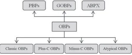
structural characteristics. Thus, OBPs that bind to pheromones are referred as pheromone-binding proteins (PBPs), such as BmorPBP1 (Kriegeret al., 1996). General odorant-binding pro-teins (GOBPs), such as GOBP1 and GOBP2, which are present in both females and males of tobacco hawk mothManduca sexta (Vogtet al., 1991), are proposed to bind to general odorants. Less well described are antennal-binding proteins X (ABPx), which are first reported in B. mori (Krieger et al., 1996), although their ligand binding specificity has only been stud-ied recently (Heet al., 2010). As shown in Fig. 1, an alterna-tive classification for OBPs consists of ‘Classic’ OBPs, ‘Plus-C’ OBPs, ‘Minus-C’ OBPs and ‘Atypical’ OBPs (Hekmat-Scafe et al., 2002; Zhouet al., 2004). The main sequence differences are the number of cysteine (Cys) residues and their conserva-tion. According to this, ‘Classic’ OBPs have six conserved Cys residues at specific positions and include PBPs, GOBPs and ABPx, whereas ‘Plus-C’ OBPs have two additional Cys and a conserved proline (Hekmat-Scafe et al., 2002; Zhou et al., 2004). ‘Minus-C’ OBPs have less than six Cys and ‘Atypical’ OBPs are characterized by having six Cys residues as in the ‘Classic’ OBPs but with additional cysteines in the C-terminal region (Xuet al., 2003). For OBPs with the six conserved Cys residues, there is also a classification according to chain length and C-terminus length (Tegoni et al., 2004): (i) long-chain OBPs with approximately 140 amino acids (e.g. OBPs of the mothsB. mori andA. polyphemus); (ii) medium-chain OBPs with approximately 120 amino acids (e.g. some OBPs of the mosquitoAnopheles gambiaeand the beeApis mellifera); and (iii) short-chain OBPs with approximately 100 amino acids (e.g. OBPs from the cockroachLeucophaea maderae). More recently, a novel subclass of OBPs, according to their C-termini and 3D structure, called C8 OBP class, was proposed by Lagardeet al. (2011). The findings for the A. gambiaeOBP7 (AgamOBP7) suggest a more evolved protein as a result of an increase in structural complexity with eight Cys residues, four disulphide bridges and a C-terminus slightly longer than Classic OBPs. Moreover, it is proposed that AgamOBP7 may have evolved from Classic OBPs, which served as a basal group of OBPs according to phylogenetic analyses (Vieira & Rozas, 2011). The focus in the present review is on Classic OBPs because they all have well conserved characteristics such as six Cys residues, three disulphide bridges and an average molecular weight of approximately 14 kDa. Furthermore, most of the well-studied OBPs such as PBPs and GOBPs of lepidopteran species and the OBP (LUSH) for the pheromone (11-Z-vaccenyl acetate) of Drosophila melanogasterare Classic OBPs.
Function and specificity.In insects, perception of the envi-ronment, such as host plants, prey and potential mates is guided mainly by chemical signals, which are termed semiochemi-cals. If a semiochemical is a small hydrophobic molecule, it cannot easily cross a polar environment, such as the insect sensillum lymph. It is considered that OBPs are the key com-ponent to solubilize and transport these molecules across the lymph to reach the ORs. For hydrophobic ligands such as lep-idopteran sex pheromones at least, it has become clear that OBPs play an important role in ligand capturing and transport to achieve the ligand–OR interaction, as well as in contributing
Fig. 1. Classification scheme of insect odorant binding pro-teins (OBPs) according to their primary protein sequence. ABPX, antennal-binding protein X; GOBP, general odorant-binding protein; PBP, pheromone-binding protein.
to the subsequent signal transduction (Kaissling, 2013). How-ever, experimental evidence suggests that there are three pri-mary functions for OBPs, and these may play dynamic roles in ligand selectivity: (i) ligand scavenging, responsible for the protection of ORs from saturation, which was proposed using a kinetic model forLymantria disparPBP2 (LdisPBP2) (Gong et al., 2009); (ii) ligand desorption from the cuticular wax layer of olfactory pores to the lymph, as suggested for LdisPBP1 and LdisPBP2 (Kowcun et al., 2001); and (iii) ligand recog-nition, as proposed forD. melanogasterOBP (LUSH) acting as an activator of ORNs tuned to pheromones (Laughlinet al., 2008). By contrast, Gomez-Diazet al.(2013) report that a high concentration of the sex pheromone (Z)-11-octadecenyl acetate (cis-vaccenyl acetate) ofD. melanogasteractivates ORs in the absence of LUSH, indicating that LUSH may not have a direct role as the activator of ORNs. It is also proposed that OBPs pro-tect odourant molecules from degradation by odorant-degrading enzymes during their transport and transfer to the ORs (Ishida & Leal, 2002; Leal, 2005, 2013).
bombykal may exist inB. mori. The interaction with ORs and other downstream events is not yet well understood. Apart from lepidopteran PBPs, the ligand specificity of insect OBPs has so far not been demonstrated conclusively. Nevertheless, the sex pheromone specificity of lepidopteran PBPs has provided interesting opportunities to study protein–ligand interactions by using OBPs as attractive targets for homology modelling and ligand screening. Currently, competitive binding assays using N-phenyl-1-naphthylamine as a fluorescence probe are used to study the ligand binding affinity to OBPs (Lescopet al., 2009; Zhou, 2010). Only a few studies have provided the binding affinities in connection with structural data. Hydrogen bonds appear to have an important role in specific interactions for the recognition of some ligands, as reported for LUSH and its alcohol-binding site (Kruse et al., 2003), and for other lepi-dopteran OBPs (Zhouet al., 2009). The first attempt to correlate binding affinity with structural data was made in a study by Sandleret al.(2000), in which BmorPBP1 was crystallized as a complex with bombykol. It was reported that Ser56 forms a hydrogen bond with the hydroxyl of bombykol, which plays a role in the specificity of the PBP. However, a more in-depth study was performed on the bombykol-BmorPBP complex by Klusak et al.(2003), usingab initiomethods, which suggests that not only hydrogen bonds, but also cation-π and π-πinteractions have an important role in the bombykol binding. The ab initio calculations indicate that bombykol could adopt two conforma-tions (A and B). A single hydrogen bond with Ser56 is formed in conformation A, whereas two hydrogen bonds, involving Ser56 and Met61, are formed in conformation B. BmorPBP1 is shown to bind to analogues of bombykol, when measured with Chip-assisted high-throughput electrospray ionization mass spectrometry analysis, where the compounds having differ-ent chain lengths, such as (10E,12Z)-hexadecadienyl acetate and (10E,12Z)-octadecadien-1-ol, bind to the protein with a higher affinity than bombykol (Hooperet al., 2009; Heet al., 2010). Similarly, Campanacci et al. (2001) report that PBP1 ofMamestra brassicae (MbraPBP1) and ApolPBP1 is unable to discriminate pheromones from certain other compounds. By competitive binding assays using 1-aminoanthracene, fatty acids show significant binding affinity. Zhouet al.(2009) and He et al. (2010) report that BmorPBP1 binds well to both bombykol and bombykal, whereas a GOBP (BmorGOBP2) binds differently to these compounds. It is suggested that the binding of bombykol to BmorGOBP2 involves hydrogen bond-ing to Arg110 rather than to Ser56 as found for BmorPBP1. In addition, the hydroxyl group of bombykol may form an additional hydrogen bond with Glu98 via a water molecule, as predicted through MD simulations (Gräteret al., 2006a,b), whereas bombykal cannot form such additional hydrogen bond. This explains the ligand discrimination by BmorGOBP2 at the structural level. Interestingly, Zhou et al.(2009) also demon-strate by crystallography that there is no conformational change among the BmorGOBP2 structures bound with sex pheromone components and their analogues, which is consistent with a recent study onDrosophilaLUSH (Gomez-Diazet al., 2013). A crystallized structure of an OBP ofA. gambiae(AgamOBP1) shows numerous contacts withN,N-diethyl-3-methylbenzamide (DEET) at its binding site (e.g. van der Waals interactions).
Hydrogen bonds are formed via water molecules with Trp114, Cys95 and Gly92 (Tsitsanou et al., 2012). Although DEET appears to have a specific interaction with the OBP, it was recently reported that eugenyl acetate could be a better repellent than DEET as a result of a better affinity with the binding site of AgamOBP1, as shown by molecular modelling studies (Affonso et al., 2013). However, binding assays and robust behavioural bioassays are necessary to corroborate those findings.
Although OBPs are divergent across insect species, and even within same species (Pelosi et al., 2006), the general structural characteristics of OBPs are conserved, such as six α-helical domains and three disulphide bridges (from the six Cys residues). These features are well conserved in the GOBP of the honeybeeA. mellifera(ASP2). However, ASP2 shows a broad specificity for ligands compared with PBPs (Lescopet al., 2009). Structurally different compounds, such as 2-isobutyl-3-methoxypyrazine, isoamyl acetate, 1,8-cineol and 2-heptanone show significant affinity for ASP2 through weak and nonspecific interactions. Therefore, further evidence and robust bioassay techniques are necessary to establish the dis-criminatory capability of insect OBPs.
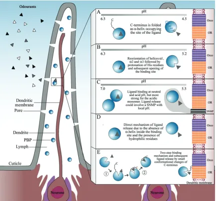
binds more strongly to the AtraPBP1 at neutral pH than at acid pH. However, the deletion of the C-terminus of AtraPBP1 increases the pheromone-binding affinity by 100-fold at pH 5.0 and 1.5-fold at pH 7.0. There is probably an occupation of the binding site of AtraPBP1 by its C-terminus at neutral pH, as well as acid pH, which decreases the binding affin-ity of (Z11,Z13)-hexadecadienal. Furthermore, three wild-type PBPs (PBP1, PBP2 and PBP3) of two sibling species Heli-coverpa armigeraandHelicoverpa assultashow a decrease in the binding affinity at acid pH. The mutant-type PBPs with-out C-terminal segment have a similar binding affinity at the same pH (Guoet al., 2012). Zubkovet al.(2005) found that the C-terminus of ApolPBP1 does not have a major role for the dis-placement of ligands. Instead, there is a reorientation of helices α1,α3 andα4 at acid pH, which causes protonation of histi-dine residues (His69, His70 and His95) inside the binding site, leading to an opening of the binding site, and the pheromone is released near the dendritic membrane. Pesentiet al.(2009) report that theA. melliferaPBP (ASP1) has a contradictory con-formational change compared with the observed in BmorPBP1. Thus, ligands bind to this protein at low pH (4.0) and the release occurs at pH 7.0. However, binding is also possible at neutral pH; therefore, an uncommon conformational change was pro-posed. At pH 7.0, ASP1 can form dimers through the absence of the C-terminus from the core of the protein and the participation of the N-terminus to form a more stable dimer structure. This dimeric form binds ligands but with lower affinity than the acidic monomer. There is another more direct ligand release mecha-nism forL. maderaePBP (LmaPBP) as suggested by Lartigue et al.(2003). This mechanism, in contrast to that for BmorPBP1, is based on the absence of a helix inside of the PBP and the presence of a significant amount of hydrophilic residues in the binding site.
Kinetics studies have been undertaken forL. disparLdisPBP2 using fluorescence binding assays and tryptophan anisotropy measurements to obtain the association in timescale and mul-timerization, respectively (Gong et al., 2009). The results indicate that LdisPBP2 binds (+)- and (−)-disparlure in both a rapid step and a slow step. The slow step could be more important for obtaining an active ligand for ORs as a result of an internal binding, which results in a more stable ligand–OBP complex. By contrast, the rapid step appears to be related to binding at an external site, where the initial interactions between ligand and OBP occur. Moreover, within a few hours, there is an increased presence of LdisPBP2 as dimer, which has a smaller binding capacity compared with monomer. Subse-quently, a more detailed binding mechanism was proposed. It appears that two steps are necessary to obtain a final complex. First, LdisPBPs and the ligand form an intermediate complex through diffusion-controlled collision. The second and final step involves the relocation of the ligand from an external binding site to a different internal one (Gonget al., 2010). In Fig. 2, a schematic representation is provided that summarizes the most outstanding research on lepidoptera OBPs, and their ligand binding and release mechanisms.
Structure prediction of OBPs by homology modelling. Approximately 4 years ago, Zhou (2010) reported that 47
crystal structures of insect OBPs were deposited in the Protein Data Bank (PDB) (http://www.rcsb.org/pdb). At the beginning of 2014, a search with ‘odorant binding protein’ as keywords returned the structures of OBPs in only eight insect species, despite several hundred OBPs having been reported in an enor-mous number of insect species. To date, more than 60 crystal structures have been determined by X-ray crystallography. Only 13 of them have a high resolution (<1.5 Å), 45 have a lower resolution of 1.5–2.5 Å and three have a resolution of 2.5–3.0 Å. Thus, A. mellifera, D. melanogaster, A. gambiae and B. mori have the greatest number of crystal structures, including both OBPs and PBPs in some cases. These crystal structures were solved under different conditions, such as ligand–OBP complex, apo-OBP form and at different pHs. The PBPs from moths often have seven α-helices and three conserved disulphide bridges. The high sequence identity provides a similar global fold among these OBPs, which is crucial for homology modelling, and the structural studies of these OBPs have provided some outstanding information for computer-assisted modelling of these proteins. For example, early studies on homology modelling with OBPs were carried out by Honson & Plettner (2006) aiming to determine the three-dimensional arrangement of disulphide bridges on L. disparPBPs (LmarPBP1 and LmarPBP2). Based on the crystal structure of BmorPBP1, the homology models revealed that the most exposed and accessible disulphide bridge is C2–C5 (i.e. Cys50–Cys108), which is the most easily reduced disulphide bridge by cyanylation using tris(2-carboxyethyl)phosphine, 2-mercaptoethanol and dithiothreitol. The structural models of these proteins suggest that the easy reduction of C2–C5 is the result of a steric property of Cys side chains that are more exposed. Moreover, the conformation of C2–C5 could be influenced strongly by electronic effects from a nearby aspartic acid (Asp106), which stabilizes C5 through a proton transfer.
Fig. 2. Schematic representation of the mechanisms of ligand release of moth pheromone-binding proteins (PBPs). Background section represents a commonly occurring multipore sensillum present in moths, such asBombyx moriandAntheraea polyphemus. From the external environment, different odorants ( , and ) can enter to sensilla by diffusion through cuticle pores. PBPs (blue spheres) are secreted by auxiliary cells and located in the sensillium lymph. These proteins are proposed as the first filter of selection and discrimination of odorants, which are represented by the ligand–PBP complex (grey triangle-sphere) in the lymph. (A) Ligand binding and release mechanism proposed forB. moriPBP (BmorPBP) (Horstet al., 2001; Lautenschlageret al., 2005; Leal, 2005). (B) Ligand binding and release mechanism proposed forA. polyphemusPBP (ApolPBP1) (Zubkovet al., 2005). (C) Uncommon ligand binding and release mechanism proposed forApis melliferaASP1 (Pesentiet al., 2009). (D) Ligand binding and release mechanism proposed forLeucophaea maderaePBP (LmaPBP) (Lartigueet al., 2003). (E) Ligand binding and release mechanism proposed for
Lymantria disparPBPs (LdisPBPs) (Gonget al., 2009, 2010). Inactivation or degradation (by odorant-degrading enzymes) is not represented in this scheme because this is not part of the main discussion of this review. OR, olfactory receptor; Orco, OR co-receptor; SNMP, Sensory neuron membrane protein.
chain A’ as template, for the modelling of aphid OBPs (Qiao et al., 2009). Similarly, other studies have used the same template to predict the 3D structure of the OBPs of A. pisum andSitobion avenae(Sunet al., 2011; Sunet al., 2012a; Zhong et al., 2012) and of the plant bugAdelphocoris lineolatus(Wang et al., 2013a). Moreover, a template can be used to model not only a full-length, but also a part of target (Chang & Swaan, 2006). For example, for modelling of Culex quinquefasciatus OBP2, Paramasivan et al. (2007) used two templates. The
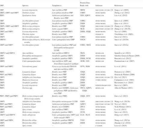
2006) are popular and freely available software for modelling processes, both of which offer friendly platforms for protein homology modelling. swiss-model is suitable for beginners as an automated and online server, and does not need to be downloaded and installed. Instead, the server offers the capa-bility to build homology models within a workspace through a web connection (Arnold et al., 2006). On the other hand, the modeller software must be downloaded and installed. Although modeller calculates a model automatically, it must be provided with an alignment between the target and template, as well as user scripts as commands. Once the model has been built, other software can be used to visualize the model, such as pymol (http://www.pymol.org), ras-mol (http://www.rasras-mol.org), chimera (http://www.cgl.ucsf. edu/chimera/) and swiss-pdbviewer (http://www.spdbv.vital-it. ch/), amongst others. Homology modelling studies of insect OBPs to date are listed in Table 1, including templates and the software used. Apart from homology modelling, there are other types of computing approaches in protein structure predictions, according to several reviews on the subject (Sánchez et al., 2000; Schwede et al., 2007; Ravna & Sylte, 2012). These include threading methods, ab initiomethods and integrative or hybrid methods. However, homology modelling is the most precise and reliable method currently being used (Bordoli & Schwede, 2012; Ravna & Sylte, 2012). There are numerous reviews on homology modelling that have a similar content structure, each highlighting the steps, precision and limitations of the technique, together with an apparently endless list of available modelling software. Therefore, below, the important concepts in homology modelling are only summarized, with a focus on the ligand affinities of insect OBPs.
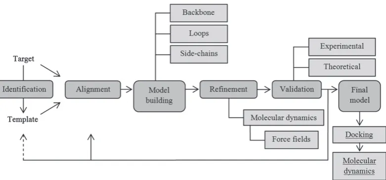
Protein structure modelling based on homology between target and template consists of four main steps: template iden-tification, target-template alignment, model building and model refinement and validation (Fig. 3). Template identification is the first step in obtaining a homology model. Such identifica-tion is usually carried out with basic local alignment search tool blast (http://www.ncbi.nlm.nih.gov/BLAST/). This tool provides regions of similarity and an identity percentage of nucleotide or protein sequences between target and templates. To determine a ligand–protein interaction, a template must have some characteristics, such as high resolution, sharing regions or substructures with target, and be bound as holo-protein (i.e. ligand–protein interaction as biochemically active complex) (Orry & Abagyan, 2012). The percentage sequence identity is an important indicator for establishing the best template. Thus, 30% or more sequence homology is considered good to obtain a precise model. If the sequence identity is less than 30%, the quality of the model decreases (Schwedeet al., 2007). Based on the above, the sequence identities of the targets LdisPBP1 and LdisPBP2, with the template BmorPBP (1DQE), are 61% and 48%, respectively (Honsonet al., 2003), which has provided a good homology model for both PBPs. However, the study by Honson et al. (2003) does not pro-vide an evaluation on the quality of the predicted models. This is commonly performed with procheck (http://www.ebi. ac.uk/thornton-srv/software/PROCHECK/), comprising soft-ware that evaluates the stereochemical quality of the predicted
Table 1. Summary of homology modelling studies to determine odorant-binding protein structures of insects.
OBP Species Template(s)
Protein Data
Bank code Software References
OBP Locusta migratoria Apis melliferaPBP 3BFH discovery studio 2.0 Jianget al.(2009)
OBP2, OBP3 and OBP7 Sitobion avenae Leucophaea maderaePBP 1ORG swiss-model Zhonget al.(2012)
PBP1 Spodoptera litura Antheraea polyphemusand Bombyx moriPBP
1IS8, 1QWV modeller Liuet al.(2012)
OBP Acyrthosiphon pisum Leucophaea maderaePBP 1ORG swiss-model Qiaoet al.(2009) OBP4 Anopheles gambiae Anopheles gambiaeOBP1 2ERB swiss-model Qiaoet al.(2011)
OBP1, OBP2 and OBP3 Helicoverpa armigera Bombyx moriPBP 1DQE discovery studio 2.0 Zhanget al.(2012a)
OBP5 Helicoverpa armigera Aedes aegyptiOBP1 3K1E discovery studio 2.0 Zhanget al.(2012b)
OBP2 and OBP3 Locusta migratoria Anopheles gambiaeOBP1 2ERB, 1DQE swiss-model Yuet al.(2009)
OBP Phormia regina Bombyx moriPBP – insight ii Tsuchiharaet al.(2005) OBP3 Acyrthosiphon pisum Leucophaea maderaePBP 1ORG swiss-model Sunet al.(2011)
OBP10 Helicoverpa armigera,Helicoverpa
assulta
Culex quinquefasciatusOBP 3OGN swiss-model Sunet al.(2012b)
OBP3 and OBP7 Acyrthosiphon pisum Leucophaea maderaePBP and
Drosophila melanogaster LUSH
1ORG, 3B6X swiss-model Sunet al.(2012a)
OBP15 and OBP21 Apis mellifera Apis melliferaOBP14 3RZS modeller Spinelliet al.(2012)
OBP Drosophila melanogaster Anopheles gambiaeOBP1 2ERB swiss-model Sánchez-Gracia & Rozas (2008) OBP1 Anopheles gambiae Anopheles gambiaeOBP1 2ERB, 3N7H swiss-model Rusconiet al.(2012) OBP2 Culex quinquefasciatus Apis melliferaASP1 and
Penicillium roquefortisynthase
1R5R, 1DI1 modeller Paramasivanet al.(2007)
OBP1 and OBP2 Scleroderma guani Pyrococcus horikoshiiPH1010
andApis melliferaASP1
3D76, 3BJH swiss-model Liet al.(2011)
PBP1 Lymantria dispar Bombyx moriPBP 1DQE swiss-model Honsonet al.(2003)
PBP1 and PBP2 Lymantria dispar Bombyx moriPBP 1DQE swiss-model Honson & Plettner (2006)
OBP1 Adelphocoris lineolatus Bombyx moriPBP 1DQE discovery studio 2.0 Guet al.(2011) PBP1 Mamestra brassicae Bombyx moriPBP 1DQE turbo-frodo Campanacciet al.(2001) OBP1 Anopheles gambiae Anopheles gambiaeOBP1 2ERB – Biessmannet al.(2010) OBP1 Locusta migratoria Bombyx moriPBP 1DQE insight/homology Banet al.(2003) PBP3 Ostriniaspp. Bombyx moriGOBP2,Antheraea
polyphemusPBP andBombyx moriPBP
2WCJ, 1QWV, 1DQE
modeller Allen & Wanner (2011)
PBP1, PBP2 and PBP3 Helicoverpa armigeraand
Helicoverpa assulta
Bombyx moriPBPs 1DQE, 2FJY swiss-model Guoet al.(2012)
OBP5 Adelphocoris lineolatus Drosophila melanogasterLUSH 1OOI discovery studio 2.0 Wanget al.(2013b)
PBP1 and PBP2 Lymantria dispar Antheraea polyphemusPBP 1QWV discovery studio 3.0 Yuet al.(2012)
PBP1 and PBP2 Lymantria dispar Bombyx moriGOBP2 2WCJ swiss-model Yu & Plettner (2013)
OBP2 Apis cerana Apis melliferaOBP2 1TUJ swiss-model Liet al.(2013) OBP7 Helicoverpa armigera Anopheles gambiaeOBP1 3N7H swiss-model Sunet al.(2013)
OBP37 and OBP39 Aedes albopictus Culex quinquefasciatus OBP1 and
Aedes aegypti OBP1
2L2C, 3K1E swiss-model Denget al.(2013)
OBP3 and OBP4 Holotrichia oblita Anopheles gambiaeOBP20 3VB1 swiss-model Wanget al.(2013a)
OBP1 Holotrichia oblita Anopheles gambiaeOBP1 2ERB swiss-model Zhuanget al.(2014)
ASP2 Apis cerana – – i-tasser Luet al.(2014)
ASP,A. mellifera; OBP, odorant-binding protein; PBP, pheromone-binding protein.
energy of a particular protein conformation through calculations by force fields, which is usually performed with the software amber (Weiner & Kollman, 1981), gromacs (Berendsenet al., 1995) and namd (Phillips et al., 2005). It is noteworthy that MD simulations are used not only for refinement of homology models, but also to determine structural changes in proteins. For example, MD simulation has been used to assess the binding dynamics and specificity of insect PBPs. Thus, Gräter et al. (2006b) report a similar binding affinity for bombykol and bom-bykal to BmorPBP1 through MD simulations that considered entropic contributions to the free energy of the protein/ligand binding complexes. Although bombykol acts as hydrogen-bond donor and bombykal as hydrogen-bond acceptor, the MD simulation shows that the binding modes of both bombykol and bombykal are similar, forming and breaking hydrogen
bonds in a reversible way. More recently, Chu et al. (2013) provide a putative pH-induced ligand-releasing mechanism, which is predicted using MD simulation. Their findings suggest that the OBP1 of C. quinquefasciatus (CquiOBP1) releases the mosquito oviposition pheromone at low pH as a result of the cleavage of hydrogen bonds in the binding site. These simulations have been performed on crystal structures of OBPs. However, Yuet al.(2012) report a constant pH MD simulation on the modelled structures of L. dispar PBPs (LdisPBPs) at both pH 7.3 and pH 5.5. Subsequently, the findings were cor-roborated by circular dichroism, where conformational changes are revealed (Yuet al., 2012).
Fig. 3. General building scheme for protein structures by homology modelling, as applied to insect odorant-binding proteins to date. Underlined section indicates an optional step when ligand binding is studied.
classical mechanics or molecular mechanics (MM) works to find the global minimum of free energy in a protein struc-ture to describe a conformation, considering atoms and bonds as single particles and springs, respectively (Bordner, 2012). All refinement processes on homology models of insect OBPs have used MM. An example is the predicted structure of the salivary OBP2 of the mosquito C. quinquefasciatus, where GROMOS96 (Scottet al., 1999) was used as force field based on MM in the refinement step (Paramasivan et al., 2007). Similarly, a refined homology model of Locusta migrato-ria OBP1 (LmigOBP1) was obtained by Jiang et al. (2009) using charmm force field software for macromolecular energy minimization and dynamics calculation (Brooks et al., 1983). Despite these studies on the refinement of predicted OBPs structures, there is no information available on how long these simulations were required to complete. Currently, MD at long-timescale (e.g. nanoseconds) appears to be useful for refinement of homology models from proteins of small or medium size (Fan & Mark, 2004; Ravalet al., 2012), such as insect OBPs.
Model validation is a crucial step to ensure its quality. There are two approaches for model validation: experimental and the-oretical. The experimental approach compares the results from modelling studies with biological observations, and establishes the model quality. If the biological results are in accordance with the homology model, it is considered correct (Chang & Swaan, 2006; Ravna & Sylte, 2012). The theoretical method for vali-dation analyzes the stereochemical quality of the model. Thus, when the model is not satisfactory, the steps are repeated from template identification or target-template alignment forward on (Martí-Renomet al., 2000). Therefore, all the steps described above are usually repeated iteratively until the best homology model is obtained.
Insect OBPs are flexible and this property is likely to be very important for ligand recognition patterns and function. To date, optional steps are being developed after the validation of mul-tiple homology models, such as docking and MD simulations
of ligand–protein complexes. Docking (also called molecular docking) is discussed in more detail below. It is important to note that homology models and experimental protein struc-tures are only a snapshot of a specific conformation. There-fore, all results that are determined represent only one state of the protein without information about its dynamic. Thus, studies on drug discovery have been directed towards deter-mining the stability of the ligand–protein complexes (Alonso et al., 2006). An example is the identification of an inhibitor binding site in human sirtuin 2 (SIRT2), which is a protein from histone deacetylase family (Sakkiah et al., 2013). Five well known inhibitors (suramin, mol-6, sirtinol, 67 and nf675) were docked to SIRT2. The plotted RMSD values reveal that the five complexes between the inhibitors and SIRT2 were sta-ble after approximately 10 ns of simulations. The stability of certain protein structures or ligand–protein complexes is com-monly observed when RMSD values become unchanged with time between the predicted structural conformations and sur-rounding environment (i.e. water molecules and ions) (Sokkar et al., 2011).
progress of modelling techniques. If the template selection for a full-length target is difficult because the sequence identity is too low (less than 30%) or the template resolution is too low, the modelling of some conserved regions is feasible. For example, homology modelling of α4β2 nicotinic acetylcholine receptor (nAChR) with homopentameric acetylcholine binding protein (AChBP) from the sea slug Aplysia californica as template gave a sequence identity of 18–20%. Despite such low identity, the shared features between the target nAChR and template AChBP, such as a similar fold and highly conserved residues in the binding site (Hansenet al. (2005), allowed an nAChR model to be predicted using AChBP as a suitable template (Iturriaga-Vásquezet al., 2010).
It is important to obtain precise alignment between targets and templates. Distant evolutionary relationships, specific regions that do not align reliably and MSA programmes themselves could lead to imprecision in the alignment. Arnoldet al.(2006) note that, when the imprecision of a target-template align-ment cannot be determined, visual and manual intervention can improve the model quality significantly. If the precision of a sequence alignment decreases, the percentage of equiva-lent Cαatoms for the superimposed target and template struc-tures increases, as measured by RMSD. Raval et al. (2012) note that the results of homology modelling have a low res-olution by imprecise refinement during MD simulation with errors reflected in the values of 3 Å for atomic coordinates. The timescale of MD application and the precision of force fields could also be the source of errors. However, the main failure of MD within the refinement process appears to be caused by force field errors (Ravalet al., 2012). A restricted conformational sampling for the initial homology model was proposed to improve the predicted structures. This conforma-tional sampling emerges from the MD (referred to the pro-tein motion possibly with surrounding solvent) and the use of energy functions (force fields) (Bordner, 2012), compris-ing samplcompris-ing steps to find the global optimal structures with lowest energy.
Ligand binding affinity to homology models of OBPs: methods and interaction. Many types of biological effects in vertebrates and invertebrates are dependent on ligand–protein interactions. The transport function of OBPs is carried out through interactions between ligands and the binding sites of these proteins. As mentioned previously, homology modelling, as a quick and inexpensive method, allows the prediction of the binding site in protein structures. Once a good qual-ity homology model is obtained, it is possible to simulate molecular docking of ligands, which predicts how and where small molecules could be bound to the protein model. Before docking, an important step is the identification of the binding pocket to find the cavities suitable as potential binding sites for ligands. For this, a ligand structure preparation is neces-sary, a step that can be achieved by searching for ligands in databases (e.g. PubChem; http://www.pubchem.ncbi.nlm.nih. gov/) or by drawing them de novo using appropriate soft-ware, such as chem3d (http://www.cambridgesoft.com) or spartan (http://www.wavefun.com). Then, molecular dock-ing is applied over a sufficient number of times to achieve
Table 2. Molecular docking on homology models of insect odorant-binding proteins.
OBP Ligands Software Reference
Anopheles gambiaeOBP1 Indole quanta-charmm Biessmannet al.(2010)
Locusta migratoriaOBP Pentadecanol cdock Jianget al.(2009)
Helicoverpa armigeraOBP1, OBP2 and OBP3
(Z)-9-Hexadecenal and (Z)-11-hexadecenal – Zhanget al.(2012a)
Lymantria disparPBP1 Aziridine – Honsonet al.(2003)
Anopheles gambiaeOBP1 N-Phenyl-1-naphylamine (generic ligand) autodock 4.0 Rusconiet al.(2012)
Lymantria disparPBP1 and PBP2
Palmitic acidN-butyl ester,
bis(3,4-epoxycyclohexylmethyl) adipate and l-trans-epoxysuccinyl-isoleucyl-proline methyl ester propylamide
cdock Yuet al.(2012)
Lymantria disparPBP1 and PBP2
(+) and (−) disparlure molecular operating environment (moe) Yu & Plettner (2013)
Apis ceranaOBP2 N-Phenyl-1-naphylamine (generic ligand) molegro virtual docker 4.2 Liet al.(2013)
Adelphocoris lineolatus OBP5 cis-Nerolidol cdock Wanget al.(2013b)
Holotrichia oblita OBP1 Hexyl benzoate,β-ionone, cinnamaldehyde and myrcene
autodock 4.0 Zhuanget al.(2014)
Apis cerana ASP2 Linalool, geraniol,β-ionone, 4-allylveratrole, phenylacetaldehyde, dibutyl phthalate, isoamylacetate, methyl-p-hydroxyl benzoate and butanedione
molegro virtual docker 4.2 Luet al.(2014)
OBP, odorant-binding protein; PBP, pheromone-binding protein.
of active semiochemicals using a computational reverse chem-ical ecology approach on a GOBP of the oriental fruit fly Bactrocera dorsalis.
Although the docking method is a powerful tool for predict-ing ligand–protein interactions, other experimental methods are being used in conjunction with homology modelling, such as fluorescence competitive binding assays (FBA), radioactively labelled ligand (RLL) studies and site-directed mutagenesis. Fluorescence competitive binding assays and RLL comprise two technologies for receptor-ligand binding investigations. By employing radio-isotopic labels such as3H, RLL can be used in the determination of receptor distribution and subtypes, screen-ing of ligands and quantification. However, this type of bind-ing assay has major disadvantages (e.g. radioactive wastes, high costs, health hazards and requirement of special licenses) com-pared with FBA, which is a widely used method to determine the affinity to a wide range of compounds of insect OBPs (Zhou et al., 2004; de Jong et al., 2005). Fluorescence competitive binding assays provide a ligand screening with the use of flu-orophores (commonly 1-N-phenylnaphthylamine) and quantifi-cation through displacement of the fluorophore by ligands. An example is the FBA approach used by Zhanget al., 2012b for 113 compounds, using homology modelling as a complement. Although these binding assays indicate that (E)-β-farnesene, ethyl butyrate, ethyl heptanoate and acetic acid 2-methylbutyl ester are the best ligands for H. armigeraOBP5, the homol-ogy 3D model reveals key residues in the OBP5 binding site, which may have an important role in ligand specificity. Further-more, the protein structure prediction by homology modelling, along with mutagenesis, allows examination of ligand–protein interactions, the role of nonconserved residues in the binding sites and ligand binding modes (Cavasotto & Phatak, 2009). The ligand binding affinity can be altered through site-directed muta-genesis, as inA. gambiaeOBP1 (Rusconiet al., 2012). Also,
physicochemical properties can be changed through mutations of the residues in the binding site, such as Phe59, Met84, His111, Trp114, Tyr122, Phe123 and Leu124. Thus, the mutated OBPs exhibit a lower binding affinity in the binding assays. More recently, Zhuanget al.(2014) report that Tyr111 of the scarab beetle Holotrichia oblitaOBP (HoblOBP1) is a key residue in the binding site of the protein. Through homology mod-elling, molecular docking and subsequent site-directed mutage-nesis along with fluorescence binding assays, it was possible to demonstrate the role of Tyr111 in the binding ofβ-ionone, myrcene, hexyl benzoate and cinnamaldehyde.
Conclusions
to predict best ligands), may represent a great leap forward in the search for super-ligands to manipulate insect behaviours. Finally, it is worth noting that OBP homology models as well as crystal and NMR structures provide only static representa-tions of dynamic olfaction systems. Once molecular modelling is successfully performed, further robust bioassays need to be carried out and super-ligands need to be screened with insects to identify potential pest control agents.
Acknowledgements
We would like to thank Dr Andrés Avila for valuable and helpful comments on an earlier version of the manuscript. This work was supported by CONICYT (21110933). We also thank two reviewers and the Editor, Dr Rob Weaver, for their professional and valuable comments that improved this work. Rothamsted Research receives grant-aided support from the Biotechnology and Biological Sciences Research Council (BBSRC) of the U.K.
References
Affonso, R., Guimarães, A.P., Oliveira, A.A.et al.(2013) Applications of molecular modeling in the design of new insect repellents targeting the odorant binding protein ofAnopheles gambiae.Journal of the Brazilian Chemical Society,24, 473–482.
Allen, J.E. & Wanner, K.W. (2011) Asian corn borer pheromone binding protein 3, a candidate for evolving specificity to the 12-tetradecenyl acetate sex pheromone.Insect Biochemistry and Molecular Biology, 41, 141–149.
Alonso, H., Bliznyuk, A.A. & Gready, J.E. (2006) Combinding dock-ing and molecular dynamic simulations in drug design.Medicinal Research Reviews,26, 531–568.
Arnold, K., Bordoli, L., Kopp, J. & Schwede, T. (2006) The SWISS-MODEL Workspace: a web-based environment for protein structure homology modelling.Bioinformatics,22, 195–201.
Ban, L., Scaloni, A., D’Ambrosio, C.et al.(2003) Biochemical char-acterization and bacterial expression of an odorant-binding protein fromLocusta migratoria.Cellular and Molecular Life Sciences,60, 390–400.
Berendsen, H.J.C., van der Spoel, D. & van Drunen, R. (1995) GRO-MACS: a message-passing parallel molecular dynamics implementa-tion.Computer Physics Communications,91, 43–56.
Bette, S., Breer, H. & Krieger, J. (2002) Probing a pheromone binding protein of the silkmothAntheraea polyphemusby endogenous tryp-tophan fluorescence.Insect Biochemistry and Molecular Biology,32, 241–246.
Biessmann, H., Andronopoulou, E., Biessmann, M.R.et al.(2010) The
Anopheles gambiaeodorant binding protein 1 (AgamOBP1) mediates indole recognition in the antennae of female mosquitoes.PLoS ONE, 5, e9471. DOI:10.1371/journal.pone.0009471.
Bordner, A.J. (2012) Force fields for homology modeling.Homology Modeling: Methods and Protocols (ed. by A. J. W. Orry and R. Abagyan), pp. 83-106. Molsoft L.L.C., San Diego, California. Bordoli, L. & Schwede, T. (2012) Automated protein structure modeling
with SWISS-MODEL Workspace and the protein model portal.
Homology Modeling: Methods and Protocols(ed. by A. J. W. Orry and R. Abagyan), pp. 107-136. Molsoft L.L.C., San Diego, California. Briand, L., Eloit, C., Nespoulous, C. et al. (2002) Evidence of an odorant-binding protein in the human olfactory mucus: location, structural characterization, and odorant-binding properties. Biochem-istry,41, 7241–7252.
Brimau, F., Cornard, J.-P., Le Danvie, C.et al.(2010) Binding speci-ficity of recombinante odorant-binding protein isoforms is driven by phosphorylation.Journal of Chemical Ecology,36, 801–813. Brooks, B.R., Bruccoleri, R.E., Olafson, B.D.et al.(1983) CHARMM:
a program for macromolecular energy minimization and dynamics calculations.Journal of Computational Chemistry,4, 187–217. Campanacci, V., Krieger, J., Bette, S. et al. (2001) Revisiting the
specificity of Mamestra brassicae and Antheraea polyphemus
pheromone-binding proteins with a fluorescence binding assay.
Journal of Biological Chemistry,276, 20078–20084.
Cavasotto, C.N. & Phatak, S.S. (2009) Homology modeling in drug discovery: current trends and applications.Drug Discovery Today,14, 676–683.
Chang, C. & Swaan, P.W. (2006) Computational approaches to modeling drug transporters.European Journal of Pharmaceutical Sciences,27, 411–424.
Chu, W.-T., Zhang, J.-L., Zheng, Q.-C. et al. (2013) Constant pH molecular dynamics (CpHMD) and molecular docking studies of CquiOBP1 pH-induced ligand releasing mechanism. Journal of Molecular Modeling,19, 1301–1309.
Damberger, F.F., Ishida, Y., Leal, W.S. & Wüthrich, K. (2007) Structural basis of ligand binding and release in insect pheromone-binding proteins: NMR structure ofAntheraea polyphemusPBP1 at pH 4.5.
Journal of Molecular Biology,373, 811–819.
Deng, Y., Yan, H., Gu, J. et al. (2013) Molecular and functional characterization of odorant-binding protein genes in an inva-sive vector mosquito, Aedes albopictus. PLoS ONE, 8, e68836. DOI:10.1371/journal.pone.0068836.
Durrant, J.D. & McCammon, J.A. (2011) Molecular dynam-ics simulations and drug discovery. BMC Biology, 9, 71. DOI:10.1186/1741-7007-9-71.
Edgar, R.C. (2004) MUSCLE: multiple sequence alignment with high accuracy and high throughput.Nucleic Acids Research, 32, 1792–1797.
Eswar, N., Marti-Renom, M.A., Webb, B.et al.(2006) Comparative protein structure modeling with MODELLER.Current Protocols in Bioinformatics(ed. by A. Baxevanis), pp. 5.6.1-5.6.30. John Wiley & Sons, Inc., New York, New York.
Fan, H. & Mark, A.E. (2004) Refinement of homology-based protein structures by molecular dynamics simulation techniques. Protein Science,13, 211–220.
Gajendrarao, P., Krishnamoorthy, N., Sakkiah, S.et al.(2010) Molecular modeling study on orphan human protein CYP4A22 for identification of potential ligand binding site.Journal of Molecular Graphics and Modelling,28, 524–532.
Ginalski, K. (2006) Comparative modeling for protein structure predic-tion.Current Opinion in Structural Biology,16, 172–177. Gomez-Diaz, C., Reina, J.H., Cambillau, C. & Benton, R. (2013)
Ligands for pheromone-sensing neurons are not conformationally activated odorant binding proteins. PLoS Biology, 11, e1001546. DOI:10.1371/journal.pbio.1001546.
Gong, Y., Pace, T.C.S., Castillo, C.et al.(2009) Ligand-interaction kinetics of the pheromone-binding protein from the gypsy moth,L. dispar: insights into the mechanism of binding and release.Chemistry & Biology,16, 162–172.
Gong, Y., Tang, H., Bohne, C. & Plettner, E. (2010) Binding conforma-tion and kinetics of two pheromone-binding proteins from the gypsy mothLymantria dispar with biological and nonbiological ligands.
Biochemistry,49, 793–801.
Gräter, F., Xu, W., Leal, W. & Grubmüller, H. (2006b) Pheromone discrimination by the pheromone-binding protein ofBombyx mori.
Structure,14, 1577–1586.
Große-Wilde, E., Svatoš, A. & Krieger, J. (2006) A pheromone-binding protein mediates the bombykol-induced activation of a pheromone receptorin vitro.Chemical Senses,31, 547–555.
Grolli, S., Merli, E., Conti, V.et al.(2006) Odorant binding protein has the biochemical properties of a scavenger for 4-hydroxy-2-nonenal in mammalian nasal mucosa.FEBS Journal,273, 5131–5142. Gu, S., Wang, W., Wang, G.et al.(2011) Functional characterization
and immunolocalization of odorant binding protein 1 in the lucerne plant bug,Adelphocoris lineolatus(Goeze).Archives of Biochemistry and Physiology,77, 81–98.
Guo, H., Huang, L., Pelosi, P. & Wang, C. (2012) Three pheromone-binding proteins help segregation between twoHelicoverpaspecies utilizing the same pheromone components.Insect Biochemistry and Molecular Biology,42, 708–716.
Hang, N.T. (2008) Comparison of multiple sequence alignment pro-grams in practise. Master of Bioinformatics Thesis, University of Århus.
Hansen, S.B., Sulzenbacher, G., Huxford, T.et al.(2005) Structures of
AplysiaAChBP complexes with nicotinic agonists and antagonists reveal distinctive binding interfaces and conformations.EMBO Jour-nal,24, 3635–3646.
He, X., Tzotzos, G., Woodcock, C. et al. (2010) Binding of the general odorant binding protein ofBombyx moriBmorGOBP2 to the moth sex pheromone components.Journal of Chemical Ecology,36, 1293–1305.
Hekmat-Scafe, D.S., Scafe, C.R., McKinney, A.J. & Tanouye, M.A. (2002) Genome-wide analysis of the odorant-binding protein gene family in Drosophila melanogaster. Genome Research, 12, 1357–1369.
Honson, N. & Plettner, E. (2006) Disulfide connectivity and reduction in pheromone-binding proteins of the gypsy moth,Lymantria dispar.
Naturwissenschaften,93, 267–277.
Honson, N., Johnson, M.A., Oliver, J.E.et al.(2003) Structure–activity studies with pheromone-binding proteins of the gypsy moth, Lyman-tria dispar.Chemical Senses,28, 479–489.
Honson, N., Gong, Y. & Plettner, E. (2005) Structure and function of insect odorant and pheromone-binding proteins (OBPs and PBPs) and chemosensory-specific proteins (CSPs). Recent Advances in Phytochemistry,39, 268–277.
Hooper, A.M., Dufour, S., He, X.et al.(2009) High-throughput ESI-MS analysis of binding between theBombyx moripheromone-binding protein BmorPBP1, its pheromone components and some analogues.
Chemical Communications,14, 5725–5727.
Horst, R., Damberger, F., Luginbühl, P.et al.(2001) NMR structure reveals intramolecular regulation mechanism for pheromone binding and release.Proceedings of the National Academy of Sciences of the United States of America,98, 14374–14379.
Ishida, Y. & Leal, W.S. (2002) Cloning of putative odorant-degrading enzyme and integumental esterase cDNAs from the wild silkmoth,
Antheraea polyphemus.Insect Biochemistry and Molecular Biology, 32, 1775–1780.
Iturriaga-Vásquez, P., Carbone, A., García-Beltrán, O. et al. (2010) Molecular determinants for competitive Inhibition ofα4β2 nicotinic acetylcholine receptors.Molecular Pharmacology,78, 366–375. Jacquin-Joly, E. & Merlin, C. (2004) Insects olfactory receptors:
contributions of molecular biology to chemical ecology.Journal of Chemical Ecology,30, 2359–2397.
Jacquin-Joly, E., Vogt, R.G., Francois, M.C. & Nagnan-Le Meillour, P. (2001) Functional and expression pattern analysis of chemosensory proteins expressed in antennae and pheromonal gland ofMamestra brassicae.Chemical Senses,26, 833–844.
Jayanthi, P.D.K., Kempraj, V., Aurade, R.M.et al.(2014) Computa-tional reverse chemical ecology: virtual screening and predicting behaviorally active semiochemicals forBactrocera dorsalis.BMC Genomics,15, 209. (also cited as P.D.et al., 2014)
Jiang, Q., Wang, W., Zhang, Z. & Zhang, L. (2009) Binding specificity of locust odorant binding protein and its key binding site for initial recognition of alcohols.Insect Biochemistry and Molecular Biology, 39, 440–447.
de Jong, L.A.A., Uges, D.R.A., Franke, J.P. & Bischoff, R. (2005) Receptor-ligand binding assays: technologies and applications. Jour-nal of Chromatography B,829, 1–25.
Kaissling, K.-E. (2013) Kinetics of olfactory responses might largely depend on the odorant-receptor interaction and the odorant deactiva-tion postulated for flux detectors.Journal of Comparative Physiology A,199, 879–896.
Katre, U.V., Mazumder, S., Prusti, R.K. & Mohanty, S. (2009) Ligand binding turns moth pheromone-binding protein into a pH sensor.
Journal of Biological Chemistry,284, 32167–32177.
Klein, U. (1987) Sensillum-lymph proteins from antennal olfactory hairs of the mothAntheraea polyphemus(Saturniidae). Insect Biochem-istry,17, 1193–1204.
Klusak, V., Havlas, Z., Rulíšek, L.et al.(2003) Sexual attraction in the silkworm moth: nature of binding of bombykol in pheromone binding protein-an ab initio study.Chemistry & Biology,10, 331–340. Kolb, P., Ferreira, R.S., Irwin, J.J. & Shoichet, B.K. (2009) Docking
and chemoinformatic screens for new ligands and targets.Current Opinion in Biotechnology,20, 429–436.
Kowcun, A., Honson, N. & Plettner, E. (2001) Olfaction in the gypsy moth,Lymantria dispar: effect of pH, ionic strength, and reductants on pheromone transport by pheromone-binding proteins.Journal of Biological Chemistry,276, 44770–44776.
Krieger, J., von Nickisch-Rosenegk, E., Mameli, M.et al.(1996) Bind-ing proteins from the antennae ofBombyx mori.Insect Biochemistry and Molecular Biology,26, 297–307.
Kruse, S.W., Zhao, R., Smith, D.P. & Jones, D.N.M. (2003) Structure of a specific alcohol-binding site defined by the odorant binding protein LUSH fromDrosophila melanogaster.Nature Structural Biology,10, 694–700.
Lagarde, A., Spinelli, S., Tegoni, M.et al.(2011) The crystal structure of odorant binding protein 7 fromAnopheles gambiaeexhibits an outstanding adaptability of its binding site.Journal of Molecular Biology,414, 401–412.
Larsson, P., Wallner, B., Lindahl, E. & Elofsson, A. (2008) Using mul-tiple templates to improve quality of homology models in automated homology modeling.Protein Science,17, 990–1002.
Lartigue, A., Gruez, A., Spinelli, S.et al.(2003) The crystal structure of a cockroach pheromone-binding protein suggests a new ligand binding and release mechanism.Journal of Biological Chemistry, 278, 30213–30218.
Laughlin, J.D., Ha, T.S., Jones, D.N.M. & Smith, P. (2008) Activation of pheromone-sensitive neurons is mediated by conformational acti-vation of pheromone-binding protein.Cell,133, 1255–1265. Lautenschlager, C., Leal, W.S. & Clardy, J. (2005) Coil-to-helix
tran-sition and ligand release ofBombyx moripheromone-binding pro-tein.Biochemical and Biophysical Research Communications,335, 1044–1050.
Leach, A.R. (2001) Protein structure prediction, sequence analysis and protein folding.Molecular Modeling Principles and Applications(ed. by A. R. Leach), pp. 509-558. Pearson Education Limited, U.K. Leal, W.S. (2003) Proteins that make sense.Insect Pheromone
Biochem-istry and Molecular Biology(ed. by G. J. Blomquist and R. G. Vogt), pp. 447-476. Elsevier, U.K.
Leal, W.S. (2013) Odorant reception in insects: roles of receptors, bind-ing proteins, and degradbind-ing enzymes.Annual Review of Entomology, 58, 373–391.
Leite, N.R., Krogh, R., Xu, W. et al. (2009) Structure of an odorant-binding protein from the mosquito Aedes aegypti sug-gests a binding pocket covered by a pH-sensitive ‘Lid’.PLoS ONE, 4, e8006. DOI:10.1371/journal.pone.0008006.
Lescop, E., Briand, L., Pernollet, J. & Guittet, E. (2009) Structural basis of the broad specificity of a general odorant-binding protein from honeybee.Biochemistry,48, 2431–2441.
Levinthal, C. (1966) Molecular model-building by computer.Scientific American,214, 42–52.
Li, X., Lu, D., Liu, X.et al.(2011) Ultrastructural characterization of olfactory sensilla and immunolocalization of odorant binding and chemosensory proteins from an ectoparasitoid Scleroderma guani
(Hymenoptera: Bethylidae).International Journal of Biological Sci-ences,7, 848–868.
Li, H.L., Zhang, L.Y., Zhuang, S.L.et al.(2013) Fluorescence inves-tigation on the interaction of a prevalent competitive fluorescent probe with entomic odorant binding protein.Spectroscopy Letters,46, 527–534.
Liu, N., He, P. & Dong, S. (2012) Binding properties of pheromone-binding protein 1 from the common cutwormSpodoptera litura. Comparative Biochemistry and Physiology - Part B, 161, 295–302.
Lu, Y., Li, H., Zhuang, S. et al. (2014) Olfactory biosensor using odorant-binding proteins from honeybee: ligands of floral odors and pheromones detection by electrochemical impedance.Sensors and Actuators B: Chemical,193, 420–427.
Löbel, D., Jacob, M., Völkner, M. & Breer, H. (2002) Odorants of different chemical classes interact with distinct odorant binding protein subtypes.Chemical Senses,27, 39–44.
Maida, R., Steinbrecht, A., Ziegelberger, G. & Pelosi, P. (1993) The pheromone binding protein ofBombyx mori: purification, character-ization and immunocytochemical localcharacter-ization. Insect Biochemistry and Molecular Biology,23, 243–253.
Maida, R., Krieger, J., Gebauer, T.et al. (2000) Three pheromone-binding proteins in olfactory sensilla of the two silkmoth species
Antheraea polyphemusandAntheraea pernyi.European Journal of Biochemistry,267, 2899–2908.
Maida, R., Ziegelberger, G. & Kaissling, K.-E. (2003) Ligand binding to six recombinant pheromone-binding proteins ofAntheraea polyphe-musandAntheraea pernyi.Journal of Comparative Physiology B, 173, 565–573.
Mangoni, M., Roccatano, D. & Di Nola, A. (1999) Docking of flexible ligands to flexible receptors in solution by molecular dynamics simulation.Proteins,35, 153–162.
Martí-Renom, M.A., Stuart, A.C., Fiser, A.et al.(2000) Comparative protein structure modeling of genes and genomes.Annual Review of Biophysics and Biomolecular Structure,29, 291–325.
McCammon, J.A., Gelin, B.R. & Karplus, M. (1977) Dynamics of folded proteins.Nature,267, 585–590.
Metropolis, N., Rosenbluth, A.W., Rosenbluth, M.N. & Teller, A.H. (1953) Equation of state calculations by fast computing machines.
Journal of Chemical Physics,21, 1087–1092.
Morris, G.M., Goodsell, D.S., Halliday, R.S.et al.(1998) Automated docking using a lamarckian genetic algorithm and an empirical binding free energy function.Journal of Computational Chemistry, 19, 1639–1662.
Nespoulous, C., Briand, L., Delage, M.-M. et al. (2004) Odorant binding and conformational changes of a rat odorant-binding protein.
Chemical Senses,29, 189–198.
Notredame, C., Higgins, D.G. & Heringa, J. (2000) T-Coffee: a novel method for fast and accurate multiple sequence alignment.Journal of Molecular Biology,302, 205–217.
Orry, A.J.W. & Abagyan, R. (2012) Preparation and refinement of model protein-ligand complexes.Homology Modeling: Methods and Protocols (ed. by A. J. W. Orry and R. Abagyan), pp. 351-373. Molsoft L.L.C., San Diego, California.
Paas, Y., Devillers-Thiéry, A., Teichberg, V.I.et al.(2000) How well can molecular modeling predict the crystal structure: the case of the ligand-binding domain of glutamate receptors.Trends in Pharmaco-logical Sciences,21, 87–92.
Paramasivan, R., Sivaperumal, R., Dhananjeyan, K.J. et al. (2007) Prediction of 3-dimensional structure of salivary odorant-binding protein-2 of the mosquito Culex Quinquefasciatus, the vector of human lymphatic filariasis.In Silico Biology,7, 1–6.
Pelosi, P. (2001) The role of perireceptor events in vertebrate olfaction.
Cellular and Molecular Life Sciences,58, 503–509.
Pelosi, P., Calvello, M. & Ban, L. (2006) Diversity of odorant-binding proteins and chemosensory proteins in insects.Chemical Senses,30, 291–292.
Pesenti, M.E., Spinelli, S., Bezirard, V. et al. (2009) Queen bee pheromone binding protein pH-induced domain swapping favors pheromone release.Journal of Molecular Biology,390, 981–990. Phillips, J.C., Braun, R., Wang, W.et al.(2005) Scalable molecular
dynamics with NAMD.Journal of Computational Chemistry, 26, 1781–1802.
Pophof, B. (2004) Pheromone-binding proteins contribute to the activa-tion of olfactory receptor neurons in the silkmothsAntheraea polyphe-musandBombyx mori.Chemical Senses,29, 117–125.
Qiao, H., Tuccori, E., He, X. et al.(2009) Discrimination of alarm pheromone (E)-β-farnesene by aphid odorant-binding proteins.Insect Biochemistry and Molecular Biology,39, 414–419.
Qiao, H., He, X., Schymura, D.et al.(2011) Cooperative interactions between odorant-binding proteins ofAnopheles gambiae.Cellular and Molecular Life Sciences,68, 1799–1813.
Rarey, M., Kramer, B., Lengauer, T. & Klebe, G. (1996) A fast flexible docking method using an incremental construction algorithm.Journal of Molecular Biology,261, 470–489.
Raval, A., Piana, S., Eastwood, M.P.et al.(2012) Refinement of protein structure homology models via long, all-atom molecular dynamics simulations.Proteins,80, 2071–2079.
Ravna, A.W. & Sylte, I. (2012) Homology Modeling of transporter proteins (carriers and ion channels).Homology Modeling: Methods and Protocols(ed. by A. J. W. Orry and R. Abagyan), pp. 281-299. Molsoft L.L.C., San Diego, California.
Rusconi, B., Maranhao, A.C., Fuhrer, J.P.et al.(2012) Mapping the
Anopheles gambiaeodorant binding protein 1 (AgamOBP1) using modeling techniques, site directed mutagenesis, circular dichroism and ligand binding assays.Biochimica et Biophysica Acta, 1824, 947–953.
Sakkiah, S., Arooj, M., Kumar, M.R. et al.(2013) Identification of inhibitor binding site in human Sirtuin 2 using molecular docking and dynamics simulations.PLoS ONE,8, e51429. DOI:10.1371/journal. pone.0051429.
Sánchez, R., Pieper, U., Melo, F.et al.(2000) Protein structure modeling for structural genomics.Nature Structural Biology,7, 986–990. Sánchez-Gracia, A. & Rozas, J. (2008) Divergent evolution and
molecu-lar adaptation in theDrosophilaodorant-binding protein family: infer-ences from sequence variation at theOS-EandOS-Fgenes.BMC Evolutionary Biology,8, 323.