P R O T O C O L
Open Access
Digoxin versus placebo, no intervention, or
other medical interventions for atrial
fibrillation and atrial flutter: a protocol for a
systematic review with meta-analysis and
Trial Sequential Analysis
Naqash J. Sethi
1*, Sanam Safi
1, Joshua Feinberg
1, Emil E. Nielsen
1, Christian Gluud
1,2and Janus C. Jakobsen
1,2,3Abstract
Background:Atrial fibrillation is the most common arrhythmia of the heart with a prevalence of approximately 2% in the western world. Atrial flutter, another arrhythmia, occurs less often with an incidence of approximately 200,000 new patients per year in the USA. Patients with atrial fibrillation and atrial flutter have an increased risk of death and morbidities. In the management of atrial fibrillation and atrial flutter, it is often necessary to use medical interventions to lower the heart rate. Lowering the heart rate may theoretically prevent the development of heart failure and tachycardia-mediated cardiomyopathy. The evidence on the benefits and harms of digoxin compared with placebo or with other medical interventions is unclear. This protocol for a systematic review aims at identifying the beneficial and harmful effects of digoxin compared with placebo, no intervention, or with other medical interventions for atrial fibrillation and atrial flutter.
Methods:This protocol for a systematic review was conducted following the recommendations of Cochrane and the eight-step assessment procedure suggested by Jakobsen and colleagues. We plan to include all relevant randomised clinical trials comparing digoxin with placebo, no intervention, or with other medical interventions. We plan to search the Cochrane Central Register of Controlled Trials (CENTRAL), MEDLINE, EMBASE, LILACS, Science Citation Index Expanded on Web of Science, and BIOSIS to identify relevant trials. Any eligible trial will be assessed and classified as either at high risk of bias or low risk of bias, and our primary conclusions will be based on trials with low risk of bias. We will perform our meta-analyses of the extracted data using Review Manager 5.3 and Trial Sequential Analysis ver. 0.9.5.5 beta. For both our primary and secondary outcomes, we will create a‘Summary of Findings’table based on GRADE assessments of the quality of the evidence.
Discussion:The results of this systematic review have the potential to benefit millions of patients worldwide as well as healthcare economy.
Systematic review registration:PROSPERO CRD42016052935
Keywords:Atrial fibrillation, Atrial flutter, Digoxin, Medical therapy, Systematic review, Meta-analysis, Trial Sequential Analysis
* Correspondence:[email protected]
1Copenhagen Trial Unit, Centre for Clinical Intervention Research,
Department 7812, Rigshospitalet, Copenhagen University Hospital, Copenhagen, Denmark
Full list of author information is available at the end of the article
Background
Atrial fibrillation is the most common arrhythmia of the heart with a prevalence of approximately 2% in the western world [1, 2]. Atrial flutter, another arrhythmia, occurs less often with an incidence of approximately 200,000 new patients per year in the USA [3]. The prevalence of both atrial fibrillation and atrial flutter are increasing possibly because of a greater life expectancy in the general population, an increased prevalence of risk factors for atrial fibrillation and atrial flutter, and an improved ability to suspect and diagnose the arrhyth-mias [1, 4, 5]. Atrial fibrillation and atrial flutter are as-sociated with an increased risk of death and morbidities [6–12]. The risks of both cerebral stroke and heart fail-ure are increased nearly fivefold in patients with atrial fibrillation and atrial flutter, and an estimated 20% of every stroke may be due to atrial fibrillation [6–11]. Atrial fibrillation and atrial flutter also have a significant impact on healthcare costs and account for approxi-mately 1% of the National Health Service budget in the UK and approximately 26 billion dollars of annual expenses in the USA [13, 14].
Definition and classification
The atriums of the heart receive blood returning from the body and pump it further ahead to the ventricles. Atrial fibrillation and atrial flutter are defined as abnor-mal heart rhythms that arise from improper electrical activity of the heart which lead to ineffective mechanical contraction [15–17]. The ineffective mechanical contrac-tion stresses the muscle cells of the heart which over time may cause heart failure [18, 19]. Persistent rapid rates can also cause or worsen a tachycardia-mediated cardiomyopathy [20].
Atrial fibrillation and atrial flutter can be asymptom-atic or lead to symptoms such as palpitations, dyspnoea, and dizziness [21]. Atrial fibrillation may be diagnosed using an electrocardiogram as (1) irregular R-R intervals (when atrioventricular conduction is present), (2) absence of distinct repeating P-waves, and (3) irregular atrial activity [16, 17]. Atrial flutter may be diagnosed using an electrocardiogram as characteristic flutter waves (F-waves) at a regular atrial rate of 250 to 350 beats per minute. The flutter waves may resemble P-waves or have a‘saw-tooth’shape [22].
Atrial fibrillation may either be non-valvular or valvu-lar, where the latter form is characterised by rheumatic mitral stenosis, mechanical heart valve, tissue heart valve, or mitral valve repair [1]. However, the definition of the terms non-valvular and valvular lacks consistency in both trials and guidelines [23, 24]. A paper has pro-posed a new term ‘mechanical and rheumatic mitral valvular atrial fibrillation’, as the authors report that only
mechanical valves and mitral stenosis have special needs in regard to antithrombotic treatment [24].
The development of atrial fibrillation is associated with various risk factors, e.g. ageing, obesity, smoking, hyper-tension, diabetes, and other cardiac diseases (valvular or other structural heart diseases) [17, 25]. The develop-ment of atrial flutter is presumably associated with pro-longed PR interval on an electrocardiogram and some of the same risk factors as atrial fibrillation [8]. However, it has not been demonstrated that atrial flutter is associ-ated with either obesity, diabetes, hypertension, or valvu-lar heart disease [8]. Both atrial fibrillation and atrial flutter may also occur in patients with no risk factors (so-called lone atrial fibrillation or lone atrial flutter) [3].
Based on the duration of the arrhythmia, atrial fibrilla-tion may be divided into five different forms [15–17]:
Recent-onset atrial fibrillation Paroxysmal atrial fibrillation Persistent atrial fibrillation
Long-standing persistent atrial fibrillation Permanent atrial fibrillation
Based on the re-entrant circuit, atrial flutter may be divided into two different forms:
Typical atrial flutter is a macro-reentrant atrial tachycardia that can be subdivided based on the rotation of the circuit to counterclockwise atrial flutter (90% of patients) or clockwise atrial flutter (10% of patients) [26].
Atypical atrial flutter is defined as any atrial tachycardia with an electrocardiogram pattern of continuous undulation of the atrial complex, different from typical atrial flutter, at a rate of≥240 beats per minute [26].
Pathophysiology
The pathogenesis of atrial fibrillation is thought to be an interaction between a trigger for initiation and an abnor-mal tissue substrate for maintenance [25].
The trigger for initiation is often a rapidly firing focus most often located in the left atrium and the proximal parts of the pulmonary veins [27]. The abnormal tissue substrate for maintenance is often a result of an under-lying heart disease like coronary heart disease, valvular heart disease, cardiomyopathies, or heart failure [16]. The pathogenesis of the abnormal tissue substrate is induced by inflammation [28], fibrosis [29], or hyper-trophy [30].
Nevertheless, the electric remodelling is often reversible if sinus rhythm is restored, though it can become permanent if atrial fibrillation persists [31].
Atrial flutter is classified as a macro-reentrant tachycar-dia. The macro-reentrant tachycardia occurs when an electrical impulse recurrently moves in a self-perpetuating circuit within the heart, rather than moving from one end of the heart to the other and terminating [26].
Antithrombotic treatment
As mentioned (see the‘Background’section), the risk of stroke is increased nearly fivefold in patients with atrial fibrillation and atrial flutter [10]. Antithrombotic treat-ment is necessary to reduce the risk of stroke in high-risk patients with atrial fibrillation and atrial flutter, regardless of the management strategy of the heart dis-ease [16]. The risk of stroke in patients with atrial fibril-lation and atrial flutter can be estimated by the CHA2DS2-VASc score [32], while the risk of bleeding can be estimated by the HAS-BLED score [33]. Com-bined, these scores may help the physician determine the patient’s need for antithrombotic treatment [16].
Antithrombotic drugs aim at reducing the formation of thrombi by affecting different clotting processes. Depending on the mechanism, the drugs are divided into either anticoagulants or antiplatelet drugs. The classifi-cation, mechanism, and examples of anticoagulants and antiplatelet drugs are summarised in Table 1.
The comparative efficacy and safety between anticoag-ulants and antiplatelet drugs has been assessed. Two systematic reviews have shown that warfarin (vitamin dependent antagonist) and apixaban (non-vitamin K-dependent antagonist) are both superior to antiplatelet
drugs for preventing stroke, with a comparable rate of major bleeding and intracranial haemorrhage [34, 35].
The comparative benefits and harms between warfarin and non-vitamin K-dependent antagonists has been assessed. Ruff et al. showed in a systematic review that the non-vitamin K-dependent antagonists compared with warfarin significantly reduced the risk of all-cause mortality by 10%, stroke by 19%, and intracranial haem-orrhage by 52% [36]. However, the risk of gastrointes-tinal bleeding was increased by 25% by the non-vitamin K-dependent antagonists [36].
Description of the intervention
As mentioned (see the‘Background’section), atrial fibrilla-tion and atrial flutter cause the atriums and ventricles to beat rapidly and irregularly which stresses the muscle cells of the heart. An intervention to control the heart rate, mainly digoxin, beta blockers, non-dihydropyridine cal-cium channel blockers, amiodarone, or sotalol, is therefore often necessary to reduce the heart rate and consequently prevent excessive tachycardia and limit symptoms in nearly all patients with atrial fibrillation and atrial flutter [16, 22]. Lowering the heart rate might also theoretically prevent the development of heart failure and tachycardia-mediated cardiomyopathy [18, 20, 37].
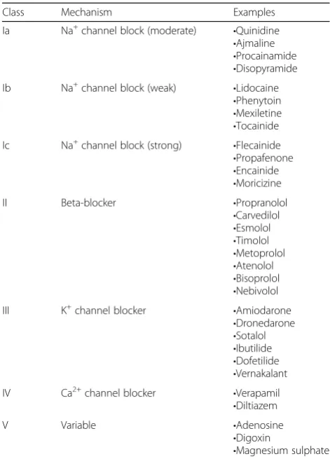
The drugs used as treatment for atrial fibrillation and atrial flutter are classified according to two different classifications: the Vaughan Williams classification and the Sicilian Gambit classification.
The Vaughan Williams classification classifies the drugs in five different classes according to their general effect [38]. The Vaughan Williams classification is summarised in Table2.
The Sicilian Gambit classification places a greater focus on the underlying mechanism of the drugs and classifies each drug according to the effects on bio-cellular channels, receptors, and pumps. We will not describe this classification in detail but refer to the work by the European Society of
Cardiology [39].
We will in this systematic review use the Vaughan Williams classification as it is the most commonly used classification.
Digoxin
Digoxin (Vaughan Williams class V) is thought to in-crease the contractility of the heart (inotropic effect) and decreases the heart rate (chronotropic effect). The con-tractility of the heart is increased by inhibiting the Na
+
/K+ ATPase. The inhibited Na+/K+ATPase results in a higher concentration of intracellular calcium which leads to an increase in the left ventricular systolic function
Table 1The classification, mechanism, and examples of anticoagulants and antiplatelet drugs
Class Mechanism Examples
Anticoagulants Affect the coagulation cascade to prevent blood coagulation [112]
Vitamin K-dependent antagonists •Warfarin
Non-vitamin K-dependent antagonists
•Dabigatran •Rivaroxaban •Apixaban •Edoxaban Heparin
•Unfractionated heparin •Low-molecular-weight heparin
Antiplatelet drugs
Theoretically affect the migration and aggregation of platelets, consequently aiming at inhibiting thrombus formation [112]
[40]. The heart rate is decreased by inducing vagal activation which presumably slows down the conduction of the atrioventricular node, consequently lowering the heart rate [41].
The Atrial Fibrillation Follow-up Investigation of Rhythm Management (AFFIRM) trial showed that digoxin (used without beta-blockers or non-dihydropyridine calcium channel blockers) achieved rate control (rest
≤80 beats/min) in 58% of the patients given digoxin as first therapy [42].
Other medical interventions
Beta-blockers (Vaughan Williams class II) block the sympathetic activity in the atrioventricular node, conse-quently decreasing the heart rate [41]. The different beta-blockers are illustrated in Table 2. Beta-blocker monotherapy is often the recommended first-line ther-apy for heart rate control in atrial fibrillation and atrial flutter [43]. The AFFIRM trial showed that beta-blockers (with or without digoxin) achieved rate control (rest
≤80 beats/min) in 70% of the patients given beta-blockers as first therapy [42].
Non-dihydropyridine calcium channel blockers (Vaughan Williams class IV) slow the atrioventricular node
conduction by blocking calcium channels and thus lower the heart rate [41]. The main non-dihydropyridine calcium channel blockers are illustrated in Table 2. Non-dihydropyridine calcium channel blockers are reasonable in controlling the heart rate [44] but are not recommended in patients with concomitant heart failure because of their negative inotropic effects [45]. The AFFIRM trial showed that non-dihydropyridine calcium channel blockers (with or without digoxin achieved rate control (rest ≤80 beats/ min) in 54% of the patients given non-dihydropyridine cal-cium channel blockers as first therapy [42].
Amiodarone (Vaughan Williams class III) may also control the heart rate, as it exhibits beta and calcium channel blockade in addition to its antiarrhythmic activ-ity. However, amiodarone has extensive non-cardiac ad-verse events and is only used if other rate control drugs are not effective enough, not well tolerated, or contrain-dicated [16].
Sotalol (Vaughan Williams class III) may also control the heart rate, as it exhibits beta blockade in addition to its antiarrhythmic activity. However, sotalol may induce life-threatening arrhythmias such as torsades de pointes [46]. Accordingly, it is seldom used.
Why is it important to do this review?
Digoxin is widely used for heart rate control in patients with atrial fibrillation and atrial flutter. Guidelines recommend using digoxin as the primary drug for heart rate control in patients with atrial fibrillation and atrial flutter who have concomitant heart failure and reduced ejection fraction [16, 43, 47]. Digoxin is also recom-mended for acute heart rate control in patients with pre-served ejection fraction [16, 43, 47]. Nevertheless, a report from 2014 has shown a dramatic decrease in overall digoxin treatment visits in the USA, from 12.9 million visits in 1997 to 1.87 million visits in 2012 [48].
According to the newest guideline by the European Society of Cardiology, there are no head-to-head rando-mised trials of digoxin versus other medical interven-tions in relation to heart rate control in patients with atrial fibrillation or atrial flutter [43]. However, our group found several randomised trials by performing a preliminary search in Medical Literature Analysis and Retrieval System Online (MEDLINE) [49–52]. Siu et al. compared diltiazem, digoxin, and amiodarone and showed superior rate control in the diltiazem group [49]. Joseph et al. compared sotalol, amiodarone, and digoxin and showed superior rate control in the sotalol group [50]. Tisdale et al. compared diltiazem with digoxin and showed superior rate control in the diltiazem group [51]. Tse et al. compared digoxin with amioda-rone and showed similar rate control efficacy in the compared groups [52].
Table 2The Vaughan Williams classification
Class Mechanism Examples
Ia Na+channel block (moderate) •Quinidine •Ajmaline •Procainamide •Disopyramide Ib Na+channel block (weak) •Lidocaine
•Phenytoin •Mexiletine •Tocainide Ic Na+channel block (strong) •Flecainide
•Propafenone •Encainide •Moricizine
II Beta-blocker •Propranolol
•Carvedilol •Esmolol •Timolol •Metoprolol •Atenolol •Bisoprolol •Nebivolol III K+channel blocker •Amiodarone
•Dronedarone •Sotalol •Ibutilide •Dofetilide •Vernakalant IV Ca2+channel blocker •Verapamil
•Diltiazem
V Variable •Adenosine
•Digoxin
During recent years, systematic reviews of observa-tional studies have compared digoxin versus no digoxin (the latter participants usually receiving some other treatment for heart rate control) in patients with atrial fibrillation or atrial flutter and showed that digoxin seemed to increase the risk of all-cause mortality regard-less of concomitant heart failure [53–56]. A systematic review of both observational studies and randomised clinical trials showed similar findings on all-cause mor-tality when assessing the observational studies. However, they did not show any difference on all-cause mortality when assessing the randomised clinical trials. They reported that the observed association between digoxin and mortality in observational studies might be a result of confounding that cannot be mitigated by statistical adjustment [57].
In 2014, a systematic review of randomised trials assessed the drugs used for heart rate control and included 16 randomised clinical trials. Eight of them were head-to-head trials comparing digoxin with other medical heart rate control interventions. The authors reported that the data were inconclusive as to whether any one drug was safer or more effective than the others. However, the review authors did not use any method to deal with risks of random errors [58]. Therefore, it is still unclear whether digoxin offers more benefits and less harms compared with placebo, no intervention, or with other medical interventions in controlling the heart rate. No former systematic review comparing digoxin with placebo, no intervention, or with other medical interven-tions for atrial fibrillation and atrial flutter has taken account both risks of systematic errors and risks of ran-dom errors (Cochrane methodology, Trial Sequential Analysis, and the Grades of Recommendation, Assess-ment, DevelopAssess-ment, and Evaluation (GRADE) assess-ment). In the present systematic review, we will collect and present current evidence of digoxin versus placebo, no intervention, or with other medical interventions for atrial fibrillation and atrial flutter.
Objectives
This review has two objectives:
1. To assess the beneficial and harmful effects of digoxin compared with placebo or no intervention for atrial fibrillation and atrial flutter
2. To assess the beneficial and harmful effects of digoxin compared with other medical interventions for atrial fibrillation and atrial flutter
Methods
This systematic review protocol has been developed based on Preferred Reporting Items for Systematic
Reviews and Meta-Analysis Protocols (PRISMA-P)
guidelines for reporting systematic reviews evaluating healthcare interventions [59, 60]. A PRISMA-P checklist file is attached (Additional file 1).
Criteria for considering studies for this review
Types of studies
Randomised clinical trials irrespective of trial design, set-ting, publication status, publication year, and language. We will not specifically search for non-randomised stud-ies. However, if we during our literature searches identify non-randomised studies (quasi-randomised studies or observational studies) with adequate reports of harmful effects, then we will narratively report these results. By focusing on randomised clinical trials, we are aware that the present review will be biased towards focusing on benefits and less on harms. Trials that only include a subset of eligible participants will only be included if (1) separate data on the eligible participants are available or (2) the majority of participants are eligible. We will document difficult decisions in the review, and sensitiv-ity analyses will assess the impact of these decisions on the findings of the review. Randomised clinical trials will be included irrespective of the reporting of one of the outcomes in this protocol.
Types of participants
Patients with atrial fibrillation or atrial flutter. We will accept the definitions used by the trialists. Patients will be included irrespective of age, sex, and comorbidities.
Types of interventions
We will include four types of trials:
Digoxin compared with placebo Digoxin compared with no intervention
Digoxin added to a co-intervention compared with a similar co-intervention
Digoxin compared with any type of medical intervention other than digoxin (e.g. beta-blockers, non-dihydropyridine calcium channel blockers, ami-odarone, or sotalol)
We will as experimental intervention accept digoxin irrespective of dose, route of administration, and dur-ation of administrdur-ation.
All control interventions will be included irrespective of dose, route of administration, and duration.
Types of outcome measures
We will for all outcomes, beside heart rate control and conversion to sinus rhythm, use the trial results at maximal follow-up. We will for heart rate control and conversion to sinus rhythm use the trial results reported within 48 h to assess the acute effects of digoxin versus placebo, no intervention, or other medical interventions in patients with atrial fibrillation or atrial flutter. We will, additionally, conduct sensitivity analyses excluding all trials with short (less than 12 months) or long (more than 24 months) follow-up in the primary meta-analyses of the outcome results at maximal follow-up (i.e. all-cause mortality, serious adverse events, quality of life, heart failure, and stroke).
Primary outcomes
1. All-cause mortality.
2. Serious adverse events. We will define a serious adverse event as any untoward medical occurrence that resulted in death, was life-threatening, required hospitalisation or prolongation of existing hospital-isation, and resulted in persistent or significant disability or jeopardised the patient [61]. 3. Quality of life measured on any valid scale.
Secondary outcomes
1. Heart failure (as defined by the trialists) 2. Stroke (as defined by the trialists) 3. Heart rate control
4. Conversion to sinus rhythm (as defined by the trialists)
All outcomes, except quality of life and heart rate control, will be analysed as proportions of participants in each group.
Search methods for identification of studies
Electronic searches
We will search the Cochrane Central Register of Controlled Trials (CENTRAL), MEDLINE, EMBASE, LILACS, Science Citation Index Expanded on Web of Science, and BIOSIS in order to identify relevant trials. The preliminary search strategy for MEDLINE (Ovid) is given in Additional file 2.
We will search all databases from their inception to the present, and we will impose no restriction on language of publication or publication status.
Searching other resources
The reference lists of relevant publications will be checked for any unidentified randomised trials. We will
contact authors of included studies, and major
pharmaceutical companies, by email asking for unpub-lished randomised trials. Further, we will search for ongoing trials on:
ClinicalTrials.gov (www.clinicaltrials.gov); Google Scholar (https://scholar.google.dk/);
The Turning Research into Practice (TRIP) Database (https://ww.tripdatabase.com/);
European Medicines Agency (EMA) (http:// www.ema.europa.eu/ema/);
United States Food and Drug Administration (FDA) (www.fda.gov);
China Food and Drug Administration (CFDA) (http://eng.sfda.gov.cn/WS03/CL0755/); Medicines and Healthcare products Regulatory
Agency
(https://www.gov.uk/government/organisations/ medicines-and-healthcare-products-regulatory-agency);
The World Health Organization (WHO) International Clinical Trials Registry Platform (ICTRP) search portal (http://apps.who.int/ trialsearch/).
Additionally, we will hand search conference abstracts from cardiology conferences for relevant trials.
We will also include unpublished and grey literature trials if we identify these, and assess relevant retraction statements and errata for included studies.
Data collection and analysis
We will perform the review following the recommenda-tions of Cochrane [62]. The analyses will be performed using Review Manager 5.3 [63] and Trial Sequential Analysis ver. 0.0.5.5 beta [64]. In case of Review Man-ager statistical software not being sufficient, we will use STATA 14 [65].
Selection of studies
Two authors (NJS and SS) will independently screen titles and abstracts. We will retrieve all relevant full-text study reports/publications, and two review authors (NJS and SS) will independently screen the full-text and iden-tify and record reasons for exclusion of the ineligible studies. We will resolve any disagreement through discussion, or if required, we will consult a third person (JCJ). Trial selection will be displayed in an adapted flow diagram as per the Preferred Reporting Items for Systematic Reviews and Meta-Analyses (PRISMA) state-ment [66].
Data extraction and management
will be resolved by discussion with a fifth author (JCJ). We will assess duplicate publications and companion pa-pers of a trial together in order to evaluate all available data simultaneously (maximise data extraction, correct bias assessment). We will contact the trial authors by email to specify any additional data, which may not have been reported sufficiently or at all in the publication.
Trial characteristics
Bias risk components (as defined below); trial design (parallel, factorial, or crossover); number of intervention arms; length of follow-up; estimation of sample size; inclusion and exclusion criteria.
Participants’characteristics and diagnosis
Number of randomised participants; number of analysed participants; number of participants lost to follow-up/ withdrawals/crossover; compliance with medication; age range (mean or median) and sex ratio; type of arrhythmia (atrial fibrillation or atrial flutter); baseline numbers of cardiovascular risk factors (i.e. diabetes mel-litus, hypertension, hyperlipidaemia, or smoking); base-line number of participants with heart failure; basebase-line number of participants with valvular heart disease; base-line number of participants with previous myocardial in-farction; baseline number of participants with previous revascularisation; and baseline number of participants with previous angina.
Intervention characteristics
Dose of intervention, duration of intervention, and mode of administration.
Control characteristics
Type of control intervention, dose of intervention, duration of intervention, and mode of administration.
Co-intervention characteristics
Type of co-intervention, dose of co-intervention, dur-ation of co-intervention, and mode of administrdur-ation.
Outcomes
All outcomes listed above will be extracted from each randomised clinical trial, and we will identify if out-comes are incomplete or selectively reported according to the criteria described later in ‘incomplete outcome data’ bias domain and‘selective outcome reporting’ bias domain.
Notes
Funding of the trial and notable conflicts of interest of trial authors will be extracted, if available.
We will note in the ‘Characteristics of included stud-ies’ table if outcome data were not reported in a usable
way. Two review authors (NJS and SS) will independ-ently transfer data into the Review Manager file [63]. Disagreements will be resolved through discussion, or if required, we will consult with a third author (JCJ).
Assessment of risk of bias in included studies
We will use the instructions given in the Cochrane Handbook for Systematic Reviews of Interventions [62] in our evaluation of the methodology and hence the risk of bias of the included trials. We will evaluate the meth-odology in respect of:
Random sequence generation Allocation concealment
Blinding of participants and personnel Blinding of outcome assessment Incomplete outcome data Selective outcome reporting Other risks of bias
Overall risk of bias
These domains enable classification of randomised trials with low risk of bias and high risk of bias. The latter trials tend to overestimate positive intervention effects (benefits) and underestimate negative effects (harms) [67–73].
We will classify the trials according to the following criteria:
Random sequence generation
Low risk: If sequence generation was achieved using computer random number generator or a random number table. Drawing lots, tossing a coin, shuffling cards, and throwing dice were also considered adequate if performed by an independent adjudicator.
Unclear risk: If the method of randomisation was not specified, but the trial was still presented as being randomised.
High risk: If the allocation sequence is not randomised or only quasi-randomised. These trials will be excluded.
Allocation concealment
Low risk: If the allocation of patients was performed by a central independent unit, on-site locked com-puter, identical-looking numbered sealed envelopes, drug bottles, or containers prepared by an independ-ent pharmacist or investigator.
Uncertain risk: If the trial was classified as
High risk: If the allocation sequence was familiar to the investigators who assigned participants.
Blinding of participants and personnel
Low risk: If the participants and the personnel were blinded to intervention allocation and this was described.
Uncertain risk: If the procedure of blinding was insufficiently described.
High risk: If blinding of participants and the personnel was not performed.
Blinding of outcome assessment
Low risk of bias: If it was mentioned that outcome assessors were blinded and this was described. Uncertain risk of bias: If it was not mentioned if the
outcome assessors in the trial were blinded, or the extent of blinding was insufficiently described. High risk of bias: If no blinding or incomplete blinding of outcome assessors was performed.
Incomplete outcome data
Low risk of bias: If missing data were unlikely to make treatment effects depart from plausible values. This could either be (1) there were no drop-outs or withdrawals for all outcomes or (2) the numbers and reasons for the withdrawals and drop-outs for all outcomes were clearly stated and could be described as being similar to both groups. Generally, the trial is judged as at a low risk of bias due to incomplete outcome data if drop-outs are less than 5%. How-ever, the 5% cut-off is not definitive.
Uncertain risk of bias: If there was insufficient information to assess whether missing data were likely to induce bias on the results.
High risk of bias: If the results were likely to be biased due to missing data either because the pattern of drop-outs could be described as being dif-ferent in the two intervention groups or the trial used improper methods in dealing with the missing data (e.g. last observation carried forward).
Selective outcome reporting
Low risk of bias: If a protocol was published before or at the time the trial was begun and the outcomes specified in the protocol were reported on. If there is no protocol or the protocol was published after the trial has begun, reporting of all-cause mortality and serious adverse events will grant the trial a grade of low risk of bias.
Uncertain risk of bias: If no protocol was published and the outcomes all-cause mortality and serious adverse events were not reported on.
High risk of bias: If the outcomes in the protocol were not reported on.
Other risks of bias
Low risk of bias: If the trial appears to be free of other components (for example, academic bias or for-profit bias) that could put it at risk of bias. Unclear risk of bias: If the trial may or may not be
free of other components that could put it at risk of bias.
High risk of bias: If there are other factors in the trial that could put it at risk of bias (for example, authors conducted trials on the same topic, for-profit bias, etc.).
Overall risk of bias
Low risk of bias: The trial will be classified as overall ‘low risk of bias’only if all of the bias domains described in the above paragraphs are classified as ‘low risk of bias’.
High risk of bias: The trial will be classified‘high risk of bias’if any of the bias risk domains described in the above are classified as‘unclear’or‘high risk of bias’.
We will assess the domains ‘blinding of outcome assessment’,‘incomplete outcome data’, and‘selective out-come reporting’ for each outcome result. Thus, we will assess the bias risk for each outcome assessed in addition to each trial. Our primary conclusions will be based on the results of both our primary and secondary outcome results with overall low risk of bias. If no results with overall low risk of bias are found, we will base our primary conclusions on the outcome results irrespective of overall risk of bias. We will present our primary conclusions in the‘Summary of Findings’table.
Differences between the protocol and the review
We will conduct the review according to this published protocol and report any deviations from it in the‘ Differ-ences between the protocol and the review’ section of the systematic review.
Measures of treatment effect
Dichotomous outcomes We will calculate risk ratios
Continuous outcomes We will calculate the mean dif-ferences (MDs) and if necessary, as a hypothesis generat-ing analysis, the standardised mean difference (SMD) with 95% CI for continuous outcomes. We will also report Trial Sequential Analysis-adjusted CIs (see para-graphs below).
Dealing with missing data
We will, as first option, contact all trial authors to obtain any relevant missing information and data (i.e. for data extraction and for assessment of risk of bias, as specified above).
Dichotomous outcomes We will not impute missing
values for any outcomes in our primary analysis. In two of our sensitivity analyses (see paragraph below), we will impute data.
Continuous outcomes We will primarily analyse scores
assessed at single time points. If only change from base-line scores are reported, we will analyse the results to-gether with follow-up scores [62]. If standard deviations (SDs) are not reported, we will calculate the SDs using trial data, if possible. We will not use intention-to-treat data if the original report did not contain such data. We will not impute missing values for any outcomes in our primary analysis. In our sensitivity analysis (see para-graph below) for continuous outcomes, we will impute data.
Assessment of heterogeneity
We will primarily investigate forest plots to visually assess any sign of heterogeneity. We will secondly assess the presence of statistical heterogeneity by chi2 test (threshold P< 0.10) and measure the quantities of het-erogeneity by theI2-statistic [74, 75].
We will follow the recommendations for threshold by theCochrane Handbook for Systemic Reviews of Interven-tions[62]:
0 to 40%: might not be important
30 to 60%: may represent moderate heterogeneity 50 to 90%: may represent substantial heterogeneity 75 to 100%: may represent considerable
heterogeneity
We will investigate possible heterogeneity through subgroup analyses. Ultimately, we may decide that a meta-analysis should be avoided [62].
Assessment of reporting biases
We will use a funnel plot to assess reporting bias if ten or more trials are included. We will visually inspect fun-nel plots to assess the risk of bias. We are aware of the
limitations of a funnel plot, i.e. a funnel plot assesses bias due to small sample size and asymmetry of a funnel plot is not necessarily caused by reporting bias. From this information, we assess possible reporting bias. For dichotomous outcomes, we will test asymmetry with the Harbord test [76] if τ2 is less than 0.1 and with the Rücker test if τ2 is more than 0.1. For continuous out-comes, we will use the regression asymmetry test [77] and the adjusted rank correlation [78].
Unit of analysis issues
We will only include randomised clinical trials. For trials using crossover design, only data from the first period will be included [62, 79]. There will therefore not be any unit of analysis issues. We will not include cluster ran-domised trials, as these have a high risk of biased results due to confounding [62].
Data synthesis
Meta-analysis
We will undertake this meta-analysis according to the recommendations stated in the Cochrane Handbook for Systematic Reviews of Interventions[62], Keus et al. [80], and the eight-step assessment suggested by Jakobsen et al. for better validation of meta-analytic results in sys-tematic reviews [81]. We will use the statistical software Review Manager 5.3 [63] provided by Cochrane to analyse data.
are crossed [81]. Our primary conclusion will be based on results with low risk of bias [81].
Where multiple trial intervention groups are reported in a single trial, we will include only the relevant groups. If two comparisons are combined in the same meta-analysis, we will halve the control group to avoid double-counting [81, 85].
Trials with a factorial design will be included. In case of, e.g. a 2 × 2 factorial designed trial, the two groups receiving digoxin will be considered experimental groups, while the two groups receiving other medical rate control interventions will be considered control groups.
If quantitative synthesis is not appropriate, we will report the results in a narrative way.
Trial Sequential Analysis
Cumulative meta-analyses are at risk of producing ran-dom errors due to sparse data and multiple testing of ac-cumulating data [64, 85–94]. Therefore, Trial Sequential Analysis [64] can be applied to control these risks (http://www.ctu.dk/tsa/) [91]. Similar to a sample size calculation in a randomised clinical trial, Trial Sequential Analysis estimates the required information size (that is the number of participants needed in a meta-analysis to detect or reject a certain intervention effect) in order to minimise random errors [89]. The required information size takes into account the anticipated intervention effect, the variance of the anticipated difference in inter-vention effects, the acceptable risk of falsely rejecting the null hypothesis (alpha), the acceptable risk of falsely confirming the null hypothesis (beta), and the vari-ance of the intervention effect estimated between tri-als [81, 89, 95]. We tried to base the anticipated intervention effects on empirical data [81]. However, no suitable empirical data could be found, and in-stead, we pragmatically hypothesised the anticipated intervention effects. When analysing dichotomous outcomes, we pragmatically anticipated an interven-tion effect of 15% relative risk reducinterven-tion. When
analysing continuous outcomes, we pragmatically
anticipated an intervention effect equal to the mean difference of the observed SD/2 [96].
Trial Sequential Analysis enables testing for signifi-cance to be conducted each time a new trial is included in the meta-analysis. On the basis of the required infor-mation size, trial sequential monitoring boundaries are constructed. This enables one to determine the statistical inference concerning cumulative meta-analysis that has not yet reached the diversity-adjusted required informa-tion size [86, 89].
Firm evidence for benefit or harm may be established if a trial sequential monitoring boundary is crossed before reaching the diversity-adjusted required information size,
in which case further trials may turn out to be superflu-ous. In contrast, if a boundary is not surpassed, one may conclude that it is necessary to continue with further trials before a certain intervention effect can be detected or rejected. Firm evidence for lack of the postulated interven-tion effect can also be assessed with Trial Sequential Ana-lysis. This occurs when the cumulative Z-score crosses the trial sequential boundaries for futility.
For dichotomous outcomes, we will estimate the required information size based on a relative risk reduc-tion of 15%, the observed proporreduc-tion of participants with an outcome in the control group, an alpha of 2.5% for our primary outcomes and 2.0% for our secondary out-comes (see the ‘Meta-analysis’ section), a beta of 10%, and diversity as suggested by the trials in the meta-analysis (diversity-adjusted required information size) [81, 89]. Additionally, we will calculate Trial Sequential Analysis-adjusted CIs.
For continuous outcomes, we will estimate the required information size based on a minimal clinically important difference of SD/2, the standard deviation observed in the control group, an alpha of 2.5% for our primary outcomes and 2.0% for our secondary outcomes (see ‘ Meta-ana-lysis’), a beta of 10%, and a diversity as suggested by the trials in the meta-analysis (diversity adjusted required in-formation size) [81, 89]. Additionally, we will calculate Trial Sequential Analysis-adjusted CIs.
Subgroup analysis and investigation of heterogeneity
Subgroup analysis
We will perform the following subgroup analyses when analysing the primary outcomes (all-cause mortality, serious adverse event, and quality of life).
1. High risk of bias trials compared to low risk of bias trials
2. Comparison of different types of control interventions
3. Participants with atrial fibrillation compared to participants with atrial flutter
4. Age of participants: 0 to 59 years, 60 to 79 years, and above 80 years
5. Duration of atrial fibrillation: recent-onset atrial fibrillation (as defined by the trialists), paroxysmal atrial fibrillation (less than 7 days of onset), persist-ent atrial fibrillation (more than 7 days and less than 1 year of onset), and long-standing persistent atrial fibrillation (more than 1 year of onset).
6. Men compared to women
7. Participants with heart failure compared to participants without heart failure
We will use the formal test for subgroup differences in Review Manager [63].
Sensitivity analysis
To assess the potential impact of the missing data for dichotomous outcomes, we will perform the two follow-ing sensitivity analyses on both the primary and second-ary outcomes.
‘Best-worst-case’scenario: We will assume that all participants lost to follow-up in the experimental group have survived, had no serious adverse event, had no heart failure, had no stroke, and converted to sinus rhythm and that all those participants lost to follow-up in the control group have not survived, had a serious adverse event, had a heart failure, had a stroke, and did not convert to sinus rhythm. ‘Worst-best-case’scenario: We will assume that all
participants lost to follow-up in the experimental group have not survived, had a serious adverse event, had a heart failure, had a stroke, and did not convert to sinus rhythm and that all those partici-pants lost to follow-up in the control group have survived, had no serious adverse event, had no heart failure, had no stroke, and converted to sinus rhythm.
We will present results of both scenarios in our review.
When analysing quality of life or heart rate control, a ‘beneficial outcome’ will be the group mean plus two standard deviations (SDs) (we will secondly use one SD in another sensitivity analysis) of the group mean and a ‘harmful outcome’ will be the group mean minus two SDs (we will secondly use one SD in another sensitivity analysis) of the group mean [81].
To assess the potential impact of missing SDs for con-tinuous outcomes, we will perform the following sensi-tivity analysis.
Where SDs are missing and it is not possible to calculate them, we will impute SDs from trials with similar populations and low risk of bias. If we find no such trials, we will impute SDs from trials with a similar population.
We will present results of this scenario in our review. Other post hoc sensitivity analyses might be warranted if unexpected clinical or statistical heterogeneity is iden-tified during the analysis of the review results [81].
‘Summary of Findings’table
We will create a‘Summary of Findings’table using each of the prespecified primary and secondary outcomes
(all-cause mortality, serious adverse event, quality of life, stroke, heart failure, heart rate control, and conversion to sinus rhythm). We will use the five GRADE consider-ations (bias risk of the trials, consistency of effect, im-precision, indirectness, and publication bias) to assess the quality of a body of evidence as it relates to the stud-ies which contribute data to the meta-analyses for the prespecified outcomes [81, 97–99]. We will use methods and recommendations described in Chapter 8 (Section 8.5) and Chapter 12 of the Cochrane Handbook for Sys-tematic Reviews of Interventions [62] using GRADEpro software. We will justify all decisions to downgrade the quality of studies using footnotes, and we will make comments to aid the reader’s understanding of the review where necessary. Firstly, we will present our results in the ‘Summary of Findings’ table based on the results from the trials with low risk of bias; secondly, we will present the results based on all trials.
Discussion
Observational evidence has indicated that digoxin might increase the risk of serious adverse events in patients with atrial fibrillation—on the other hand, digoxin is a recommended and commonly used intervention. More-over, clinical guidelines postulate that no head-to-head randomised clinical trials of digoxin versus other med-ical interventions in relation to heart rate control in pa-tients with atrial fibrillation or atrial flutter exists—still, we can identify several trials on the topic by searching PubMed. The present systematic review aims at compar-ing digoxin with placebo, no intervention, or with other medical interventions in patients with atrial fibrillation and atrial flutter. The outcomes will be all-cause mortal-ity, serious adverse events, quality of life, stroke, heart failure, heart rate control, and conversion to sinus rhythm. Due to the millions affected around the World, there is an urgent need of this systematic review.
This protocol has a number of strengths. The prede-fined methodology is based on the Cochrane Handbook for Systematic Reviews of Interventions [62], the eight-step assessment suggested by Jakobsen et al. [81], Trial Sequential Analysis [64], and GRADE assessment [97– 99]. Hence, this protocol takes into account both risks of systematic errors and risk of random errors.
we do not expect to identify many relevant randomised clinical trials and we want to increase the statistical power. We have predefined a number of subgroup ana-lyses and sensitivity anaana-lyses to assess if the effects differ between trials using different types of control interven-tions, and we will thoroughly consider if it is valid to perform meta-analysis of all included trials. A further limitation is the large number of subgroup analyses which increases the risk of a type 1 error. We have adjusted our thresholds for significance according to the number of primary outcomes, but we have also included multiple subgroup analyses and assess all outcomes at two time points. The large risk of type 1 error will be taken into account when interpreting the results of the review.
Additional files
Additional file 1:PRISMA-P 2015 checklist. (DOCX 30 kb)
Additional file 2:The preliminary search strategy for MEDLINE (Ovid).
(PDF 221 kb)
Abbreviations
AFFIRM:The Atrial Fibrillation Follow-up Investigation of Rhythm Management; CENTRAL: The Cochrane Central Register of Controlled Trials; CFDA: China Food and Drug Administration; CI: Confidence interval; EMA: European Medicines Agency; EMBASE: Excerpta Medica database; GRADE: The Grading of Recommendations Assessment Development, and Evaluation; LILACS: Latin American and Caribbean Health Sciences Literature; MD: Mean difference; MEDLINE: Medical Literature Analysis and Retrieval System Online; PRISMA: Preferred Reporting Items for Systematic Reviews and Meta-Analyses; PRISMA-P: Preferred Reporting Items for Systematic Reviews and Meta-Analysis Protocols; PROSPERO: International Prospective Register of Systematic Reviews; RR: Risk ratio; SMD: Standardised mean difference; TRIP: Turning Research into Practice; WHO: World Health Organization
Acknowledgements
The expert help from Sarah Louise Klingenberg (Information Specialist, The Cochrane Hepato-Biliary Group, Copenhagen Trial Unit, Copenhagen, Denmark) in making the search strategy is hugely appreciated. We thankSystematic Reviewsto provide us with the PRISMA-P checklist to optimise the protocol. The PRISMA-P checklist is given in Additional file 1.
Funding
The Copenhagen Trial Unit, Centre for Clinical Intervention Research, paid the salary for all authors during the writing of this review. Otherwise, we have received no funding.
Availability of data and materials
Not applicable.
Authors’contributions
NJS drafted the protocol. JCJ, SS, JF, EEN, and CG amended the protocol. All authors read and approved the final manuscript.
Competing interests
The authors declare that they have no competing interests.
Consent for publication
Not applicable.
Ethics approval and consent to participate
Not applicable.
Sources of support
The making of this protocol was supported by Copenhagen Trial Unit, Centre for Clinical Intervention Research, Rigshospitalet, Copenhagen, Denmark.
Publisher’s Note
Springer Nature remains neutral with regard to jurisdictional claims in published maps and institutional affiliations.
Author details 1
Copenhagen Trial Unit, Centre for Clinical Intervention Research, Department 7812, Rigshospitalet, Copenhagen University Hospital, Copenhagen, Denmark.2The Cochrane Hepato-Biliary Group, Copenhagen Trial Unit, Centre for Clinical Intervention Research, Department 7812, Rigshospitalet, Copenhagen University Hospital, Copenhagen, Denmark.
3Department of Cardiology, Holbæk Hospital, Holbæk, Denmark.
Received: 21 December 2016 Accepted: 28 March 2017
References
1. Pistoia F, Sacco S, Tiseo C, Degan D, Ornello R, Carolei A. The epidemiology of atrial fibrillation and stroke. Cardiol Clin. 2016;34(2):255–68.
2. Camm AJ, Lip GY, De Caterina R, et al. 2012 focused update of the ESC Guidelines for the management of atrial fibrillation: an update of the 2010 ESC Guidelines for the management of atrial fibrillation. Europace. 2012; 14(10):1385–413.
3. Granada J, Uribe W, Chyou PH, et al. Incidence and predictors of atrial flutter in the general population. J Am Coll Cardiol. 2000;36(7):2242–6. 4. Chugh SS, Havmoeller R, Narayanan K, et al. Worldwide epidemiology of
atrial fibrillation: a global burden of disease 2010 study. Circulation. 2014; 129(8):837–47.
5. Fitzmaurice DA, Hobbs FD, Jowett S, et al. Screening versus routine practice in detection of atrial fibrillation in patients aged 65 or over: cluster randomised controlled trial. BMJ. 2007;335(7616):383.
6. Stewart S, Hart CL, Hole DJ, McMurray JJ. A population-based study of the long-term risks associated with atrial fibrillation: 20-year follow-up of the Renfrew/Paisley study. Am J Med. 2002;113(5):359–64.
7. Benjamin EJ, Wolf PA, D’Agostino RB, Silbershatz H, Kannel WB, Levy D. Impact of atrial fibrillation on the risk of death: the Framingham Heart Study. Circulation. 1998;98(10):946–52.
8. Rahman F, Wang N, Yin X, et al. Atrial flutter: clinical risk factors and adverse outcomes in the Framingham Heart Study. Heart Rhythm.
2016;13(1):233–40.
9. Healey JS, Oldgren J, Ezekowitz M, et al. Occurrence of death and stroke in patients in 47 countries 1 year after presenting with atrial fibrillation: a cohort study. Lancet. 2016;388(10050):1161–9.
10. Wolf PA, Abbott RD, Kannel WB. Atrial fibrillation as an independent risk factor for stroke: the Framingham Study. Stroke. 1991;22(8):983–8. 11. Odutayo A, Wong CX, Hsiao AJ, Hopewell S, Altman DG, Emdin CA. Atrial
fibrillation and risks of cardiovascular disease, renal disease, and death: systematic review and meta-analysis. BMJ. 2016;354:i4482.
12. Go AS, Hylek EM, Phillips KA, et al. Prevalence of diagnosed atrial fibrillation in adults: national implications for rhythm management and stroke prevention: the Anticoagulation and Risk Factors in Atrial Fibrillation (ATRIA) Study. JAMA. 2001;285(18):2370–5.
13. Stewart S, Murphy NF, Walker A, McGuire A, McMurray JJ. Cost of an emerging epidemic: an economic analysis of atrial fibrillation in the UK. Heart. 2004;90(3):286–92.
14. Mozaffarian D, Benjamin EJ, Go AS, et al. Heart disease and stroke statistics—2016 update: a report from the American Heart Association. Circulation. 2016;133(4):e38–360.
15. Calkins H, Kuck KH, Cappato R, et al. 2012 HRS/EHRA/ECAS expert consensus statement on catheter and surgical ablation of atrial fibrillation:
recommendations for patient selection, procedural techniques, patient management and follow-up, definitions, endpoints, and research trial design. Europace. 2012;14(4):528–606.
Guidelines and the Heart Rhythm Society. J Am Coll Cardiol. 2014;64(21):e1–76.
17. Camm AJ, Kirchhof P, Lip GY, et al. Guidelines for the management of atrial fibrillation: the task force for the management of atrial fibrillation of the European Society of Cardiology (ESC). Europace. 2010;12(10):1360–420. 18. Wyse DG. Therapeutic considerations in applying rate control therapy for
atrial fibrillation. J Cardiovasc Pharmacol. 2008;52(1):11–7.
19. Daoud EG, Weiss R, Bahu M, et al. Effect of an irregular ventricular rhythm on cardiac output. Am J Cardiol. 1996;78(12):1433–6.
20. Gopinathannair R, Etheridge SP, Marchlinski FE, Spinale FG, Lakkireddy D, Olshansky B. Arrhythmia-induced cardiomyopathies: mechanisms, recognition, and management. J Am Coll Cardiol. 2015;66(15):1714–28. 21. Lip GY, Fauchier L, Freedman SB, et al. Atrial fibrillation. Nat Rev Dis Primers.
2016;2:16016.
22. Page RL, Joglar JA, Caldwell MA, et al. 2015 ACC/AHA/HRS guideline for the management of adult patients with supraventricular tachycardia: a report of the American College of Cardiology/American Heart Association Task Force on Clinical Practice Guidelines and the Heart Rhythm Society. J Am Coll Cardiol. 2016;67(13):e27–115.
23. Fauchier L, Philippart R, Clementy N, et al. How to define valvular atrial fibrillation? Arch Cardiovasc Dis. 2015;108(10):530–9.
24. De Caterina R, Camm AJ. What is‘valvular’atrial fibrillation? A reappraisal. Eur Heart J. 2014;35(47):3328–35.
25. Markides V, Schilling RJ. Atrial fibrillation: classification, pathophysiology, mechanisms and drug treatment. Heart. 2003;89(8):939–43.
26. Saoudi N, Cosio F, Waldo A, et al. Classification of atrial flutter and regular atrial tachycardia according to electrophysiologic mechanism and anatomic bases: a statement from a joint expert group from the Working Group of Arrhythmias of the European Society of Cardiology and the North American Society of Pacing and Electrophysiology. J Cardiovasc Electrophysiol. 2001; 12(7):852–66.
27. Haissaguerre M, Jais P, Shah DC, et al. Spontaneous initiation of atrial fibrillation by ectopic beats originating in the pulmonary veins. NEJM. 1998; 339(10):659–66.
28. Aviles RJ, Martin DO, Apperson-Hansen C, et al. Inflammation as a risk factor for atrial fibrillation. Circulation. 2003;108(24):3006–10.
29. Burstein B, Nattel S. Atrial fibrosis: mechanisms and clinical relevance in atrial fibrillation. J Am Coll Cardiol. 2008;51(8):802–9.
30. Morillo CA, Klein GJ, Jones DL, Guiraudon CM. Chronic rapid atrial pacing. Structural, functional, and electrophysiological characteristics of a new model of sustained atrial fibrillation. Circulation. 1995;91(5):1588–95. 31. Wijffels MC, Kirchhof CJ, Dorland R, Allessie MA. Atrial fibrillation begets
atrial fibrillation. A study in awake chronically instrumented goats. Circulation. 1995;92(7):1954–68.
32. Lip GY, Nieuwlaat R, Pisters R, Lane DA, Crijns HJ. Refining clinical risk stratification for predicting stroke and thromboembolism in atrial fibrillation using a novel risk factor-based approach: the euro heart survey on atrial fibrillation. Chest. 2010;137(2):263–72.
33. Pisters R, Lane DA, Nieuwlaat R, de Vos CB, Crijns HJ, Lip GY. A novel user-friendly score (HAS-BLED) to assess 1-year risk of major bleeding in patients with atrial fibrillation: the Euro Heart Survey. Chest. 2010;138(5):1093–100. 34. Connolly SJ, Eikelboom J, Joyner C, et al. Apixaban in patients with atrial
fibrillation. NEJM. 2011;364(9):806–17.
35. Shi XX, Ren GH, Wang J, et al. Effectiveness and safety of warfarin and anti-platelet drugs for the primary prevention of stroke in patients with non-valvular atrial fibrillation: a meta-analysis. Int J Clin Exp Med. 2015;8(6):8384–97. 36. Ruff CT, Giugliano RP, Braunwald E, et al. Comparison of the efficacy and safety
of new oral anticoagulants with warfarin in patients with atrial fibrillation: a meta-analysis of randomised trials. Lancet. 2014;383(9921):955–62. 37. Shinbane JS, Wood MA, Jensen DN, Ellenbogen KA, Fitzpatrick AP,
Scheinman MM. Tachycardia-induced cardiomyopathy: a review of animal models and clinical studies. J Am Coll Cardiol. 1997;29(4):709–15. 38. Vaughan Williams EM. A classification of antiarrhythmic actions reassessed
after a decade of new drugs. J Clin Pharmacol. 1984;24(4):129–47. 39. Task Force of The Working Group on Arrhythmias of the European Society
of Cardiology. The Sicilian gambit. A new approach to the classification of antiarrhythmic drugs based on their actions on arrhythmogenic mechanisms. Circulation. 1991;84(4):1831–51.
40. Hauptman PJ, Kelly RA. Digitalis. Circulation. 1999;99(9):1265–70. 41. Van Gelder IC, Rienstra M, Crijns HJ, Olshansky B. Rate control in atrial
fibrillation. Lancet. 2016;388(10046):818–28.
42. Olshansky B, Rosenfeld LE, Warner AL, et al. The Atrial Fibrillation Follow-up Investigation of Rhythm Management (AFFIRM) study: approaches to control rate in atrial fibrillation. J Am Coll Cardiol. 2004;43(7):1201–8. 43. Kirchhof P, Benussi S, Kotecha D, et al. 2016 ESC guidelines for the
management of atrial fibrillation developed in collaboration with EACTS. Europace. 2016;18(11):1609–78.
44. Ulimoen SR, Enger S, Carlson J, et al. Comparison of four single-drug regimens on ventricular rate and arrhythmia-related symptoms in patients with permanent atrial fibrillation. Am J Cardiol. 2013;111(2):225–30. 45. Goldstein RE, Boccuzzi SJ, Cruess D, Nattel S. Diltiazem increases late-onset
congestive heart failure in postinfarction patients with early reduction in ejection fraction. The Adverse Experience Committee; and the Multicenter Diltiazem Postinfarction Research Group. Circulation. 1991;83(1):52–60. 46. Kuck KH, Kunze KP, Roewer N, Bleifeld W. Sotalol-induced torsade de
pointes. Am Heart J. 1984;107(1):179–80.
47. Ponikowski P, Voors AA, Anker SD, et al. 2016 ESC guidelines for the diagnosis and treatment of acute and chronic heart failure: the task force for the diagnosis and treatment of acute and chronic heart failure of the European Society of Cardiology (ESC). Developed with the special contribution of the Heart Failure Association (HFA) of the ESC. Eur Heart J. 2016;37(27):2129–200.
48. Goldberger ZD, Alexander GC. Digitalis use in contemporary clinical practice: refitting the foxglove. JAMA Intern Med. 2014;174(1):151–4. 49. Siu CW, Lau CP, Lee WL, Lam KF, Tse HF. Intravenous diltiazem is superior to
intravenous amiodarone or digoxin for achieving ventricular rate control in patients with acute uncomplicated atrial fibrillation. Crit Care Med. 2009; 37(7):2174–9. quiz 80.
50. Joseph AP, Ward MR. A prospective, randomized controlled trial comparing the efficacy and safety of sotalol, amiodarone, and digoxin for the reversion of new-onset atrial fibrillation. Ann Emerg Med. 2000;36(1):1–9.
51. Tisdale JE, Padhi ID, Goldberg AD, et al. A randomized, double-blind comparison of intravenous diltiazem and digoxin for atrial fibrillation after coronary artery bypass surgery. Am Heart J. 1998;135(5 Pt 1):739–47. 52. Tse HF, Lam YM, Lau CP, Cheung BM, Kumana CR. Comparison of
digoxin versus low-dose amiodarone for ventricular rate control in patients with chronic atrial fibrillation. Clin Exp Pharmacol Physiol. 2001; 28(5–6):446–50.
53. Ouyang AJ, Lv YN, Zhong HL, et al. Meta-analysis of digoxin use and risk of mortality in patients with atrial fibrillation. Am J Cardiol. 2015;115(7):901–6. 54. Wang ZQ, Zhang R, Chen MT, et al. Digoxin is associated with increased
all-cause mortality in patients with atrial fibrillation regardless of concomitant heart failure: a meta-analysis. J Cardiovasc Pharmacol. 2015;66(3):270–5. 55. Vamos M, Erath JW, Hohnloser SH. Digoxin-associated mortality: a
systematic review and meta-analysis of the literature. Eur Heart J. 2015; 36(28):1831–8.
56. Qureshi W, O’Neal WT, Soliman EZ, Al-Mallah MH. Systematic review and meta-analysis of mortality and digoxin use in atrial fibrillation. Cardiol J. 2016;23(3):333–43.
57. Ziff OJ, Lane DA, Samra M, et al. Safety and efficacy of digoxin: systematic review and meta-analysis of observational and controlled trial data. BMJ. 2015;351:h4451.
58. Al-Khatib SM, Allen LaPointe NM, Chatterjee R, et al. Rate- and rhythm-control therapies in patients with atrial fibrillation: a systematic review. Ann Intern Med. 2014;160(11):760–73.
59. Moher D, Shamseer L, Clarke M, et al. Preferred Reporting Items for Systematic Review and Meta-Analysis Protocols (PRISMA-P) 2015 statement. Syst Rev. 2015;4:1.
60. Shamseer L, Moher D, Clarke M, et al. Preferred Reporting Items for Systematic Review and Meta-Analysis Protocols (PRISMA-P) 2015: elaboration and explanation. BMJ. 2015;349:g7647.
61. International conference on harmonisation of technical requirements for registration of pharmaceuticals for human use (ICH) adopts consolidated guideline on good clinical practice in the conduct of clinical trials on medicinal products for human use. International Digest of Health Legislation. 1997;48(2):231–4.
62. Higgins J, Green S. Cochrane Handbook for Systematic Reviews of Interventions version 5.1.0 The Cochrane Collaboration. 2011. www. handbook.cochrane.org.
65. StataCorp. Stata: Release 14. Statistical Software. College Station, TX: StataCorp LP. 2014. http://www.stata.com.
66. Moher D, Liberati A, Tetzlaff J, Altman DG. Preferred Reporting Items for Systematic Reviews and Meta-Analyses: the PRISMA statement. Ann Intern Med. 2009;151(4):264–9. w64.
67. Gluud LL. Bias in clinical intervention research. Am J Epidemiol. 2006;163(6): 493–501.
68. Kjaergard LL, Villumsen J, Gluud C. Reported methodologic quality and discrepancies between large and small randomized trials in meta-analyses. Ann Intern Med. 2001;135(11):982–9.
69. Moher D, Pham B, Jones A, et al. Does quality of reports of randomised trials affect estimates of intervention efficacy reported in meta-analyses? Lancet. 1998;352(9128):609–13.
70. Schulz KF, Chalmers I, Hayes RJ, Altman DG. Empirical evidence of bias. Dimensions of methodological quality associated with estimates of treatment effects in controlled trials. JAMA. 1995;273(5):408–12. 71. Wood L, Egger M, Gluud LL, et al. Empirical evidence of bias in treatment
effect estimates in controlled trials with different interventions and outcomes: meta-epidemiological study. BMJ. 2008;336(7644):601–5. 72. Savovic J, Jones H, Altman D, et al. Influence of reported study design
characteristics on intervention effect estimates from randomised controlled trials: combined analysis of meta-epidemiological studies. Health Technol Assess. 2012;16(35):1–82.
73. Lundh A, Lexchin J, Mintzes B, Schroll JB, Bero L. Industry sponsorship and research outcome. Cochrane Database Syst Rev. 2017;2:Mr000033. 74. Higgins JP, Thompson SG. Quantifying heterogeneity in a meta-analysis.
Stat Med. 2002;21(11):1539–58.
75. Higgins JP, Thompson SG, Deeks JJ, Altman DG. Measuring inconsistency in meta-analyses. BMJ. 2003;327(7414):557–60.
76. Harbord RM, Egger M, Sterne JA. A modified test for small-study effects in meta-analyses of controlled trials with binary endpoints. Stat Med. 2006; 25(20):3443–57.
77. Egger M, Davey Smith G, Schneider M, Minder C. Bias in meta-analysis detected by a simple, graphical test. BMJ. 1997;315(7109):629–34. 78. Begg CB, Mazumdar M. Operating characteristics of a rank correlation test
for publication bias. Biometrics. 1994;50(4):1088–101.
79. Elbourne DR, Altman DG, Higgins JP, Curtin F, Worthington HV, Vail A. Meta-analyses involving cross-over trials: methodological issues. Int J Epidemiol. 2002;31(1):140–9.
80. Keus F, Wetterslev J, Gluud C, van Laarhoven CJ. Evidence at a glance: error matrix approach for overviewing available evidence. BMC Med Res Methodol. 2010;10:90.
81. Jakobsen JC, Wetterslev J, Winkel P, Lange T, Gluud C. Thresholds for statistical and clinical significance in systematic reviews with meta-analytic methods. BMC Med Res Methodol. 2014;14:120.
82. DerSimonian R, Laird N. Meta-analysis in clinical trials. Control Clin Trials. 1986;7(3):177–88.
83. Demets DL. Methods for combining randomized clinical trials: strengths and limitations. Stat Med. 1987;6(3):341–50.
84. Jakobsen JC, Wetterslev J, Lange T, Gluud C. Viewpoint: taking into account risks of random errors when analysing multiple outcomes in systematic reviews. Cochrane Database Syst Rev. 2016;3:Ed000111.
85. Brok J, Thorlund K, Gluud C, Wetterslev J. Trial sequential analysis reveals insufficient information size and potentially false positive results in many meta-analyses. J Clin Epidemiol. 2008;61(8):763–9.
86. Wetterslev J, Thorlund K, Brok J, Gluud C. Trial sequential analysis may establish when firm evidence is reached in cumulative meta-analysis. J Clin Epidemiol. 2008;61(1):64–75.
87. Brok J, Thorlund K, Wetterslev J, Gluud C. Apparently conclusive meta-analyses may be inconclusive—trial sequential analysis adjustment of random error risk due to repetitive testing of accumulating data in apparently conclusive neonatal meta-analyses. Int J Epidemiol. 2009;38(1): 287–98.
88. Thorlund K, Devereaux PJ, Wetterslev J, et al. Can trial sequential monitoring boundaries reduce spurious inferences from meta-analyses? Int J Epidemiol. 2009;38(1):276–86.
89. Wetterslev J, Thorlund K, Brok J, Gluud C. Estimating required information size by quantifying diversity in random-effects model meta-analyses. BMC Med Res Methodol. 2009;9:86.
90. Thorlund K, Anema A, Mills E. Interpreting meta-analysis according to the adequacy of sample size. An example using isoniazid chemoprophylaxis for
tuberculosis in purified protein derivative negative HIV-infected individuals. Clin Epidemiol. 2010;2:57–66.
91. Thorlund K EJ, Wetterslev J, Brok J, Imberger G, Gluud C. User manual for Trial Sequential Analysis (TSA). 2011. http://www.ctu.dk/tsa/files/tsa_manual. pdf.
92. Imberger G, Gluud C, Boylan J, Wetterslev J. Systematic reviews of anesthesiologic interventions reported as statistically significant: problems with power, precision, and type 1 error protection. Anesth Analg. 2015; 121(6):1611–22.
93. Imberger G, Thorlund K, Gluud C, Wetterslev J. False-positive findings in Cochrane meta-analyses with and without application of trial sequential analysis: an empirical review. BMJ Open. 2016;6(8):e011890.
94. Wetterslev J, Jakobsen JC, Gluud C. Trial Sequential Analysis in systematic reviews with meta-analysis. BMC Med Res Methodol. 2017;17(1):39. 95. Turner RM, Bird SM, Higgins JP. The impact of study size on meta-analyses:
examination of underpowered studies in Cochrane reviews. PLoS One. 2013; 8(3):e59202.
96. Norman GR, Sloan JA, Wyrwich KW. Interpretation of changes in health-related quality of life: the remarkable universality of half a standard deviation. Med Care. 2003;41(5):582–92.
97. Guyatt GH, Oxman AD, Vist GE, et al. GRADE: an emerging consensus on rating quality of evidence and strength of recommendations. BMJ. 2008; 336(7650):924–6.
98. Guyatt GH, Oxman AD, Schunemann HJ, Tugwell P, Knottnerus A. GRADE guidelines: a new series of articles in the Journal of Clinical Epidemiology. J Clin Epidemiol. 2011;64(4):380–2.
99. Schunemann HJ, Best D, Vist G, Oxman AD. Letters, numbers, symbols and words: how to communicate grades of evidence and recommendations. CMAJ. 2003;169(7):677–80.
• We accept pre-submission inquiries
• Our selector tool helps you to find the most relevant journal • We provide round the clock customer support
• Convenient online submission • Thorough peer review
• Inclusion in PubMed and all major indexing services • Maximum visibility for your research
Submit your manuscript at www.biomedcentral.com/submit