Dr. Ushir Yogesh V. Associate Professor,
Dept. of Pharmacognosy and Phytochemistry, S. M. B. T. College of Pharmacy, Dhamangaon, Tal. Igatpuri, Nashik, India
E-mail: [email protected] Address for correspondence
Access this article online www.japer.in
Phytochemical Compositions and
in-vitro
Anti-inflammatory
Activity of
Plectranthus mollis
INTRODUCTION
The mechanism of inflammation injury is attributed,
in part, to release of Reactive Oxygen Species (ROS)
from activated neutrophil and macrophages. Thus free
radicals are important mediators that provoke or
sustain inflammatory processes and consequently,
their neutralization by antioxidants and radical
scavengers can attenuate inflammation (Catherine et
al., 2006). The inflammation leads to Rheumatoid
arthritis which is a major ailment among rheumatic
disorders (Jadhav and Kadam, 2009).
The Plectranthus species ethanobotanically claimed for the treatment of RA (Anonymous 2003). So, the efforts
begin to check the efficient fraction for the anti-
inflammatory activity & constituents responsible for
it.
MATERIALS AND METHODS
Plant material Collection
Place Jyotiba (Maharashtra; India) where Plectranthus
mollis grown widely. Plant collection was carried out two times, mansoon & autumn season, to accurately
reflect the chemical composition of the respective
plant.
Preparation of plant extracts
The dried powdered of aerial parts (Stem, leaves,
flowers and seeds) of the Plectranthus mollis (PMA) was extracted with chloroform using Soxhlet
apparatus for 5 hrs. It gives 10.38% chloroform
extract (PMACE). The crude extract is stored in
desiccators along with calcium carbonate for use in
research work.
Chemicals and reagents
All applied chemicals were of the great purity
available and supplied by the Sigma–Aldrich Chemical
Company. Hydrocortisone sodium Succinate,
equivalent to 100 mg hydrocortisone (PrimacortTM
Macleods Pharmaceutical Ltd., Daman, India),
obtained from shoppers shop, Nashik, India.
Chromatographic Separation of Fractions
The PMACE (1.5g) was separated on a silica gel
(60-120#) column by gradiantly with a hexane/EtOAc
gradient with reference to polarity index (Wellsow et
al., 2006; Rao et al., 2009). The loaded PMACE was
yield six fractions (F1–F6). Fraction number F3 (31.5
mg) was recrystalised by ethanol to afford PMC-1. Research
Research Research
Research ArticleArticleArticleArticle
The objective of study was to isolate phytochemicals from Plectranthus mollis
and evaluate for anti-inflammatory activity. First time for Plectranthusmollis the study are carried out here for the mentioned activity. The constituents are PMC-1 and PMC-2 were isolated from Plectranthus mollis. The identification of isolated compounds was carried out by various physical, chemical and instrumental techniques. The PMC-1 is identified as β- sitosterol and PMC-2 is identified as stigmasterol in Plectranthus mollis . In-vitro anti-inflammatory models were studied; as inhibition of protein denaturation, effect of membrane stabilization of human RBC. We conclude that, the chloroform fraction of Plectranthus mollis
shows strong anti-inflammatory activity at different concentration. The chloroform fraction showed dose dependent activity, when compare to aspirin and, hydrocortisone as a standard.
Keywords: Anti- inflammatory activity, Plectranthus mollis, constituents, protein
denaturation, membrane stabilization. ABSTRACT
ABSTRACT ABSTRACT ABSTRACT Ushir Yogesh V.*, Morankar
Pravin G., Dhake Avinash S
S.M.B.T. College of Pharmacy, Dhamangaon, Nashik, India
Further in continuation of elution of PMACE yielded
five fractions (F16–F21) was rechromatographed over
a silica gel G preparative TLC in CHCl3/MeOH (90/6)
solvent system. From this mixture one pure
compound was obtained as; PMC-2.
Characterization of isolated compounds
The isolated compounds were characterized with help
of physical, chemical and instrumental techniques.
Characterization of compound PMC-1
Is a Pale yellowish needles, soluble in Chloroform,
methanol, ethyl acetate and ethanol, gives positive
Salkovaski test, mp 132-134oC. UV (CHCl
3) λmax:
250nm; IR (Zn Selenoide-3275 Hydraulic Press Tech):
3434 (O-H stretch), 2964.35 H stretch), 2872.21
(C-H stretch), 2861.85 (C-(C-H stretch), 1574 (C-(C-H bend),
1460 (C=C stretch), 1231 (O-H bend), 1000-900 (C-C
stretch) cm-1; LC-ESI-MS (m/e): 414, 383, 274, 247,
219, 180, 161, 158; 1H-NMR (CDCl
3 400MHz): δ 4.34
(tdd), 5.23 (1H, m), 1.23 (3H, s), 1.39 (3H,s), 1.06
(3H,s), 1.29 (3H,d), 1.29 (3H,d), 1.25 (3H,s), 1.23
(3H,s), 0.88 (3H,s).
Characterization of compound PMC-1
Is a off white color powder, Soluble in Chloroform,
methanol, ethyl acetate and ethanol, gives positive
Salkovaski test, mp 175-177oC. UV(CHCl
3) λmax:
250nm; IR (Zn Selenoide-3275 Hydraulic Press Tech):
3458 (O-H stretch), 2935 (C-H stretch), 2890 (C-H
stretch), 2862.87 (C-H stretch), 1665 (C-H bending),
1459 (C=C stretch), 1241.55 (O-H bend), 1000-900
(C-C stretch) cm-1; LC-ESI-MS (m/e): 412, 367, 329, 301,
275, 246, 179, 160, 157; 1H-NMR (CDCl
3 400MHz): δ
5.02 (tdd), 5.35 (1H, m), 1.17 (3H, s), 1.24 (3H,s), 1.17
(3H,s), 5.34 (1H,m), 5.34 (1H,m), 1.02 (3H,s), 1.01
(3H,s), 0.69 (3H,s).
In- Vitro Anti-inflammatory Activity
The anti-inflammatory activity tested for fractions
rich in PMC-1 and PMC-2. The two in vitro models are used as;
HRBC Membrane Stabilization
The test mixtures (4.5 mL) consisted of 2 mL
hypotonic saline (0.25% NaCl), 1 ml 0.15 M phosphate
buffer (PH 7.4) and 1 mL test solution (50, 100, 200,
400, 600 and 800 µg mL-1 of final volume) in normal
saline. 0.5 mL of 10% Human RBC (HRBC) in normal
saline was added. Hydrocortisone sodium was used
as the reference drug. For control tests, 1 mL of
isotonic saline was used instead of test solution while
product control tests lacked red blood cells. The
mixtures were incubated at 56 0C for 30 minutes. The
tubes were cooled under running tap water for 20
minutes. The mixtures were centrifuged and the
absorbance of the supernatants read at 560 nm (Okoli
and Akah, 2004; Aher et al., 2009). Percent membrane
stabilizing activity was calculated as follows; %
Protection = 100 — (Optical density of drug treated
sample/Optical density of control x 100)
Inhibition of protein denaturation
The test mixture (0.5 mL) consisted of 0.45 mL bovine
serum albumin (5% BSA aqueous solution) and 0.05
mL of test sample (50, 100, 200, 400, 600 and 800 µg
mL-1 of final volume). PH was adjusted at 6.3 using a
small amount of 1 N HCl. Aspirin was used as the
reference drug. The samples were incubated at 37o C
for 20 min and then heated at 57o C for 3min. After
cooling the samples, 2.5 mL phosphate buffer saline
(PH 6.3) was added to each tube. Turbidity was
measured spectrophotometrically at 660 nm (Grant et
al., 1990). For control tests 0.05 mL distilled water
was used instead of sample while product control
tests lacked BSA. The percentage inhibition of protein
denaturation was calculated as follows; % inhibition =
(Absorbance of Control – Absorbance of
Sample)/Absorbance of Control x 100
Statistical Analysis
All the results of pharmacological study are reported
as Mean + Standard deviation (S.D.)
RESULTS
Isolation of Phytoconstituents
The isolated constituents were characterized with
help of instrumental techniques as UV
spectrophotometry, IR spectrophotometry, mass
spectrometry and NMR spectroscopy. Co-TLC of the
also used as criteria for their identification. PMA
offered two compounds; PMC-1 (β-sitosterol) and
PMC-2 (stigmasterol) from CHCl3 extract fractions.
Structures of isolate compound are given in Figure 1.
In-Vitro Anti-inflammatory Activity
Membrane Stabilization of HRBC
Maximum inhibition, 97.67% was observed at
800μg/ml of PMC-1 and 97.53% was observed at 800
µgmL-1 of PMC-2. Hydrocortisone, a standard anti-
inflammatory drug showed the maximum inhibition,
98.29% at the concentration of 800µgmL-1.
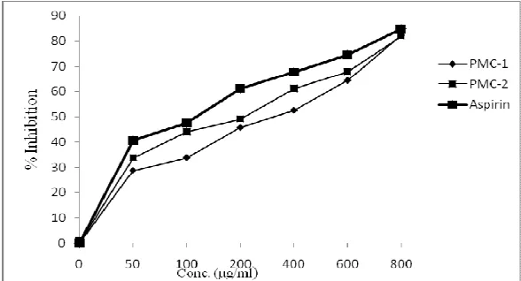
Inhibition of Protein Denaturation
Maximum inhibition, 82.38% was observed at 800
µgmL-1 of PMC-1 and 81.87 % was observed at
800μg/ml of PMC-2. Aspirin, a standard anti-
inflammatory drug showed the maximum inhibition,
84.75% at the concentration of 800µgmL-1.
PMC-1 (β-sitosterol) PMC-1 (stigmasterol)
Figure 1: Structures of isolated compounds
Figure 2: Effect of PMC-1 and PMC-2 on Membrane Stabilization of RBC
DISCUSSION
The over anabolism of ROS leads to cell injury by
damaging the lipid peroxidation of membranes and
macromolecules. In addition, ROS propagate
inflammation by stimulating the release of the
cytokines such as IL-1, TNF-α, and IF-γ, which
stimulate precipitation of extra neutrophil and
macrophages. Most clinically important medicine
belongs to cyclo-per-hydro-phenanthrene compounds
or NSAIDs anti-inflammatory chemical therapeutics
for treatment of inflammation related diseases.
Though these have great activity and long term
administration is required for treatments of long term
diseases. Furthermore, these drugs have various and
severe side effects. Therefore, medicinal plant agents
with very less side effects are desirable to
replacement to chemical therapeutics (Luo et al.,
2002).
The ROS propagate inflammation by enhancing the
inflammatory mediators, so its stabilization or
inhibition is necessary. The presence of β-sitosterol
and stigmasterol in Plectranthus mollis stabilize ROS present. Most of the investigators have reported that
denaturation of the protein is one of the cause of
rheumatoid arthritis (Qiuhong et al., 2013).
Production of auto-antigens in certain rheumatic
diseases may be due to in vivo denaturation of proteins. The mechanism of denaturation probably
involves alteration in electrostatic, hydrogen,
hydrophobic and disulphide bonding. The β-sitosterol
and stigmasterol present in PMACE able to inhibit the
denaturation of protein by inhibiting and stabilizing
the ROS. Recent studies have shown that many
phytosterol contribute significantly to the
anti-inflammatory activity of many plants (Vadivu and
Lakshmi, 2008). The lysosomal enzymes released
during inflammation produce a variety of disorders.
The extra cellular activity of these enzymes is said to
be related to acute or chronic inflammation. The
steroidal drugs act either by inhibiting these
lysosomal enzymes or by stabilizing the lysosomal
membrane (Wellsow et al., 2006). Since HRBC
membrane are similar to cell lysosome membrane the
prevention of hypotonicity induced HRBC membrane
lysis is taken as a measure of anti- inflammatory
activity of drugs. We can say that, the β-sitosterol and
stigmasterol in Plectranthus mollis are act as an anti-inflammatory agent by neutralizing lysosomal
enzymes or by stabilizing the lysosomal membrane.
In both models as Plectranthus mollis shows promising effects. So, from the results of the present
study it can be stated that Plectranthus mollis is capable of controlling the production of auto-antigens
due to denaturation of proteins in rheumatic diseases
either by preventing synthesis of free radicals. The
presence of constituent’s β-sitosterol and stigmasterol
are responsible for stated activity.
REFERENCES
1. Aher, A.N., Pal, S.C., Yadav, S.K., Patil, U.K., 2009.
Antioxidant activity of isolated phytoconstitu1ents
from Casuarina equisetifolia (Casuarinaceae). J. Plant
Sci. 4 (1), 15-20.
2. Anonymous, 2003. A Dictionary of India Raw material
and Industrial product; ‘The wealth of India’ Vol-VI,
Council of Industrial and Scientific Research, New
Delhi.
3. Catherine, W.L., Monique, S.J., Alan, J.P., Deshpande, V.,
2006. Plectranthus: A review of ethnobotanical uses. J.
Ethnopharm. 103, 1–24.
4. Grant, N.H., Alburn, H.E., Kryzanauskas, C., 1970.
Stabilisation of serum albumin by anti-inflammatory
drugs. Biochem. Pharmacol. 19, 715-722.
5. Jadhav, V. M., Kadam, V.J., 2009. In-vitro anti-arthritic
activity of Abutilon indicum (Linn.) Sweet. J. Pharma.
Res. 2(4), 644-645.
6. Okoli, C.O., Akah, P.A., 2004. Mechanism of the
anti-inflammatory activity of the leaf extracts of Culcasia
scandens P. beauv (Araceae). Pharmacol. Biochem. Beha. 79, 473-481.
7. Qiuhong, W., Haixue, K., Yang, S., Yanping, S., Jian, F.,
Rui, G., Kelvin, C., 2013. Naturally derived
anti-inflammatory compounds from Chinese medicinal
8. Luo, X.D., Basile, M.J., Kennelly, E.J., 2002. Polyphenolic
antioxidants from the fruits of Chrysophyllum cainito L.
(star apple). J. Agri. Food Chem. 50, 1379-1382.
9. Rao, Y.K., Fang, S.H., Hsieh, S.C., Yeh, T.H., Tzeng, Y.M.,
2009. The constituents of Anisomeles indica and their
anti-inflammatory activities. J. Ethnopharmacol. 121,
292–296.
10.Vadivu, R., Lakshmi, K.S., 2008. In vitro and in vivo anti
inflammatory activity of leaves of Symplocos
cochinchinensis (Lour) Moore spp Laurina. Bang. J.
Pharmacol. 3, 121-124.
11.Wellsow, J., Grayer, R.J., Veitch, N.C., Kokubun, T.,
Roberto, L., Kite, G.C., Simmonds, S.J., 2006.
Insect-antifeedant and antibacterial activity of diterpenoids
from species of Plectranthus. Phytochem. 67, 1818–
1825.
How to cite this article:
Ushir Yogesh V.*, Morankar Pravin G, and Dhake Avinash S;
Phytochemical Compositions and in-vitro Anti-inflammatory
Activity of Plectranthus mollis; J. Adv. Pharm. Edu. & Res. 2013: 3(2): 85-89.