W
OJCIECHR
ZESZUTKO1, T
OMASZK
ONOPKA2, W
ACŁAWK
OPEĆ3Anti−Neutrophil Cytoplasmic Antibodies
and Autoantibodies to Human Heat Shock Protein 60
in Periodontitis Patients
Autoprzeciwciała dla antygenów cytoplazmatycznych neutrofili
oraz ludzkiego białka szoku termicznego 60 u pacjentów
z zapaleniem przyzębia
1 Department of Pathological Anatomy, Silesian Piasts University of Medicine in Wrocław 2 Department of Periodontology
3 Laboratory of Immunology, Department of Nephrology, Silesian Piasts University of Medicine in Wrocław,
Poland
Adv Clin Exp Med 2006, 15, 4, 599–605 ISSN 1230−025X
ORIGINAL PAPERS
© Copyright by Silesian Piasts University of Medicine in Wrocław
Abstract
Background.The autoimmune response may contribute to the pathogenesis of periodontitis. Changing levels of anti−neutrophil cytoplasmic autoantibodies (ANCAs) are serologic markers for many infective and connective tis− sue diseases. Hsp 60 may also be an important factor in the initiation of an autoimmune response.
Objectives. Investigating the presence of ANCAs (p−ANCA to myeloperoxidase and c−ANCA to proteinase 3) and antibodies to human heat shock protein 60 (Hsp 60) in the sera of 73 patients with periodontitis and 13 control subjects.
Material and Methods. The study group consisted of 73 patients aged 19–50 years: 31 with generalized aggres− sive periodontitis, 13 with localized aggressive periodontitis, and 29 with chronic periodontitis. The control group comprised 13 periodontally healthy subjects. The serum levels of p−ANCA, c−ANCA, and anti−Hsp 60 were detect− ed by ELISA in each subject.
Results.Elevated levels of p−ANCA were demonstrated only in five patients with generalized aggressive periodon− titis. Elevated levels of antibodies to Hsp 60 were found only in 12.9% of patients with generalized aggressive peri− odontitis, 7.6% of patients with localized aggressive periodontitis, and 13.7% of patients with chronic periodontits.
Conclusions.There was no relationship between the significantly elevated levels of p−ANCA and antibodies to Hsp 60 and clinical measurements of periodontitis. These results do not support evidence for a primary role of autoimmunity in periodontal disease. It seems probable that the destruction of periodontal tissues may sometimes be the primary driving factor in the induction of the enhanced autoimmune response in periodontitis (Adv Clin Exp Med 2006, 15, 4, 599–605).
Key words: antibodies, anti−neutrophil cytoplasmic, heat shock protein 60, autoimmunity, periodontitis.
Streszczenie
Wprowadzenie.Odpowiedź autoodpornościowa może uczestniczyć w etiopatogenezie zapalenia przyzębia. Zmie− nione stężenia autoprzeciwciał dla cytoplazmy neutrofili (ANCA) są markerami serologicznymi dla wielu chorób zapalnych i tkanki łacznej. Przeciwciała dla białka szoku termicznego 60 (HSP 60) mogą być również istotnym czynnikiem w inicjowaniu reakcji autoimmunologicznej.
Cel pracy. Zbadanie występowania ANCA (p−ANCA antymieloperoksydazie i c−ANCA antyproteinazie 3) oraz prze− ciwciał dla Hsp 60 w surowicy 73 pacjentów z zapaleniem przyzębia i u 13 osób z grupy kontrolnej.
Materiał i metody. Grupa badana składała się z 73 pacjentów w wieku 19–50 lat: 31 z uogólnionym agresywnym zapaleniem przyzębia, 13 z umiejscowionym agresywnym zapaleniem przyzębia i 29 z przewlekłym zapaleniem przyzębia. Grupa kontrolna liczyła 13 osób ze zdrowym klinicznie przyzębiem. Stężenia surowicze p−ANCA, c−ANCA i przeciwciał dla HSP 60 badano u każdego pacjenta metodą ELISA.
Research on autoimmunity in periodontal dis− ease was initiated by Fits et al. [1], who discovered autoantibodies to type I collagen in the sera of patients with adult periodontitis (AP) in 1986. In later experiments, the role of collagen−I was con− firmed as the main autoantibody in periodontitis [2–7]. Higher levels of IgG and IgA anti−collagen−I antibodies were found in the inflamed gingival tis− sue than in serum, which was explained by the endogenous synthesis of these autoantibodies in the periods of gingival tissues damage [4] and that collagen type I−specific T−cell clones could be iden− tified in the inflamed gingival tissue of periodonti− tis patients [6]. CD5+ B cells seem to be the main
source of anti−collagen antibodies, and with the increase in inflamed gingival tissue a switch occurs from natural IgM to IgG and IgA [7]. Govze and Herzberg [8] reported the presence of antibodies of subclass IgG anti−desmosomal proteins (desmo− plakin 1 and 3) and also glycoproteins (desmoglein 1, 2, and 3) in 90% of sera and in all analyzed sam− ples of gingival crevicular fluid from patients with periodontitis. In view of these observations it was suggested that some of the already discovered autoantigens, for example collagen I and desmoso− mal proteins, and glycoproteins uncovered or drawn from inflamed gingival tissues, may initiate polyclonal activation of populations of B lympho− cytes and the synthesis of antibodies typical to these fragments [3, 5–8]. These findings point to a secondary character of autoimmunity caused by the destruction of gingival tissues.
Other autoantibodies in the sera of patients with AP were found by Novo and Viera [9]. They discovered the presence of anti−neutrophil cyto− plasmic antibodies (ANCAs) directed against “a granular extract” of granulocytes in 75% of analyzed patients and, to a lesser degree, against lactoferrin. The increased levels of ANCA are a useful diagnostic serologic marker for vasculitis, glomerulonephritis, pulmonary renal syndrome, Wegener’s granulomatosis, and connective tissue diseases. In the course of these diseases, activation of neutrophils occurs, with the release of the con− tents of the granules (myeloperoxidase−MPO, pro− teinase 3−PR3, elastase, lactoferrin, cathepsin G, eosinophil peroxidase) which are antigenic [10]. The main neutrophil cytoplasmic antigens for ANCA are MPO (p−ANCA) and PR3 (c−ANCA).
Novo and Viera [9] suggested an important role of autoimmunity in the pathogenesis of periodontitis, manifesting itself in the form of anti−MPO− induced complement cell lysis.
The heat shock proteins (Hsp) have gained wide interest as potential autoantigens due to their high degree of amino−acid sequence identity in eukaryotic and prokaryotic cells [11–19]. This molecular mimicry may be particularly important for the initiation of an autoimmune response. Hsp 60 may be especially important in the causal relationship between microbial infections and autoimmunity [20]. In juvenile chronic arthritis, rheumatoid arthritis, and Crohn’s disease, a spe− cific anti−Hsp antibody is presented on the cells of the host (endogenous Hsp) or on the infectious microorganisms (exogenous Hsp) [11, 13, 16]. Hsp are found in bacteria that are generally con− sidered pathogenic to periodontium (Actinobacil− lus actinomycetemcomitans, Porphyromonas gin− givalis, Prevotella intermedia, Treponema denti− cola) [20–23]. They have also been identified in the basal cells of inflamed gingivae of periodonti− tis patients [24, 25]. The homology between the A. actinomycetemcomitans 64−kDa protein and human Hsp 60 is 48% [22]. It is possible that anti− bodies to the exogenous Hsp of periodontal pathogens may be cross−reacted and directed towards the endogenous Hsp of host cells.
The purpose of this study was to investigate the levels of p−ANCA, c−ANCA, and antibodies to human Hsp 60 in the sera of patients with peri− odontitis. This study was undertaken to evaluate the role of these autoantibodies in immune mech− anisms in periodontitis.
Material and Methods
Experimental and
Control Subjects
The study group comprised 73 patients aged 19–50 years (mean age: 35.1, male to female ratio: 26 : 47), referred to the Department of Perio− dontology, Silesian Piasts University of Medicine in Wrocław. All participants in the study signed an informed consent form. The patients were system−
Wnioski.Nie wykazano związku między istotnie podwyższonymi stężeniami p−NCA i przeciwciałami dla HSP 60 a wskaźnikami klinicznymi zapalenia przyzębia. Wyniki te nie potwierdzają pierwotnej roli procesów autoodpor− nościowych w zapaleniach przyzębia. Wydaje się, że zniszczenie tkanek przyzębia może być niekiedy czynnikiem wywołującym reakcję autoimmunologiczną w zapaleniu przyzębia (Adv Clin Exp Med 2006, 15, 4, 599–605).
ically healthy, with no signs of symptomatic infec− tion. None of the patients had been taking any medication during the three months before the examination which could cause any changes in the microflora, immunological system, or inflammato− ry response. This study group consisted of 31 patients (aged 19–39 years, mean age: 28.3, 19 women) with generalized aggressive periodontitis (GAP), 13 patients (aged 19–29 years, mean age: 24.3, 10 women) with localized aggressive peri− odontitis (LAP), and 29 patients (aged 35–50 years, mean age: 40.7, 18 women) with chronic periodontitis (CP) (Table 1). The diagnoses were determined on the basis of clinical and radi− ographic criteria according to the International Workshop for the Classification of Periodontal Diseases and Conditions in 1999 [26]. The control group consisted of 13 periodontally healthy sub− jects aged 24 to 45 years (mean age: 32.3), eight of whom were women.
The clinical state of the periodontium was indi− cated by means of the Silness and Löe (1964) Plaque Index (PI), the Löe and Silness (1963) Gingival Index (GI), the Saxer et al. (1977) Papilla Bleeding Index (PBI), and the Sulcus Fluid Rate (SFR) according Löe and Holm−Pedersen (1965), and the pocket depth (PD) was measured using a periodontal probe from four surfaces of each tooth.
Sera
Five milliliters of blood were taken from the elbow vein. It was incubated at room temperature for 30 minutes to coagulate it, then it was cen− trifuged for 10 minutes at 1000 × g. Sera were stored at –70°C until used.
Measurement of ANCA
Levels of p−ANCA and c−ANCA were detected by means of enzyme−linked immunosorbent assay (ELISA) from Millenia Diagnostic Products
Corporation (Los Angeles, CA, USA) test sets. The investigation was performed according to the rec− ommendations of the manufacturer of the antibody. The results of the test were expressed in units per milliliter (U/ml). The norm for p−ANCA is less than 8 U/ml and for c−ANCA less than 5 U/ml.
Measurement
of Hsp 60 Antibodies
ELISA was also used to mark the antibody anti−heat shock protein 60 (Hsp 60, human recom− binant, Stressgen Biotechnology Co., Victoria, Canada). The recombinant Hsp was used in the lipophilated form. According to the manufacturer it had a purity above 90% (SDS−PAGE). In the preliminary tests, the optimal concentration of protein on the polystyrene plates and the optimal dilution of the tested sera were determined in the sera of 12 patients with periodontitis. On the basis of these tests, 5 µg of heat shock proteins and a dilution of 1 : 5 of the tested sera were consid− ered as the optimal concentration and dilution in the ELISA test, respectively.
Experimental Procedures
in the ELISA Test
Ninety−six polystyrene plates were used (Nunc−Immuno Plate, Poly Sorp). Each of the plates was filled with 100 µl phosphate buffer, pH 7.5, each containing 5 µg of heat shock protein. The plates were then incubated overnight at 4°C, then washed three times using phosphate−buffered saline (PBS) to which 0.05% Tween 20 was added. The buffer was also used to wash the plates in the successive steps of the ELISA test. The test sera were diluted with 1 : 5 PBS containing 1% bovine serum albumin and 0.05% Tween 20.
One hundred µl of the test sera was added to each of the plates and incubated for two hours at 37°C, after which they were washed three times with PBS. To each of the plates was added 100 µl of rabbit antibody anti−human IgG−alkaline phos− phatase (DAKO, A/S, Glostrup, Denmark; a 1 : 500 dilution in PBS with 0.05% Tween 20) and it was incubated for 2 hours at 37°C. The plates were then washed three times with PBS and 100 µl of substrate (p−nitrophenyl phosphate, Sigma Che− mical Co., St. Louis, USA; diluted with 1 M die− thanolamine buffer, pH 9.8, containing 0.05 mM magnesium chloride) was added to each plate. The plates were incubated for 1 hour at 37°C, after which the absorbance was measured at 450 nm using an automated Stat Fax 2100 reader.
Table 1.Division of the examined groups according to diagnosis
Tabela 1. Podział badanych grup pod względem rozpoz− nania
Examined group Control group (Grupa badana) (Grupa kontrolna) 31 patients with GAP –
13 patients with LAP – 29 patients with CP –
Determination of the Specificity
of Anti−Hsp 60 Antibodies
Test sera taken from four patients with periodon− titis, which was highly reactive with Hsp 60 in ELISA (high absorbance), were incubated with Hsp60 (80µg Hsp 60 + 120 µl patient sera, 120 µg Hsp 60 + 120 µl patient sera) for one night at 4°C, after which ELISA of Hsp 60 antigen was repeated. The braking percent in the ELISA test was calculated from the dif− ference in the optical densities of the serum before and after incubation with antigen Hsp 60.
Statistical Analysis
Serum antibody levels between two groups and means of the clinical measurement were eval− uated using the Mann−Whitney test. Ap−value of < 0.05 was considered to indicate a statistically significant difference.
Results
Of the 73 patients with periodontitis, a signif− icant level of p−ANCA was observed only in 5 subjects (6.8%). These were exclusively patients with GAP (significant antibody levels of anti− MPO neutrophils were found in 16.1% of the test− ed patients with GAP). It was observed that the clinical state of the periodontium in the GAP group, where the level of the p−ANCA antibody was higher than 8 U/ml, was not worse compared with patients with GAP in whom the level of this antibody was not of a notable value (Table 2). A significant level of p−ANCA in the blood sera was not noted in any of the subjects of the control group. In the patients with periodontitis, the level of c−ANCA was not higher than the noted values.
The presence of anti−HSP 60 antibody higher
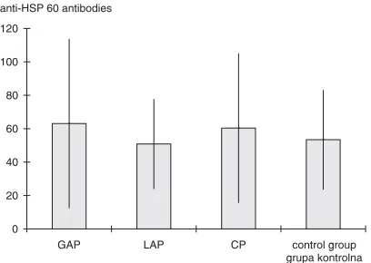
than 2 SDabove the mean control value was shown in 12.9% of the patients with GAP (4 patients), 7.6% with LAP (1 patient), and 13.7% with CP (4 patients). The levels of anti−HSP 60 antibodies in patients with periodontitis and in the control group are shown in Fig. 1. No significant difference was found in the levels of these antibodies between the analyzed groups. It was not established whether the clinical state of the periodontal tissues in patients with anti−HSP 60 antibodies levels more than 2 SD above the mean control value differed greatly from patients in whom the level of this antibody was less than this limit (Table 3).
Discussion
Described results reveal a higher level than the established norms for p−ANCA in only 5 of the 73 patients using commercially available tests for
Table 2.Means of the clinical measurements in GAP patients with blood serum p−ANCA levels above and below 8 U/ml
Tabela 2. Średnie wskażników klinicznych u pacjentów chorych na uogólnione agre− sywne zapalenie przyzębia, u których stężenie p−ANCA w surowicy krwi > i < 8 U/ml
Clinical ANCA > 8 U/ml ANCA < 8 U/ml Z value in p
measurement (n = 5) (n = 26) Mann−Whitney
(Pomiar test
kliniczny) (Wartości Z w teście
Manna−Whitneya)
PI 1.11 ± 0.53 0.98 ± 0.45 0.8 0.42
GI 2.06 ± 0.72 1.82 ± 0.64 0.91 0.36
PBI 2.38 ± 1.09 2.21 ± 0.96 0.37 0.7
SFR (mm) 5.8 ± 0.75 6.2 ± 2.65 0.18 0.85
PD (mm) 5.19 ± 1.12 4.44 ± 1.16 1.5 0.13
0 20 40 60 80 100 120
GAP LAP CP control group
grupa kontrolna anti−HSP 60 antibodies
Fig. 1.Levels of antibodies to human Hsp 60. Antibody levels are expressed as absorbance values for each group. There were no significant differences in the levels of antibodies to Hsp 60 between periodon− titis patients and control group (p> 0.05) in Mann− −Whitney test
determining p−ANCA and c−ANCA. In the control tests in these 5 patients, one had a negative test and the other four subjects revealed a significant drop from the values at the beginning. It is important to note that positive p−ANCA tests was found in patients with GAP, i.e. those with the most pro− gressive periodontitis. Hence the question whether these autoantibodies are of significant importance in the pathogenesis of periodontal diseases. In pre− sented observations only the anti−MPO antibodies were identified in several cases and in amounts just slightly exceeding the established norms. The pres− ence of these autoantibodies can be explained by the immunological response to the myeloperoxi− dase of neutrophils, which intensively infiltrate the inflamed periodontal tissues. Novo et al. [27] recently found ANCA−positive sera in patients with systemic lupus erythematosus, rheumatoid arthritis, and periodontitis. This result confirms a well−known association between ANCA and these systemic conditions.
Presented results demonstrate that ANCA does not play an important role in the etiopatho− genesis of periodontitis, as was suggested by Novo and Viera [9]. It must be taken into consideration that these authors used a neutrophil granular extract as the antigen in ELISA, omitting many controls which in this type of study are the basis for excluding non−specific reactions. Presented studies confirm that the autoimmune response is not a primary etiological factor in periodontitis. In some patients (mainly, it seems, in the generalized and progressive form of periodontitis) the pres− ence of translocation of ANCA−reactive proteins to the neutrophil surface and degranulation can be detected, which may be one of the secondary mechanisms enhancing the vicious circle of this pathology.
Presented studies did not reveal a major dif− ference in the levels of antibody to human Hsp 60 between patients with different types of periodon−
titis and the control group. This finding indicates that periodontitis does not have any effect on the production of specific autoantibodies to Hsp 60. This could be due to intraepithelial localization in the gingival tissue, the Hsp 60 epitopes not being sufficiently available to cause specific humoral immunological response. Presented results can be interpreted as another suggestion for the lack of an important component of the autoimmunological process in the initiation and support of the gingival tissue damage in periodontal diseases. Elevated levels of antibodies to human Hsp 60 in the sera of periodontitis patients compared with controls have been reported by Tabeta et al. [28]. They also sug− gested the cross−reactivity of antibodies to human Hsp 60 (a self antigen) andP. gingivalisGroEL (an exogenous bacterial Hsp 60 antigen). Significant associations between the concentration of antibod− ies to human Hsp and clinical indices of periodon− titis were described by Lopatin et al. [29]. These authors also suggested that anti−Hsp antibodies may play a protective role in the development of periodontal disease and their levels are lowest in more advanced periodontitis. The differences between these results, also own observations, may be attributable to differences in periodontal dis− ease advancement and activity at the time of sam− pling these antibodies.
In light of today’s knowledge, the authors expect that in some periodontitis cases (especially in GAP), the autoimmunological process may take part in tissue destruction, but only as a secondary process in relation to earlier destruction of the attachment apparatus and the presence of peri− odontopathic bacteria as a consequence of peri− odontal diseases. Perhaps in refractory forms of periodontitis the autoimmunological mechanisms replace the dental plaque in the stimulation of dis− ease−causing changes. Predominantly type I colla− gen and desmosomal proteins in the junctional epithelium were indicated as potential autoanti− Table 3.Means of the clinical measurements in periodontitis patients with blood serum levels of
antibodies to Hsp 60 above and below the mean control value + 2 SD
Tabela 3. Średnie wskażników klinicznych u pacjentów chorych na zapalenie przyzębia, u których stężenie przeciwciał Hsp 60 w surowicy krwi > i < wartości średniej w grupie kontrolnej + 2 SD
Clinical Hsp 60 > control Anti−Hsp 60 < Z value in p
measurement mean + 2 SD control mean Mann−Whitney
(Pomiar (n = 9) + 2 SD test
kliniczny) (n = 64) (Wartości Z w teście
Manna−Whitneya)
PI 1.34 ± 0.71 1.08 ± 0.69 1.15 0.24
GI 1.89 ± 0.58 1.61 ± 0.68 1.15 0.24
PBI 2.01 ± 0.83 1.97 ± 0.99 0.12 0.96
SFR (mm) 5.42 ± 1.73 5.12 ± 2.33 0.91 0.35
genes in periodontitis [3, 4, 8]. Described examina− tions do not confirm that, apart from special cases, the role of autoantigens in periodontitis could be fulfilled by myeloperoxidase and proteinase 3 from the azurophilic granules of neutrophils. The possi−
bility of occasional participation of antibodies to human Hsp 60 in a secondary autoimmune response in periodontitis cannot be excluded, although presented studies did not provide suffi− cient evidence of this.
References
[1] Fits A, Singh G, Dolby AE:Antibody to collagen type I in periodontal disease. J Periodontol 1986, 57, 693–698.
[2] Hirsch HZ, Tarkowski A, Miller EJ, Gay S, Koopman WJ, Mestecky J: Autoimmunity to collagen in adult periodontal disease. J Oral Pathol 1988, 17, 456–459.
[3] Anusaksathien O, Singh G, Matthews N, Dolby AE:Autoimmunity to collagen in adult periodontal disease: Immunoglobulin classes in sera and tissue. J Periodont Res 1992, 27, 55–61.
[4] Anusaksathien O, Singh G, Peters TJ, Dolby AE: Immunity to self−antigens in periodontal disease. J Periodontol 1992, 63, 194–199.
[5] Hahn C−L, Schenkein H, Tew JG:Polyclonal B cell activators and in vitro induction of auto−antibody reactive with collagen. J Periodont Res 1997, 32, 608–613.
[6] Wasseaar A, Reinhardus C, Thepen T, Abraham−Inpijn L, Kievits F: Cloning, characterization, and antigen specificity of T−lymphocyte subsets extracted from gingival tissue of chronic adult periodontitis patients. Infect Immun 1995, 63, 2147–2153.
[7] Sugawara M, Yamashita K, Yoshie H, Hara K: Detection of, and anti−collagen antibody produced by, CD5−pos− itive B cells in inflamed gingival tissues. J Periodont Res 1992, 27, 489–498.
[8] Govze Y, Herzberg MC: Serum and gingival crevicular fluid anti−desmosomal antibodies in periodontitis. J Periodontol 1993, 64, 603–608.
[9] Novo E, Viera N:Antineutrophil cytoplasmic antibodies: a missing link in the pathogenesis of periodontal dis− ease? J Periodont Res 1996, 31, 365–368.
[10] Lesavre P:Antineutrophil cytoplasmic autoantibodies antigen specificity. Am J Kidney Dis 1991, 18, 159–163.
[11] Boog CJP, De Graeff−Meeder ER, Lucassen MA, Van der Zee R, Voorhorst−Ogink MM, Van Kooten PJS, Geuze Hj, Van Eden W:Two monoclonal antibodies generated against human hsp 60 show reactivity with syn− ovial membranes of patients with juvenile chronic arthritis. J Exp Med 1992, 175, 1805–1810.
[12] Elsaghier A, Prantera C, Bothamley G, Wilkins E, Jindal S, Ivanyi J:Disease association of antibodies to human and mycobacterial hsp70 and hsp60 stress proteins. Clin Exp Immunol 1992, 89, 305–309.
[13] Jarjour WN, Jeffries BD, Davis JS, Welch WJ, Mimura T, Winfield JB: Autoantibodies to human stress pro− teins. A survey of various rheumatic and other inflammatory diseases. Arthritis Rheum 1991, 34, 1133–1138.
[14] Kaufmann SHE: Heat shock proteins and the immune response. Immunol Today 1990, 11, 129–136.
[15] Kaufmann SHE, Schoel B, Embden JDA, Koga T, Wand−Wurttenberger A, Munk ME, Steinhoff U: Heat− shock protein 60: Implications for pathogenesis of and protection against bacterial infections. Immun Rev 1991, 12, 67–90.
[16] Nualláin EM, Monaghan H, Reen DJ: Antibody response of restricted isotype to heat shock proteins in juvenile chronic arthritis. Scand J Immunol 1993, 38, 83–88.
[17] Res PCM, Thole JER, De Vries RRP:Heat shock proteins in immunopathology. Curr Opin Immunol 1991, 3, 924–929.
[18] Yi Y, Yang X, Brunham RC:Autoimmunity to heat shock protein 60 and antigen−specific production of inter− leukin−10. Infect Immun 1997, 65, 1669–1674.
[19] Van Eden W, Van der Zee R, Paul AGA, Prakken BJ, Wendling U, Anderton SM, Wauben MHD:Do heat shock proteins control the balance of T−cell regulation in inflammatory diseases? Immunol Today 1998, 19, 303–307.
[20] Yamazaki K, Ohsawa Y, Tabeta K, Ito H, Ueki K, Oda T, Yoshie H, Seymour GJ:Accumulation of human heat shock protein 60−reactive T cell in the gingival tissues of periodontitis patients. Infect Immun 2002, 70, 2492–2501.
[21] Lokensgard J, Bakken V, Schenck K:Heat shock response in Actinobacillus actinomycetemcomitans. FEMS Immunol Med Microbiol 1994, 8, 321–328.
[22] Nakano Y, Inai Y, Yamashita Y, Nagaoka S, Kusuzaki−Nagira T, Nishihara T, Okahashi N, Koga T:
Molecular and immunological characterization of a 64−kDa protein of Actinobacillus actinomycetemcomitans. Oral Microbiol Immunol 1995, 10, 151–159.
[23] Vayssier C, Mayrand D, Grenier D: Detection of stress proteins in Porphyromonas gingivalisand other oral bac− teria by Western immunoblotting analysis. FEMS Microbiol Let 1994, 121, 303–308.
[24] Ando T, Kato T, Ishihara K, Ogiuchi H, Okuda K:Heat shock proteins in the human periodontal disease process. Microbiol Immunol 1995, 39, 321–327.
[25] Konopka T: Role of cytokines, adhesion molecules, heat shock proteins, and autoimmunity response in peri− odontitis. Wrocław Medical University, 1998, 4. Habilitation thesis.
[27] Novo E, Garcia−MacGregor E, Viera N, Chaparro N, Crozzoli Y: Periodontitis and anti−neutrophil cytoplas− mic antibodies in systemic lupus erythematosus and rheumatoid arthritis: a comparative study. J Periodontol 1999, 70, 185–188.
[28] Tabeta K, Yamazaki K, Hotokezaka H, Yoshie H, Hara K:Elevated humoral immune response to heat shock protein (hsp60) family in periodontitis patients. Clin Exp Immunol 2000, 120, 285–293.
[29] Lopatin DE, Shelburne CE, Poperin NV, Kowalski C, Bagramian RA:Humoral immunity to stress proteins and periodontal disease. J Periodontol 1999, 70, 1185–1193.
Address for correspondence:
Tomasz Konopka
Department of Periodontology
Silesian Piasts University of Medicine in Wrocław Krakowska 26
50−425 Wrocław Poland
Phone/fax: +48 71 784 03 81 E−mail: [email protected] Conflict of interest: None declared Received: 3.01.2006
Revised: 9.05.2006 Accepted: 15.05.2006
Praca wpłynęła do Redakcji: 3.01.2006 r. Po recenzji: 9.05.2006 r.