Kasim Caglayan
1, A, D, F, Nilsen Erdogan
2, C, Bahattin Avci
3, C, Bulent Gungor
4, B, E, F,
Hamza Cinar
4, B, D, Nilden Arslan
5, CEffect of Beta-Glucan on Intestinal Anastomoses
in a Rat Model
Wpływ betaglukanu na zespolenia jelitowe w modelu szczurzym
1 Bozok University, Faculty of Medicine, Department of Surgery, Yozgat, Turkey 2 Bozok University, Faculty of Medicine, Department of Pathology, Yozgat, Turkey
3 Ondokuz Mayis University, Faculty of Medicine, Department of Biochemistry, Samsun, Turkey 4 Ondokuz Mayis University, Faculty of Medicine, Department of Surgery, Samsun, Turkey 5 Ondokuz Mayis University, Faculty of Medicine, Department of Public Health, Samsun, Turkey
A – research concept and design; B – collection and/or assembly of data; C – data analysis and interpretation;
D – writing the article; E – critical revision of the article; F – final approval of article; G – other
Abstract
Objectives. The authors aimed to investigate the effect of â-Glucan on healing of an experimental left-sided colon anastomosis model.
Material and Methods. Twenty adult male Wistar albino rats were randomized into two groups which had colonic transection and end-to-end anastomosis. Group I (Control): anastomosis group, received no treatment (n = 10); group II, anastomosis + β-Glucan (50 mg/kg/day within seven days after surgical procedure). Bursting pressure, hydroxyproline levels and histopathological characteristics of the anastomosis were analyzed.
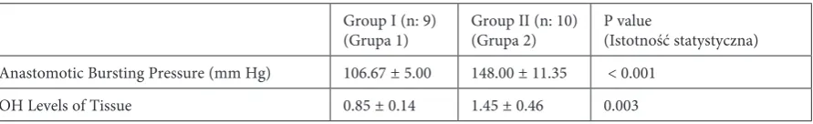
Results. The average burst pressure of Groups I and II were 106.67 ± 5.00 and 148.00 ± 11.35 mm Hg and hydroxy-proline levels were 0.85 ± 0.14 and 1.45 ± 0.46 µg/mg, respectively. Both the burst pressure and hydroxyhydroxy-proline levels in group II were statistically significantly higher (p < 0.05). Histopathological examination revealed less epithelial damage in group II (p < 0.05). Though not statistically significant, less edema and damage to the submu-cosal-muscular layer was seen in Group II (p = 0079).
Conclusions. Due to significant increases in anastomotic bursting pressures and tissue hydroxyproline levels and considering the inhibitory effect of β-Glucan on epithelial damage, edema, and submucosal-muscular layer damage, β-Glucan was thought to contribute to the healing of the anastomosis (Adv Clin Exp Med 2013, 22, 2, 157–163).
Key words: colon anastomosis, β-Glucan, burst pressure, hydroxyproline.
Streszczenie
Cel pracy. Autorzy zbadali wpływ β-glukanu na gojenie eksperymentalnego modelu lewostronnego zespolenia okrężnicy.
Materiał i metody. Dwadzieścia dorosłych samców szczurów albinosów szczepu Wistar podzielono losowo na dwie grupy, u których wykonano przecięcie okrężnicy i zespolenie koniec do końca. Grupa I (kontrolna): wyko-nano zespolenie, bez leczenia (n = 10), grupa II, wykowyko-nano zespolenie i podano β-glukan (50 mg / kg / dzień w ciągu siedmiu dni po zabiegu). Oceniono ciśnienie rozrywające, stężenie hydroksyproliny i cechy histopatolo-giczne zespolenia.
Wyniki. Średnie ciśnienie rozrywające grupy I i II wynosiło 106,67 ± 5,00 i 148,00 ± 11,35 mm Hg, a stężenie hydroksyproliny wynosiło 0,85 ± 0,14 i 1,45 ± 0,46 ng / mg. Zarówno ciśnienie rozrywające, jak i stężenie hydrok-syproliny w grupie II były statystycznie istotnie większe (p < 0,05). Badanie histopatologiczne wykazało mniejsze uszkodzenie nabłonka w grupie II (p < 0,05). Chociaż nie było to istotne statystycznie, mniejszy obrzęk i uszkodze-nie warstwy podśluzówkowo-mięśniowej zaobserwowano w grupie II (p = 0.079).
Adv Clin Exp Med 2013, 22, 2, 157–163 ISSN 1899–5276
OrIGINAl PAPErS
Anastomotic leakage after colorectal surgery is a major cause of morbidity and mortality. It is also associated with high rates of local recurrences and survival after curative resection of colorectal cancers. Anastomotic leak rates range from 0.5% to 30% in the literature [1–3]. Although there are differences among surgeons, the ratio is expressed as 3.4–6% amongst colorectal cancer surgery ex-perienced surgeons [4]. Many factors, such as poor blood supply of the anastomosis, location of anastomosis, lack of technical capacity, colonic bacteria (bacterial flora of the colon, colonic bac-teria distribution or content), inflammation, age, obesity, smoking, hypoalbuminemia, cardiovascu-lar disease (CVD) and chemotherapy, and use of drugs such as dexamethasone, adversely affect the healing of the anastomosis [1, 4].
β-Glucan is a glucose polymer found in the cell walls of yeast, fungi and cereals. It has benefi-cial effects on the immune system and is accepted as a booster for the immune system, and it has no toxic or side effects [5, 6]. The biological activity of β-Glucan is by binding to β-Glucan receptors on monocytes, leukocytes and macrophages, increas-ing the release of cytokines and arachidonic acid metabolites, and stimulation of hematopoiesis [7, 8]. The immune reaction and the formation of col-lagen in the early period is important for wound healing and this happens with fibroblast prolifera-tion and collagen synthesis [9]. A study by Hyun Joo Son et al. [10] showed an in-vitro positive ef-fect of β-Glucan use on fibroblast proliferation. For this reason, β-Glucan use is thought to have a positive effect in preventing intestinal anasto-motic leaks with its effects of strengthening the immune system and enhancing fibroblast prolif-eration, and this study was planned.
The aim of this study is to investigate, in an experimental rat model, the effects of the β-Glucan on the healing of left colon anastomoses.
Material and Methods
Animals
The study was approved by the Ondokuz Mayýs University local Ethics Committee for An-imal Experiments (Date: Dec. 27, 2010, number: 2010/78). Twenty Wistar Albino male rats, weight
ranging 250–300 g, were used in the study. All rats were fed ad libitum with standard rat chow and tap water. All subjects were kept in 12 hour dark-ness and 12 hour light before and after study (at a standard temperature of 22°C).
All animals were given water only on the first postoperative day; standard rat chow and water ad libitum were provided on the second postopera-tive day. There was no difference in food intake between groups.
Experimental Groups
The rats were randomly divided into two groups, each containing ten rats. Group I (control): anastomosis group, received no treatment. Group II (β-Glucan): 50 mg/kg was suspended in 2 ml of sa-line and given by intragastric gavage to the glucan groups within seven days after surgical procedure (Mustafa Nevzat Company, Istanbul, Turkey). The dose of β-glucan was decided by taking the studies of Sener et al. [8, 11] into consideration.
Surgical Procedure
50 mg/kg ketamine (Ketalar, Pfizer, Turkey) with 10 mg/kg xylazine HCl (rompun, Bayer, Tur-key) were given to all animals for anesthesia. The abdominal skins of all subjects were shaved and painted with iodine povidone under general an-esthesia. All surgical procedures were performed under sterile conditions. laparotomy was per-formed with a midline incision of approximately 3 cm. After the left colon was transected horizon-tally, 4 cm proximal to the peritoneal reflection, an end-to-end anastomosis was performed with a single layer manner with 5/0 polyprolene su-tures (Prolone, Ethicon). The fascia and skin were closed separately by 4-0 silk. In the control group, one rat died on the sixth day. This animal was ex-cluded from the study. All the rats were sacrified with a high dose of anesthetic on the 7th day after surgery. After being sacrified, a repeat laparotomy was immediately made through the same midline incision and the peritoneal cavity was opened.
Measurement of Bursting Pressure
cath-eter placement. A three-way cannula was placed at the tip of the catheter. One end was connected to intraluminal side and the other end was connected to the manometer. Methylene blue, diluted with saline, was administered at 6 ml/min speed with an infusion pump. The pressure when the blue-colored fluid was seen or the time when a sudden fall in pressure was seen had been defined as the burst pressure and was recorded in mm Hg.
Measurement of Hydroxyproline Level
Tissue samples were put into an appropriate PBS (Phosphate Buffer Solution, 10 mM, pH 7.2) after being homogenized manually in porcelain mortar and pestle with liquid nitrogen. Tissue samples were stored frozen at –80°C in individual aliquots after being sonicated for 1 min at +4°C in 220V (Fisher Sonic Dismembrator; Mosel 300). The homogenates were centrifuged at +4°C for 5 minutes with 15000 g (Sigma 3K30, S. No: 76262, Germany) and the supernatants were used for
analysis. Hydroxyproline levels were determined using a commercially available Hydroxyproline As-say Kit (BioVision Incorporated, Milpitas, USA), based on an analysis of chromogenin as a result of the reaction at 560 nm spectrophotometrically. The range of the kit was 0.1–2 μg. The amount of protein in tissue homogenates was determined by the lowry method and hydroxyproline level was expressed as µg per mg. protein (µg/mg.prot). The principle of the lowry method is based on the re-activity of the peptide nitrogen[s] with the copper [II] ions under alkaline conditions and the subse-quent reduction of the Folin-Ciocalteay phospho-molybdicphosphotungstic acid to heteropolymo-lybdenum blue by the copper-catalyzed oxidation of aromatic acids [13].
Histopathologic
Evaluation
Tissue samples were taken from the anasto-mosis region, and histopathological examination
Table 1. Scores used to analyze healing of the anastomosis line in ileum semi-quantitatively Tabela 1. Punktacja stosowana do półilościowej analizy gojenia linii zespolenia w jelicie krętym
Score (Punk- tacja)
Necrosis
(Martwica) PMN lymphocytes (limfocyty) Macrophages (Makrofagi) Edema (Obrzęk) Mucosal epithelium (Nabłonek błony śluzowej)
Submucosal-muscular layer (Warstwa podśluzówkowo- -mięśniowa)
0 none normal
number normal num-ber normal num-ber none normal glan-dular good bridging
1 small
patches slight in-crease slight increase slight increase some normal cubic average bridging
2 some
patches marked infiltration marked infil-tration marked infil-tration marked incomplete cubic poor bridging
3 massive massive
infiltration massive infil-tration massive infil-tration severe absent no bridging
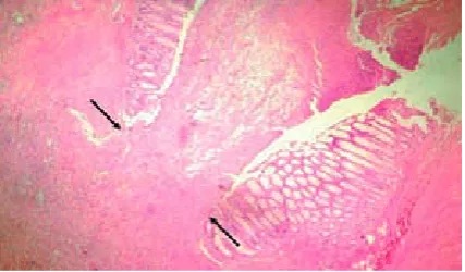
Fig. 1. The figure shows a tissue segment of the anasto-mosis line in the control group (H&E ×40)
Ryc. 1. rycina ukazuje część tkanki linii zespolenia w grupie kontrolnej (H & E ×40)
Fig. 2. The figure shows a tissue segment of the anas-tomosis line in the β-Glucan group. A) H&E ×40, B) H&E ×100
was performed by a pathologist blinded for all groups. Tissue samples were fixed for 12 hours in a 10% buffered neutral formaline solution. After a routine follow-up, samples were embedded in paraffin blocks and sections of 4–5 µm thickness were stained with hematoxylin and eosin (H&E) and finally examined under a light microscope.
A semi-quantitative scale as described by Biert et al. [14], containing various parameters involved in wound healing, was used (Table 1). The amount of necrosis was expressed as none (0 points), only small patches (1 point), some patch-es (2 points) or massive (3 points). In the anasto-motic area, accumulation of polymorphonuclear cells (pmns), macrophages and lymphocytes was also assessed in terms of none or normal num-ber (0 points), slight increase (1 point), marked infiltration (2 points) and massive infiltration (3 points). Edema, expressed as the ratio of max-imum thickness of the wall at the anastomosis and the thickness of the normal intestinal wall as present at the end of the section, was established in terms of none (0 points), some (1–1.5× normal thickness; 1 point), marked (1.5–2× normal thick-ness; 2 points) and severe (> 2x normal thickthick-ness; 3 points). Healing of the mucosa was expressed as normal, i.e. mucosa with restored glandular epithelium (0 points), intact mucosa with cubic epithelium but without glands (1 point), mu-cosa only partially covered by cubic epithelium (2 points) and mucosa completely devoid of epi-thelial coverage (3 points). Submucosal-muscular repair was assessed in terms of good (0 points), average (1 point), poor (2 points) or no (3 points) fibroblast stretching and bridging the anastomot-ic wound. This way, in each anastomosis, two ob-servations from the mesenterial and two from the anti-mesenterial side were obtained.
Statistical Analysis
The data was encoded, then transmitted to a computer and analyzed using the SPSS15.0 pack-age program. Descriptive characteristics of the da-ta were expressed as mean ± sda-tandard deviation. The Mann-Whitney U test was used to compare
the groups. Statistical significance level was con-sidered as p < 0.05.
Results
Anastomotic Bursting Pressure
and Hydroxyproline Content
Anastomosis bursting pressure was found to be 106.67 ± 5.00 mm Hg in Group I and 148.00 ± 11.35 mm Hg in Group II. The bursting pres-sure in group II was significantly greater than the bursting pressure in Group I (p < 0.001). When the groups were compared in terms of peri-anas-tomotic tissue hydroxyproline levels, it was mea-sured at 0.85 ± 0.14 µg/mg.prot in group I and 1.45 ± 0.46 µg/mg.prot in group II. The hydroxy-proline level in Group II was significantly greater than in Group I (p = 0.003). When the differences between groups in terms of anastomotic bursting pressures and tissue hydroxyproline levels were taken into account, it is possible the use of 50 mg/ kg β-Glucan for 7 days in the postoperative period contributes to anastomotic healing. The bursting pressures and tissue hydroxyproline levels of the groups are shown in Table 2.Histology
Histopathological examination of tissue sam-ples taken from the peri-anastomotic region in the group using β-Glucan showed less necrosis in the anastomosis (p = 0.024). However, in terms of in-flammation, lymphocytes and macrophage infiltra-tion were statistically significantly less in the con-trol group (p = 0.022 and p = 0.023). less mucosal epithelial damage was found in group II and this difference was found to be statistically significant (p = 0.012). Tissue polymorphonuclear leukocyte levels (PNl) were found to be similar between the two groups and the difference was insignificant (p = 0.274). Edema and submucosal-muscular lay-er damage wlay-ere less in group II, but the difflay-erence was not significant (p = 0079). Histopathologic ex-amination results are given in Table 3.
OH levels of Tissue 0.85 ± 0.14 1.45 ± 0.46 0.003
Discussion
Anastomotic leakage is an important compli-cation of colon surgery and is associated with mor-bidity and mortality [15]. Despite improvements in perioperative and surgical techniques, the levels of gastrointestinal anastomotic leaks have not fall-en to negligible levels [16]. For this reason, anas-tomotic leaks still continue to be an annoying situ-ation and surgeons are expected to prevent leaks. Anastomotic healing is a complex process involv-ing a series of biological events. In this process, co-ordination of cellular activity and humoral factors are necessary [9]. The degree of inflammatory re-sponse, mucosal re-epithelialization rate, amount of newly synthesized collagen, resistance and im-pact affect anastomotic healing [17]. Anastomosis strength is associated with collagen fibers and their maturation with the submucosal layer [17]. Fibro-blast proliferation and collagen synthesis occurs in the submucosal layer [9]. The synthesis of collagen by fibroblasts is at maximum 5–7 days. Therefore, taking into account the experimental models in the literature, this study terminated on the 7th day [17, 18]. Similarly, Kosmidis et al. [18], in their study, showed the key role of myofibroblast in the healing process of colonic cancer anastomosis and expressed that the detachment of anastomosis is reduced after 7 days.
In the literature, β-Glucan was found to have a positive contribution to the wound healing pro-cess, indirectly by increasing the release after bind-ing to specific receptors on macrophages and by means of glucan-specific receptors expressed on human fibroblasts [7, 19]. Similarly, β-Glucan is expressed to stimulate monocytes, macrophages and natural killer (NK) cells [20]. In another clini-cal study, Demir et al. [20] expressed enhancement in proliferation and stimulation of peripheral
blood monocytes after short term (two weeks) use of β-Glucan without any evidence of side effects in breast cancer patients. In this present study, the histopathologic examination of tissue samples from the anastomosis region revealed statisti-cally significantly increased macrophage, neutro-phil and lymphocyte infiltration in rats fed with β-glucan compared to the control group. Hyun Joo Son et al. [10] stated in their study that there were positive effects on fibroblast proliferation.
Anastomotic bursting pressure and peri-anastomotic tissue hydroxyproline levels are the parameters used in experimental studies related to colonic cancer [9]. Collagen has an important role in all stages of wound healing and tissue strengthening with integrity [21]. Strength of the anastomosis and quality depends on the strength of synthesized collagen [22]. lack of collagen leads to weak anastomosis [23]. In this study, a statisti-cally significant increase in hydroxyproline levels, which is a parameter of collagen synthesis, was de-termined in the peri-anastomotic tissue samples of β-glucan-fed rats compared to the control group. likewise, the anastomotic bursting pressure of the rats fed with β-glucan was statistically signifi-cantly higher than in the control group. Accord-ing to these results, the authors can say that it has a positive effect on anastomotic bursting pressure and hydroxyproline levels. The authors think the significant increase in both the burst pressure and hydroxyproline levels is probably the result of an incremental effect of β-glucan on fibroblast prolif-eration. There are studies showing a positive effect of β-glucan on fibroblast proliferation in the lit-erature [10]. Again, in the study of Son et al. [24], it was speculated that a direct interaction of the β-glucan with fibroblasts was proposed as a pos-sible mechanism of wound repair while the effect of β-glucan on repair involved a macrophage
re-Table 3. Histopathologic examination results of peri-anastomotic tissue Tabela 3. Wyniki badań histopatologicznych tkanek w obrębie zespolenia
Group I
(Grupa 1) Group II (Grupa 2) P value (Istotność statystyczna)
Necrosis (Martwica) 2.22 ± 0.667 1.50 ± 0.527 0.024
PNl 1.89 ± 0.601 2.20 ± 0.632 0.274
lymphocytes (limfocyty) 1.56 ± 0.527 2.30 ± 0.675 0.022
Macrophages (Makrofagi) 1.44 ± 0.527 2.10 ± 0.568 0.023
Edema (Obrzęk) 1.89 ± 0.601 1.40 ± 0.516 0.079
Mucosal epithelium (Nabłonek błony śluzowej) 1.89 ± 0.333 1.30 ± 0.483 0.012 Submucosal-muscular layer
the anastomotic leakage rates. To authors’ knowl-edge, in this first experimental study of β-glucan on anastomotic healing, when significant increases
to have a clearer idea of the effects of β-glucan on anastomotic healing when the results of similar tests are compared.
References
[1] Konishi T, Watanabe T, Kishimoto J, Nagawa H: risk factors for anastomotic leakage after surgery for colorectal cancer: results of prospective surveillance. J Am Coll Surg 2006, 202, 439–444.
[2] Hyman NH, Osler T, Cataldo P, Burns EH, Shackford SR: Anastomotic leaks after bowel resection: What does peer review teach us about the relationship to postoperative mortality? J Am Coll Surg 2009, 208, 48–52.
[3] den Dulk M, Noter SL, Hendriks ER, Brouwers MA, van der Vlies CH, Oostenbroek RJ, Menon AG, Steup WH, van de Velde CJ: Improved diagnosis and treatment of anastomotic leakage after colorectal surgery. Eur J Surg Oncol 2009, 35, 420–426.
[4] Chambers WM, Mortensen NJ: Postoperative leakage and abscess formation after colorectal surgery. Best Pract res Clin Gastroenterol 2004, 18, 865–880.
[5] Engstad CS, Engsad RE, Olsen JO, Osterud B: The effect of soluble beta-1,3-glucan and lipopolysaccharide on cytokine production and coagulation activation in whole blood. Int Immunopharmacol 2002, 2, 1585–1597.
[6] Brown GD, Gordon S: Fungal β-glucans and mammalian immunity. Immunity 2003, 19, 311–315.
[7] Demirbas A: β-Glucan and mineral nutrient contents of cereals grown in Turkey. Food Chem 2005, 90, 773–777.
[8] Sener G, Ekşioğlu-Demiralp E, Cetiner M, Ercan F, Yeğen BC: Beta-glucan ameliorates methotrexate-induced oxidative organ injury via its antioxidant and immunomodulatory effects. Eur J Pharmacol 2006, 542, 170–178.
[9] Inan A, Koca C, Sen M: Effects of diclofenac sodium on bursting pressures of anastomoses and hydroxyproline contents of perianastomotic tissues in a laboratory study. Int J Surg 2006, 4, 222–227.
[10] Son HJ, Han DW, Baek HS, Lim HR, Lee MH, Woo YI, Park JC: Stimulated TNF-a release in macrophage and enhanced migration of dermal fibroblast by β-glucan. Curr Applied Phys 2007, 7S1, 33–36.
[11] Sener G, Toklu H, Ercan F, Erkanli G: Protective effect of beta-glucan againstoxidative organ injury in a rat model of sepsis. Int Immunophramacol 2005, 5, 1387–1396.
[12] Li Y, Bao Y, Jiang T, Tan L, Gao Y, Li J: Effect of the combination of fibrin glue and growth hormone on incom-plete intestinal anastomoses in a rat model of intra-abdominal sepsis. J Surg res 2006, 131, 111–117.
[13] Lowry OH, Rosebrough NJ, Farr AL, Randall RJ: Protein measurement with the Folin phenol reagent, J Biol Chem 1951, 193, 265–275.
[14] Biert J, Seifert WF, Verhofstad AA, Wobbes T, de Man BM, Hoogenhout J, Hendriks T: A semiquantitative histological analysis of repair of anastomoses in the rat colon after combined preoperative irradiation and local hyperthermia. radiat res 1998, 149, 372–377.
[15] Kiyama T, Onda M, Tokunaga A, Yoshiyuki T, Barbul A: Effect of early postoperative feeding on the healing of colonic anastomoses in the presence of intra-abdominal sepsis in rats. Dis Colon rectum 2000, 43, S54–S58.
[16] Law WL, Choi HK, Lee YM, Ho JW, Seto CL: Anastomotic leakage is associated with poor long-term outcome in patients after curative colorectal resection for malignancy. J Gastrointest Surg 2007, 11, 8–15.
[17] Aslan A, Temiz M, Hakverdi S, Polat G, Tumer C, Temiz A, Canbolant E: Effect of mesalamine on healing in experimental colon anastomosis: a randomised experimental study. Int J Surg 2008, 6, 40–44.
[18] Kosmidis C, Efthimiadis C, Anthimidis G, Basdanis G, Apostolidis S, Hytiroglou P, Vasiliadou K, Prousalidis J, Fahantidis E: Myofibroblasts and colonic anastomosis healing in Wistar rats. BMC Surg 2011, 11, 6.
[19] Wei D, Williams D, Browder W: Activation of AP-1 and SP1 correlates with wound growth factor gene expression in glucan-treated human fibroblasts. Int Immunopharmacol 2002, 2, 1163–1172.
[20] Demir G, Klein HO, Mandel-Molinas N, Tuzuner N: Beta glucan induces proliferation and activation of mono-cytes in peripheral blood of patients with advanced breast cancer. Int Immunopharmacol 2007, 7, 113–116.
[21] Teke Z, Aytekin FO, Aydin C, Kabay B, Yenisey C, Sacar S, Simsek NG, Tekin K: Effects of pyrrolidine dithio-carbamate on healing of colonic anastomoses in the cecal ligation and puncture model of intraperitoneal sepsis in rats. World J Surg 2007, 31, 200–209.
[22] Küper MA, Schölzl N, Traub F, Mayer P, Weinreich J, Coerper S, Steurer W, Königrainer A, Beckert S:
Everolimus interferes with the inflammatory phase of healing in experimental colonic anastomoses. J Surg res 2011, 167, 158–165.
[23] Ilhan YS, Bulbuller N, Kirkil C, Ozercan R, Seckin D: The effect of an angiotensin converting enzyme inhibitor on intestinal wound healing.J Surg res 2005, 128, 61–65.
Address for correspondence:
Kasim Caglayan
Bozok University, Faculty of Medicine Department of Surgery, Yozgat Turkey
Tel.: +90-354-2127949
E-mail: [email protected]
Conflict of interest: None declared