This is an open access journal, and articles are distributed under the terms of the Creative Commons Attribution-Non Commercial-ShareAlike 4.0 License, which allows others to remix, tweak, and build upon the work non-commercially, as long as appropriate credit is given and the new creations are licensed under the identical terms.
© 2018 Journal of Advanced Pharmacy Education & Research | Published by SPER Publication
97
Transfusion Therapy: an overview
Eman Al Mussaed
Department of Basic Sciences, Hematopathology Division, Princess Nourah Bint Abdulrahman University, College of Medicine, Riyadh, Saudi Arabia.
Correspondence: Eman Al Mussaed, Department of Basic Sciences, Hematopathology Division, Princess Nourah Bint Abdulrahman University, College of Medicine, Riyadh, Saudi Arabia, Email:[email protected].
ABSTRACT
Transfusion therapy is now a vital medical specialty with well-established practices, principles and guidelines, and often lifesaving part of the treatment of many patients, but carries a significant risk if not performed according to the national guidelines. Transfusion therapy is common in clinical practices and has undergone significant progress over the last decade, particularly with the establishment of an international approach and cooperation among regulatory authorities. Recently, accrediting and regulatory agencies have made specific clinical practice guidelines of blood transfusion. Transfusion should only be given when there is no alternative, as it is potentially hazardous. Blood transfusions have been linked with risks and complications such as transfusion reactions and transmission of pathogens. Such complications have been largely minimized through advancements in blood banking. However, despite all precautions to prevent the complications such as screening for antibodies and markers of infective agents, there is still a very small risk of transfusion reactions and transmission of pathogens. This is part of the reasons why caution is advised in the use of blood and blood components. The physicians should order blood transfusions after conducting a detailed analysis of the patient’s clinical presentation, laboratory values, and consultations with transfusion medicine professionals to make sure that blood transfusions are indicated and appropriate. The inappropriate use of blood components puts patients at unnecessary risk of complications and wasting the resources. This review aims to simplify the understanding of blood transfusion, and focuses on clinical practice guidelines and recommendations for transfusion of blood components. In addition, we discussed the underlying arguments for these recommendations, and updated the multi-disciplinary guideline on transfusion policy of blood and blood components.
Keywords:
Blood transfusion, Blood banking, Transfusion.
Introduction
Red blood cell transfusions
Red cell transfusion is a commonly used treatment to cure anemia and improve the oxygen carrying capacity of blood [1, 2].
For many decades, the decision to transfuse red blood cells (RBC) was based upon the "10/30 rule" to maintain a blood hemoglobin (Hb) concentration above 10 g/dL and hematocrit above 30 percent [3]. However, due to the risk of transfusion
reactions and transmission of pathogens, in addition to wasting the resources, a re-examination of transfusion practices in the 1980s. In 1988 consensus conference of the National Institutes of Health (NIH) on perioperative RBC transfusions suggested that no single criterion should be used as an indication for red cell
therapy, and multiple factors related to the patient's clinical status and oxygen delivery should be considered [4].
During the subsequent 25 years, a large body of clinical evidence was generated, resulting in the publication of many guidelines for RBC transfusion in different settings. A common main topic of published guidelines is the need to balance the benefit of treating anemia with the desire to avoid unnecessary transfusion, with its associated costs and potential hazards. Comparison of different guidelines reveals substantial similarities. The most recently published guidelines from the AABB are based on a systematic review of randomized, controlled trials evaluating transfusion thresholds [5].
RBC transfusion is usually indicated if Hb <6 g/dL, and rarely indicated if Hb >10 g/dL. For patients with Hb 6-10 g/dL, other factors such as age, comorbidities, and risk of ischemia should be considered when making decisions [5-7]. The decision to transfuse
RBCs should be based on a clinical assessment of the patient. As more studies addressing RBC transfusion become available, it becomes increasingly clear that liberal transfusion strategies (i.e., giving more blood; transfusing at a higher Hb level) are not necessarily associated with superior outcomes and may expose patients to unnecessary risks, whereas restrictive transfusion strategy (i.e., giving less blood; transfusing at a lower Hb level)
Access this article online
Website: www.japer.in E-ISSN: 2249-3379
How to cite this article: Eman Al Mussaed. Transfusion Therapy: an
overview. J Adv Pharm Edu Res 2018;8(4):97-104.
decreases transfusion requirements without increasing adverse events [5, 6, 8].
The guidelines recommend adhering to a restrictive transfusion strategy and considering transfusion when Hb is 7-8 g/dL in hospitalized, stable patients. This recommendation is based on evidence from clinical trials comparing outcomes in liberal versus restrictive transfusion strategies in patient population [9, 10]. A
restrictive transfusion strategy is also recommended for patients with preexisting cardiovascular disease (CVD). In this group, transfusion should be considered when Hb levels are < 8 g/dL or for symptoms such as chest pain, tachycardia or congestive heart failure [6].
For critical care patients including ischemic stroke, acute coronary syndrome and sepsis, transfusion should be considered
with Hb level ≤ 8 g/dL [11]. Also, adult critical care medical and
surgical inpatients being treated for sepsis during the first 6 hours
of resuscitation may be transfused with an Hb level of ≤ 10 g/dL [12]. Stable, non-bleeding medical and surgical inpatients patients
are considered candidates for RBC transfusion when the Hb level
is ≤ 7 g/dL [13].
RBC transfusion is indicated in patients who are actively bleeding and should be based on clinical assessment of the patient in addition to laboratory testing. However, a recent study examining transfusion in patients with active upper gastrointestinal bleeding showed superior outcomes in patients treated with a restrictive transfusion strategy (< 7 g/dL) [14]. In
such group, in active bleeding laboratory monitoring of the Hb level should be performed to assess the response to transfusion and the need for ongoing blood component support. The overall use of RBC transfusions in clinical practice remains relatively high and still varies widely among many centers and practitioners. However, the triggers in most centers can be summarized in Table 1 [5, 15].
Table 1: Indication for transfusion of RBCs among majority of centers.
Hb Level Patient group
≤ 7 g/dL Stable, medical and surgical patients with no bleeding
< 8 g/dL Patients with ischemic stroke and active acute coronary disease < 10 g/dL Patients treated for sepsis during the first 6 hours of resuscitation.
Regarding oncology patient, the approach to blood transfusion may differ depending on the goals of therapy. In patients undergoing cancer therapy with curative intent should be transfused similarly to other medical patients, with transfusion for symptoms and consideration of a threshold of 7 - 8 g/dL in the absence of symptoms. However, we believe the use of transfusion in oncology patients should be made on individualized approach basis.
In summary, the evidence supports that restrictive transfusion strategies are at least as good as (and likely better than) liberal transfusion approaches with regards to clinical outcomes of the patients [5, 10].
The risks of allogeneic red cell transfusion include those common to all blood components, such as transfusion-transmitted infection or errors in the processing, administration and those
specific to red cells, such as storage age lesion [2]. These risks are
summarized in the Table 2.
Table 2: Early and late complications of allogeneic red cell transfusion
Early
Anaphylactic transfusion reactions Febrile non-hemolytic transfusion reactions Transfusion-associated circulatory overload (TACO)
Transfusion-related acute lung injury (TRALI) Citrate toxicity
hyperkalemia Hypothermia Air embolism
Clotting abnormalities (after massive transfusion) Circulatory overload
Late
Transmission of infection
a. Viral (hepatitis A, B, C, HIV, CMV) b. Bacterial (Treponeum pallidum, Salmonella)
c. Parasites (malaria, toxoplasma) Transfusion-associated graft-versus-host disease (ta-GVHD)
Transfusion- related iron overload Alloimmunization Post transfusion purpura (PTP)
Platelet Transfusions
Hemostasis depends on an adequate number of functional platelets, together with an intact coagulation (clotting factor) system. There are two ways that platelets can be collected: by isolation from a unit of donated blood, (random donor pooled platelets) or by apheresis from a donor in the blood bank (apheresis or single donor platelets).
Pooled platelets: A single unit of platelets can be isolated from every unit of donated blood, by centrifuging the blood within the closed collection system to separate the platelets from RBCs. The number of platelets per unit varies according to the platelet count of the donor; a yield of 7 x 10¹⁰ platelets is typical [16]. Since this
number is inadequate to raise the platelet count in an adult recipient, four to six units are pooled to allow transfusion of 3 to 4 x 10¹¹ platelets per transfusion. Advantages of pooled platelets include lower cost and ease of collection and processing. The major disadvantage is recipient exposure to multiple donors in a single transfusion and logistic issues related to bacterial testing
[17].
Apheresis (single donor) platelets: Platelets can also be collected from volunteer donors in the blood bank, in a one-to-one and half hour apheresis procedure. A typical apheresis platelet unit provides the equivalent of six or more units of platelets from whole blood (i.e., 3 to 6 x 10¹¹ platelets). Advantages of single donor platelets are exposure of the recipient to a single donor rather than multiple donors, and the ability to match donor and recipient characteristics such as HLA type, cytomegalovirus status, and blood type for certain recipients [17].
reactions, febrile non-hemolytic transfusion reactions, transfusion-associated graft-versus-host disease (ta-GVHD), and post-transfusion purpura [18-21].
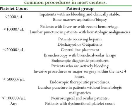
Indications for Platelet Transfusion: Platelets can be transfused therapeutically (to control active bleeding or in preparation for an invasive procedure that would cause bleeding), or prophylactically (to prevent spontaneous bleeding). Actively bleeding patients with thrombocytopenia should be transfused with platelets immediately to keep platelet counts above 50,000/µL in most bleeding situations, and above 100,000/µL if there is disseminated intravascular coagulation or central nervous system bleeding. Fever, infection or inflammation, coagulopathy, and acquired or inherited platelet function defect should be also considered as contributing factors to bleeding. The dose and frequency of platelet transfusions will depend on the platelet count and the severity of bleeding. Typical platelet count thresholds that are used for some common invasive procedures in most centers are summarized in Table 3 [5, 22-25].
The threshold for prophylactic transfusion varies depending on the patient and on the clinical scenario. Studies of patients with thrombocytopenia suggest that patients can bleed even with platelet counts greater than 50,000/μL. However, bleeding is much more likely at platelet counts less than 5000/μL. Among
individuals with platelet counts between 5000/μL and
50,000/μL, clinical findings can be helpful in decision-making regarding platelet transfusion such as age, co-existing inflammation, infection, and fever and the underlying condition responsible for a patient's thrombocytopenia [26].
The initial guidelines recommended transfusion of nonbleeding patients at the level of 20000/μL. This value was concluded from the observation that there is significantly increased risk of bleeding when the platelet count is < 5000/μL, and several studies has shown the risk of bleeding does not seem to change between 10000/μL and a count of 20000/μL [27]. Recently,
several important randomized trials and systematic reviews have further clarified platelet transfusion triggers, including therapeutic versus prophylactic platelet transfusion, platelet dosing, whether to use single donor versus random donor platelets, whether to include leukoreduction, irradiation, and whether to match HLA, ABO, and Rh type [5, 27].
Table 3. Platelet count thresholds that are used for common procedures in most centers.
Platelet Count Patient group
<5000/µL Inpatients with no bleeding and clinically stable. Bone marrow aspiration/biopsy
<10000/µL Lumbar puncture in patients with hematologic malignancies Patients with fever or with recent hemorrhage.
<20000/µL
Patients receiving heparin Discharged or Outpatients Central line placement Bronchoscopy with bronchoalveolar lavage
Endoscopic diagnostic procedures
< 50000/µL
Patients who are actively bleeding Invasive procedures or major surgery within the next 4
hours.
Endoscopic therapeutic procedures. Lumbar puncture in patients without hematologic
malignancies < 100000/µL Neurosurgical and ocular patients.
Any Patients with dysfunctional platelet count
There is no trigger for patients with dysfunctional platelets due to underlying platelet function disease or medication affecting platelet function. However, in both situations, transfusion medical service (TMS) physician is involved in helping to establish the dose and frequency of transfusion if multiple transfusions are required. For the common bedside procedures such as central line placement, lumbar puncture, and BM biopsy, the threshold is provider and service dependent and falls between 20000 and 50000/μL [5].
Granulocyte transfusions
Functioning granulocytes are a vital component of the defense system against bacterial and fungal infections in humans. In spite of modern antimicrobials and supportive therapy, bacterial, fungal, and viral infections are still major complications in patients with prolonged disease-related or therapy-related neutropenia. Neutropenia is one of the most frequent side effects of aggressive treatments, and the risk of infection increases rapidly when the granulocyte count falls below 500 cells/μL [28].
Granulocyte transfusions are not commonly used in clinical practices, and are used in neutropenic patients in certain conditions. The efficacy of this therapy has still not been
completely proven [28]. The use of granulocyte
colony-stimulating factor (G-CSF) for donor stimulation has revived interest in granulocyte transfusion. G-CSF use has increased the granulocyte yield by approximately four fold. Multiple recent studies have shown that granulocyte transfusions can be helpful in controlling severe infections progressing despite the use of appropriate antibiotics [29, 30].
The decision on whether to use granulocyte transfusions therapeutically should be made after a detailed assessment of the clinical state of the patient; granulocyte transfusions may be recommended for patients who meet the following criteria: severe isolated neutropenia (absolute neutrophil count <0.5×10⁹/L) or acquired bone marrow suppression with an expected duration of profound neutropenia of more than 10–15 days, or any bacteremia, fungemia, and invasive bacterial or fungal infection in neutropenic patients unresponsive to proper antimicrobial therapy. Another possible group may be patients with granulocyte dysfunctions, such as chronic granulomatous disease, during episodes of severe, life-threatening infection [28].
CMV IgG negative granulocytes should ideally be provided for recipients who are at risk of CMV disease (infants, pregnant women, CMV negative recipients of CMV negative allogeneic bone marrow transplants). HLA compatible apheresis granulocytes should be considered for alloimmunised patients who have previously documented refractoriness to platelet transfusions due to HLA antibodies or who have had severe transfusion reactions as a result of such antibodies [31].
Plasma transfusions:
approximately 250 ml. Both FFP and FP24 are considered clinically equivalent and are typically transfused using a weight-based dosing of 10 to 20 ml/kg of recipient’s weight. Once thawed, either product must be transfused within 24 hours or be relabeled as “thawed plasma” to allow for refrigerated storage for up to 5 days [32]. Risks associated with plasma transfusion include
allergic reactions, TACO, TRALI, and transfusion-transmitted infections. Indications of plasma transfusion can be summarized for the following clinical conditions [33-35]:
1. Active bleeding or risk of bleeding due to deficiency of multiple coagulation factors.
2. Severe bleeding due to warfarin therapy or emergency
reversal of warfarin effect in settings, where prothrombin complex concentrate with adequate levels of factor VII is not available.
3. Massive transfusion with coagulopathic bleeding.
4. Bleeding or prophylaxis of bleeding for a known single coagulation factor deficiency for which no concentrate is available.
5. Replacement fluid when performing plasma exchange,
particularly in the treatment of thrombotic thrombocytopenic purpura (TTP).
6. Rare specific plasma protein deficiencies in a setting where concentrates are not readily available such as hereditary C1-inhibitor, antithrombin, protein C and protein S deficiencies and in heparin resistance [36].
Frozen Plasma should not be used for increasing blood volume or albumin concentration, coagulopathy that can be corrected with administration of Vitamin K and in normalizing abnormal coagulation screen results, in the absence of bleeding. A common reason for plasma transfusion is to normalize an elevated international normalized ratio (INR) before a planned surgery or invasive procedure, in spite of lack of evidence demonstrating clinical benefit [37, 38]. The faulty assumptions in this situation are
the elevated INR correlates with a risk for bleeding, and plasma transfusion will normalize the INR and reduce this risk [39]. The
analysis of available studies demonstrated that a mildly elevated INR is not predictive of an elevated risk for bleeding [40]. Further,
for mild prolongation of the INR (1.1-1.85), transfusion of plasma has not been shown to significantly improve the INR value
[41]. If plasma transfusion is indicated to correct an elevated INR,
a post transfusion INR must be obtained before ordering
additional plasma. Patients with an INR ≥ 2.0 (≥ 1.5 for
neurosurgical patients) are considered appropriate candidates for plasma transfusion [5].
In patients with liver disease, Frozen Plasma may be used to treat multiple coagulation factors prior to an invasive procedure that would create a risk of bleeding. However, the response may be unpredictable and complete normalization of the hemostatic defect does not occur. Patients with liver disease may safely
undergo operative or invasive procedures when the PT is ≤1.5
times mid-range normal. Analysis of factor levels over an INR range of 1.3 to 1.9 demonstrated mean factor levels that were adequate to support hemostasis [42].
Cryoprecipitate Transfusion
A cryoprecipitate unit is prepared by thawing one unit of FFP between 1-6oC and recovering the cold insoluble precipitate. The cryoprecipitate is refrozen within 1 hour. If several units of cryoprecipitate have been pooled into one bag, it was labeled Cryoprecipitated Antihemophilic Factor (AHF), Pooled. Cryoprecipitate contains concentrated levels of fibrinogen, Factor VIII:C, Factor VIII: vWF (von Willebrand factor), Factor XIII, and fibronectin. Each unit of cryoprecipitate should contain at least 80 IU Factor VIII: C and 150 mg of fibrinogen in 5-20 mL of plasma. Compatibility testing is unnecessary. Cytomegalovirus testing and leukoreduction are not required. If a large volume of ABO-incompatible cryoprecipitate is used, the recipient may develop a positive Direct antiglobulin test (DAT) and, very rarely, mild hemolysis [43].
Indications and Contra-indications: Cryoprecipitate is indicated for bleeding associated with fibrinogen deficiencies in the following conditions [43]:
1. Hypofibrinogenemia / dysfibrinogenemia: with fibrinogen levels of <100 to 120 mg/dL or reduced functional levels of fibrinogen.
2. Uremic bleeding, if 1-deamino-8-D-arginine vasopressin (DDAVP) and other modalities are not available
3. Massive Transfusion: when the fibrinogen level is
documented to be <100 mg/dL.
4. When it is not possible to give enough FFP to provide adequate levels of fibrinogen without volume overloading the patient.
5. Congenital afibrinogenemia / Congenital and
acquireddysfibrinogenemia: in case of bleeding or risk of bleeding associated with a fibrinogen level of <100 mg/dL by a quantitative or functional assay.
6. For hemophilia A or vWD, cryoprecipitate should only be used if appropriate recombinant or virus- inactivated Factor VIII or Factor VIII: vWF concentrates are not available. DDAVP is the treatment choice for type 1 vWd
[44].
Cryoprecipitate is indicated as well in Factor XIII deficiency which is a rare disease characterized by bleeding and poor wound healing. Factor XIII has a half-life of 4 to 14 days, and only about 1-5% activity levels are needed to control bleeding. Virus inactivated Factor XIII concentrates are preferred for the treatment of Factor XIII deficient patients if available. Cryoprecipitate can be given in doses of one bag per 10-20 kg of body weight every 3 to 4 weeks [42, 45].
Blood Substitute and Current Research and
Trials
artificial blood or blood surrogates) is a substance used to mimic and fulfill some functions of biological blood. Blood substitutes are coming up and several sources of hemoglobin-based oxygen carriers are currently in Phase III clinical trials [46, 47]. Intravenous
immunoglobulin has been used for many years. Cryopreservation and lyophilization of platelets and platelet substitutes are all being studied as remedies for the short shelf-life of the platelet components [46].
A-Red Cell Substitutes: There are different types of red cell substitutes currently being studied for use in transfusion medicine. The goal is to enhance the oxygen-carrying capacity for patients suffering from acute anemia due to blood loss. All of these have shown some promises for use in patients who refuse blood transfusion or in situations where blood is not readily available. The main categories of oxygen-carrying blood substitutes are hemoglobin-based oxygen carriers (HBOC) and perfluorocarbon-based oxygen carriers (PFBOC) [3, 48]. Oxygen
therapeutics are in clinical trials in the U.S. and Europe, and Hemopure is available in South Africa [47, 49].
1. Hemoglobin-Based Oxygen Carriers: These are purified cell-free hemoglobins, where the globulin portion of the molecule has been modified chemically by conjugation, cross-linking or polymerization. Modification increases the oxygen-releasing ability of the haemoglobin. The bovine HBOC has completed phase III clinical trials and has been approved in South Africa for treatment of perioperative anaemia in adult surgical patients [47, 49, 50].
2. Perfluorocarbon-Based Red Cell Substitutes:
These consist of carbon backbones highly substituted with fluorine. They can dissolve large amounts of oxygen. The perfluorocarbon (PFC) are biologically inert; however, the phospholipids are required to emulsify them. The only perfluorocarbon that remains in clinical trials is Oxygent, an emulsion of perfluoroocytl bromide and egg yolk phospholipid. Oxygent is being studied for use in perioperative period to allow more extensive hemodilution [51, 52].
B-Platelets Substitutes: Many experimental approaches have been explored to produce hemostatically active novel human platelet products and substitutes capable of long-term storage. These various products are designed to replace the use of allogeneic donor platelets with modified or artificial platelets.
1. Cryopreserved platelets: Platelets suspended in dimethylsulfoxide (DMSO) at -80°C have been preserved up to 10 years. During the thawing and post-thaw processing, however, these platelets develop functional and morphologic defects. There are investigations under way to develop methods that do not require processing after freezing, and can be directly infused after thawing. Because of the complexities of storing, processing and thawing frozen platelets, the current use is limited
[46, 53, 54].
2. Lyophilized platelets: Lyophilized platelets are created after treatment with a paraformaldehyde solution and then freeze-dried. Specific advantages of this product include storage measured in years instead of days, reduced storage space and true sterility. Once rehydrated, they appear to retain
structural integrity and attach only to damaged subendothelial surfaces [55-57].
3. Infusible Platelet Membranes (IPMs): Infusible Platelet Membranes (IPMs) are manufactured from outdated platelet units. Platelet-derived microparticles (microvesicles) are the particles that are formed spontaneously from a platelet during collection and processing of components. They appear to have the ability to function as a platelet. They are procoagulant-active, adhere to the vascular subendothelium and enhance platelet adhesion to form a primary hemostatic plug. One indication for IPMs may be in patients who are refractory to platelet transfusions and for whom finding human leukocytic antigen (HLA)-matched platelet apheresis donors is difficult. One problem appears to be a relatively short life (less than 24 h) in vivo[58, 59].
Conclusions and Recommendations
Transfusion of blood and blood components has been a routine practice for more than half a century. An assessment of the value of transfusion based on well-designed and appropriately powered randomized, controlled trials is the first step in optimizing transfusion practices. Systematic reviews provide the second step whereas the third step is the development of clinical practice guidelines. Such clinical practice guidelines are typically supported by professional organizations and/or health authorities. Although implementation of clinical practice guidelines can be challenging, such guidelines are necessary for the practice of evidence-based medicine, which optimizes patient care and improves patient outcomes. To establish a stronger scientific basis for transfusion practice, future research is essential to provide meaningful evidence and development of algorithms framing the indications and effectiveness of blood components and hemoglobin-based oxygen carriers or other synthetic blood substitutes to reduce transfusion requirements.
Competing interest
The author indicated no potential conflicts of interest.
References
1. Takei T, Amin NA, Schmid G, Dhingra-Kumar N and
Rugg D (2009). Progress in global blood safety for HIV. Journal of Acquired Immune Deficiency Syndromes, 52: S127–31.
2. Shah A, Stanworth SJ and McKechnie S (2015). Evidence and triggers for the transfusion of blood and bloodproducts. Anesthesia, 70 (suppl 1): 3-5.
3. Wang J and KandKlein HG (2010). Red blood cell
transfusion in the treatment and management of anaemia: the search for the elusive transfusion trigger, 98: 2-11. 4. Consensus conference (1988). Perioperative red blood cell
transfusion. JAMA, 260: 2700-3.
6. Carson J, Carless PA and Hebert P (2012). Transfusion thresholds and other strategies for guiding allogeneic red blood cell transfusion. Cochrane Database Syst Rev., 18: CD002042.
7. Shander A, Gross I, Hill S, Javidroozi M and Sledge S (2013). A new perspective on best transfusion practices. Blood Transfusion, 11: 193-202.
8. Cholette J, Swartz M, Rubenstein J, Henrichs K, Wang H, Powers K, Daugherty L, Alfieris G, Gensini F and Blumberg N (2016). Outcomes Using a Conservative Versus Liberal Red Blood Cell Transfusion Strategy in Infants Requiring Cardiac Operation. Ann Thorac Surg., pii: S0003-4975: 30544-6.
9. Carson J, Terrin M, Noveck H, Sanders D, Chaitman B, Rhoads G, Nemo G, Dragert K, Lauren Beaupre L, Hildebrand K, Macaulay W, Lewis C, Cook D, Dobbin G, Zakriya K, Apple F, Horney R and Magaziner J, for the FOCUS Investigators (2011). Liberal or Restrictive Transfusion in High-Risk Patients after Hip Surgery. N Engl J Med, 365: 2453-62.
10. Mazer CD, Whitlock RP, Fergusson DA, Belley-Cote E, Connolly K, Khanykin B, Gregory AJ, de Médicis É, Carrier FM, McGuinness S, Young PJ, Byrne K, Villar JC, Royse A, Grocott HP, Seeberger MD, Mehta C, Lellouche F, Hare GMT, Painter TW, Fremes S, Syed S, Bagshaw SM, Hwang NC, Royse C, Hall J, Dai D, Mistry N, Thorpe K, Verma S, Jüni P, Shehata N; TRICS Investigators and Perioperative Anesthesia Clinical Trials Group. Six-Month Outcomes after Restrictive or Liberal Transfusion for Cardiac Surgery. N Engl J Med. 2018 Sep 27;379(13):1224-1233.
11. Task Force for Diagnosis and Treatment of
Non-ST-Segment Elevation Acute Coronary Syndromes of European Society of Cardiology, Bassand JP, Hamm CW, Ardissino D, Boersma E, Budaj A, Fernández-Avilés F, Fox KA, Hasdai D, Ohman EM, Wallentin L, Wijns W. (2007). Guidelines for the diagnosis and treatment of non-ST-segment elevation acute coronary syndromes. Eur Heart J., 28: 1598-660.
12. Rivers E, Nguyen B, Havstad S, Ressler J, Muzzin A,
Knoblich B, Peterson E, Tomlanovich M and Early Goal-Directed Therapy Collaborative Group (2001). Early goal-directed therapy in the treatment of severe sepsis and septic shock. N Engl J Med., 345: 1368-77.
13. Hébert P, Wells G, Blajchman M, Marshall J, Martin C, Pagliarello G, Tweeddale M, Schweitzer I, Yetisir E, and the Transfusion Requirements in Critical Care Investigators for the Canadian Critical Care Trials Group (1999). N Engl J Med, 340: 409-417.
14. Villanueva C, Colomo A, Bosch A, Concepción M,
Hernandez-Gea V, Aracil C, Graupera I, Poca M, Alvarez-Urturi C, Gordillo J, Guarner-Argente C, Santaló M, Muñiz E and Guarner C (2013). Transfusion strategies for acute upper gastrointestinal bleeding. N Engl J Med., 368: 11-21.
15. Likosky DS, FitzGerald DC, Groom RC, Jones DK, Baker RA, Shann KG, Mazer CD, Spiess BD and Body SC (2010). Effect of the perioperative blood transfusion and blood conservation in cardiac surgery clinical practice guidelines of the Society of Thoracic Surgeons and the Society of Cardiovascular Anesthesiologists upon clinical practices. Anesth Analg. 111: 316-23.
16. Slichter SJ (2007a). Evidence-based platelet transfusion guidelines. Hematology Am Soc Hematol Educ Program, 2007: 172-8.
17. McCullough J (2010). Overview of platelet transfusion. Semin Hematol., 47: 235-42.
18. Blumberg N, Heal JM, Gettings KF, Phipps RP, Masel D, Refaai MA, Kirkley SA and Fialkow LB (2010). An association between decreased cardiopulmonary complications (transfusion-related acute lung injury and transfusion-associated circulatory overload) and implementation of universal leukoreduction of blood transfusions. Transfusion, 50: 2738-44.
19. King KE and Ness PM (2011). How do we prevent
transfusion-associated graft-versus-host disease in children? Transfusion, 51: 916-20.
20. Kleinman S, Reed W and Stassinopoulos A (2013). A
patient-oriented risk-benefit analysis of pathogen-inactivated blood components: application to apheresis platelets in the United States. Transfusion, 53: 1603-18.
21. Daurat A, Roger C, Gris J, Daurat G, Feissel M, Le
Manach Y, Lefrant J and Muller L (2016). Apheresis platelets are more frequently associated with adverse reactions than pooledplatelets both in recipients and in donors: a study from French hemovigilance data. Transfusion, 56: 1295-303.
22. Van Veen JJ, Nokes TJ and Makris M (2010). The risk of spinal haematoma following neuraxial anaesthesia or lumbar puncture in thrombocytopenic individuals. Br J Haematol., 148: 15-25.
23. Astwood E and Vora A (2011). Personal practice: how we manage the risk of bleeding and thrombosis in children and young adults with acute lymphoblastic leukaemia. Br J Haematol., 152: 505-11.
24. Zeidler K, Arn K, Senn O, Schanz U and Stussi G (2011). Optimal pre-procedural platelet transfusion threshold for central venous catheter insertions in patients with thrombocytopenia. Transfusion, 51: 2269-76.
25. Nandagopal L, Veeraputhiran M, Jain T, Soubani AO and Schiffer CA (2016). Bronchoscopy can be done safely in patients with thrombocytopenia. Transfusion. 56: 344-8.
26. Slichter SJ, Kaufman RM, Assmann SF, McCullough J,
27. Slichter SJ (2004). Relationship between platelet count and bleeding risk in thrombocytopenic patients. Transfus Med Rev, 18: 153–167.
28. Drewniak A, Kuijpers TW (2009). Granulocyte
transfusion therapy: randomization after all? Haematologica, 94:1644-8.
29. Atallah E and Schiffer CA (2006). Granulocyte transfusion. Curr Opin Hematol. 13:45-9.
30. Quillen K, Wong E, Scheinberg P, Young NS, Walsh TJ, Wu CO, Leitman SF (2009). Granulocyte transfusions in severe aplastic anemia: an eleven-year experience. Haematologica. 94:1661-8.
31. Ljungman P (2004). Risk of cytomegalovirus transmission by blood products to immunocompromised patients and means for reduction. Br J Haematol. 125:107-16.
32. Benjamin RJ and McLaughlin LS (2012). Plasma
components: properties, differences, and uses. Transfusion, 5: 9S-19S.
33. Szczepiorkowski ZM, Winters JL, Bandarenko N, Kim
HC, Linenberger ML, Marques MB, Sarode R, Schwartz J, Weinstein R, Shaz BH and Apheresis Applications Committee of the American Society for Apheresis (2010). Guidelines on the use of therapeutic apheresis in clinical practice--evidence-based approach from the Apheresis Applications Committee of the American Society for Apheresis. J Clin Apher., 25: 83-177.
34. Roback JD, Caldwell S, Carson J, Davenport R, Drew MJ, Eder A, Fung M, Hamilton M, Hess JR, Luban N, Perkins JG, Sachais BS, Shander A, Silverman T, Snyder E, Tormey C, Waters J, Djulbegovic B; American Association for the Study of Liver; American Academy of Pediatrics; United States Army; American Society of Anesthesiology and American Society of Hematology (2010). Evidence-based practice guidelines for plasma transfusion, 50: 1227-39. 35. Yang L, Stanworth S, Hopewell S, Doree C and Murphy M
(2012). Is fresh-frozen plasma clinically effective? An update of a systematic review of randomized controlled trials. Transfusion, 52: 1673-86.
36. American Society of Anesthesiologists Task Force on
Perioperative Blood Transfusion and Adjuvant Therapies (2006). Practice guidelines for perioperative blood transfusion and adjuvant therapies: an updated report by the American Society of Anesthesiologists Task Force on Perioperative Blood Transfusion and Adjuvant Therapies. Anesthesiology, 105: 198-208.
37. Stanworth SJ, Estcourt LJ, Powter G, Kahan BC, Dyer C, Choo L, Bakrania L, Llewelyn C, Littlewood T, Soutar R, Norfolk D, Copplestone A, Smith N, Kerr P, Jones G, Raj K, Westerman DA, Szer J, Jackson N, Bardy PG, Plews D, Lyons S, Bielby L, Wood EM, Murphy MF; TOPPS Investigators (2013). A no-prophylaxis platelet-transfusion strategy for hematologic cancers. N Engl J Med. 368: 1771-80.
38. Desborough M and Stanworth S (2012). Plasma transfusion for bedside, radiologically guided, and operating room invasive procedures. Transfusion, 52: 20S-9S.
39. Tinmouth A (2012). Evidence for a rationale use of frozen plasma for the treatment and prevention of bleeding. Transfus Apher Sci., 46: 293-298.
40. Segal JB, Dzik W and Transfusion Medicine/Hemostasis Clinical Trials N (2005). Paucity of studies to support that abnormal coagulation test results predict bleeding in the setting of invasive procedures: an evidence-based review. Transfusion, 45: 1413-1425.
41. Abdel-Wahab OI, Healy B and Dzik WH (2006). Effect of fresh-frozen plasma transfusion on prothrombin time and bleeding in patients with mild coagulation abnormalities. Transfusion. 46: 1279-85.
42. Deitcher SR (2002). Interpretation of the international normalized ratio in patients with liver disease. Lancet, 359: 47-48.
43. AABB, America’s Blood Centers and the American Red
Cross (2002). Circular of Information for the Use of Human Blood and Blood Components. July2002. http://www.aabb.org/tm/coi/ Documents/ coi 1113.pdf. Accessed 10/10/16
44. Mannucci PM (2001). How I treat patients with von
Willebrand disease. Blood, 97: 1915-9.
45. Development Task Force of the College of American
Pathologists (1994). Practice parameter for the use of fresh-frozen plasma, cryoprecipitate and platelets. JAMA, 271: 777-81.
46. Arya RC, Wander G and Gupta P (2011). Blood
componenttherapy: Which, when and how much. J Anaesthesiol Clin Pharmacol., 27: 278-84.
47. Palmer AF and Intaglietta M (2014). Blood substitutes. Annu Rev Biomed Eng., 16: 77-101
48. Jiin-Yu Chen, Michelle Scerbo, and George Kramer
(2009). A Review of Blood Substitutes: Examining The History, Clinical Trial Results, and Ethics of Hemoglobin-Based Oxygen Carriers. Clinics (Sao Paulo), 64: 803–813.
49. Standl T (2001). Haemoglobin-based erythrocyte
transfusion substitutes. Expert Opin Biol Ther, 1: 831-43.
50. Van Hemelrijck J, Levien LJ, Veeckman L, Pitman A,
Zafirelis Z and Standl T (2014). A safety and efficacy evaluation of hemoglobin-based oxygen carrier HBOC-201 in a randomized, multicenter red blood cell controlled trial in noncardiac surgery patients. Anesth Analg., 119: 766-76.
51. Water JA, Trouwborst A and Spense RK (1996). A pilot study of the effects of perflubron emulsion, AF0104, on mixed venous oxygen tension in anesthetized surgical patients. Anesth Analg, 82: 103-7.
52. Stephan C, Schlawne C, Grass S, Waack IN, Ferenz KB, Bachmann M, Barnert S, Schubert R, Bastmeyer M, de Groot H, Mayer C (2014). Artificial oxygen carriers based on perfluorodecalin-filled poly(n-butyl-cyanoacrylate) nanocapsules. J Microencapsul. 31: 284-92
53. Johnson L, Coorey CP and Marks DC (2014). The
54. Cid J, Escolar G, Galan A, Lopez-Vilchez I, Molina P, Diaz-Ricart M, Lozano M and Dumont LJ (2016). In vitro evaluation of the hemostatic effectiveness of cryopreserved platelets. Transfusion, 56: 580-6.
55. Bode AP and Read MS (200). Lyophilized platelets:
Continued development. Transfus Sci, 22: 99-105. 56. Bode AP and Fischer TH (2007). Lyophilized platelets:
fifty years in the making. Artif Cells Blood Substit Immobil Biotechnol., 35: 125-33.
57. Ramachandran N. and Hiles M.C. (2014). Use of
sterilized, lyophilized platelets for multiple applications. Cytotherapy, 16: S60–S61
58. Chao FC, Kim BK, Houranieh AM, Liang FH, Konrad
MW, Swisher SN and Tullis JL (1996). Infusible platelet membrane microvesicles: a potential transfusion substitute for platelets. Transfusion, 36: 536-42.