R E S E A R C H
Open Access
eEF1B
γ
binds the Che-1 and TP53 gene
promoters and their transcripts
Cinzia Pisani
1*, Annalisa Onori
1, Francesca Gabanella
2,3, Francesca Delle Monache
1, Antonella Borreca
2,3,
Martine Ammassari-Teule
2,3, Maurizio Fanciulli
4, Maria Grazia Di Certo
2,3, Claudio Passananti
1and Nicoletta Corbi
1*Abstract
Background:We have previously shown that the eukaryotic elongation factor subunit 1B gamma (eEF1Bγ) interacts with the RNA polymerase II (pol II) alpha-like subunit“C”(POLR2C), alone or complexed, in the pol II enzyme. Moreover, we demonstrated that eEF1Bγbinds the promoter region and the 3’UTR mRNA of the vimentin gene. These events contribute to localize the vimentin transcript and consequentially its translation, promoting a proper mitochondrial network.
Methods:With the intent of identifying additional transcripts that complex with the eEF1Bγprotein, we performed a series of ribonucleoprotein immunoprecipitation (RIP) assays using a mitochondria-enriched heavy membrane (HM) fraction.
Results:Among the eEF1Bγcomplexed transcripts, we found the mRNA encoding the Che-1/AATF multifunctional protein. As reported by other research groups, we found the tumor suppressor p53 transcript complexed with the eEF1Bγprotein. Here, we show for the first time that eEF1Bγbinds not only Che-1 and p53 transcripts but also their promoters. Remarkably, we demonstrate that both the Che-1 transcript and its translated product localize also to the mitochondria and that eEF1Bγdepletion strongly perturbs the mitochondrial network and the correct localization of Che-1. In a doxorubicin (Dox)-induced DNA damage assay we show that eEF1Bγdepletion significantly decreases p53 protein accumulation and slightly impacts on Che-1 accumulation. Importantly, Che-1 and p53 proteins are components of the DNA damage response machinery that maintains genome integrity and prevents tumorigenesis.
Conclusions:Our data support the notion that eEF1Bγ, besides its canonical role in translation, is an RNA-binding protein and a key player in cellular stress responses. We suggest for eEF1Bγa role as primordial transcription/translation factor that links fundamental steps from transcription control to local translation.
Keywords:Translation elongation factor, eEF1Bγ, Che-1, AATF, p53, RIP assay, RNA binding protein, Mitochondria, POLR2C, DNA damage
Background
The eukaryotic elongation factor 1 subunit gamma (eEF1Bγ), also known as the pancreatic tumor-related protein, is a part of the eEF1 multiprotein macromol-ecular complex. The eEF1 holoenzyme plays a role in protein synthesis by recruiting the aminoacyl-tRNAs to the A site of the ribosome [1]. Using the current no-menclature for higher eukaryotes, eEF1 consists of two different sub-complexes: eEF1A and eEF1B. eEF1A
(formerly eEF1α) is a single polypeptide, whereas eEF1B is a multimer of eEF1Bα(formerly eEF1β), eEF1Bδ(formerly eEF1δ), and eEF1Bγ(formerly eEF1γ). There is evidence to indicate that eEF1Bγstimulates, but is not required for, the catalytic activity of eEF1Bα [2, 3]. Indeed, eEF1Bγ appears dispensable for translation, and its absence does not seem to affect the global rate of translational elongation [4, 5]. Nevertheless, multiple non-canonical roles for eEF1Bγ are emerging, some of which can be regulated by phosphoryl-ation driven by several protein kinases [6]. A role of eEFB1γ in the oxidative stress response pathways is justified by the presence in the N terminus of a conserved sequence resem-bling the glutathione-binding region of the theta class of
* Correspondence:[email protected];[email protected] 1CNR-Institute of Molecular Biology and Pathology, Department of Molecular
Medicine, Sapienza University, Viale Regina Elena 291, 00161 Rome, Italy Full list of author information is available at the end of the article
glutathione S-transferase (GST) enzymes, which are in-volved in the detoxification of oxygen radicals. The over-expression of the eEF1Bγ gene product has been reported in several tumors, including pancreatic, breast, colon, and gastric tumors [7–11]. There is growing evidence that the elongation step is also regulated in response to environ-mental cues, supporting the idea that deregulation of trans-lational control serves as a common mechanism by which diverse oncogenic pathways promote cellular transform-ation and tumor development [12, 13]. In a wider context, aberrant proliferation of cancer cells is supported by adap-tation to nutrient microenvironment mediated by a dy-namic metabolic reprogramming [14]. Importantly, the level of eEF1Bγupregulation was shown to positively cor-relate with tumor aggressiveness, presumably due to an al-tered redox balance [2, 15, 16]. eEF1Bγ displays an affinity for membrane and cytoskeleton elements, and it can prop-erly anchor the different subunits of the EF1 complex to the cytoskeleton [2, 6, 17]. Interestingly, Al-Maghrebi et al. (2002) demonstrated the RNA-binding properties of eEF1Bγ by showing for the first time its binding to the 3’ UTR of vimentin mRNA [18], suggesting that eEF1Bγ could exert many of its biological functions through the binding of a pool of mRNAs. In addition, hu-man eEF1Bγwas recently identified in a proteomic screen as a member of the pre-mRNA 3’ end cleavage complex [19]. In this context, eEF1Bγ could participate in the an-choring and translation of a set of mRNAs that are prefer-entially translated on cytoskeletal- or membrane-bound ribosomes, such as vimentin mRNA. Vimentin has been recently reported to have a regulatory role in supporting the morphology, organization and function of mitochon-dria [20, 21]. Importantly we previously demonstrated that eEF1Bγ partially co-localizes with mitochondria [5]. Yoo’s research group showed that hCdc73, a component of the human RNA polymerase II-associated factor complex (PAFc), binds and destabilizes p53 mRNA via eEF1Bγ, thus acting as a binding platform [22]. They proposed that mis-regulation of this interaction may lead to tumor progres-sion. Liu et al. reported a new role for eEF1Bγ in the activation of the NF-Kb signaling pathway, through target-ing the mitochondrial antiviral adaptor protein (MAVS), which bridges viral RNA recognition and downstream sig-nal activation [23]. The Esposito research group showed that the TNF receptor associated protein (TRAP1), a mito-chondrial member of the HSP90 family, which is involved in the protection of oxidative stress, selectively binds eEF1Bγ, and, remarkably, both TRAP1 and eEF1Bγare co-upregulated in human colorectal cancers [24]. We have pre-viously shown that eEF1Bγinteracts with the RNA polymer-ase II (pol II) alpha-like subunit “C” (POLR2C), alone or complexed, in pol II [25–27]. The POLR2C/POLR2J hetero-dimer (also called RPB3/RPB11) is reminiscent of theα sub-unit homodimer of bacterial RNA polymerase [28]. In
bacteria, the alpha subunit homodimer associates withσ fac-tors that mediate promoter recognition [29–31]. Moreover, eEF1Bγ has been described to bind the vimentin 3’UTR, and we have shown that it also binds the promoter region of the vimentin gene [5, 18]. These results suggest that eEF1Bγ has a role in shuttling/nursing vimentin mRNA (and pre-sumably a specific set of mRNAs) from their gene locus to their appropriate cellular compartment for translation. On the basis of eEF1Bγsub-cellular localization and its involve-ment in RNA metabolism and mitochondria/cytoskeleton organization, herein, using a mitochondria-enriched heavy membrane (HM) fraction, we identified, by ribonucleopro-tein complex immunoprecipitation (RIP assay), several novel transcripts that complexed with eEF1Bγ. Among the isolated mRNAs, we found genes involved in translation and in mitochondrial/cytoskeleton metabolism. In particular, we found the mRNA of the pol II binding protein Che-1/AATF, and we confirmed the presence of the p53 transcript [22]. Che-1 plays a role in multiple fundamental processes, in-cluding control of transcription, cell cycle regulation, DNA damage responses and apoptosis [32–34]. Recent studies suggest that Che-1 protein level dysregulation could be rele-vant for the transformation process. Che-1 is found upregu-lated in several leukemia cell lines and in patient with chronic lymphocytic leukemia [33].
Loss or mutation of the oncosuppressor p53 is strongly associated with the susceptibility to cancer and to malig-nant tumor progression [35, 36]. Notably, Che-1 directly binds p53 and is an important regulator of p53 activity [37]. Moreover, Che-1 enhances the oncogenic potential of the mutated forms of the oncosuppressor p53 (mtp53) [34, 38]. Here, we show for the first time that eEF1Bγ binds to the Che-1 and TP53 promoter regions. In addition, we describe a novel mitochondrial localization for the Che-1 protein, and we show that Che-Che-1 needs mitochondrial integrity for correct localization. We suggest a role for eEF1Bγ as a primordial transcription/translation factor that links the fundamental steps between transcription control and local translation.
Methods
Constructs
The myc-tagged pCS2-eEF1Bγ(Myc-eEF1Bγ) (UniProtKB: P26641) construct and its derived deletion mutants were generated by PCR amplification or sub-cloning [5].
Cell culture and transfections
HeLa human cervical cancer cells and HCT116 human colon carcinoma cells were grown in Dulbecco’s modified Eagle’s medium (DMEM) supplemented with 10 % foetal bovine serum (Gibco-BRL, Grand Island, NY, USA). hSH-SY5Y neuroblastoma cells were grown in DMEM supple-mented with 15 % foetal bovine serum. All cell cultures were maintained at 37 °C in a humidified atmosphere of 5 % CO2. Transient transfections were performed using
Lipofectamine or Lipofectamine 2000 reagents (Thermo Fisher Scientific, Inc., Waltham, MA, USA), according to the manufacturer’s instructions.
The siRNA-mediated interference experiments for eEF1Bγ expression were performed by transfecting SMART pool-specific or non-pool-specific control pool double-stranded RNA oligonucleotides (GE Healthcare Dharmacon Inc., Lafayette, CO, USA) using Lipofectamine 2000.
Drug treatment with doxorubicin (1 μM) (Sigma-Aldrich Co., St. Louis, MO, USA) was carried out by in-cubating cells with the indicate concentration of the drug in fresh media for either 1 h or 2 h before analysis.
Sub-cellular fractionation
The mitochondria-enriched heavy membrane (HM) frac-tion were obtained as previously described [39]. Briefly, HeLa cells (~6 × 10⁶) were harvested in lysis buffer (250 mM sucrose, 20 mM Hepes, pH 7.5, 10 mM KCl, 1.5 mM MgCl2, 1 mM EDTA, 1 mM EGTA) and complete
protease inhibitor (Roche, Indianapolis, IN, USA). The cells were disrupted by twelve passages through a 25-gauge needle. The HM fractions were obtained by centrifugation at 10,000gfor 10 min. The HM pellet was resuspended in high stringency buffer (50 mM Tris–HCl pH 7.4, 250 mM NaCl, 5 mM EDTA, 10 % glycerol, 0.5 % Igepal-CA 630) plus a proteinase inhibitor cocktail (Complete™, Roche, In-dianapolis, IN, USA).
The mitochondrial fraction was purified from HeLa and hSH-SY5Y cells (~2 × 10⁷) using a Qproteome Mitochon-dria Isolation Kit (Qiagen, Hilden, Germany) as previously described [5].
Immunoblotting
Whole-cell lysate was obtained as previously described, and sub-cellular fractionations (see above) were analyzed by western blotting [5]. The publicly available software ImageJ (National Institutes of Health, USA) was used to quantify the densitometry of the immunoblot bands.
RIP assay
Mitochondria-enriched heavy membrane (HM) fraction or whole-cell extracts were prepared as above in the presence of RNase inhibitors (Thermo Fisher Scientific, Inc., Waltham, MA, USA). For the immunoprecipitation assay, the protein lysate was pre-cleared for 1 h at 4 °C
with Protein A/G-Agarose beads (Roche, Indianapolis, IN, USA) and then immunoprecipitated overnight with the anti-eEF1Bγrabbit polyclonal antibody or with anti-myc monoclonal antibody. A “no-antibody” immunopre-cipitation was performed as a negative control. The beads were washed five times for 5 min at 4 °C with a high strin-gency buffer and once in PBS buffer. The beads containing the immunoprecipitate samples were collected and resus-pended in buffer R (50 mM Tris–HCl pH 7, 10 mM DTT, 5 mM EDTA, 1 % SDS) [40]. A portion of immunoprecip-itation was processed for western blot analysis. RNA was extracted using TRIzol® reagent (Thermo Fisher Scientific, Inc., Waltham, MA, USA) according to the manufacturer’s instructions. RNAs were converted to cDNAs and ran-domly amplified using a Full Spectrum™ Complete Tran-scriptome RNA Amplification Kit (System Biosciences, Mountain View, CA, USA) according to the manufac-turer’s protocol. cDNA was run on a 2 % agarose gel, and the portion between 200 and 500 bp was isolated and cloned into pGEM-T vectors using a pGEM®-T Easy Vec-tor System (Promega, Madison, WI, USA) according to the manufacturer’s instructions. The obtained clones were analyzed by EcoRI digestion, and the selected clones were sequenced by Eurofins MWG Services.
RNA extraction, retrotranscription and quantitative real-time PCR (qPCR)
Total RNA from HeLa and hSH-SY5Y cells was extracted using TRIzol® reagent according to the manufacturer’s in-structions and was then reverse transcribed using a High Capacity cDNA Reverse Transcription kit (Thermo Fisher Scientific, Inc., Waltham, MA, USA). A quantitative real-time PCR (qPCR) assay was performed in triplicate in a 96-well format in an ABI Prism 7000 Sequence Detection System (Applied Biosystems, Foster City, CA, USA) using the SYBR Green PCR Master mix. GAPDH or MT-ND2 was used for the normalization of mRNA, and the relative expression was calculated using the comparative Ct method (2-ΔΔCt). Primer sequences used in this study are shown in Additional file 1: Table S1.
Chromatin immunoprecipitation (ChIP) assay
Polysome profile analysis
The polysomal profile analysis was performed as previ-ously described [42]. Briefly, cells were homogenized in lysis buffer (10 mM Tris–HCl pH 7.5, 100 mM NaCl, 10 mM MgCl2, 1 % Triton X-100, 30 U/ml RNasin). Ly-sates were incubated on ice for 5 min and then centri-fuged at 12,000 rpm for 5 min at 4 °C. Supernatants were immediately loaded onto a 10 ml 15–50 % (w/v) sucrose gradient and centrifuged at 37,000 rpm for 180 min at 4 °C at in a Beckman SW41 rotor. For EDTA treatments, 100 mM EDTA was added to the cytoplas-mic extracts before stratification on a sucrose gradient. Free ribosomal subunits (60S and 40S), monosomes (80S), large polysomes, and the very light mRNPs were detected by UV absorbance at 254 nm using a BioLogic LP system (BioRad Inc., Hercules, CA, USA). Each gra-dient was collected in 9 fractions, and the proteins were precipitated with a mix containing 50 % ethanol, 25 % methanol and 25 % acetone and were then processed for western blot analysis.
Immunofluorescence and confocal laser scanning microscopy
Cells were fixed with 4 % formaldehyde in PBS, perme-abilized in 0.2 % Igepal-CA 630 (Sigma Chemical Co., St. Louis, MO, USA) for 10 min, and blocked with 1 % BSA in PBS at room temperature. Samples were incubated se-quentially with the appropriate primary and secondary antibodies. Slides were mounted with ProLong with DAPI (Thermo Fisher Scientific, Inc., Waltham, MA, USA) or Hoechst 33258 solution (Sigma Chemical Co., St. Louis, MO, USA). To label mitochondria, cells were incubated with 250 nM of MitoTracker® Red CMXRos M7512 (Thermo Fisher Scientific, Inc., Waltham, MA, USA) according to the manufacturer’s instructions and then were fixed and incubated with anti-Che-1 rat poly-clonal antibodies. Slides were examined by conventional epifluorescence microscopy (Olympus BX51). Images were captured using a digital camera SPOT RT3 and merged using the IAS2000 software. For confocal laser scanning microscopy, slides were examined with a confocal system TCS-SP5 (Leica Microsystem, GmbH Wetzlar, Germany).
RNA-FISH combined with immunofluorescence
HeLa cells were processed for immunofluorescence in the presence of RNase inhibitors, with secondary anti-body incubation subsequent to FISH to prevent denatur-ation of the antibodies. FISH was performed using a FITC labeled oligonucleotide probe (Additional file 1: Table S1) [43, 44]. The cells were fixed in 4 % parafor-maldehyde in PBS (pH 7.4) for 30 min and washed three times with PBS and 0.2 % Igepal-CA 630 for 5 min. The slides were then permeabilized by treatment with 70 %
ethanol overnight at 4 °C. The cells were rehydrated for 5 min in 50 % formamide, 2× SCC (300 mM NaCl, 30 mM sodium citrate, pH 7.0) and pre-hybridized with hybridization buffer (50 % formamide, 10 % dextran sul-fate, 2 mM vanadyl-ribonucleoside complex, 40μgE. coli tRNA, 2× SSC) for 1 h at 37 °C. Then, cells were incubated overnight with 30 ng FITC labeled DNA oligonucleotide probe in 40μl hybridization buffer at 37 °C. The coverslips were washed twice in 2× SCC/50 % formamide at 37 °C and twice in 1× SCC at room temperature. The coverslips were incubated with anti-FITC primary antibodies (to amplify the signal) and the appropriate secondary anti-bodies in 1 % blocking reagent solution (Roche, Indianapo-lis, IN, USA) at room temperature and mounted with ProLong with DAPI. The slides were examined by conven-tional epifluorescence microscopy (Olympus BX51). The images were captured using a digital camera SPOT RT3 and merged using IAS2000 software. Co-localization ana-lysis was performed using Image J software (Image J, Colo-calization Coloc 2, Intensity correlation quotient (ICQ)). The ICQ value is calculated based on Li's intensity correl-ation analysis, which is considered a stable method for co-localization analysis as it allows the discrimination of coincidental events in a heterogeneous situation. ICQ var-ies from−0.5 (exclusion) to 0.5 (complete co-localization) [45]. Region-of-Interest (ROI) were drawn around single cells. Background and threshold correction were applied for each ROI.
Antibodies
times with 250 μg of the purified His tag Che-1 protein every week using Freund’s adjuvants (Difco, Detroit, MI, USA); antiserum was collected 5 days after the last injection. All procedures were carried out in accordance with the eth-ical guidelines for animal care of the European Community Council (directive 2010/63EU). Housing of the animals meets the behavioral needing of the specie and was supervised by the Responsible Veterinarian. The secondary antibodies conjugated to horseradish peroxidase were pur-chased from GE Healthcare (GE Healthcare, Chicago, IL, USA). Alexa-Fluor-488 or Alexa-Fluor-594-conjugated sec-ondary antibodies were purchased from Thermo Fisher Sci-entific (Thermo Fisher SciSci-entific, Inc., Waltham, MA, USA).
Results
eEF1Bγbinds specific mRNAs and their gene promoter regions
The eEF1Bγprotein has been shown to associate with both the vimentin promoter and vimentin 3' UTR mRNA, thus
influencing the cellular shape and mitochondria localization [5, 18]. Here, with the intent of identifying additional tran-scripts complexed with the eEF1Bγprotein, we performed a series of ribonucleoprotein immunoprecipitation (RIP) assays. A scheme of the RIP assay protocol is shown in Fig. 1a. The HeLa cell mitochondria-enriched HM frac-tion was immunoprecipitated with anti-eEF1Bγpolyclonal antibodies (Fig. 1b). The co-immunoprecipitated RNA was purified and retro-transcribed. The cDNA output was randomly amplified, size selected and cloned. Fig. 1c shows the resulting HM-cDNA library. Interestingly, among the mRNAs complexed with eEF1Bγ, we iso-lated the Che-1/AATF mRNA. The RT-PCR analysis, shown in Fig. 1d, demonstrated the presence of Che-1 mRNA in eEF1BγRIP assay output.
Although in a specific subcellular fraction, we confirmed the presence of vimentin and p53 transcripts, as previ-ously reported [18, 22]. Additional file 2: Table S2 shows a list of selected individual clones randomly sequenced from
b
c
d
e
the mitochondria-enriched heavy membrane (HM)-cDNA library. Additional file 3: Figure S1A shows the RT-PCR validation of some of the individual clones. The finding of the Che-1 transcript in the eEF1Bγ RIP assay output is consistent with the presence of eEF1Bγon the Che-1 pro-moter that we have communicated in our previous manu-script [5]. As shown in Fig. 1e, by DNA ChIP analysis in Hela cells, we confirmed the presence of eEF1Bγ on the Che-1 promoter, and, importantly, we showed for the first time the presence of eEF1Bγon the TP53 gene promoter. The ChIP experiments were also performed on hu-man neuroblastoma hSH-SY5Y cells, confirming the data (Additional file 4: Figure S2A).
eEF1Bγco-localizes with specific mRNAs
It has been shown that in vimentin 3’ UTR mRNA, eEF1Bγ binds an RNA element named the “Y shaped structure”, which exhibits striking sequence homology across species [47, 48]. Moreover, Sasvari et al. demon-strated a role for eEF1Bγ in Tomato bushy stunt virus (TBSV) replication by interacting with a stem-loop
structure at the 3’ end of the viral RNA [49]. Interest-ingly, as shown in Fig. 2a, the human Che-1 3′ UTR mRNA is characterized by a stem-loop secondary struc-ture folded according to the dynamic programming algo-rithm originally proposed by Zuker and Stiegler [50]. To visualize and validate the interaction between eEF1Bγ and the Che-1 3’UTR, we performed an MS2 assay [51]. Figure 2b (left panel) shows a schematic representation of the MS2 assay. Briefly, the MS2-GFP system is based on two components: a fusion of the MS2 coat protein with the GFP protein carrying a nuclear localization sig-nal NLS (MS2-GFP) and a reporter transcript (Report mRNA) containing multimers of the RNA stem-loop, recognized by the MS2-GFP protein, upstream of the 3’ UTR of the mRNA of interest. MS2-GFP chimeric pro-tein, over-expressed alone in mammalian cells, shows a nuclear localization, whereas when it binds to the RNA stem-loop, it tends to move in the cytoplasmic compart-ment. As shown in Fig. 2b (right panel), MS2-GFP pro-tein expressed in HeLa cells alone or with the 3′UTR of Che-1 or vimentin was analyzed in the presence or
3’UTR of interest LUC
AA AA MS2
Binding Sites
GFP
MS2
G FP
MS 2
GF P
MS 2 G
F P
MS 2 G
F P
MS 2
a
H e L a
eEF1Bγ mRNA Che-1 merge
eEF1Bγ mRNA VIM merge
b
c
3’ UTR Che-1
C
+Myc-eEF1Bγ
MS2GFP +3’ UTR Che-1
MS2GFP +3’ UTR VIMMS2GFP
H e L a
0 0,1 0,2 0,3 0,4 0,5
eEF
1Bγ
;
3’U
TR
VIM
eEF
1
B
γ
/m
R
N
A
co
lo
cali
zati
o
n
, (
ICQ)
eEF 1Bγ
;
3’U TR
Ch e-1
absence of eEF1Bγ. Only in the presence of myc-eEF1Bγa clear dot fluorescent pattern is observed in the cytoplasmic compartment. Additional file 3: Figure S1B shows additional MS2 assays performed on different mRNAs complexed with eEF1Bγ (see Additional file 2: Table S2). As shown in Fig. 2c, to visualize co-localization of endogenous eEF1Bγprotein and endogenous Che-1 or vimentin mRNAs, we performed RNA-FISH analysis com-bined with indirect eEF1Bγ-immunofluorescence in HeLa cells. We quantified eEF1Bγ/3’UTR mRNA co-localization by measuring intensity correlation quotient (ICQ) values as shown in the bar graph.
Characterization of the eEF1BγmRNA binding property
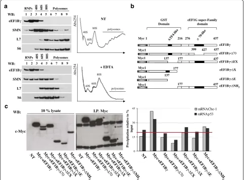
Taking into account that eEF1Bγ acts as an RNA-binding protein, we assessed the eEF1Bγ distribution in a polysomal profile in HeLa cells (Fig. 3a, right). Cyto-plasmic extracts were subjected to ultracentrifugation in a sucrose density gradient in the presence or absence of EDTA. The EDTA treatment dissociated the large and small ribosomal subunits and virtually disrupted all poly-ribosomes. In our experimental conditions, eEF1Bγ pro-tein was mainly retained in slow-sedimenting fractions that were enriched in ribonucleoprotein particles (mRNPs) (Fig. 3a, left). We also tested the distribution
b
70-l ike AT F 4-like 4371 216 276
GST Domain Myc eEF1G super-Family domain eEF1Bγ 177
Myc1 eEF1Bγ-ΔX
137 Myc1
eEF1Bγ-ΔE
Myc1 eEF1Bγ-ΔNH
2 eEF1Bγ-ΔEX eEF1Bγ-Δ70
399 427 Myc1 437 137 177 Myc1 437 177 437
c
0 15 30 45 M yc-e E F 1Bγ NT Myc
-eE F1B γ -ΔΕX M yc -eE F 1Bγ -Δ E M yc -eEF 1Bγ -Δ70 M yc -eE F 1Bγ -Δ X M yc -eE F 1Bγ -Δ N H 2 mRNAp53 mRNAChe-1 input WB: * * * c-Myc I.P. Myc M yc-e EF 1Bγ N
T Myc
-eE F1B γ -ΔΕX M yc -eE F 1Bγ -Δ E M yc -eE F1B γ -Δ70 M yc-e EF 1Bγ -Δ X M yc -eE F 1Bγ -Δ N H 2 M yc-e E F 1Bγ γ N
T Myc
-eE F1B γ -ΔΕX M yc -eE F 1Bγ -Δ E M yc -eE F1B γ -Δ70 M yc-e EF 1Bγ -Δ X M yc -eE F 1B -Δ N H 2 eEF1Bγ
WB: 1 2 3 4 5 6 7 8 9 Polysomes RNPs 40S 60S 80S
S6 L7 SMN
1 2 3 4 5 6 7 8 9 Polysomes RNPs 40S 60S 80S
eEF1Bγ WB: S6 L7 SMN
a
Abs254 NT 40S 60S 80S polysomes Abs254 + EDTA 40S60S 80S polysomesof the survival motor neuron (SMN) protein because it has been demonstrated that this RNA-related protein as-sociates with polyribosomes [42, 52]. The protein com-position of each collected fraction was validated using S6 and L7 antibodies to monitor the small and large ribosomal subunits, respectively. The eEF1Bγ protein is a multi-domain polypeptide that harbors a GST like main on the N-terminus and an eEF1G super-family do-main at the carboxyl terminus (ref: pFAM 00147). In addition, in the carboxyl terminus, there is a region with 74 % homology to the sigma-70 factors ECF subfamily signature (ref: PDOC00814). To identify the eEF1Bγ do-main/s responsible for Che-1 and p53 mRNA interac-tions, a series of eEF1Bγ deletion mutants fused to the myc tag was constructed (Fig. 3b). HeLa cells were tran-siently transfected with myc-eEF1Bγor with its deletion mutants to perform a RIP assay analysis. The immuno-precipitations, performed using myc-tag antibodies, were analyzed by western blotting as shown in Fig. 3c (left panel), and co-immunoprecipitated mRNAs were ex-tracted and converted to cDNA. Che-1 and p53 mRNAs co-immunoprecipitated with the indicated constructs were plotted in a graph. The data are expressed as per-cent precipitation relative to the input mRNAs. The mean background level is illustrated by the horizontal line in the graph [53]. By RT-PCR, the cDNA output confirmed the presence of both Che-1 and p53 tran-scripts in full-length eEF1Bγ and their limited presence in deletion mutants eEF1Bγ−ΔNH2and eEF1Bγ−ΔX.
Che-1 protein mitochondria localization and eEF1Bγ depletion effects
We previously demonstrated that eEF1Bγcontributes to govern the correct localization of vimentin intermediate-filament protein, which is known to be involved in cell morphology and organelle positioning [5]. Because the Che-1 protein was observed in both the nucleus and cytoplasmic organelles [33, 54, 55], we examined Che-1 sub cellular localization in more detail. In Fig. 4a (left panel), using the rat Che-1 antibody in a dual-label im-munofluorescence assay with mitochondrion-selective dye MitoTracker (red), we detected novel localization of Che-1 protein in mitochondria. To further verify the Che-1 mitochondrial localization, we used the mito-chondrial marker Tom20 in a dual-label immunofluores-cence assay in hSH-SY5Y cells. Extensive co-localization between endogenous Che-1 and Tom20 is revealed by the merged-color image (Additional file 4: Figure S2B). West-ern blot analysis of the mitochondria-enriched heavy mem-brane (HM) fraction, prepared from HeLa cells, clearly confirmed a Che-1 mitochondrial association (Fig. 4a, right panel). Che-1 mitochondrial localization was also observed in the mitochondrial fraction prepared from hSH-SY5Y cells (Additional file 4: Figure S2C). To investigate the
possible effects of eEF1Bγ depletion on mitochondrial Che-1 expression levels, quantitative real time PCR (qPCR) and western blot analyses were performed with HeLa cells. In our experimental conditions, eEF1Bγ knockdown did not produce any significant change in Che-1 expression levels (both RNA and protein levels) in mitochondria-enriched HM fractions (Fig. 4b, c). The same results were obtained using a purified mitochondrial fraction (Fig. 4b, c). We also checked transcript and protein levels of Che-1 upon eEF1Bγ depletion by analyzing the whole-cell lysate. Histograms presented in Additional file 3: Figure S1C and S1D show almost no changes in Che-1 levels. Similar re-sults were obtained when qPCR was performed on repre-sentative mRNAs co-immunoprecipitated with eEF1Bγ (Additional file 2: Table S2 and Additional file 3: Figure S1C). Equivalent results were obtained for eEF1Bγ siRNA in the hSH-SY5Y cell line (Additional file 4: Figure S2D and E). Next, we investigated the mitochondrial localization of Che-1 by indirect immunofluorescence with both anti-Tom20 antibodies and rat anti-Che-1 antibodies in HeLa cells treated with eEF1Bγ siRNA. Figure 4d shows the results of mitochondrial fragmentation, swelling and disorganization. The mitochondrial network was severely compromised (fragmented) and the co-localization Che-1/ Tom20 was partially lost.
eEF1Bγin cellular responses to genotoxic stress
Because our data on eEF1Bγ depletion indicated almost no changes in the Che-1 and p53 levels, we investigated the possible impact of eEF1Bγin stress pathways shared by Che-1 and p53, such as genotoxic stress induced by treatment with doxorubicin (Dox). To this end, we ex-amined the effect of eEF1Bγdepletion on Che-1 and p53 mRNA and protein levels in HCT116 cells during Dox-induced DNA damage. As shown in Fig. 5a, quantitative real time PCR (qPCR) analysis indicated that HCT116 cells transiently transfected with either siRNA-eEF1Bγ or siRNA-Control and treated with 1 μM Dox at one hour and two hours did not display significant changes of both p53 and Che-1 mRNA levels. Only a slightly de-crease of Che-1 and p53 transcripts was detected when Dox treatment was coupled with eEF1Bγ depletion. Western blot analysis indicated that HCT116 cells tran-siently transfected with either siRNA-eEF1Bγ or siRNA-Control and treated with 1 μM Dox at one hour and two hours produced an evident decrease of p53 protein accumulation and a slight decrease of Che-1 protein ac-cumulation (Fig. 5b) [33, 56].
Discussion
shown that it also binds the promoter region of the vimentin gene [5, 18]. Here, to identify further func-tional pathways in which eEF1Bγis involved, we put our efforts in the isolation and characterization of additional mRNAs recognized by eEF1Bγprotein, using the RIP assay technology. To this end, we focused on the mitochondria-enriched heavy membrane (HM) subcellular fraction with the idea of assessing eEF1Bγinvolvement in mitochondrial and cytoskeletal metabolisms. Among the isolated mRNAs, we mainly found genes involved in cytoskeleton transport/ organization, translation and mitochondrial metabol-ism. We confirmed the presence of vimentin and p53
transcripts, already reported [18, 22]. By serendipity, we found the mRNA of the pol-II binding protein Che-1/ AATF. The human Che-1 3′UTR is characterized by the presence of a conserved RNA stem-loop structure that could be the target of eEF1Bγprotein. By use of different imaging techniques (MS2-GFP and FISH combined with immunofluorescence), we visualized the co-localization/ interaction of endogenous eEF1Bγwith endogenous Che-1 or vimentin mRNAs in peculiar granules accumulated in the cytoplasm around the nucleus.
The polysomal profile analysis reveals that the eEF1Bγ protein is mainly present in the ribosome free
mRNPs-a
H e L a
hoechst merge Mitotraker
Che-1 Che-1
Mitotracker
Merge
b
MIT O
HeLa Che-1
HSP60 Tom20
WC L
WB:
eEF1Bγ
eEF1Bγ
Tom20 Hax1 Che-1
WC L
siCT RL
sieE
F1B
γ
HeLa HM HeLa MITO NT siC
TRL
sieE
F1B
γ
d
c
WB:
0 0,4 0,8 1,2 1,6
Fo
ld
o
f
in
duct
io
n
/M
T-ND2
siCTRL
sieEF1Bγ
HeLa MITO
Che -1
eEF 1Bγ 0
0,4 0,8 1,2 1,6
Fo
ld
o
f
in
duct
io
n
/G
APDH
Che -1
eEF 1Bγ siCTRL sieEF1Bγ
HeLa HM
sieE
F
1
B
γ
siControl
Che-1 Tom20 DAPI Merge
H e L a
enriched fractions. These data together support the no-tion that the ability of eEF1Bγ to bind selected mRNAs is fundamental to carrying out its non-canonical roles. A further eEF1Bγ non-canonical role resides in its ability to recognize specific gene promoters. We have previ-ously shown that eEF1Bγ binds the promoter region of the vimentin gene, and here, we showed that eEF1Bγ is also found in both Che-1 and TP53 promoters regions. These results suggest a role for eEF1Bγ in nursing/traf-ficking selected mRNAs from the gene locus to the local product translation site.
These findings are consistent with the following no-tions: 1) eEF1Bγ binds the p53 transcript and controls its stability, and we show here that eEF1Bγ also binds Che-1 mRNA; 2) Fanciulli and colleagues demonstrated that Che-1 directly interacts with p53 and is involved in regulating p53 expression [37]; and 3) both Che-1 and eEF1Bγdirectly bind to the alpha-like pol II heterodimer. More precisely, eEF1Bγ binds to the subunit POLR2C (RPB3), whereas Che-1 contacts the small subunit POLR2J (RPB11). These two subunits form a core subassembly unit
of pol II and are considered the functional counterpart of the bacterial RNA polymerase alpha subunit homodimer. In bacteria, the alpha subunit homodimer associate withσ factors that mediate promoter recognition [29–31].
With the aim of characterizing the eEF1Bγ protein do-main responsible for mRNA binding, we used a series of eEF1Bγdeletion mutants in RIP assay. In our assays, only two deletion mutants retained minimal Che-1 and p53 mRNA binding ability, thus suggesting that eEF1Bγ pro-tein integrity is required for proper RNA binding activity.
Data reported in the literature have indicated a very wide Che-1 protein distribution, including the nucleolus, nucleus, cytoplasm, Golgi apparatus, centrosome and focal adhesion [33, 54, 55]. We have shown for the first time that Che-1 localizes at the mitochondria. Indeed, Che-1 has been reported to ameliorate mitochondrial dysfunc-tion associated with the accumuladysfunc-tion of superoxide [57]. Is it possible that eEF1Bγis important for transportation of the Che-1 mRNA to the mitochondria, and once the mRNA is there, eEF1Bγ is dispensable for its translation. Garg’s research group recently demonstrated that Che-1 0
0,4 0,8 1,2 1,6
NT
si-Control
1h 2h
Fo
ld
o
f
in
ductio
n
/G
APDH
Che-1 p53 eEF1Bγ
si-eEF1Bγ
NT 1h 2h
WB:
0 0,5 1 1,5 2 2,5 3
N T
si-Control
1h 2h
si-eEF1Bγ
NT1h 2h N
T
si-Control
1h 2h
si-eEF1Bγ
NT1h 2h
Den
si
tomet
ri
c
u
n
its
Che-1
0 0,5 1 1,5 2 2,5 3
p53
NT
NT 1h 2h 1h 2h Dox Dox
si-Control
si-eEF1Bγ
α-tubulin Che-1
eEF1Bγ
p53
β-actin
b
HCT116
HCT116
cooperates with miR-2909 in the regulation of mitochon-drial uncoupling protein 2 (UCP2), a critical protein whose dysregulation is involved in the pathogenesis of a number of human diseases, including cancer [58, 59]. The connection between eEF1Bγ, Che-1 and p53 proteins and their transcripts indicated, for these genes, involvement in closely related pathways. Indeed, p53 is involved in regula-tion of the mitochondrial metabolism, playing multiple roles depending on its wild-type/mutation status and translocation into the mitochondria [60–62]. As wells as for Che-1, eEF1Bγcould also participate to p53 localization and/or translation. In this scenario, eEF1Bγaffecting Che-1 and p53 RNA metabolism, could be an important player within functional networks interconnecting Che-1 and p53 proteins. The depletion of eEF1Bγ induces mitochondrial fragmentation and disorganization; this phenomenon cor-relates with an aberrant Che-1 protein sub-cellular distri-bution, as we already described for vimentin intermediate filaments [5]. Because in a steady-state condition eEF1Bγ depletion produces almost no changes in Che-1 levels, we investigated the possible impact of eEF1Bγin stress path-ways in which both Che-1 and p53 are involved such as DNA damage [33, 63, 64]. In a Dox-induced genotoxic stress in HCT116 cells, eEF1Bγ depletion decreases p53 protein accumulation and slightly impacts also on Che-1 accumulation. Importantly, Che-1 and p53 proteins are ef-fectors of the DNA damage response machinery that is re-sponsible for maintaining genome integrity and preventing tumorigenesis. Our data are in agreement with the role of eEF1Bγ in cellular stress responses, suggesting that the DNA damage response pathway will be fundamental in further investigations of non-canonical eEF1Bγ functions, pointing also at elucidating eEF1Bγ role in tumorigenesis and cancer progression.
Conclusions
Using the RIP assay with eEF1Bγ in the mitochondria-enriched HM fraction, we isolated several novel mRNAs involved in cytoskeleton transport/organization, transla-tion and mitochondrial metabolism. Among the eEF1Bγ complexed transcripts, we found the mRNA that encodes Che-1 protein and we confirmed the presence of the p53 transcript. Importantly, we demonstrated that eEF1Bγ binds to both Che-1 and TP53 gene promoters. We de-scribed for the first time Che-1 mitochondrial localization. In a Dox-induced DNA damage assay, we show that eEF1Bγdepletion significantly decreases p53 protein mulation and slightly impacts also on Che-1 protein accu-mulation. Taking into account that eEF1Bγ is able: 1) to bind directly pol II, 2) to bind to target gene promoters, 3) to bind their transcripts, 4) to accompany the mRNAs to the correct translation site and 5) to participate/enhance translation elongation through its detoxification GST-domain and through its ability to anchor cytoskeleton, we
suggest for eEF1Bγ a role in cellular stress responses as primordial transcription/translation factor that links fundamental steps from transcription control to local translation.
Additional files
Additional file 1: Table S1.Oligos used in the present study. (DOC 56 kb)
Additional file 2: Table S2.Different mRNAs associated to eEF1Bγ. (DOC 42 kb)
Additional file 3: Figure S1.A. RIP assay output was analyzed by semi-quantitative RT-PCR with specific primers to validate some of mRNAs co-immunoprecipitated with eEF1Bγand they are listed in Table S2.B. The myc-eEF1Bγand MS2-GFP fusion proteins were expressed in HeLa cells with the report mRNA carrying both the MS2 binding site and the indicated 3′UTR.C. RIP assay-eEF1BγmRNAs (Additional file 2: Table S2) analyzed by quantitative real time PCR (qPCR) in HeLa whole-cell lysates treated with siRNA as shown. The gene expression ratio between mRNAs and GAPDH are shown as the mean ± SD from three independent experiments performed in triplicate.D. Representative western blot of HeLa whole-cell lysates treated or un-treated with siRNA as shown. The antibodies that were used are indicated. Densitometric analysis represents the mean ± S.D. of 3 independent experiments (right panel). (PDF 208 kb)
Additional file 4: Figure S2.A. Chromatin immunoprecipitation was performed in hSH-SY5Y cells using anti-eEF1Bγrabbit polyclonal antibodies or no-Ab as a control. Immunoprecipitates from each sample were analyzed by PCR performed with primers specific for the human Che-1 promoter and for the human TP53 promoter. The thymidine kinase human promoter was amplified as a negative control. A sample representing linear amplification of the total input chromatin (input) was included in the PCR as a control.
B. Co-localization of endogenous Che-1, performed with the anti-Che-1 rat polyclonal antibody (green), and the mitochondrial marker Tom20 (red), in hSH-SY5Y cells. Extensive co-localization (yellow) between Che-1 and Tom20 is visualized by the merged-color image. The boxed area represents a high magnification image of co-localization. Nuclei were labeled with DAPI (blue). Scale bars: 10μm.C. Western blot analysis of hSH-SY5Y whole-cell lysate and mitochondrial enriched fraction. The quality of mitochondrial-enriched fractions was monitored using anti-HSP60 monoclonal antibodies and anti-Tom20 rabbit polyclonal antibodies.
D. Quantitative real time PCR (qPCR) analysis of the eEF1Bγand Che-1 mRNAs in hSH-SY5Y cells (siRNA-Control and siRNA-eEF1Bγ). The gene expression ratio between eEF1Bγand GAPDH and between Che-1 and GAPDH are shown as the mean ± SD from three independent experiments performed in triplicate.E. Representative Western blot of hSH-SY5Y whole-cell lysates treated or un-treated with siRNA as shown. The antibodies that were used are indicated. (PDF 199 kb)
Abbreviations
Dox:Doxorubicin; eEF1Bγ: Eukaryotic Elongation Factor subunit 1B gamma; HM: Heavy membrane; MIT: Mitochondria; pol II: RNA polymerase II; POLR2C: RNA polymerase II (pol II) alpha-like subunit“C”; RIP assay: Ribonucleoprotein (RNP) immunoprecipitation assay
Acknowledgments
We thank Dr. R. G. Ruscitti for her precious constant assistance. We thank Dr F. Florenzano for valuable confocal microscopy assistance. This manuscript was edited for proper English language by Nature Publishing Group Language Editing; Certificate Verification Key: 0E6E-6013-7491-6303-B085.
Funding
This work was supported by Telethon-Italy (grant no. GGP14703), Associazione Italiana per la Ricerca sul Cancro (AIRC) Project grant no 15255 and FARMM-Onlus.
Availability of data and materials
Authors’contributions
Conceived and designed the experiments: CPi, NC, MGDC CPa. Performed the experiments: CPi, FG, FDM, AB, MGDC, AO. Analyzed the data: CPi, NC, CPa, AO, MGDC, MF. Contributed reagents/materials/tools: MF, MAT. Wrote the paper: CPi, CPa, NC. All authors read and approved the final manuscript.
Competing interests
The authors declared that they have no competing interests.
Consent for publication
Not applicable.
Ethics approval and consent to participate
Not applicable.
Author details
1CNR-Institute of Molecular Biology and Pathology, Department of Molecular
Medicine, Sapienza University, Viale Regina Elena 291, 00161 Rome, Italy.
2CNR -Institute of Cell Biology and Neurobiology, Rome, Italy.3IRCCS
Fondazione Santa Lucia, Rome, Italy.4Department of Research, Advanced
Diagnostic, and Technological Innovation, SAFU Laboratory, Regina Elena Cancer Institute, Rome, Italy.
Received: 17 May 2016 Accepted: 10 September 2016
References
1. Merrick WC, Nyborg J. The protein biosynthesis elongation cycle. In Translational Control of Gene Expression. CSHL Press NY (Sonenberg N, Hershey J, Mathews M (Eds)). 2000; 89–125.
2. Le Sourd F, Boulben S, Le Bouffant R, Cormier P, Morales J, Belle R, Mulner-Lorillon O. eEF1B: At the dawn of the 21st century. Biochim Biophys Acta. 2006;1759(1–2):13–31.
3. Trosiuk TV, Shalak VF, Szczepanowski RH, Negrutskii BS, El'skaya AV. A non-catalytic N-terminal domain negatively influences the nucleotide exchange activity of translation elongation factor 1Bα. FEBS J. 2016;283(3):484–97. 4. Esposito AM, Kinzy TG. The eukaryotic translation elongation Factor
1Bgamma has a non-guanine nucleotide exchange factor role in protein metabolism. J Biol Chem. 2010;285(49):37995–8004.
5. Corbi N, Batassa EM, Pisani C, Onori A, Di Certo MG, Strimpakos G, Fanciulli M, Mattei E, Passananti C. The eEF1γsubunit contacts RNA polymerase II and binds vimentin promoter region. PLoS One. 2010;5(12):e14481. 6. Sasikumar AN, Perez WB, Kinzy TG. The many roles of the eukaryotic
elongation factor 1 complex. Wiley Interdiscip Rev RNA. 2012;3(4):543–55. 7. Veremieva M, Kapustian L, Khoruzhenko A, Zakharychev V, Negrutskii B,
El’skaya A. Independent overexpression of the subunits of translation elongation factor complex eEF1H in human lung cancer. BMC Cancer. 2014;14:913.
8. Al-Maghrebi M, Anim JT, Olalu AA. Up-regulation of eukaryotic elongation factor-1 subunits in breast carcinoma. Anticancer Res. 2005;25(3c):2573–7. 9. Chi K, Jones DV, Frazier ML. Expression of an elongation factor 1
gamma-related sequence in adenocarcinomas of the colon. Gastroenterology. 1992;103(1):98–102.
10. Mimori K, Mori M, Tanaka S, Akiyoshi T, Sugimachi K. The overexpression of elongation factor 1 gamma mRNA in gastric carcinoma. Cancer. 1995;75(6 Suppl):1446–9.
11. Lew Y, Jones DV, Mars WM, Evans D, Byrd D, Frazier ML. Expression of elongation factor-1 gamma-related sequence in human pancreatic cancer. Pancreas. 1992;7(2):144–52.
12. Ruggero D. Translational control in cancer etiology. Cold Spring Harb Perspect Biol. 2013;5(2).
13. Chen B, Tan Z, Gao J, Wu W, Liu L, Jin W, Cao Y, Zhao S, Zhang W, Qiu Z, Liu D, Mo X, Li W. Hyperphosphorylation of ribosomal protein S6 predicts unfavorable clinical survival in non-small cell lung cancer. J Exp Clin Cancer Res. 2015;34:126.
14. Yoshida GJ. Metabolic reprogramming: the emerging concept and associated therapeutic strategies. J Exp Clin Cancer Res. 2015;34:111. 15. Udensi UK, Tchounwou PB. Dual effect of oxidative stress on leukemia
cancer induction and treatment. J Exp Clin Cancer Res. 2014;33:106.
16. Mimori K, Mori M, Inoue H, Ueo H, Mafune K, Akiyoshi T, Sugimachi K. Elongation factor 1 gamma mRNA expression in oesophageal carcinoma. Gut. 1996;38(1):66–70.
17. Kim S, Kellner J, Lee CH, Coulombe PA. Interaction between the keratin cytoskeleton and eEF1Bgamma affects protein synthesis in epithelial cells. Nat Struct Mol Biol. 2007;14(10):982–3.
18. Al-Maghrebi M, Brulé H, Padkina M, Allen C, Holmes WM, Zehner ZE. The 3’ untranslated region of human vimentin mRNA interacts with protein complexes containing eEF-1gamma and HAX-1. Nucleic Acids Res. 2002;30(23):5017–28.
19. Shi Y, Di Giammartino DC, Taylor D, Sarkeshik A, Rice WJ, Yates 3rd JR, Frank J, Manley JL. Molecular architecture of the human pre-mRNA 3’processing complex. Mol Cell. 2009;33(3):365–76.
20. Tang HL, Lung HL, Wu KC, Le AH, Tang HM, Fung MC. Vimentin supports mitochondrial morphology and organization. Biochem J. 2008;410(1):141–6.
21. Chernoivanenko IS, Matveeva EA, Gelfand VI, Goldman RD, Minin AA. Mitochondrial membrane potential is regulated by vimentin intermediate filaments. FASEB J. 2015;29(3):820–7.
22. Jo JH, Chung TM, Youn H, Yoo JY. Cytoplasmic parafibromin/hCdc73 targets and destabilizes p53 mRNA to control p53-mediated apoptosis. Nat Commun. 2014;5:5433.
23. Liu D, Sheng C, Gao S, Jiang W, Li J, Yao C, Chen H, Wu J, Chen S, Huang W. eEF1Bγis a positive regulator of NF-кB signaling pathway. Biochem Biophys Res Commun. 2014;446(2):523–8.
24. Matassa DS, Amoroso MR, Agliarulo I, Maddalena F, Sisinni L, Paladino S, Romano S, Romano MF, Sagar V, Loreni F, Landriscina M, Esposito F. Translational control in the stress adaptive response of cancer cells: a novel role for the heat shock protein TRAP1. Cell Death Dis. 2013;4:e851. 25. Sainsbury S, Bernecky C, Cramer P. Structural basis of transcription initiation
by RNA polymerase II. Nat Rev Mol Cell Biol. 2015;16(3):129–43. 26. Kornberg RD. Mediator and the mechanism of transcriptional activation.
Trends Biochem Sci. 2005;30(5):235–9.
27. Fanciulli M, Bruno T, Di Padova M, De Angelis R, Lovari S, Floridi A, Passananti C. The interacting RNA polymerase II subunits, hRPB11 and hRPB3, are coordinately expressed in adult human tissues and down-regulated by doxorubicin. FEBS Lett. 1998;427(2):236–40.
28. Davis JA, Takagi Y, Kornberg RD, Asturias FA. Structure of the yeast RNA polymerase II holoenzyme: Mediator conformation and polymerase interaction. Mol Cell. 2002;10(2):409–15.
29. Woychik NA, McKune K, Lane WS, Young RA. Yeast RNA polymerase II subunit RPB11 is related to a subunit shared by RNA polymerase I and III. Gene Expr. 1993;3(1):77–82.
30. Corbi N, Di Padova M, De Angelis R, Bruno T, Libri V, Iezzi S, Floridi A, Fanciulli M, Passananti C. The alpha-like RNA polymerase II core subunit 3 (RPB3) is involved in tissue-specific transcription and muscle differentiation via interaction with the myogenic factor myogenin. FASEB J. 2002;16(12):1639–41.
31. Harden TT, Wells CD, Friedman LJ, Landick R, Hochschild A, Kondev J, Gelles J. Bacterial RNA polymerase can retainσ70 throughout transcription. Proc Natl Acad Sci U S A. 2016;113(3):602–7.
32. Passananti C, Fanciulli M. The anti-apoptotic factor Che-1/AATF links transcriptional regulation, cell cycle control, and DNA damage response. Cell Div. 2007;2:21.
33. Iezzi S, Fanciulli M. Discovering Che-1/AATF: a new attractive target for cancer therapy. Front Genet. 2015;6:141.
34. Bruno T, Iezzi S, Fanciulli M. Che-1/AATF: A Critical Cofactor for Both Wild-Type- and Mutant-p53 Proteins. Front Oncol. 2016;6:34.
35. Vousden KH, Lane DP. p53 in health and disease. Nat Rev Mol Cell Biol. 2007;8(4):275–83.
36. Muller PA, Vousden KH. p53 mutations in cancer. Nat Cell Biol. 2013;15(1):2–8. 37. Desantis A, Bruno T, Catena V, De Nicola F, Goeman F, Iezzi S, Sorino C,
Gentileschi MP, Germoni S, Monteleone V, Pellegrino M, Kann M, De Meo PD, Pallocca M, Höpker K, Moretti F, Mattei E, Reinhardt HC, Floridi A, Passananti C, Benzing T, Blandino G, Fanciulli M. Che-1 modulates the decision between cell cycle arrest and apoptosis by its binding to p53. Cell Death Dis. 2015;6:e1764.
39. Karbowski M, Neutzner A, Youle RJ. The mitochondrial E3 ubiquitin ligase MARCH5 is required for Drp1 dependent mitochondrial division. J Cell Biol. 2007;178(1):71–84.
40. Keene JD, Komisarow JM, Friedersdorf MB. RIP-Chip: the isolation and identification of mRNAs, microRNAs and protein components of ribonucleoprotein complexes from cell extracts. Nat Protoc. 2006;1(1):302–7. 41. Onori A, Pisani C, Strimpakos G, Monaco L, Mattei E, Passananti C, Corbi N.
UtroUp is a novel six zinc finger artificial transcription factor that recognises 18 base pairs of the utrophin promoter and efficiently drives utrophin upregulation. BMC Mol Biol. 2013;14:3.
42. Gabanella F, Pisani C, Borreca A, Farioli-Vecchioli S, Ciotti MT, Ingegnere T, Onori A, Ammassari-Teule M, Corbi N, Canu N, Monaco L, Passananti C, Di Certo MG. SMN affects membrane remodelling and anchoring of the protein synthesis machinery. J Cell Sci. 2016;129(4):804–16.
43. Raj A, Tyagi S. Detection of individual endogenous RNA transcripts in situ using multiple singly labeled probes. Methods Enzymol. 2010;472:365–86.
44. Grünwald D, Singer RH, Czaplinski K. Cell biology of mRNA decay. Methods Enzymol. 2008;448:553–77.
45. Li Q, Lau A, Morris TJ, Guo L, Fordyce CB, Stanley EF. A syntaxin 1, Galpha(o), and N-type calcium channel complex at a presynaptic nerve terminal: analysis by quantitative immunocolocalization. J Neurosci. 2004;24(16):4070–81. 46. Fanciulli M, Bruno T, Di Padova M, De Angelis R, Iezzi S, Iacobini C, Floridi A,
Passananti C. Identification of a novel partner of RNA polymerase II subunit 11, Che-1, which interacts with and affects the growth suppression function of Rb. FASEB J. 2000;14(7):904–12.
47. Zehner ZE, Shepherd RK, Gabryszuk J, Fu TF, Al-Ali M, Holmes WM. RNA-protein interactions within the 3‘untranslated region of vimentin mRNA. Nucleic Acids Res. 1997;25(16):3362–70.
48. Bermano G, Shepherd RK, Zehner ZE, Hesketh JE. Perinuclear mRNA localisation by vimentin 3'-untranslated region requires a 100 nucleotide sequence and intermediate filaments. FEBS Lett. 2001;497(2–3):77–81. 49. Sasvari Z, Izotova L, Kinzy TG, Nagy PD. Synergistic roles of eukaryotic translation elongation factors 1Bγand 1A in stimulation of tombusvirus minus-strand synthesis. PLoS Pathog. 2011;7(12):e1002438.
50. Gruber AR, Lorenz R, Bernhart SH, Neuböck R, Hofacker IL. The Vienna RNA websuite. Nucleic Acids Res. 2008;36(Web Server issue):W70–4.
51. Querido E, Chartrand P. Using fluorescent proteins to study mRNA trafficking in living cells. Methods Cell Biol. 2008;85:273–92.
52. Sanchez G, Dury AY, Murray LM, Biondi O, Tadesse H, El Fatimy R, Kothary R, Charbonnier F, Khandjian EW, Côté J. A novel function for the survival motoneuron protein as a translational regulator. Hum Mol Genet. 2013;22(4):668–84.
53. Dahl JA, Collas P. A rapid micro chromatin immunoprecipitation assay (microChIP). Nat Protoc. 2008;3(6):1032–45.
54. Sorino C, Bruno T, Desantis A, Di Certo MG, Iezzi S, De Nicola F, Catena V, Floridi A, Chessa L, Passananti C, Cundari E, Fanciulli M. Centrosomal Che-1 protein is involved in the regulation of mitosis and DNA damage response by mediating pericentrin (PCNT)-dependent Chk1 protein localization. J Biol Chem. 2013;288(32):23348–57.
55. Ishigaki S, Fonseca SG, Oslowski CM, Jurczyk A, Shearstone JR, Zhu LJ, Permutt MA, Greiner DL, Bortell R, Urano F. AATF mediates an antiapoptotic effect of the unfolded protein response through transcriptional regulation of AKT1. Cell Death Differ. 2010;17(5):774–86.
56. Williams AB, Schumacher B. p53 in the DNA-Damage-Repair Process. Cold Spring Harb Perspect Med. 2016;6(5).
57. Xie J, Guo Q. Apoptosis antagonizing transcription factor protects renal tubule cells against oxidative damage and apoptosis induced by ischemia-reperfusion. J Am Soc Nephrol. 2006;17(12):3336–46.
58. Robbins D, Zhao Y. New aspects of mitochondrial Uncoupling Proteins (UCPs) and their roles in tumorigenesis. Int J Mol Sci. 2011;12(8):5285–93.
59. Kaul D, Sharma S, Garg A. Mitochondrial uncoupling protein (UCP2) gene expression is regulated by miR-2909. Blood Cells Mol Dis. 2015;55(1):89–93. 60. Kamp WM, Wang PY, Hwang PM. TP53 mutation, mitochondria and cancer.
Curr Opin Genet Dev. 2016;38:16–22.
61. Park JH, Zhuang J, Li J, Hwang PM. p53 as guardian of the mitochondrial genome. FEBS Lett. 2016;590(7):924–34.
62. Qin LS, Jia PF, Zhang ZQ, Zhang SM. ROS-p53-cyclophilin-D signaling mediates salinomycin-induced glioma cell necrosis. J Exp Clin Cancer Res. 2015;34:57.
63. Dutertre M, Lambert S, Carreira A, Amor-Guéret M, Vagner S. DNA damage: RNA-binding proteins protect from near and far. Trends Biochem Sci. 2014;39(3):141–9.
64. Truitt ML, Ruggero D. New frontiers in translational control of the cancer genome. Nat Rev Cancer. 2016;16(5):288–304.
65. The ViennaRNA Web Services. http://rna.tbi.univie.ac.at. Accessed 12 May 2016.
• We accept pre-submission inquiries
• Our selector tool helps you to find the most relevant journal
• We provide round the clock customer support
• Convenient online submission
• Thorough peer review
• Inclusion in PubMed and all major indexing services
• Maximum visibility for your research
Submit your manuscript at www.biomedcentral.com/submit

![Fig. 2 eEF1Bγ co-localizes with specific mRNAs a The human Che-1 3′ UTR was folded according to the computer algorithm of Zuker and Stieglerto yield a structure of minimum free energy [65]](https://thumb-us.123doks.com/thumbv2/123dok_us/9093425.1902107/6.595.58.537.354.612/localizes-specific-according-computer-algorithm-stieglerto-structure-minimum.webp)