Najia et al. World Journal of Pharmaceutical and Life Sciences
CASE PRESENTATION
Dr. Sami Alnajjar, Dr. Najia Al Hojaili, Dr. Attia Al Zahrani, Dr. Laila Alabasi, Dr. Sahar Ali, Dr. Hamid Mansour and Dr. Mohammad Al Thobiti
Saudi Aradia.
Article Received on 13/01/2019 Article Revised on 03/02/2019 Article Accepted on 24/02/2019
What is your Diagnosis?
Hypoxic ischemic encephalopathy
transient metabolic disturbances
Focal ischemic injury (arterial infarction –
venous infarction)
Intra cranial Hge
Infection (congenital infection, meningitis,
septicemia)
Inborn error of metabolism (pyridoxine
dependency, glycine encephalopathy, maple syrup urine disease)
Brain anomaly
Epileptic syndromes (benign familial neonatal
seizures, benign idiopathic neonatal seizures)
Maternal drug withdrawal
Kernicterus
On 3rd day of life baby came to ER after discharge
from the other hospital
Baby was admitted as a case of neonatal seizure with fever for investigation full sepsis work up done started amika-vanco CT was requested to be done in the morning
On 4th day of life
LP :- WBC 1320 poly 57.3% mono 42.7% (D/C amika, start cefotaxime) Glucose 0.5 Protein 183 RBCs 5000
CBC and chemistry were acceptable
Brain ultrasound was done in the morning :showed
large left cerebral parenchymal Hge with normal size lat ventricle and no midline shift
CT was done:- multi- focal left hemispheric acute cerebral Hge and a focal hypoattenuating area in left aspect of the brain stem for clinical correlation.
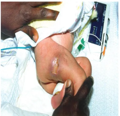
1.30 pm, baby developed cyanosis of the left foot and ankle (order to put hot fomentation and observe).
World Journal of Pharmaceutical and Life Sciences
WJPLS
www.wjpls.org SJIF Impact Factor: 5.088
*Corresponding Author: Dr. Najia Al Hojaili
Saudi Aradia.
ABSTRACT
File number:- 236888
Date of birth :- 24/6/1439
Full term male – 2.5 kg – to G6P4+1 uneventful pregnancy, product of C.S in Alahli Al Saudi hosp. because
of bad CTG and fetal distress MSAF, delivered sleepy required only tactile stimulation then picked up
A/S:- 5/1min 8/5min.
Admitted in NICU and kept in NPO, IVF, IV antibiotic.
All investigation were within normal Bl. gas normal – CXR clear lung field.
On 2nd day of life developed convulsion in form of blinking of eyes, frothy oral secretion, with desaturation so loaded by phenytoin, baby was stable maintain saturation on oxyhood 3L/min.
By around 4.00 pm cyanosis increased and there was line of demarcation – edema - order to shift the baby to
NICU2 and urgent Doppler ultrasound – surgical
consultation – hematology consultation - vascular
surgery consultation
1. Urgent Doppler ultrasound :- normal wave of
DPA, PTA and popliteal artery
2. Surgical consultation:- start hot compresses – line of demarcation to be observed - no surgical intervention indicated
3. Vascular consultation:- left foot area red and blue – no definite history of canulation or pricking – discoloration of the whole foot – triphasic Doppler signals of DPA and PTA left side Swelling of left foot? Compartment S – recommendation baby has good vascularity of left foot limb elevation – no vascular surgery intervention
4. Hematology consultation:- conservative
Cefotaxime was D/C and started meropenam, ID consultation done they ordered for meropenam 10 days and vanco for 5 days
A discussion between our COD and hematology
consultant resulted in to give FFP after taking sample for protein C, S and factor XIII
On the next 2 days there was some sort of improvement in the color of the foot and there was no need to start heparin
On the 7th day baby was irritable and in pain full examination was done and there was Lt testis swelling , hard painful in touch
Urgent Doppler ultrasound done showed Rt testis
normal size , echogenicity and blood supply while Lt testis enlarged in size 9x9 mm mixed
echogenicity with only venous Bl supply.
no arterial vascularity detected, suggesting
testicular torsion
Urgent pedia-surgery consultation was done and
baby went to OR
There was no torsion thrombosis
Gangrenous Lt testis, part of it sluphed during manipulation and sent to histopathology
Immature testicular tissue with hemorrhagic infarction
On the 10th day factor V Leiden, VIII,homocysteine level
CT angio was requested showed Lt intra cerebral hemorrhagic infarction with normal cerebral vein and sinuses no sign of venous thrombosis seen
Factor XIII within normal levels
Anti-thrombin III within normal levels
Factor V leiden within normal levels
Factor VIII within normal levels
Homocysteine level within normal levels
Protein S activity
Protein C activity
Baby was discharged at 19 day old, hemodynamicstable to follow up
Introduction
Anticoagulant protein C pathway
Anticoagulant effect at
Thrombosis Occurring the downstream damage at the vascular injury.
The anticoagulant effects of protein C
Protein C System - 3 abnormalities • Protein C deficiency
• Protein S deficiency
• Mutation of factor V cleavage site (activated protein C resistance)
Hereditary Protein C deficiency
• AD
Most patients heterozygous
Rare severe homozygous - purpura fulminansThe protein C pathway. APC = activated protein C; PC = protein C; S = protein S; T = thrombin; TM = thrombomodulin; Va = factor Va; VIII = factor VIIIa.
APC Resistance - Mutant Factor V (Factor V Leiden)
Activated Protein C (APC) destroys factor Va by cleaving it at arginine 506
Some patients have a mutated factor V with a glutamine at position 506, this prevents APC from cleaving factor Va and destroying it
Defect is termed Factor V Leiden or APC resistance
Increased risk of venous thrombosisEpidemiology incidence
In the United States and worldwide, protein C deficiency by plasma level alone is found in 1 in 200 to 1 in 500 persons in the general population.
Severe homozygous or compound heterozygousprotein C deficiency occurs in approximately 1 in 500,000 to 1 in 750,000 live births.
Clinical Presentation
Physical:- Patients with symptomatic hereditary protein C deficiency may present with VTE or WISN.
Homozygotes and compound heterozygotes frequently present with NPF during the first hours of life.
Venous thromboembolism
• Deep venous thrombosis of the lower extremity. • A chronic condition associated with swelling, pain,
discoloration, and venous insufficiency of the lower extremity.
Clinical Presentation cont Warfarin-induced skin necrosis
The skin lesions of WISN occur on the extremities, torso, breasts, and penis.
They begin as erythematous macules and, if appropriate therapy is not initiated promptly, evolve to become purpuric and necrotic bullae.Clinical Presentation cont… Neonatal purpura fulminans
Affected neonates present with diffuse ecchymoses which, similar to the lesions of WISN, progress to form necrotic bullae if appropriate therapy is not rapidly instituted.A patient with neonatal purpura fulminans.
Clinical Presentation cont
• May be presented by intracranial Hge
Ultrasound showing that the left adrenal gland was enlarged and heterogeneous, consistent with a left adrenal haematoma (arrow) measuring 35.1 mm × 24.4 mm.
DDx
Congenital & Acquired hypercoagulable states.
Congenital Acquired
1. 2. 3. 4. 5. 6. 7.
Protein C deficiency Protein S deficiency Antithrombin Ⅲ deficiency Factor V Leiden
Prothrombin gene G20210A mutation
Hyper-homocysteinemia Dysfibrinolysis
1. 2. 3. 4. 5. 6. 7.
Antiphospholipid antibody syndrome
Malignancy Surgery / Trauma Liver disease Vit K deficiency DIC
Severe sepsis specially Gm -ve
Diagnosis
In fact, testing for an inherited hypercoagulable state is costly & likely to uncover an abnormality in more than 60% of patients presenting with idiopathic VTEs.
Although, the remaining 40% will haveunremarkable test results, this does not imply a true absence of a hypercoagulable state.
A stepwise approach to thrombophilia testing
Workup Assays
• A variety of immunologic and functional protein C assays are available.
Immunologic assays
• Immunologic methods for the measurement of
protein C antigen include enzyme-linked
immunosorbent assays (ELISAs),
radioimmunoassays (RIAs), and
electroimmunoassays.
Functional assays
• Activated protein C (aPC) activity can be measured by means of a clotting assay or a chromogenic substrate. The adult reference range for protein C activity tends to be slightly lower than the immunologic normal range.
Workup cont Screening
The timing of testing with respect to acute thrombosis and warfarin therapy deserves special mention.
Acute thrombosis
• The levels of protein C, protein S, and antithrombin are reduced in the setting of acute thrombosis. Therefore, these levels should generally not be assessed at the time of presentation with acute VTE. However, a normal protein C activity in this setting essentially rules out hereditary protein C deficiency.
Warfarin
• Because protein C is a vitamin K–dependent protein, its levels are reduced with warfarin administration.
• Therefore, it is recommended that protein C testing not be performed unless the patient has been off vitamin K antagonist therapy for at least 2 weeks. If the patient has a severe thrombotic diathesis that does not permit discontinuation of anticoagulation.
Treatment
• A substantial proportion of individuals with
protein C deficiency remain asymptomatic
throughout life and require no specific therapy. • However, thromboprophylaxis may be considered
in such individuals, particularly if there is a strong family history of thrombosis
Treatment
• A case report by Milleret and colleagues describes 2 years of successful prophylaxis in a patient with neonatal severe protein C deficiency, using warfarin oral suspension. The international normalized ratio (INR) was measured by home monitoring, with a target INR of 2.5 to 3.5.
• For those patients who do develop clinical
manifestations of hereditary protein C deficiency,
treatment depends on the particular clinical syndrome: venous thromboembolism (VTE), warfarin-induced skin necrosis (WISN), or neonatal purpura fulminans (NPF).
Treatment
Venous thromboembolism
• VTE in patients with protein C deficiency is managed in much the same way as it is for patients with VTE due to other causes,
• Because the risk of recurrent VTE in protein C – deficient patients may be as high as 60%,long-term anticoagulation is often recommended, particularly following a spontaneous thromboembolic event.
Treatment
Warfarin-induced skin necrosis
• WISN is a medical emergency that requires
treatment as soon as it is recognized.
• Therapy consists of immediate discontinuation of warfarin, administration of vitamin K, and initiation of therapeutic doses of heparin.
• If the patient is protein C deficient, exogenous protein C should be administered, either in the form of fresh frozen plasma (FFP) or, preferably, as purified protein C concentrate (Ceprotin).Treatment
Neonatal purpura fulminans
• Like WISN, NPF is a medical emergency that requires rapid normalization of plasma protein C activity. Although fresh frozen plasma has been used as a source of exogenous protein C in the treatment of NPF, frequent administration is required to maintain adequate plasma levels, thereby limiting its usefulness in this setting.
• Highly purified protein C concentrate (Ceprotin) represents an attractive alternative that does not subject patients to the high volume and protein load of fresh frozen plasma.
Treatment
Neonatal purpura fulminans cont
• After treatment of the acute phase of NPF, patients are transitioned to anticoagulation therapy, on which they must remain indefinitely. Warfarin may be used in this setting, provided that exogenous protein C is administered during its initiation in order to avoid the development of WISN.For patients with breakthrough thrombosis despite anticoagulation. • Living donor liver transplantations have been
successfully performed in NPF, resulting in a permanent cure.
Treatment
• Heparin
• Enoxaparin (Lovenox)
• Dalteparin (Fragmin)
• Warfarin
• Protein C concentrate (Ceprotin)
Figure 1a: Five-day-old newborn with homozygous protein C defi ciency and purpura fulminans. Irregularly formed hypoperfused or necrotic skin lesions surrounded by aninfl ammatory border.
Figure 1b: The same child 6 days after beginning of protein C replacement with Ceprotin®.
Figure 1c: The same child 2 months later. All figures from Dreyfus M, Masterson M, David M, et al 1995. Replacement therapy with a monoclonal antibody purified protein C concentrate in newborns with severe congenital protein C deficiency.
Medication Ceprotin®
• Ceprotin® is a highly purified plasma-derived concentrate of human protein C zymogen
• Early case reports on the treatment of newborns with severe protein C deficiency and neonatal purpura fulminans demonstrated an impressive response to substitution therapy with protein C concentrates
Medication Protexel®
• Protexel® (LFB, Les Ulis, France) is a protein C zymogen concentrate, derived from human plasma (Radosevich et al 2003).
Medication
Drotrecogin alpha activated (Xigris®)
• Drotrecogin alpha (activated) (Xigris®, Eli Lilly Co.) is available as is a recombinant analogue to the physiologic human activated protein C.
• A report was published by a Japanese group, who used another concentrate of activated protein C to treat a female newborn who developed purpura fulminans on the third day after birth due to homozygous protein C deficiency (Nakayama et al 2000).
REFERENCES
1. Hypercoagulability syndromes: Arch intern med, 2001; 161.
2. Genetic susceptibility to venous thrombosis: N Engl J Med, 2001; 344(16).
5. Robbins pathologic basis of disease, sixth ed. Chapter, 5.
6. Sultan A. Jafarri et al, Journal of Dermatology & Dermatologic Surgery 21 104–106 Department of Pathology, King Fahad Central Hospital, Jizan, Saudi Arabia, 2017.
7. Adam Cuker, MD, MS Assistant Professor of Medicine, Assistant Professor of Pathology and Laboratory Medicine, Perelman School of Medicine at the University of Pennsylvania, September 14, 2018.