69
Original article
In silico
structural and functional analysis of copalyl diphosphate synthase
enzyme in Andrographis paniculata (Burm. f.) Wall. ex Nees:
A plant of immense pharmaceutical value
Aayet i Shailaj a, Byreddi Bhav ani Ve nkata Bindu, Mot e Srinath and C har u C handra Giri Centre for Plant Molecular Biology (CPMB), Osmania University, Hyderabad-500007, Telangana State, India
Received April 13, 2018: Revised May 30, 2018: Accepted June 5, 2018: Published online June 30, 2018
Abstract
Andrographis paniculata (Burm. f.) Wa ll. ex Nees(Acanthaceae) with immense medicinal importance lacks information on its biosynthetic pathway genes and their regulatory role in the production of pharmaceutically important andrographolide. Copalyl diphosphate synthase (CPS)
is involved in the production of copalyl diphosphate, a precursor for many bioactive compounds with particular reference to diterpene lactone. In this study, we elucidated the structural and functional aspects of A. paniculata CPS (ApCPS). Composition of amino acids and hydrophobic nature of ApCPS were analysed and identified as non trans-membrane protein. A chloroplast transit peptide and mitochondrial targeting peptide in ApCPS were identified. Protein secondary structure prediction has given insight on the distribution of helix (52.52%), loop (45.91%) and strands (1.56%) in ApCPS. The homology modelling of ApCPS was carried out with SWISS MODEL. The validation of 3D model using PROCHECK revealed that 91.74% of the residues have averaged 3D-1D score >= 0.2 which is structurally reliable. In Ramachandran plot, 90.9% amino acid residues were found in most favoured region. Phylogenetic tree was constructed using MEGA 7.0 by taking eudicots, monocots, gymnosperms and fungal species. Among them, ApCPS was clustered within eudicots and closely related to Sesumum indicum in Laminales. Protein-protein interaction study using STRING10 revealed that CPS interacts with gibberilic acid and terpene synthase related proteins. In Arabidopsis thaliana,CPS coexpression was seen with gibberelic acid related proteins. The present in silico analysis will be useful in understanding the structural, functional and evolutionary diversification of ApCPS.
Key words: Andrographis paniculata (Burm. f.) Wall. ex Nees, ApCPS protein, motifs and domains, domain linkers, 3D modelling, phylogenetic analysis
Copyright @ 2018 Ukaaz Publications. All rights reserved. Email: [email protected]; Website: www.ukaazpublications.com
1. Introduction
Andrographis paniculata (Burm. f.) Wall. ex Nees (Acanthaceae)is an important medicinal herb and a valuable source for important diterpene lactone, andrographolide and its derivatives. It has immense effect on various diseases and considered to be a valuable source in medicine. It has ph armacological effects such as antimicrobial (Singha et al., 2003), anti-inflammatory, anti-cancerous and immuno-stimulatory (Kumar et al., 2004; Subramanian et al.,
2 01 2; Islam et al., 20 18 ), immu no-modu latory an d an ti-atherosclerotic (Chao and Lin, 2010). This plant has shown effect on suppression of esophageal cancer and metastasis (Li et al., 2018). The demand for such valuable compound diterpene lactones of this plant is very high. However, the detailed mechanisms and the biosynthetic pathway genes are not yet elucidated clearly (Singh et al., 2018).
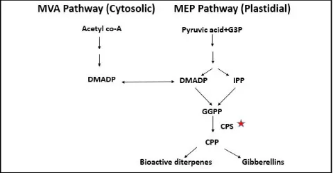
All the secondary metabolites (specifically diterpenes) of the plants have a common origin from IPP and DMAPP (Figure 1). These can
Annals of Phytomedicine 7(1): 69-77, 2018
DOI: 10.21276/ap.2018.7.1.8; Print ISSN : 2278-9839 and Online ISSN : 2393-9885 7(1):69-77 (2018) Ann. Phytomed.,
Author for cor respondence:Dr. Charu Chandra Giri Professor, Centre for Plant Molecular Biology (CPMB), Osmania University, Hyderabad-500007, Telangana State, India
E-mail:[email protected]
Tel.: +91-040-27098087
be derived either from MEP or MVA pathway which are interlinked and have connection between them (Chen et al., 2011; Vranová et al., 20 13).T he enzyme copalyl diphosph ate synthase (CPS)
catalyzes conversion of geranyl geranyl diphosphate, to copalyl diphosphate (CPP) which serves as intermediate for all diterpenoid reactions (Beale, 1990; Su et al., 2016).This CPS belongs to isomerase su per family wh ich involves in the syn thesis of terpenoids/isoprenoids. CPP is the direct precursor of gibberellic acid synthesis, other phytoalexins and labdane-related diterpenoids in plants (Prisic et al., 2004; Harris et al., 2005).
AEM000 24.1 . Expasy Compu te pI/Mw tool is u sed for th e estimation of pI (isoelectric point) and Mw (molecular weight) for the given protein sequence of ApCPS (Kyte and Doolittle, 1982). For the visualization of hydrophobicity for a peptide sequence the hydropathy plots were developed using Kyte and Doolittle (1982) method for each amino acid. The amino acid composition was shown (in %) using ProtParam tool in Expasy online server (Walker, 2005).
2.2 Prediction of signal peptide sequence
ChloroP was used for finding the choloplast transit peptide (cTP) and TargetP (Emmanuelsson et al., 2000), iPSORT (Bannai et al.,
2001, 2002) used to find out the subcellular location and signal peptide sequences in ApCPS.
2.3 Elucidation of secondary structure of ApCPS and prediction of trans-membrane helices
The secondary structure of ApCPS representing the families of related proteins was characterised using PredictProtein tool. Solvent accessibility and trans-membrane helix prediction was done by this tool. The analysis of trans-membrane helices in ApCPS was done using HMMTOP (Hidden and Markov Model Topology of Proteins) as per Tusnady and Simon (2001) and Tied Mixture Hidden Markov Model (TMHMM). The secondary structure prediction of ApCPS
was carried out using CFFSP prediction server (Chou and Fasman 1974; Dor et al., 2006).
2.4 Domain and motif analysis of ApCPS
The prediction of domain in ApCPS was carried out using Interpro and NCBI-CD search also used for identifying the super family of
ApCPS (Marchler-Bauer et al., 2016).
The Motif analysis was done using multiple Em for motif elicitation
(MEME: version 4.10.2). Motif Alignment and Search Tool was used for computation of pairwise correlation between each pair of motifs. Motifs with correlations below 0.60 have little effect on the accuracy of the E-values computed by MAST. DLP-SVM Domain prediction tool was used for identifying the Domain linkers (Ebina
et al., 2009).
2.5 Tertiary structure prediction / 3D modelling and validation of ApCPS
The tertiary structure (3D) modelling of ApCPS enzyme structure was predicted using SWISS MODEL server (Kiefer et al., 2008) and Phyre2 tool (Kelley et al., 2015). PROCHECK analysis (Laskowski
et al., 1996) was carried out for the validation and for analysing the stereochemical reliability of the 3D model using Ramachandran plot.
2.6.1 Protein protein interaction and co-expression analysis
STRING10 tool was used for analysing the retrieval of interacting proteins or genes (Szklarczyk et al., 2015). The model plant
Arabidopsis thalianaCPS was taken as reference protein sequence for the co expression study of CPS.
3. Results and Discussion
3.1 Biochemical property analysis of ApCPS protein
Amino acid sequence analysis in A. paniculata using Compute pI/ Mw tool revealed that ApCPS contained 832 amino acid residues with approximately 2.5 kb size (2496 bp). The theoretical pI value was shown as 7.07 and molecular weight was 95370.83 daltons,
i.e., 95 Kda. The similar result obtained earlier in ApCPS strengthened our finding in the present study (Garg et al., 2015).
Prot Scale helps in computing and representing the profile produced by any amino acid scale on a selected protein. The hydrophobicity plots for ApCPS with a window size of 9 and 21 shown in Figure 2A and 2B, where the relative weight of the window edges compared to the window center is 100%. The linear weight variation model is used for development of hydropathy plots without normalization. The more positive value of amino acids indicated that isoleucine, valin e an d leucin e are h ighly hydroph obic at that region (Figure 2C).
(B)
(C)
Figure 2:Hydropathy plot of ApCPS protein showing peaks for each amino acid at window size 9 (A); at window size 21 (B); The individual values for 20 amino acids using the scale Hydrophob./Kyte and Doolittle (C).
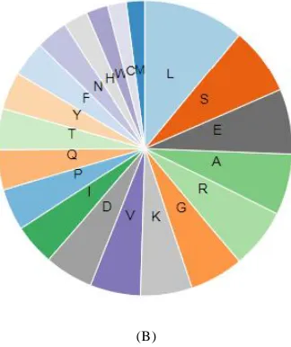
The amino acid composition and distribution showed that a highest of 11.1% residues of leucine are present in protein which is highly hydrophobic. After leucine, serine (7.5%) and glutamic acid (7.1%) were having major part of the composition (Figure 3A and 3B). The total number of 103 negatively charged residues of (Asp+Glu) and 102 positively charged residues of (Arg + Lys) were present.
(A)
(B)
Figure 3: The amino acid composition of ApCPS using ProtParam tool of ExPASy server (3A); diagrammatic representation of amino acid composition (3B) Lys (K); Ser (S); Thr (T); Ile (I); Glu (E); Pro (P); Arg (R); Gln (Q); Phe (F); Tyr (Y) His (H); Asp (N); Met (M); Cys (C); Val (V); Ala (A); Gly (G); Leu (L); Asp (D); Trp (W).
3.2 Prediction of signal peptide sequence
A chloroplast transit peptide (cTP) sequence containing 27 amino acids was identified in ApCPS. iPSORT prediction indicated that the protein sequence has no N-terminal sorting signal but having a mitochondrial targeting peptides in it (Figure 4). In an earlier study the presence of chloroplast transit peptide and mitochondrial targeting peptide in CPS was also observed in Salvia miltirriza (Su
et al., 2016).
Figure 4:Prediction of mitochondrial targeting peptide using iPSORT. 3.3ApCPS secondary structure elucidation and prediction of
trans-membrane helices
Figure 5: Secondary structure of ApCPS showing helix, sheet, turn and coil.
Figure 6: Graphical representation of secondary structure of ApCPS.
Further, analysis of protein revealed, a total of 10 protein binding regions were present in ApCPS (Figure 7). These 10 protein binding regions were at amino acid positions and residues 91(1), 139-141 (3), 189 (1), 213(1), 232 (1), 232(1), 455 (1), 469 (1), 747 (1), 827 (1), 832 (1), respectively (shown in red color: Figure 7). The helices present in the amino acid sequence were shown as brown colored boxes. Amino acid residues were categorized into buried (yellow), intermediate (light colored) and exposed portions (blue) along the sequence. The buried and exposed amino acids covered most of the amino acid composition. There were 4 disorder regions found in the seq uence at position s 1( 1), 5 6 ( 1), 82 -99 ( 18) , 7 44-74 7 ( 4)
respectively, where the length/size of residues is given in the parenthesis.
There were no trans-membrane helices and surface globular proteins observed in ApCPS based on the results of HMMTOP and TMHMM. Hence, ApCPS is considered as non-trans-membrane protein which is being translocated from nucleus to mitochondria and chloroplast. Although, surface globular proteins were not identified in the present study with ApCPS but these tools are also used in identifying the surface globular proteins in other plant species.
ApCPS.
3.4 Claasification of ApCPS protein and prediction of domains and motifs
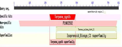
ApCPS protein belongs to isoprenoid synthase superfamily (Figure 8). The results with NCBI conserved domain search also revealed that ApCPS belongs to the isoprenoid superfamily and showed a specific hit with terpene synthase (Figure 9).Two domains, one terpene synthase N- terminal domain at 284-489 and one metal binding domain at 533-681 were identified (Figure 8).
Figure 8: Prediction of domains by Interpro
Figure 9:NCBI-CD search of ApCPS showing its superfamily 3.4.1 Predicting domain linkers by loop-length-dependent
support vector machine (DLP-SVM) tool
The domain linkers were found to be rich in proline residues which contributes for structural confirmation, and flexibility of two domains. They also acted in the prevention of unfavourable communication between the domains. Further, depending on length of the linkers the interaction between the domains varied (Bhaskara
et al., 2013). This study is also helpful in protein targeted drug development and other proteomic studies (Shatnawi et al., 2014).
Figure 10: Domain linkers (short and long) identified in ApCPS. 3.4.2 Motif analysis of ApCPS
There were 3 motifs found using MEME with Mast Alignment and Search Tool (Figure 11). The detail analysis of three motifs was characterized and the repetition in the ApCPS sequence detected Figure 12. Each of the sequence has an e value less than 10. The motif YIPAASPF occurred twice at positions 18-25 and 674-689 in the sequence. Motif MHRDWTDKGICW also repeated twice at positions 189-200 and 369-380. The third motif WQKWLRSW
occurred at positions 554-561and 674-689 along the sequence.
Figure 11:MEME results showing motifs in the ApCPS.
Figure 12:Different motifs and their positions on the sequence of
ApCPS (The boxes on the line representing the motifs). 3.5 Elucidation of tertiary structure of ApCPS by 3D homology
modelling, validation and ligand binding site prediction
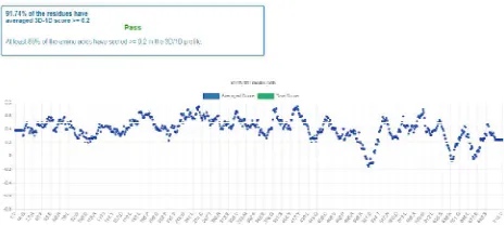
The 3D structure was predicted using Swiss Model which showed 713 amino acid coverage and with 56.81% identity (Figure 13). The elucidated 3D model was structurally validated with SAVES PROCHECK where 91.74% of the residues have averaged 3D-1D
score >= 0.2 (Figure 14). This 3D model has shown a overall quality factor (OQF) of 92.1986 (Figure 15).
Figure 13: Elucidated 3D structure of ApCPS using Swiss Model.
Figure 14: Validation of 3D structure.
Figure 15:ERRAT result showing the Quality factor for ApCPS.
In Ramachnadran plot, 90.9% amino acid residues were observed in favoured region with 8.4% additional allowed regions (Figure 16). Only 0.2% disallowed regions were found in the plot. This validation studies showing that the predicted 3D model of ApCPS
Figure 16:Ra machandra n Plot of ApCPS 3D stru ctu re. The most favoured regions are represented in red color; additional
allowed regions in yellow color. Figure 17: Predicted ligand binding site showed in blue color.
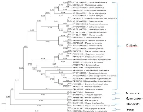
3.6 Phylogenetic analysis of ApCPS
The phylogenetic tree mainly divided into eudicots, monocots, gymnosperms and fungi. Among the 41 sequences taken for the phylogenetic construction, the ApCPS was clustered within the eudicots (Figure 18). ApCPS was shown close relationship with
Sesamum indicum and Olea europaea plants belongs to the same order Lamiales. After Lamiale plants, it was closer to the order Solanales. In an earlier study, comparable result was found with this CPS enzyme by Garg et al. (2015).
The evolutionary divergence study of this plant showed close relation to Laminales, followed by Solaneles in ApHMGR and ApDXS
enzymes related to the diterpenoid pathways (Bindu et al., 2017; Srinath et al., 2017). After the relation with eudicots, ApCPS was closely linked to monocots particularly Zea mays and Triticum aestivum and extended to gymnosperms and fungal species.
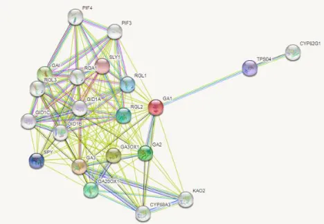
3.7 Protein interaction study
Arabidopsis thaliana CPS also called as GA1 was used for the protein interaction study (Figure 19). The protein interaction study shown that CPS mainly involved in gibberellic acid mediated signalling pathway. Further, it also involves proteins response to oxygen-containing compound which includes the enzymes such as GA1,
RGL1, RGL-2, and GA20OX1,etc. The molecular functions such as transcription factor activity and binding included interaction with
RGA1, RGL1, RGL2, RGL3 enzymes, iron ion binding included interaction with CYP88A3, KA02. Terpene synthase activity mainly involved the interaction within GA1, GA2, TPS04 enzymes. Most of the interacted proteins shown to be involved in inter cellular and intra cellular membrane bounded organelles. The CPS also shown the relation to proteins involved in diterpene biosynthesis pathway which includes GA1, TPS04, CYP 82G1, GA2, GA3OX1, GA3, GA20OX1, CYP88A3, KAO2 enzymes. These were also involved in plant hormone signal transduction and biosynthesis of secondary metabolites (Figure 20).
Figure 19:Protein-protein interactions using Arabid opsis CPS a s reference sequence using STRING 10.
Gene expression and co-expression studies will provide outline about CPS protein where the exchange of molecules and their interaction is high or low (Figure 20). CPS found to be involved in biosynthesis of gibberellin and diterpenoids in various studies (Keeling et al., 2010; Yamamura et al., 2018). This finding strengthens our result which showed the CPS co-expression mainly with
gibberellin and terpenoid synthase biosynthesis genes such as Gibberellin 3-oxidase 1, Gibberellin 20 oxidase 1 and Terpene synthase 04, etc.
Figure 20:Co-expression analysis of different proteins with reference plant CPS (Arabidopsis thalianaCPS). GA1-Gibberillic acid requiring1; GA3 - GA requiring 3; GA3OX1- Gibberellin 3-oxidase 1; GAI- DELLA protein GAI; GA2- GA REQUIRING 2; GA20OX1- Gibberellin 20 oxidase 1; RGL2- RGA-like 2;
RGL1- RGA-like 1; SPY- SPINDLY; TPS04- Terpene synthase 04; SLY1- SLEEPY1.
4. Conclusion
The present in silico analysis has given structural, functional and physicochemical aspects of protein ApCPS. The secondary structure prediction showed the alpha helices and beta sheets distribution of
ApCPS protein folding. The reliable 3D modelling of this enzyme gives information about the protein folding and can be exploited for various docking and drug targeting studies. The protein- protein interaction study will be helpful for understanding the protein at molecular level in various biosynthetic mechanisms. Evolutionary relation will give a scope for molecular biology and gene isolation studies where the primer designing for genes would be easy.
Acknowledgements
We would like to acknowledge the financial support from OU-UG C-CPEPA programme sponsored by University Grants Commission (UGC), New Delhi. Authors also thank OU-UGC-CPEPA
programme and UGC-BSR-RFSMS for research fellowships to AS, BBVB, MS.
Conflict of interest
We declare that we have no conflict of interest
References
Andersen Ranberg, J.; Kongstad, K.T.; Nielsen, M.T.; Jensen, N.B.; Pateraki, I.; Bach, S.S. and Møller, B.L. (2016). Expanding the landscape of diterpene structur al diversity throu gh ster eochemica lly contr olled combinatorial biosynthesis. Angew. Chem. Int. Ed., 55:2142-2146.
Bannai, H.; Tamada, Y.; Maruyama, O.; Nakai, K. and Miyano, S. (2001). Views: Fundamental building blocks in the process of knowledge discovery. In: Proceedings of the 14th International FLAIRS Conference, pp:233 -23 8.
Plant J., 66:212-229.
Chou, P.Y. and Fasman, G.D. (1974). Prediction of protein information. Biochem., 13:222-245.
Dor, O.; Zhou, Y. and Zhou. (2006). Achieving 80% tenfold cross-validated accuracy for secondary structure prediction by large-scale training. Proteins, 66:838-845.
Ebina, T.; Toh, H.; and Kuroda, Y. (2009). Loop length dependent SVM pr ediction of domain linkers for high throughput structur al proteomics. J. Pept. Sci., 92:1-8.
Emanuelsson, O.; Nielsen, H.; Brunak, S. and Von Heijne, G. (2000). Predicting subcellular localization of proteins based on their N-terminal amino acid sequence. J. Mol. Biol., 300:1005-1016.
Garg, A.; Agrawal, L.; Misra, R.C.; Sharma, S. and Ghosh, S. (2015). An dro gra phis pan icu lata transcr iptome pr ovides molecular insights into tissue-specific accumulation of medicinal diterpenes. BMC Genomics, 16:659.
Harris, L.J.; Saparno, A.; Johnston, A.; Prisic, S.; Xu, M.; Allard, S. and Peters, R.J. (2005). The maize An2 gene is induced by Fusarium attack and encodes an ent-copalyl diphosphate synthase. Plant Mol Biol., 59:881-894.
Islam, M.T.; Ali, E.S.; Uddin, S.J.; Islam, M.A.; Shaw, S.; Khan, I. and Gãman, M. A. (2018). Andrographolide, a diterpene lactone from Andrographis paniculata and its therapeutic promises in cancer. Cancer Lett., 420:129-145.
Kelley, L.A.; Mezulis, S.; Yates, C.M.; Wass, M.N. and Sternberg, M.J. (2015). The Phyre2 web portal for protein modeling, prediction and analysis. Nat. Protoc., 10:845-858.
Keeling, C.I.; Dullat, H.K.; Yuen, M.; Ralph, S. G.; Jancsik, S. and Bohlmann, J. (2010). Identification and functional characterization of mono-functional ent-copalyl diphosphate and ent-kaurene synthases in white spruce r eveal differ ent pa tterns for diterpene syntha se evolution for primary and secondary metabolism in gymnosperms. Plant Physiol., 152:1197-1208.
Kiefer, F.; Arnold, K.; Künzli, M.; Bordoli, L. and Schwede, T. (2008). The SWISS-MODEL Repository and associated resources. Nucleic Acids Res., 37:387-392.
Kumar R.A.; Sridevi K.; Kumar N.V.; Nanduri S. and Rajagopal S. (2004). Anticancer and immunostimulatory compou nds fr om Andrographis paniculata. J. Ethnopharmacol., 92: 291-295
K umar, S.; Steche r, G. and Tamura, K . (2 016). MEGA7: Molecular evolutionary genetics analysis version 7.0 for bigger datasets. Mol. Biol. Evol., 33:1870-1874.
Kyte, J. and Doolittle, R.F. (1982). A simple method for displaying the hydropathic character of a protein. J. Mol. Biol., 1:105-132.)
(2015). Distribution of Andrographis species in different districts of Andhra Pradesh. Proc. Natl. Acad. Sci. India, Sect. B. Biol. Sci., 85:601-606.
Parlapally, S.; Cherukupalli, N.; Bhumireddy, S.R.; Sripadi, P.; Anisetti, R.; Giri, C.C. and Reddy, V.D. (2015). Chemical profiling and antipsoriatic activity of methanolic extract of Andrographis nallamalayana J.L. Ellies. Nat. Prod. Res., 30:1256-1261.
Prisic, S.; Xu, M.; Wilderman, P.R. and Peters, R.J. (2004). Rice contains two dispa rate ent-copa lyl diphosphate synthases with distinct metabolic functions. Plant Physiol., 136:4228-4236.
Rost, B. and Sander, C. (1993). Prediction of protein secondary structure at better than 70% accuracy. J. Mol. Boil., 232:584-599.
Rost, B. and Sander, C. (1994). Combining evolutionary information and neural networks to predict protein secondary structure. Proteins: Structure, Function, and Bioinformatics, 19:55-72.
Sahay, A. and Shakya, M. (2010).In silico analysis and homology modelling of antioxida nt pr oteins of spinach. J. Proteomics Bioinfor m., 3:14 8-15 4.
Shatnawi, M.; Zaki, N.and Yoo, P.D. (2014). Protein inter-domain linker prediction using random forest and amino acid physiochemical properties. BMC bioinformatics, 15:S8.
Singh, S.; Pandey, P.; Ghosh, S. and Banerjee, S. (2018). Anti-cancer labdane diterpenoids from adventitious roots of Andrographis paniculata: augmentation of production prospect endowed with pathway gene expression. Protoplasma, pp:1-14.
Singha, P. K .; Roy, S. and Dey, S. (2003). Antimicrobia l activity of Andrographis paniculata. Fitoterapia, 74:692-694.
Srinath, M.; Shailaja, A.; Bindu, B.B.V. and Giri, C.C. (2017). Characterization of 1-deoxy-D-xylulose 5-phosphate synthase (DXS) protein in Andrographis paniculata (Burm. f.) Wall. ex. Nees: A in silico appraisal.Ann. Phytomed., 6:63-73.
Su, P.; Tong, Y.; Cheng, Q.; Hu, Y.; Zhang, M.; Yang, J and Huang, L. (2016). Functional characterization of ent-copalyl diphosphate synthase, kaurene synthase and kaurene oxidase in the Salvia miltiorrhiza gibberellin biosynthetic pathway. Sci. Rep., 6:230-257.
Subramanian R.; Asmawi M.Z. and Sadikun A. (2012). A bitter plant with a sweet future? A comprehensive review of an oriental medicinal plant: Andrographis paniculata. Phytochem. Rev., 11:39-75
Tusnády, G.E. and Simon, I. (1998). Principles governing amino acid composition of integral membr ane pr oteins: Applica tions to topology prediction. J. Mol. Biol., 283:489-506.
Vranová, E.; Coman, D.; and Gruissem, W. (2013). Network analysis of the MVA and MEP pathways for isoprenoid synthesis. Annu. Rev. Plant Biol., 64:665-700.
Walker, J.M. (Ed.). (2005). The proteomics protocols handbook. Humana Press.
Wass, M.N.; Kelley, L.A. and Sternberg, M.J. (2010). 3DLigandSite: predicting ligand-binding sites using similar structures. Nucleic Acids Res., 38:469-73.
Yamamura, Y.; Taguchi, Y.; Ichitani, K.; Umebara, I.; Ohshita, A.; Kurosaki, F. and Lee, J.B. (2018). Characterization of ent-kaurene synthase and ka urene oxidase involved in gibberellin biosynthesis fr om Scopariadulcis. J. Nat. Med., pp:1-8.
Zaheer, M. and Giri, C.C. (2015). Multiple shoot induction and jasmonic versu s salicylic acid driven elicita tion for enhanced andrographolide production in Andrographis paniculata. Plant Cell Tissue Organ Cult., 122:553-563.
Zahee r, M. an d Giri, C .C. (201 7a). Enhanced diter pene lactone (a ndr ogr apholide) production from elicited adventitious root cultures of And rogr aphis panicula ta. Res. Chem. Intermed., 43:2433–2444.