MICHAEL E. YANNONE, M.D.
Visiting
Professor of Biochemistry, Medical University of South
Carolina, Charleston, South Carolina; Professor of Obstetrics and
Gynecology, University of Iowa College of Medicine, Iowa City, Iowa
Because of the time factor involved, it would be an impossible task to present the vast body of biochemical information accumulated during the last decade on the endocrine aspects of pregnancy. However, certain key points can be made concerning the feto-placental unit and the hormone levels and effects in normal gestation. These concepts will be discussed not merely for their academic value, but also as a base to evaluate our present diagnostic and therapeutic endeavors in obstetrical practice. With the audience's indulgence, an occasional per-sonal conjecture will be interjected concerning hormonal mechanisms and clinical practices.
Feto-Placental Unit. The fetus and the placenta are incomplete steroidogenic systems. They lack the capability of synthesizing all necessary steroidal hormones from more simple precursors in contrast to the ovaries, testes, and adult adrenal cortex. To complete the task a well-coordinated interdigitation of the synthetic capabilities of fetus, placenta, and mother is required. Since the mother is involved, one wonders whether the term "feto-materno-placental unit" would not be more appropriate.
First the fetal adrenal cortex and then the placenta will be discussed; hopefully, emphasizing the information which may have practical clinical significance.
A. Adrenal Cortex. The fetal zone of the adrenal cortex persists only during gestation, con-stitutes about 80% of the gland, and is active in steroid metabolism. In the first trimester, it is believed this zone is primarily stimulated by human chorionic gonadotrophin (HCG); ACTH gaining prominence in control thereafter.
The biochemical capabilities and limitations of the fetal adrenal cortex are shown in Fig. 1. Utilizing
*
Presented at the 43rd Annual McGuire Lecture Series, December 3, 1971, at the Medical College of Virginia, Richmond.MCV QUARTERLY 8(1): 43-51, 1972
acetate as a precursor, the fetal zone more readily synthesizes the t::..5-3{3 hydroxysteroids pregneno-lone and dehydroepiandrosterone (DHEA) than the t::..4-3 ketosteroids such as cortisol and aldo-sterone. This is because of a relative block in
3{3-hydroxy dehydrogenase and t::..5 isomerase activity. Thus, the synthesis of large amounts of DHEA and its sulfate is favored, and they are transported to the placenta as such or after 16a-hydroxylation by the fetal adrenal or liver. The placenta can metabolize these androgens to estrogens as will be shown shortly.
However, such a biosynthetic lack would leave the fetus in jeopardy because of a cortisol and aldosterone deficiency. This is rectified by the fact that the fetal zone can utilize the readily available placental progesterone to produce these needed hormones. In fact, after the tenth week of gestation, the increasing placental progesterone secretion may enhance the 17,a, 21, and 11{3 hydroxylase capability of the fetal adrenal cortex to produce the corticoids. Also, the ready passage of maternal cortisol across the placenta is a secondary protective mechanism.
B. Placenta. This endocrine organ is incapable of synthesizing large quantities of progesterone and estrogens from the simple precursor acetate. How-ever, it can efficiently convert maternal plasma cholesterol to progesterone as shown in Fig. 2. This is clinically important for it means that an intact ma-ternal-placental circulation will produce essentially normal progesterone levels even though a fetal de-mise has occurred. The progesterone produced by the placenta can be metabolized by the mother and fetus to less active progestational metabolites or can be utilized by the fetal adrenal as already described. Since a 17 a-hydroxy lase deficiency in the placenta precludes the conversion of progesterone to andro-gens and subsequently estrogens, another mechanism must exist for placental estrogen synthesis.
44 YANNONE: HORMONAL CHANGES IN PREGNANCY
ACETATE
CH3 I
o;sort95
CHOLESTEROL Preonenolone
\
yH3 . / . Sulfote 1H'
C•O / C•O
~.mOH
",_,""''ID--HO HO
Pregnenolone 17-Hydroxypreonenolone
I
I
0 0
_
SJst~r·-·~-w
°"
o,so
o,so
Dehydroepiandro1 terane 16d-Hy drox
ydehydro-Sulfat• epiandros terone
t
0 Sulfatet
0__.,.
'·~(16o·hydrox=
1
~·
··
OH
HO~ HO~
DehydroepiandrosteroneI
16a-Hydroxydellydro-epiondrosterone
Relative block in 3,8-Hydroxysteraid dellydrogenose activity
\ I
PLACENTAL I \
I
PROGESTERONE_.-PROGESTERONE-+~17-HYOROXY-
~
/1
PROGESTERONE-+ ANOROSTENEOIONE21 .hydroxylotion 121-hydroxylotion
l
lljl·hydroxylot1on 11,8 -hydroxylolion lljl ·hydroxylotion
CH3 CH20H CH20H
···OH
'o
~o
c=o
~o
·.~iH
0 0 0
1sa·Hydroxy-prooesterone
Corticosterone Cortisol
~
ALDOSTERONE
0
o~
11{3-Hydroxyondrostenedione
Fig. I-Pathways of steroid metabolism in the human fetal adrenal cortex. The major secretory products are boxed in. (Reprinted
with permission from Villee. New Eng. J. Med. 281: 473, 1969.)
As depicted in Fig. 3, the placenta produces
estrogens from androgens of maternal and fetal adrenal origin. Androstenedione, DHEA, and testos-terone are converted by the placenta to estradiol
and estrone. It is estimated that the mother and
fetus each contribute about 50% of the androgens which are made into these 2 estrogens. Since the ability to 16.a-hydroxylate androgens is essentially limited to the fetal adrenal and liver, the fetus is the major contributor of 16a-hydroxylated
andro-stenedione, DHEA, and testosterone which are
converted by the placenta to estriol. The maternal contribution to estriol at term is less than 10% and
probably arises from her adrenal androgens which escape conversion to estradiol and estrone on passage through the placenta to fetus. In the fetus, however,
her androgens can be hydroxylated at the 16a
position before recirculation back to the placenta where synthesis to estriol can occur.
The above information indicates that the clinical assay in pregnancy of plasma progesterone or its major metabolite, pregnanediol, in the urine would mirror placental integrity. On the other hand,
estrogen assays, particularly urinary estriol, would reflect fetal integrity.
Hormone Secretion Patterns and Effects in Nor-mal Gestation.
ACETATE - - - - + CHOLESTEROL
20 a-o;hydropr09esleron•
Fig. 2-Principal precursors and metabolites of progesterone in the human placenta. The major steroid secreted by the placenta (progesterone) is boxed in. (Reprinted with per-mission from Villee. New Eng. J. Med. 281 :473, 1969.)
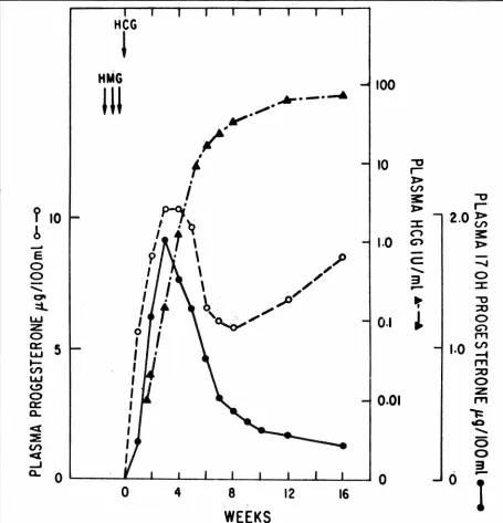
origin are seen to peak at 3-4 weeks after ovu-lation. At 6-8 weeks post-ovulation, the proges-terone reaches a nadir while the 1 7 a-hydroxy pro-gesterone continues to decline. Since the placenta lacks significant 17a:-hydroxylase capability, it is as-sumed that the secondary rise in progesterone reflects placental function. It is of interest to note that HCG-believed to maintain the corpus luteum of pregnancy-continues to rise while both hormones of corpus luteum origin are declining.
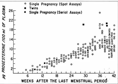
The secondary rise in placental progesterone continues to increase progressively to a maximum plateau at 36-40 weeks. This is seen from our data in Fig. 5, where the level at 8 weeks of 2 micro-grams % increases linearly to about 14 micrograms
% at term. The curve seems to parallel that of placental weight and is of similar configuration to that of urinary pregnanediol as depicted in Fig. 6.
It is important to note that no significant drop
in progesterone occurs prior to labor. However, during parturition we and others have found a
slight downward trend of mean values and this may
be a reflection of placental ischemia secondary to the uterine contractions. There is a rapid drop in the plasma progesterone after delivery of the placenta verifying the hormone's short half-life.
It has been estimated that the term placenta
produces 200-300 mgms of progesterone daily. The role of this large amount of hormone is not well defined, but possible metabolic functions are:
1. The maintenance of growth and development of the fetus; for example, it is a precursor steroid for the synthesis of corticoids by the fetal adrenal cortex.
2. An immunosuppressive agent to protect the feta-placental homotransplant.
3. A defense mechanism acting on the myo-metrium to prevent premature expulsion of the fetus.
4. Prepare the breasts for lactation.
5. Mediate a variety of biochemical events; for example, antagonize at the renal tubule the effects of the increased aldosterone con-centration found in pregnancy.
B. The Estrogens. Quantitatively, estriol is the major estrogen of human pregnancy. Its biologic potency, however, is significantly less than that of estradiol and estrone. The urinary excretion curve for estriol is presented in Fig. 7; the amount rising from low early levels to high levels at term. An
acceleration in the increase is apparent during the
last few weeks. There seems to be a good correla-tion between the estriol curve and that for the fetal weight. Klapper, reviewing the literature, found the average levels to be 10.1, 14.2, 19.8, and 26 mgms at 28, 32, 36, and 40 weeks respectively. Similar curves for urinary estradiol and estrone have been found, but the amounts are in the micro-gram range. These two estrogens, like estriol, prob-ably reflect the status of the feta-placental unit.
Munson in our group has assayed unconjugated estradiol in the plasma of normal· pregnancies. A mean value of 200 nanograms % in early pregnancy (9-16 weeks) rose to 1196 nanograms % dur-ing the last 8 weeks. A curve configuration similar to urinary estrogen excretion patterns was obtained.
I suspect that the estrogenic effects of preg-nancy are primarily due to estradiol, the most po-tent of these 3 estrogens. It mediates the growth and
function of the maternal reproductive organs and probably is important to fetal growth and develop-ment. The reason for the large mass of estriol pro-duction is not clear. There is little evidence at the moment that it is involved in the start of human labor.
46
~
0
so;
Oehydroepiondro1teront Sulfate
!''":.,. .. ,
HO~
Androstenedlone Testosterone
t
HO~HO~
ES TRONE ESTRADIOL-17,8YANNONE: HORMONAL CHANGES IN PREGNANCY
~OH
0
so;
16a-H ydrox ydehydrotpiandros ttrOflt
!
SulfateHO~~·:
16a-Hydroxydehydroepiondros terone
! (
3,/J-llydroxysf,,oid d'llydrog•11•••· isom'""' sysf'm JO~~~OH
16a-Hydroxy-androsttntdione
16a-Hydroxy-testos terone
(oromofi1i1'g sysl•m J '
r6
..
·0HHO~
ESTRIOL
Fig. 3- Synthesis of estrogens by the human placenta. The pathways for the conversion of C19 steroids to estrogens are shown. The major estrogens secreted by the placenta are boxed in. (Reprinted with permission from Villee. New Eng. J. Med. 281: 473, 1969.) results in elevated concentrations of serum
thy-roxine (also the PBI), plasma cortisol, and
testos-terone. However, the amount of free or unbound
hormone, and presumably the biologically active fraction, is unchanged. Unless this estrogen effect is taken into account, the clinician can be misled
by the elevated laboratory result. The only excep-tion to this physiologic change is for plasma cortisol in pregnancy. The large amount of placental pro-gesterone in pregnancy plasma competes with corti-sol for binding sites on transcortin and increases the free cortisol fraction. Therefore the pregnant woman is actually in a state of mild adrenal hyper-corticism.
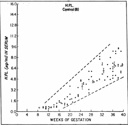
C. Human Placental Lactogen (HPL). This polypeptide hormone secreted by the syncitial trophoblast is immunologically similar to but distinct
from human growth hormone. Metabolically it ap-pears to be a weaker growth hormone. Its pattern of secretion into the blood is shown in Fig. 8; the
increasing concentration throughout pregnancy parallels the curve for placental weight. Like
pro-gesterone, its blood concentration reflects placental rather than fetal integrity. Fetal death may not
significantly drop the level for a period of time as
long as the materno-placental circulation is intact.
HCG
i
HMG
100
JU
.,,,.
~·,,,,,....
__
_..
~,
,
f
10
-0I
'
J>I
(,/')31: -0
?
10
,?-Oj~
:c
J>2.0~
r-b
I
t
("") 31:~
~
..\
1.0
c;-, J>
E
.I'
c:
0
_,
0
I
\
/ ,...
0I
\
/
3
:c
...
-at
I
;,P/
-0~
.,.
::l.
I
::u
w
I
'
. _,,,./0.1
~
0°'°"'
:z
0 c;-,0
'
f'T'1a:
5
1.0
(,/')w
I
...
~
I
f'T'1(/')
::u
LL.I
I
0 C>0
I
0.01
z
a:
I
,.,,
a..
,
--==
<(
'
'°
...
::E
I
5
(/')
<(
I
0_J
3
a..
0
0
0
-0
4
8
12
16
WEEKS
I
Fig. 4- Mean plasma levels of progesterone, 17-hydroxyprogesterone, and HCG following induction of ovulation with HMG-HCG and subsequent conception. (Reprinted with permission from Yoshimi, et al. J. Clin. Endocr. 29: 225, 1969.)
the fetus. The daily production can be as high as 400 mgms.
Possible roles of HPL are preparation of the breasts for successful lactation and indirect growth hormone effects. These include increased nitrogen retention, increased maternal fatty acid utilization sparing glucose for transmission to the fetus. The fetus, like the adult brain, utilizes essentially glucose only for its energy requirements. HPL is antagonistic to insulin and as a consequence may be an important
factor in the diabetogenic effect of pregnancy.
D. Human Chorionic Gonadotrophin (HCG).
The blood and urine levels of this glycoprotein secreted by the cytotrophoblast differ from the curves seen for the previousiy discussed horomones ;md is shown in Fig. 9. The concentration in the blood peaks at about 60 days, then decreases to % to V:3 of the peak value throughout pregnancy with a lower secondary rise at term.
tradition-48 YAN NONE: HORMONAL CHANGES IN PREGNANCY
~
0
Single Pregnancy (Spot Assays)
• Twins
~
26
• Single Pregnancy (Serial Assays)
0~
22
~
...
18
E:
~
~
"
~
14
10
<::s
~
....::
6
~
~
~
((
2
~
12
18
•
Oo • v • 0§
24
30
36
0
42
6
WEEKS AFTER THE LAST MENSTRUAL PERIOd
:
Fig. 5-The plasma progesterone concentration in normal pregnancy expressed as micrograms 3. One hundred and t1:Velve of these 179 determinations were reported previously. Note the progressive increase in concentration with advancing gestation. (Reprinted with permission from Yannone. Steroids 13: 773, 1969.)
ally is believed to maintain the corpus luteum of pregnancy. It may also regulate steroid secretion by other glands; its possible ACTH effect on the fetal adrenal cortex in early pregnancy has already been mentioned.
Clinical Implications.
A. Progesterone and Pregnancy Status. Plasma
progesterone and urinary pregnanediol have been measured in a variety of obstetric disorders to prog-nosticate dysfunction or to establish its presence. Such diagnostic attempts have been less than satis-factory. Probably the primary reason for this is that the hormone concentration can remain in the nor-mal range even with fetal death as long as the utero-placental circulation is intact. Other problems are the wide day to day variation in the same individual and the overlap of values in normal and abnormal gestation. Even when the hormone concentration is definitely subnormal, it is usually the effect of an uncorrectable dysfunction rather than the cause. Therefore exogenous progesterone substitution is
70
~50
C'>
E 40 _,
0
8
30z <(
t5
20 L.LJ0::: 0...
10
8 12 16 20 24 28 32 36 40 WEEKS OF PREGNANCY
•
Mg Estriol/24 hrs
; • ""'50
I I •
I • I
:
..
••
I •
r. .
I • • I •• • 40
I •
:
..
'
...
I I
I·
•
•
I •
I
•
•
I
I • • •
~
3o
. l
. . .
l
i: \
,: • • -,= • • ::
.,,
. .
...
,
. . .
.
,t. • • .·.: •• :.
,,
.
..
.
....
~ ••• \ ••••• - 20
,,
.
.
.:.•\
....
,, • • • . :i. • •\
,,
.
.
... .
. .
,
.. . . . . ..
,,
. .
.
.
.
. . . .
• ,*' • • • • • • .. .. ••••
,,,
. .
:
..
:
.
..
.. .. .
-~. .. .
.
.. ..
..
...
-:' • • I • ••• • •• • • • • • •
--
.
-. .. .
....
.
.
. ..- . . . . . . ·ii· ... . . \ .
..
..
.
.
,
. . .
,..
. .
_
..
, ..
.. • __ ..
..
~- .: • • • • • • • , ... :- '"' ~ 10~
,,
.
..
..
. . . .
.
.
..
---
.
..
,
...
,..
.
. . ..
.
.
·:--
.
--
. . . .
.
.._..
. ...
-. -.
,., _.--:
.
• ••:-. I• •.,
...
r._ • .,.. ••
• • .... ..
--·
....
-:-~··...
,
:
...
~.·.
...
.,..
...
-\••
..
·
... ..
---··'·
,.-- .....
., .!~.
•,...
• • •• • ••.
..
.
. .
•-
·-
1
--
'
---
.
.
... .;::.~::~:. •• • :·~ • I•' .,. ... --·- .... ,,:..1e:
.
:,.
-c.•.-i: •• :: ••
:a--"···
L'\:i..;.,!t.'- .
----~---~-I I I I I I
•
10 15 20 25 30 35 40
Weeks of pregnancy
Fig. 7- Urinary excretion of estriol-16-glucuronide during pregnancy in 31 normal women. The upper and lower dashed lines are
953 'confidence limits. (Reprinted with permission from Fuchs and Klopper (eds.). Endocrinology of Pregnancy, First Edition.
New York: Harper and Row, 1971.)
generally of little value. Similar problems are en-countered when serial assays for HPL are used as a diagnostic tool.
B. Progesterone and Adequacy of the Corpus Luteum; Abortion. Of probable clinical value are the use of serial plasma progesterone or urinary preg-nanediol assays to evaluate corpus luteum ade-quacy in certain types of infertility and possibly a few cases of first trimester abortion. Although the majority of such abortions are due to uncorrectable causes such as chromqsomal defects, a few could be due to corpus luteum dysfunction with dishar-monious transition of steroid hormone production to
the placenta. When such corpus luteum inadequacy is incriminated, correction of the infertility or
pre-vention of another abortion may be achieved by insuring an adequate corpus luteum. However, not by the use of exogenous progesterone replacement, but rather by the judicious use of ovulatory in-ductors such as Clomid® or the gonadotrophins to insure normal follicular maturation which will result in a normal corpus luteum.
C. Progesterone-Term and Premature Labor.
pre-50
mature labor. The theory most often advanced to explain the mechanism which institutes parturition is the progesterone-block concept. This maintains that progesterone suppresses myometrial contractility during gestation and labor begins upon withdrawal
of the hormone. However, our studies and those of
others have not shown a significant and consistent
drop in circulating progesterone prior to labor's onset. These findings do not exclude a protective role for progesterone as an effective decrease may occur that is not apparent in the circulating levels. One possibility that we investigated was that the concentration of free and active progesterone de-creased with advancing pregnancy because of
in-creased protein binding of the hormone. However,
we found the percentage of binding to remain constant throughout pregnancy, which meant that as the total progesterone concentration rose so did the concentration in the bound and unbound frac-tions. With this negative finding we have turned to studies which may show decreased progesterone levels within the myometrial cells. Our results are
too preliminary to draw conclusions. ·
In spite of our ignorance as to how labor starts,
we are not totally devoid of therapeutic agents. Within reason labor can be induced at term with oxytocin.
It is much less effective earlier in pregnancy.
Pros-taglandins such as F2a appear to be effective at term
as well as earlier in gestation. However, on parenteral
use in the first 2 trimesters, the therapeutic-toxic
difference becomes too narrow. This problem may
be overcome by using the prostaglandin locally in the vagina to induce an indicated abortion or premature labor. Lastly, with judicious use of intravenous alcohol in premature labor, the expul-sion of the fetus can be delayed a few days to a
few months in about 65 % of the patients.
D. Estriol and Complicated Pregnancies. The most important clinical consideration at the moment is the value of serial estriol determinations in the management of high-risk pregnancies. While it can be helpful in certain complications of pregnancy such as diabetes mellitus, toxemia, and dysmaturity to evaluate the fetal status, it is not without pitfalls. First the clinician is at the mercy of the quality control of the laboratory doing his assays. Inaccurate results can lead to false security or ill-timed
termi-nation of pregnancy. Even with a good laboratory,
the results may not always reflect the true status of
the pregnancy. Unfortunately, our knowledge of
estrogen metabolism in pregnancy is inadequate to explain such discrepancies any better than it can explain why Rh isO:.immunized jeopardized
preg-YANNONE: HORMONAL CHANGES IN PREGNANCY
nancies do not show subnormal estriol levels. Even
more disconcerting is the fact that excellent clinicians using good laboratories are reporting perinatal losses in diabetic pregnancies no better or even
160
14.4
12.8
~ 11.2 15 9.6
(lj
~
~ 8.0
~
...
~ 6.4
~
~ 4.8
3.2
1.6
00 I 0 4
/ / / / / /
H.P.L
CC?ntrol(8) / / / / /
.
/ / / / /"·
/.
....
...
. :
f
.
:
.~? • :
8 12 16 20 24 28 32 36 40
WEEKS OF GESTATION
Fig. 8-Human placental lactogen (HPL) in serum. The
80 black dots represent individual values, the open circles represent the means for 4 week' intervals and the vertical lines the standard error of the mean. The dash lines show the upper and lower limits of all determinations in the con-trols. (Reprinted with permission from Sam~an, et al. Am.
!. Obstet. Gynec. 104:781, 1969.)
100
so
e
200-~ 15
I :;j
10 s
e
s~
2
1.S
... 8 12 16 20 24 28 32 36 40
Weeks
Fig. 9-Mean serum levels of chorionic gonadotrophin in normal pregnancy. The curves are based on results reported by various authors using different bioassay and immunoassay methods. Curve 1: uterine weight increase in rats; Curve 2: ovarian hyperemia in rats; Curve 3: complement fixation; Curve 4: hemagglutination. (Reprinted with permission from Fuchs and Klopper (eds.). Endocrinology of Rreg
worse than reputable clinics have reported prior to the availability of this assay. So far there has not been any conclusive evidence that a marked reduction in fetal mortality has been obtained be-cause of estriol determination. In this light, the overzealous utilization of estriol assays as a sub-stitute for rather than an adjunct to clinical manage-ment is foolhardy.
As an adjunct to clinical judgment, the literature seems to agree on certain helpful points. These are as follows:
1. Significant day to day variation requires serial assays to determine a mean for each patient.
2. In a suspect pregnancy, weekly or biweekly assays should be done from about the 30th week up to the time of reasonable maturity (about 34th week) at which time daily or every other day determinations should be carried out.
3. In any complicated pregnancy which is 34 weeks or beyond, any precipitous fall from normal levels of urinary estriol and which is verified over 2 or more consecutive days dictates immediate delivery.
4. Chronic low estriol levels in preeclamptic toxemia or dysmaturity do not necessitate interruption unless there is a precipitous drop from the low mean. Such babies left
in utero may not grow very much, but they do achieve greater maturity.
5. Urinary estriol levels can be expected to be low in mothers with anencephalic
mon-sters or on glucocorticoid therapy for medi-cal reasons.
The development of new specific and sensitive hormone assay methods and the increase in sophisti-cation of metabolic studies has given us an impres-sive array of biochemical information concerning the physiology and endocrinology of pregnancy. However, the application of this knowledge to the improvement of obstetrical care has been difficult. This should not discourage us as medical science must like the infant learn to crawl before it can walk. Eventually we shall have all the facts needed to obtain the obstetrical results that we all desire. We have had a promising start.
REFERENCES
FUCHS, F. AND KLOPPER, A. (eds.). Endocrinology of Preg -nancy, First Edition. New York: Harper and Row, 1971. JAFFEE, R. B. Estriol in high-risk pregnancies: Has it kept its promise? Gynec. Invest. I. Suppl. 1-6, 1970.
SAMAAN, N. A. et al. Serial hormonal studies in normal and abnormal pregnancy. Am. l. Obstet. Gynec. 104:781, 1969.
VILLEE, D. B. Development of endocrine function in the human placenta and fetus. New Eng. !. Med. 281:473, 1969.
YANNONE, M. E. et al. Plasma progesterone levels in normal pregnancy, labor and the puerperium. II Clinical data. Am. !. Obstet. Gynec. 101: 1058, 1968.