IJEDR1602310
International Journal of Engineering Development and Research (www.ijedr.org)1760
Tumor Location and size Identification in Brain
Tissues Using Fuzzy C- Clustering and Artificial Bee
Colony Algorithm
Mihir A. Mishra, Pragnesh A. Patel, Kushal D. Patel Assistant Professor(s)
Computer Engineering Department, GIDC Degree Engineering College, Navsari, India
________________________________________________________________________________________________________ Abstract—Clustering approach is widely used in biomedical applications particularly for brain tumor detection in abnormal magnetic resonance (MRI) images. Fuzzy clustering using fuzzy C-means (FCM) algorithm proved to be superior over the other clustering approaches in terms of segmentation efficiency.MRI Imaging forms one of the core methods to identify Brain Tumors, and access the existence, size and thickness of the tumor. MRI Images are prone to high noise, as the whole principle works on strong electric fields.We forms the clusters using Fuzzy C-Means clustering and then identify the tumor part by removing the noise part using Artificial Bee Colony algorithm. Many image processing techniques have been proposed for brain MRI segmentation, most notably thresholding,region-growing, and clustering. Since the distribution of tissue intensities in brain images is very complex, it leads to difficulties of threshold determination.
Keywords - Clustering, MR brain tumor, Fuzzy C-means and Segmentation efficiency, Artificial Bee Colony
________________________________________________________________________________________________________
I. INTRODUCTION
In the analysis of medical images for computer-aided diagnosis and therapy, segmentation is often required as a preliminary stage. Medical image segmentation is a complex and challenging task due to the intrinsic nature of the images [1].The brain has particularly complicated structure and its precise segmentation is very important for detecting tumors, edema, and necrotic tissues, in order to prescribe appropriate therapy. Magnetic resonance imaging (MRI) is an important diagnostic imaging technique for the early detection of abnormal changes in tissues and organs [1]. It possesses good contrast resolution for different tissues and has advantages over computerized tomography (CT) for brain studies due to its superior contrast properties. Therefore, the majority of research in medical image segmentation concerns MR images [1].
MRI Imaging forms one of the core methods to identify Brain Tumors, and access the existence, size and thickness of the tumor. MRI Images are prone to high noise, as the whole principle works on strong electric fields.
Many image processing techniques have been proposed for brain MRI segmentation, most notably thresholding, region-growing, and clustering. Since the distribution of tissue intensities in brain images is very complex, it leads to difficulties of threshold determination [1].
Therefore, thresholding methods are generally restrictive and have to be combined with other methods like Region growing, Fuzzy C-means (FCM) clustering and Expectation–maximization (EM) algorithms [1].
Region growing extends thresholding by combining it with connectivity conditions or region homogeneity criteria. Successful methods require precise anatomical information to locate single or multiple seed pixels for each region and together with their associated homogeneity. Clustering is the most popular method for medical image segmentation, with Fuzzy C-Means (FCM) clustering and expectation–maximization (EM) algorithms being the typical methods [1]. The applications of the EM algorithm to brain MR image segmentation were reported by Wells and Leemput. A common disadvantage of EM algorithms is that the intensity distribution of brain images is modeled as a normal distribution, which is untrue, especially for noisy images [1].
Clustering is one of the widely used image segmentation techniques which classify patterns in such a way that samples of the same group are more similar to one another than samples belonging to different groups [2]. There has been considerable interest recently in the use of fuzzy clustering methods, which retain more information from the original image than hard clustering methods. Fuzzy C-Means algorithm is widely preferred because of its additional flexibility which allows pixels to belong to multiple classes with varying degrees of membership. But the major operational drawback of FCM technique is time consuming [2].
IJEDR1602310
International Journal of Engineering Development and Research (www.ijedr.org)1761
In this research work, the MRI images will not be rotated or any modification in the size of image will not be done after taking the images from MRI machine.The summary of this research paper is as follows.Section II provides a Literature review. Section III providesproposed Methodology. Section IV provides Experiments and results with large dataset. Section V. provides Conclusions.
II. LITERATURE REVIEW
Shan Shen, William Sandham[10], Malcolm Granat has proposed MRI Fuzzy Segmentation of Brain Tissue Using Neighborhood Attraction With Neural-Network Optimization. In this research work improvised FCM / Modified FCM offers better continuity compared to the traditional FCM algorithm, and simpler computation. Two parameters and control the degree of two factors in the neighborhood attraction. A higher leads to a stronger feature attraction and a higher leads to a stronger distance attraction. Optimized values of these parameters enable the best segmentation results to be achieved.
In this research work, new extensions to FCM are described which consider two influential factors in segmentation, both of which address issues of neighbourhood attraction (values of neighbourhood pixels). One is the feature difference between neighboring pixels in the image; the other is the relative locations of neighboring pixels. Segmentation is therefore decided not only by the pixel intensities themselves, but also by the neighboring pixel intensities and locations. Consideration of these neighboring pixels greatly restrains the influence of noise.
N. Nandha Gopal[11] has proposed an intelligent system is designed to diagnose brain tumor through MRI using image processing clustering algorithms such as Fuzzy C-Means along with intelligent optimization tools, such as Genetic Algorithm (GA), and Particle Swarm Optimization (PSO). The detection of tumor is performed in two phases: Preprocessing and Enhancement in the first phase and segmentation and classification in the second phase.The suspicious region is segmented using two algorithms GA and PSO. New CAD System is developed for verification and comparison of brain tumor detection algorithm. PSO and GA automatically determine the optimal threshold value of given image to select the initial cluster seed point then the clustering algorithm Fuzzy C-Means calculates the adaptive threshold for the brain tumor segmentation. The results are compared with the existing approaches.
Computational result indicates that the Particle Swarm Optimization algorithm improves the performances of the segmentation and can find the optimum solution faster than the other two methods. In comparison analysis, The Accuracy , Error rate & Tumor detection rate, using PSO is found out to be much higher than GA and other existing methods.
M. Shasidhar MRI Brain Image Segmentation Using Modified Fuzzy C-Means Clustering Algorithm. In this paper, the application of modified FCM algorithm for MR brain tumor detection is explored. A comprehensive feature vector space is used for the segmentation technique. Comparative analysis in terms of segmentation efficiency and convergence rate is performed between the conventional FCM and the modified FCM. Experimental results show superior results for the modified FCM algorithm in terms of the performance measures.
Average speed-ups of as much as 80 times a traditional implementation of FCM are obtained using the modified FCM algorithm, while yielding segmentation efficiency that are equivalent to those produced by the conventional technique. Thus, the modified FCM algorithm is a fast alternative to the traditional FCM technique.
Salim Bitam [11] has proposed Introduction We present a survey on bee colony algorithms and we suggest to classify them into two basic models. This classification is according to the bee behaviors criteria. Thereby, the first model represents the algorithms which are based on the foraging behavior in the bee colony. We can enumerate two sub-models in this category. The former is based on the food source searching behavior while the latter is inspired by the nest site searching behavior. In the other hand, the marriage behavior represents the second model of the algorithms inspired by bee life. We can mention here that there are a few studies based on the queen bee evolution but we considered them as an improvement of the genetic algorithms. They rely on the same principle.
A survey on bee colony research activities is presented. Similar bee researches proposed in the literature are unified in the aim to contribute in their organization and to facilitate their application for solving other problems. All recent studies related to this area are briefly described. Definitions, explanations, applications on different problems, comparisons and results of these algorithms are presented.
IJEDR1602310
International Journal of Engineering Development and Research (www.ijedr.org)1762
We implemented FCM segmentation algorithm and the proposed hybrid (ABC and FCM) segmentation algorithm to detect the tumor and its size using real brain MR images.in the proposed method, ABC algorithm used for optimizing fuzzy clustering process the result obtained shows that ABC algorithm improves the efficiency of FCM segmentation process.The improved FCM algorithm is based on the concept of data compression where the dimensionality of the input is highly reduced. The data compression includes two steps: quantization and aggregation [2]. The quantization of the feature space is performed by masking the lower 'm' bits of the feature value. The quantized output will result in the common intensity values for more than one feature vector [2]. In the process of aggregation, feature vectors which share common intensity values are grouped together. A representative feature vector is chosen from each group and they are given as input for the conventional FCM algorithm. Once the clustering is complete, the representative feature vector membership values are distributed identically to all members of the quantization level [2]. Since the modified FCM algorithm uses a reduced dataset, the convergence rate is highly improved when compared with the conventional FCM. The improved FCM algorithm uses the same steps of conventional FCM except for the change in to cluster updating and membership value updating criterions [2].
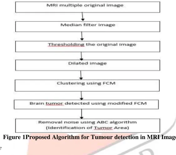
III. POPOSED ALGORITHM
Figure 1Proposed Algorithm for Tumour detection in MRI Image A. Original MRI Input Image
A set of MR brain tumor images comprising of four tumor types namely Meningioma, SLE Vasculitis and Metastase are collected from radiologists. The images used are 512*512 or 256*256 gray level images with intensity value ranges from (0 to 255) [4]. Initially, these MRI images are normalized to gray level values from (0 to 1) and the features are extracted from the normalized images. Since normalization reduces the dynamic range of the intensity values, feature extraction is made much simpler [4]. Some samples of the MRI database have been displayed in Fig. 2.
Figure 2(a, b, c) Brain Tumour image B. Median Filter
IJEDR1602310
International Journal of Engineering Development and Research (www.ijedr.org)1763
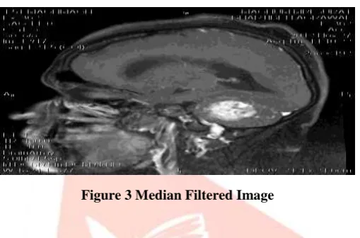
The original value of the pixel is included in the computation of the median filters are quite popularbecause for certain types of random noise, they provide excellent noise reduction capabilities with considerably less blurring than linear smoothingfilters of similar size [i]. Median filtersare particularly effective in the presence of impulse noise, also called salt and pepper noise because of its appearance as white and black dots superimposed on an image [i].Although the median filter is by far the most useful order statistics filter in image processing, it is by no means the only one, the median represents the 50th percentile of a ranked set of numbers, but recall from basic statistics that ranking lends itself to many other possibilities [i].
I = medfilt1(double(I),5);
In this research work, the 5 neighbors in ordered pixel are taken as the neighbors and the middle value of the order pixels is considered as the median and replace all the neighbor pixels with the median. Median filter is used to smooth out the MRI image at some level.
The output achieved after median filter is shown in fig 4.
Figure 3 Median Filtered Image C. Thresholding
Thresholding techniques identify a region based on the pixels with similar intensity values. This technique provides boundaries in images that contain solid objects on a contrast background [i]. Thresholding technique gives a binary output imagefrom a gray scale image. This method of segmentation applies a single fixed criterion to all pixels in the image simultaneously [i]. The key parameter in image segmentation using thresholding technique is the choice of selecting threshold value T. In case of manual thresholding method, the threshold value T can be selected by the user with the help of image histogram. This method is generally accomplished by a tool that allows the user to select the threshold value T based on choice. In case of automatic threshold selection method, the value of T can be chosen based on histogram, clustering, variance, means etc., [i].
By observing the histogram of the database, the thresholding value is considered as 0.60. I_bw = im2bw(I,0.60);
This will convert the MRI image into binary image that contains only 0s and 1s. The normalized value greater than or equal to 0.60 will be converted into 1 and the normalized value less than 0.60 is converted into 0. The output achieved after thresholding is as shown in fig 4.
Figure 4 Threshold Imag
e
D. Dilation
Useful background to this description is given in the mathematical morphology section of the Glossary. The dilation operator takes two pieces of data as inputs. The first is the image which is to be dilated [i]. The second is a (usually small) set of coordinate points known as a structuring element (also known as a kernel). It is this structuring element that determines the precise effect of the dilation on the input image [i].
IJEDR1602310
International Journal of Engineering Development and Research (www.ijedr.org)1764
Suppose that X is the set of Euclidean coordinates corresponding to the input binary image, and that K is the set of coordinates for the structuring element.Let Kx denote the translation of K so that its origin is at x.
Then the dilation of X by K is simply the set of all points x such that the intersection of Kx with X is non-empty.
The mathematical definition of grayscale dilation is identical except for the way in which the set of coordinates associated with the input image is derived. In addition, these coordinates are 3-D rather than 2-D.
In this research work, se = strel('line',11,90); I_bw = imdilate(I_bw,se);
After performing the different dilate value. One vertical line of 11 points will be consider as the strlen. If any 1 is there in the line, than the middle point is converted into 1. Dilation will help to remove out the small black portion between the white portions. The output achieved after dilate is given in fig 5.
Figure 5 Dilated Image
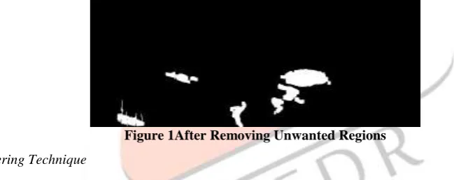
After the dilation the unwanted region part is removed by covering the 500 pixels group. If the black pixels in 500 matrix are more than white pixels than whole part is converted into black part and vice versa. The output of this step is shown in fig 7.
Figure 1After Removing Unwanted Regions E. Fuzzy C-Clustering Technique
Clustering can also be thought of as a form of data compression, where a large number of samples are converted into a small number of representative prototypes or clusters. High dimensional feature space based image segmentation is time intensive than in one dimensional feature spaces [4]. The modified FCM algorithm is based on the concept of data compression where the dimensionality of the input is highly reduced. The data compression includes two steps: quantization and aggregation. The quantization of the feature space is performed by masking the lower 'm' bits of the feature value [4]. The quantized output will result in the common intensity values for more than one feature vector. In the process of aggregation, feature vectors which share common intensity values are grouped together. A representative feature vector is chosen from each group and they are given as input for the conventional FCM algorithm. Once the clustering is complete, the representative feature vector membership values are distributed identically to all members of the quantization level. Since the modified FCM algorithm uses a reduced dataset, the convergence rate is highly improved when compared with the conventional FCM [4].
Fuzzy C-means (FCM) is a method of clustering which allows one pixel to belong to two or more clusters .The FCM algorithm attempts to partition a finite collection of pixels into a collection of "C" fuzzy clusters with respect to some given criterion [4]. Depending on the data and the application, different types of similarity measures like distance, connectivity, and intensity may be used to identify classes/clusters [4].
Steps of FCM Algorithm:
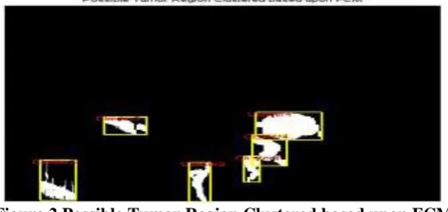
• Probable Brain Tumor regions • Multiple regions
• But major drawback is not assurance of actual brain tumor • Clustering is based upon region
• Noise and brain tumor have equal probability
IJEDR1602310
International Journal of Engineering Development and Research (www.ijedr.org)1765
Figure 2 Possible Tumor Region Clustered based upon FCMThe database of the modified FCM is given in the fig 9. Modified FCM Database
Figure 9 Modified FCM database
In above figure, “X_Cord” shows the x axis coordinate of the rectangle box. “Y_Cord” shows y axis coordinate of rectangle box. “Width “ shows the width from “X_Cord” and “Height” shows the height from “Y_Cord”, “Cetroid_X” and “Cetroid_Y” shows the x and y axis coordinate of the center point. “Area show the number of 1’s or white pixels in the rectangle.
F. Artificial Bee Colony algorithm
Artificial Bee Colony (ABC) is a novel optimizationalgorithm inspired of the natural behavior of honey bees intheir search process for the best food sources, In groupsof insects which live in colonies like the ants and bees, anindividual only can do simple task on its own, while thecooperative work of colony is the main reason determiningthe intelligent behavior of them [8].A colony of artificial bees inABC algorithm contains three groups of bees: employed,onlookerand scoutbees. Employed bees carry withthem information about their food sources, its distance anddirection from the nest, and the nectar amount of the source;scout bees are searching the environment surrounding thenest for finding new food sources; and onlooker bees waitingin the hive and finding a food source through the informationshared by employed bees. In ABC, two key behaviors aredefined: recruitment to a nectar source, and abandonment of a source [8].
The search process to discover the best solutions by the artificial bees can be summarized as follow: 1. Employed bees move randomly to find solutions in the search space.
2. Review the information obtained by the employed and scout bees with onlooker bees in colony space (note that at the first yet there is no scout bee).
3. Check the stopping criterion: stop, if satisfied condition, otherwise continue. 4. Selecting scout bees and perform the recruitment process for them.
5. Vocalize of new population of bees.
IJEDR1602310
International Journal of Engineering Development and Research (www.ijedr.org)1766
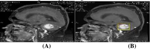
(A) (B)Figure 10(A) Wrong Tumor Detection by Modified FCM (B) Exact Tumor from Modified ABC
IV. EXPERIMENTS AND RESULTS A. Input as JPG Image In Matlab
A set of MR brain tumor images comprising of three tumor types namely Meningioma, SLE Vasculitis and Metastase are collected from radiologists. The images used are 512*512 or 256*256 gray level images with intensity value ranges from (0 to 255) [4]. Figure Shows Tumor detection in Meningioma Types Image:
(A) (B)
Figure 31Tumor detection in Meningioma Types Image
B. Comparison of FCM, Modiied FCM and ABC Algorithm
This Research work Tests on total 22 images of three tumor types namely Meningioma, SLE Vasculitis and Metastase, from that 20 images detects exact location of tumor with ABC Algorithm.
Algorithm Efficiency (%)
FCM 27.27%
Modified FCM 63.63%
Artificial Bee Colony 91.24%
Table 1 Comparison of FCM, Modied FCM and ABC Algorithm
V. CONCLUSION
In this research work, MRI image contain the noise so that it is difficult to determine the tumor part. After the Artificial Bee Colony algorithm exact brain tumor is detected for every image location and size is given in pixel with the accuracy of 91.24%. REFERENCES
[1] MRI Fuzzy Segmentation of Brain Tissue Using Neighborhood Attraction With Neural-Network Optimization by Shan Shen, William Sandham, Malcolm Granat, and Annette Sterr – IEEE 2005
[2] Improved Fuzzy C-Means Algorithm for MR Brain Image Segmentation P. Vasuda (IJCSE) International Journal on Computer Science and Engineering Vol. 02, No. 05, 2010, 1713-1715
[3] Diagnose Brain Tumor Through MRI Using Image Processing Clustering Algorithms Such As Fuzzy C Means Along With Intelligent Optimization Techniques by N. Nandha Gopal, Dr. M. Karnan – IEEE 2010
[4] MRI brain image segmentation using modified fuzzy c-means clustering algorithm by m. shasidhar, v.sudheer raja, b. vijay kumar – ieee 2011
[5] MRI brain image segmentation using modified fuzzy c-means clustering algorithm by m. shasidhar, v.sudheer raja, b. vijay kumar – ieee 2011
[6] A Survey On Bee Colony Algorithms. Salim Bitam et al./IEEE xplore 2010.
[7] Effective Fuzzy Clustering Algorithm for Abnormal MR Brain Image Segmentation D.Jude hemanth, D.Selvathi and J.Anitha – IEEE 2009
[8] An Artificial Bee Colony Optimization for MRI Fuzzy Segmentation of Brain Tissue 2011 International Conference on Management and Artificial Intelligence IPEDR vol.6 (2011) © (2011) IACSIT Press, Bali, Indonesia
[9] A comprehensive survey: artificial bee colony (ABC) algorithm and applications Dervis Karaboga · Beyza Gorkemli · Celal Ozturk · Nurhan Karaboga © Springer Science+Business Media B.V. 2012.
[10] Evaluation of Three Methods for MRI Brain Tumor Segmentation by R. B. Dubey, M. Hanmandlu, Shantaram Vasikarla – IEEE 2011