ISSN (Print) : 2320 – 3765 ISSN (Online): 2278 – 8875
I
nternational
J
ournal of
A
dvanced
R
esearch in
E
lectrical,
E
lectronics and
I
nstrumentation
E
ngineering
(A High Impact Factor, Monthly, Peer Reviewed Journal)
Website: www.ijareeie.com
Vol. 7, Issue 5, May 2018
Smart Lab Detection for Diabetic Patient
using Iris Image
Dr. Prakash H. Patil1
, Roshan M. Patil2
, Nidhi H. Manek3
, Harsh V. Ladda 4 Vice Principle, Dept. Of E&TC Engg, D.Y.P.C.O.E Ambi, Pune, India1
Dept. Of E&TC Engg, D.Y.P.C.O.E Ambi, Pune, India 2, 3, 4
ABSTRACT: Iris image analysis for clinical diagnosis is one of the most efficient non-invasive diagnosis methods for
determining health status of organs Correct and timely diagnosis is a critical, yet essential requirement of medical science. From the literature, it is found that modern technology also fails in lot of cases to diagnose disease correctly. The attempt is being made to explore the area of diagnosis from different perspectives .The approach used is a combination of ancestor's technology Iridodiagnosis with modern technology. Iridodiagnosis is an alternative branch of medical science, which can be used for diagnostic purposes the various algorithms are developed for image quality assessment, segmentation of iris, iris normalization and clinical feature classification for clinical diagnosis. The entire process shows classification accuracy of 90 ~ 92 percent between diabetic and non-diabetic subjects. This approach will be useful in the diagnosis fields, which are faster, user friendly and less time consuming.
KEYWORDS: Iridodiagnosis, iris, diabetic, SVM classification, feature extraction, retinopathy, and segmentation.
I.INTRODUCTION
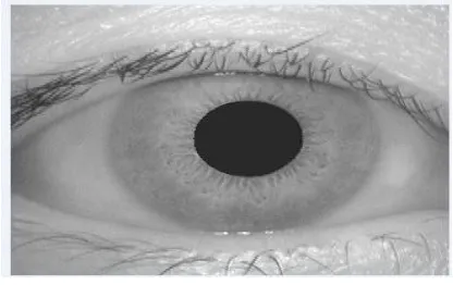
Iridology is the branch of science that deals with the study of iris i.e. colored part of the eye. The Iris is the greenish-yellow area surrounding the transparent pupil (showing as black). The white outer area is the sclera; the central transparent part is the cornea. The main intention of irido diagnosis is to collect some information about underlying disease. As technology has developed, there are various methods present for the diagnosis, which are highly reliable and accurate. Irido-diagnosis is consists on empirical science, to look into the particular area of eye for systemic health condition of the specific organ of the body. [1]
Iridology is the diagnosis of medical conditions and “pre-disease states” through abnormalities of pigmentation in the iris. The location of abnormalities on the iris is associated with the location of the medical condition in the body. The iris of the eye is divided into 60 sectors; each sector is corresponding to an inner organ. The iris is associated via multiple nerve connections to the organs. Depending on the features of the iris, classification is done and diabetic is detected. Iridodiagnosis can also be used to detect Gall Bladder Disease in the patient’s iris [4].
II.LITERATURE SURVEY
ISSN (Print) : 2320 – 3765 ISSN (Online): 2278 – 8875
I
nternational
J
ournal of
A
dvanced
R
esearch in
E
lectrical,
E
lectronics and
I
nstrumentation
E
ngineering
(A High Impact Factor, Monthly, Peer Reviewed Journal)
Website: www.ijareeie.com
Vol. 7, Issue 5, May 2018
III.PROPOSED SYSTEM
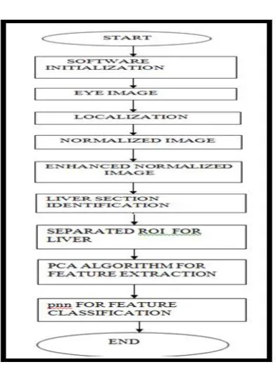
The framework followed in this paper is illustrated in the fig (1).
Fig 1: Block diagram of system
A. Elements of block diagram are as follows: a) Eye Image Acquisition
Initially the eye image is captured with the help of certain cameras, and stored in the database which contains normal as well as abnormal results of iris. Captured eye image looks as shown in fig (2). [1]
Fig. 2: Captured Eye Image b) Image preprocessing
ISSN (Print) : 2320 – 3765 ISSN (Online): 2278 – 8875
I
nternational
J
ournal of
A
dvanced
R
esearch in
E
lectrical,
E
lectronics and
I
nstrumentation
E
ngineering
(A High Impact Factor, Monthly, Peer Reviewed Journal)
Website: www.ijareeie.com
Vol. 7, Issue 5, May 2018
c) Segmentation
Segmentation is done in order to find inner and outer boundries of the iris. By subtracting pupil from sclera, we will get the iris part of an eye [5]. Once the iris region is segmented from an eye, the next step is to transform the iris region into fixed dimentions. After subtraction, we will get the iris pattern into circular shape. [4]
d) Normalization
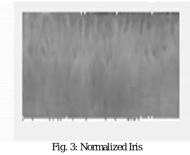
Normalization is done to convert circular iris pattern into rectangular shape as shown in fig (3).
Fig. 3: Normalized Iris e) ROI extraction
After normalization, the next step which comes into picture is ROI extraction. ROI extraction is nothing but cropping particular portion of normalized iris image according to “irido-chart” as shown in fig(4).[5]
.
Fig. 4: Irido Chart
f) Feature Extraction
Once the region of interest is find out the various features of that region are carried out. Depending on that features value we can make two different set of normal & diabetes. The different features are as listed below 1. Mean
2. Entropy
ISSN (Print) : 2320 – 3765 ISSN (Online): 2278 – 8875
I
nternational
J
ournal of
A
dvanced
R
esearch in
E
lectrical,
E
lectronics and
I
nstrumentation
E
ngineering
(A High Impact Factor, Monthly, Peer Reviewed Journal)
Website: www.ijareeie.com
Vol. 7, Issue 5, May 2018
g) SVM classification
SVM is relatively new method of classification and it expands very quickly. SVM use in medicine: SVMs are helpful in text and hypertext categorization as their application can significantly reduce the need for labeled training instances in both the standard inductive and transductive settings. SVMs can be used to solve various real world problems.
IV. COMPARATIVE ANALYSIS
ISSN (Print) : 2320 – 3765 ISSN (Online): 2278 – 8875
I
nternational
J
ournal of
A
dvanced
R
esearch in
E
lectrical,
E
lectronics and
I
nstrumentation
E
ngineering
(A High Impact Factor, Monthly, Peer Reviewed Journal)
Website: www.ijareeie.com
Vol. 7, Issue 5, May 2018
V. FLOWCHART
Fig 5: Flowchart of System
VI. APPLICATIONS
• As per diagnosis done using iris image the further treatment of the patient can be done in early stage of diabetes.
• Eye sight can be saved with proper treatment & medicine prescribed. • It analyzes iris and eye images for the purpose of medical diagnostic. • It classifies the image based on different diseases.
• It processes the IRIS and eye images for early detection of diseases.
VII. EXPERIMENTAL RESULTS
ISSN (Print) : 2320 – 3765 ISSN (Online): 2278 – 8875
I
nternational
J
ournal of
A
dvanced
R
esearch in
E
lectrical,
E
lectronics and
I
nstrumentation
E
ngineering
(A High Impact Factor, Monthly, Peer Reviewed Journal)
Website: www.ijareeie.com
Vol. 7, Issue 5, May 2018
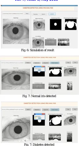
Fig. 6: Simulation of result
Fig. 7: Normal iris detected
Fig. 7: Diabetes detected
VIII. CONCLUSION
ISSN (Print) : 2320 – 3765 ISSN (Online): 2278 – 8875
I
nternational
J
ournal of
A
dvanced
R
esearch in
E
lectrical,
E
lectronics and
I
nstrumentation
E
ngineering
(A High Impact Factor, Monthly, Peer Reviewed Journal)
Website: www.ijareeie.com
Vol. 7, Issue 5, May 2018
ACKNOWLEDGEMENT
It is pleasant Endeavour to present paper on “IOT BASED SMART LAB DETECTION FOR DIABETIC
PATIENT USING IRIS IMAGE”. I take this opportunity to express my gratitude towards our guide Dr. Prakash H.
Patil (vice principle) of DYPCOE for his constant encouragement and guidance. I’m thankful to Prof. S. S. Badhe
HOD (E&TC) & Principal Dr. Abhay Pawar for their guidance, constant supervision as well as for providing necessary information regarding the project.
REFERENCES
[1] Prof. S. K. Bhatia, Priyanka Atole, Sarika Kamble, Pooja Telang “Methodology for Detecting Diabetic Presence from Iris Image Analysis” International Journal of Advanced Research in Computer Engineering & Technology (IJARCET) Volume 4 Issue 3, March 2015 PP. 776-779. [2] Ms. Pragtee Bhagvan Tathe, Mrs. Dr. M. M. Patil, Novateur “ANALYSIS OF HEALTH CONDITION BASED ON IRIS IMAGE” Publication’s International Journal of Innovation in Engineering, Research and Technology [IJIERT] ICITDCEME’15 Conference Proceedings ISSN No - 2394-3696 PP. 1-4.
[3]Jyoti Prasad, Divya Patel, Megha Jadhav, Prof. Rupali Deshmukh “IRIS BASED MEDICAL ANALYSIS BY GEOMETRIC DEFORMATION FEATURES” International Research Journal of Engineering and Technology (IRJET) e-ISSN: 2395 -0056 Volume: 03 Issue: 04 | April-2016 p-ISSN: 2395-0072 PP. 2553-2556.
[4]Piyush Samant, Ravinder Agarwal “Diagnosis of Diabetes Using Computer Methods: Soft Computing Methods for Diabetes Detection Using Iris” World Academy of Science, Engineering and Technology International Journal of Medical, Health, Biomedical, Bioengineering and Pharmaceutical Engineering Vol:11, No:2, 2017 PP. 63-68.
[5]Miss. S. B. More, Prof.N. D.Pergad“ON A METHODOLOGY FOR DETECTING DIABETIC PRESENCE FROM IRIS IMAGE ANALYSIS” International Journal of Engineering Science & Research Technology (IRJET) ISSN: 2277-9655 [More*, 4(7): July, 2015].
[6] www.ieeeprojects.com [7] www.link.springer.com [8] www.researchgate.com
[9]Dipankar Ray & Sukhendu Dey “MATLAB Programming for Engineering and Science”