Alternative Splicing of a
!
4
Subunit Proline-Rich Motif Regulates
Voltage-Dependent Gating and Toxin Block of Ca
v
2.1
Ca
2"
Channels
Thomas D. Helton,
1Douglas J. Kojetin,
2John Cavanagh,
2and William A. Horne
1Departments of
1Molecular Biomedical Sciences and
2Molecular and Structural Biochemistry, North Carolina State
University, Raleigh, North Carolina 27606
Ca2!channel !subunits modify"
1subunit gating properties
through direct interactions with intracellular linker domains. In a previous report (Helton and Horne, 2002), we showed that alternative splicing of the !4subunit had"1subunit
subtype-specific effects on Ca2!channel activation and fast inactiva-tion. We extend these findings in the present report to include effects on slow inactivation and block by the peptide toxin
#-conotoxin (CTx)-MVIIC. N-terminal deletion and site-directed mutagenesis experiments revealed that the effects of alterna-tive splicing on toxin block and all aspects of gating could be attributed to a proline-rich motif found within N-terminal !4b
amino acids 10–20. Interestingly, this motif is conserved within the third postsynaptic density-95 (PSD-95)/Discs large/zona occludens-1 domain of the distantly related membrane-associated guanylate kinase homolog, PSD-95. Sequence identity of "30% made possible the building of!4a and!4b
three-dimensional structural models using PSD-95 as the target sequence. The models (1) reveal that alternative splicing of the
!4N terminus results in dramatic differences in surface charge
distribution and (2) localize the proline-rich motif of!4bto an
extended arm structure that flanks what would be the equiva-lent of a highly modified PSD-95 carboxylate binding loop. Northern blot analysis revealed a markedly different pattern of distribution for!4aversus!4bin the human CNS. Whereas!4a
is distributed throughout evolutionarily older regions of the CNS,!4bis concentrated heavily in the forebrain. These results
raise interesting questions about the functional role that alter-native splicing of the!4subunit has played in the evolution of
complex neural networks.
Key words: calcium channel;!4subunit; PSD-95; alternative splicing; gating; N terminus;#-CTx-MVIIC
Voltage-gated Ca2!channels participate in an extensive array of
cellular activities including excitation–contraction coupling,
tran-scription, and neurotransmitter release. Neuronal Cav2 channels
are assemblies of up to five subunits,"1, "2/$,!, and%. The"1 subunit consists of four homologous repeats (I–IV) of six helices (S1–S6) that arrange to form the selectivity filter and pore. The 24 transmembrane helices are connected by a series of alternating intracellular and extracellular loops. These loops are targets for a
host of modifying proteins, including ! subunits, G-proteins,
calmodulin, and syntaxin, as well as the peptide toxins of venom-ous spiders and marine snails (Catterall, 2000). Interaction of these proteins with"1subunits typically alters the voltage
depen-dency and kinetics of channel gating, which in turn modifies Ca2!
entry into neurons.
Ultimately, gating behavior is determined by the interactions of individual amino acid side chains with the electrostatic forces within their microenvironments. This is especially true for the positively charged S4 helical segments that constitute the voltage sensors in Na!, Ca2!, and K!channels. Biophysical studies have shown that depolarization disrupts S4 side-chain interactions of
Shaker K!channels to the extent that S4 helices rotate 180° along
their axes (Cha et al., 1999; Glauner et al., 1999). This motion likely triggers a cascade of side-chain disruptions that ultimately leads to rotation and separation of the intracellular S6 segments
that form the K!channel gate (Bezanilla, 2000). Such a
mecha-nism is supported by recent studies delineating the conforma-tional changes associated with open and closed states of bacterial
two-membrane-spanning K!channels (Jiang et al., 2002b) and is
generally applicable to Na!and Ca2!channel gating.
Attempts have been made to assign specific gating functions to
individual Ca2!channel homology domains. Early chimera
stud-ies indicated that the IS6 segment was critical for setting the rate of fast inactivation (Zhang et al., 1994); however, substitution of
IIS6 and IIIS6 of the Cav2.3 channel into the slow inactivating
Cav1.2 channel caused a leftward shift in the voltage dependence
of inactivation and increased the rate of Cav1.2 channel
inactiva-tion to near Cav2.3 rates (Stotz et al., 2000). Effects on gating
have been reported for amino acid substitutions in IS3 (Zhong et al., 2001), the I-II linker (Berrou et al., 2001), IIS6 (Stotz and Zamponi, 2001), extracellular linkers IIIS3-S4 (Lin et al., 1997), and IVS3-S4 (Hans et al., 1999), and IVS6 (Berjukow et al.,
2001). Additive effects on Cav1.2 channel inactivation were
re-ported recently for individual IS6, IIS6, IIIS6, and IVS6 substi-tutions (Shi and Soldatov, 2002). Together, these data support a
structural model of "1 subunits in which individual
transmem-brane segments are interdependently entwined (Horn, 2000).
Our results indicate that this model also applies to! subunit
interactions with"1subunit intracellular linkers. We have shown previously that !4 subunit alternative splicing had "1 subtype-Received July 10, 2002; revised Aug. 22, 2002; accepted Aug. 22, 2002.
This work was supported by National Institutes of Health Grants NS32094 (W.A.H.) and GM5576 (J.C.), by a College of Veterinary Medicine State Research Support grant (W.A.H.), and by an award from the North Carolina State University Keenan Institute (J.C.).
Correspondence should be addressed to William A. Horne, Department of Molecular Biomedical Sciences, North Carolina State University, College of Vet-erinary Medicine, 4700 Hillsborough Street, Raleigh, NC 27606. E-mail: [email protected].
specific effects on voltage-dependent activation and inactivation (Helton and Horne, 2002). In this report, we extend thesefindings to include effects on slow inactivation and block by#-conotoxin
(CTx)-MVIIC; we also identify a proline-rich motif in!4bthat is
responsible for the observed differences in effects.
MATERIALS AND METHODS
Deletion mutants.Truncation of the!4bN terminus in 10 aa increments was performed using PCR and custom oligonucleotide primers [Integrat-ed DNA Technologies (IDT), Coralville, IA]. All nucleotide and amino acid positions for primers and restriction enzymes correspond to the!4b sequence (GenBank accession number U95020). Each forward primer sequence contained an idealized Kozak (1991) sequence and start codon corresponding to the beginning of each of the deleted 10 amino acids as follows: !4b #1–10F, 5$-GCCACCATGACCGCGGACGGGCCG; !4b #1–20F, 5$-GCCACCATGCAGGTGGCCCGAGGC. Both reactions in-cluded a common!4reverse primer,!4b732R (5$-TGACGGCCCCAC-TAACACC). Full-length!4bwas used as the template for these reactions. The !4b #10–20 deletion mutant was generated with the primer !4b
#10–20F
(5$-GCCACCATGTCCTCCTCCTCCTACGCCAAGAACT-CG) paired with!4b732R using the!4b#1–20 mutant as the template. Annealing temperature for PCR was 56°C with Gene ChoiceTaqDNA polymerase (PGC Scientific, Durham, NC). Correctly sized PCR frag-ments were cloned into the pT-Advantage vector (Clontech, Palo Alto, CA). PCR-based cycle sequencing (FS chemistry; Applied Biosystems, Foster City, CA) was used with an ABI Prism 310 Genetic Analyzer. The data were analyzed using ABI Prism DNA sequencing software (version 2.12; PerkinElmer Biosystems), and sequence alignments and restriction maps were generated using Lasergene Software (DNA Star, Madison, WI). Correct clones were digested withBamHI (Roche Molecular Bio-chemicals, Indianapolis, IN), and the corresponding"530 bp fragments were ligated into BamHI (nucleotide position 550) -digested !4b in pBluescript II S/K! (Stratagene, La Jolla, CA). Each !
4b deletion mutant was sequenced to confirm correct reading frame and proper N-terminal orientation.
Site-directed mutagenesis. For all !4b site-directed mutants, full-length!4bcDNA (U95020) was used as the template unless otherwise indicated. All site-directed mutagenesis reactions were performed using a QuikChange site-directed mutagenesis kit (Stratagene) and custom forward and reverse compliment oligonucleotide primers (I DT): !4bG10A,D13A, 5$ACGCCAAGAACGCGACCGCGGC -CGGGCCGCAC; !4bP15A,P18A, 5$GCGGACGGGGCGCAC TC -CGCCACCTCGCAGGTG; !4bG10A,D13A,P15A,P18A,
5$-ACGC-CAAGAACGCGACCGCGGCCGGGGCGCAC (!4bP15A,P18A
used as template); !4bG10A,P15A,
5$-TACGCCAAGAACGCGAC-CGCGGACGGGGCGCACTCCCCCACCTCGCAGGTG; !4bH16A,
5$-ACCGCGGACGGGCCGGCCTCCCCCACCTC; !4bG10A,P18A,
5$TACGCCAAGAACGCGACCGCGGACGGGCCGCAC TCC
-GCCACC TCGCAGGTG;!4bD13A,P15A,
5$-TACGCCAAGAA-CGGGACCGCGGCCGGGGCGCAC TCCCCCACC TCGCAGGTG;
!4bD13A,P18A, 5$TACGCCAAGAACGGGACCGCGGCCGGGC
-CGCAC TCCGCCACC TCGCAGGTG;!4bH16A, 5$-ACCGCGGA-CGGGCCGGCC TCCCCCACCC TCG; and !4bT11A,S17A,T19A, S20A, 5$CAAGAACGGGGCCGCGGACGGGCCGCACGCCCC -CGCCGCGCAGGTGGCC. Each of the mutant clones was sequenced to confirm reactionfidelity.
Electrophysiology.cRNAs were synthesizedin vitrousing an mMessage mMachine RNA transcription kit from Ambion (Austin, TX) (T3 or T7 depending on clone orientation in pBluescript II S/K!). Standard
Xeno-pus laevisoocyte expression methods were used to characterize! dele-tion and site-directed mutants. Briefly, full-length "1, "2/$-1, and !4 cRNAs were injected in equimolar ratios (5.6 ng of"1A, 2.4 ng of"2/$-1, and 1.6 ng of!4in 46 nl) into defolliculated oocytes (stage V–VI). The BI-2 ("1A) and"2/$-1 clones used in this study were provided by T. Tanabe (Tokyo Medical and Dental University, Tokyo, Japan). Calcium channel currents were recorded 2–4 d after oocyte injection by standard two-electrode voltage clamp using a Warner amplifier (OC-725B) at 20–22°C, and data were collected using pClamp6 software (Axon Instru-ments, Foster City, CA). Microelectrodes werefilled with 3MKCl, and the resistances of the current and voltage electrodes were 0.3–1.5 M%. Data were filtered at 2 kHz and sampled at 10 kHz. Currents were recorded in a chloride-free bath containing (in mM): 5 Ba(OH)2, 5 HEPES, 85 TEA-OH, and 2 KOH, pH adjusted to 7.4 with methanesul-fonic acid. In experiments with the peptide toxin#-CTx-MVIIC
(Pep-tide Institute Inc. Osaka, Japan), the 5 mMBa2!solution was supple-mented with 0.1 mg/ml cytochrome c to saturate nonspecific peptide binding sites. Cytochromec at 0.1 mg/ml had no noticeable effect on recorded Ba2! currents. Peptides were reconstituted according to the manufacturer’s instructions (100&Mstock solutions in sterile, deionized water). Fresh dilutions of the peptide were made immediately before use. Currents typically ranged between 0.8 and 2.5 &A, and leak currents were between 20 and 100 nA. Data were analyzed using pClamp6 software (Axon Instruments) and Excel 7.0 (Microsoft Corp., Redmond, WA). The leak and capacitive currents were subtracted on line using a standard P/4 protocol. Curve fitting was performed with SigmaPlot version 5.0 (SSPS Inc., Chicago, IL).
Slow inactivation.Oocytes were held at&80 mV for"2 min before a 300 msec reference pulse (IR) to 0 mV (!4b) or!10 mV (!4a). AfterIR, the membrane potential was stepped immediately to a conditioning pulse potential ranging from&100 mV to&20 mV (20 mV increments) and held for 5 min. During the conditioning pulse, a 300 msec test pulse (IT) to 0 mV (!4b) or!10 mV (!4a) was applied every 15 sec. Data were normalized as the ratio of the maximum current at time T (IT) divided by the maximum reference current (IR). Data werefit to the double-exponential equation IT/IR ' A1e&x /'1 ! A2e&x /'2, where IT equals current at time T,IRequals maximum reference current, x equals time in seconds, andA1andA2are components for the time constants'1and'2, respectively. The SEM is shown for each data point unless the values are smaller than the symbol.
Recovery from slow inactivation.Currents were stabilized at&80 mV or &100 mV for"2 min before a 300 msec reference pulse (IR) to 0 mV (!4b) or!10 mV (!4a). After a 100 msec step to either&80 mV or&100 mV, the oocytes were held at a conditioning pulse potential of&30 mV for 5 min. Immediately after the conditioning pulse, a 300 msec test pulse was applied (I1), and then the holding potential was stepped back to either&80 mV or&100 mV and 300 msec test pulses (I2–12) were applied at 15 sec intervals starting at time 0 for a total of 3 min. Data were normalized as the ratio of the maximum current at time T (IT) divided by the maximum reference current (IR). Data were fit to the single exponential equationIT/IR'I(!Ae&x /', where ITequals current at time point T,IRequals maximum reference current,I(equals current remaining at end of protocol, x equals time in seconds, and A is the component for the time constant'.
Voltage dependence of activation and inactivation.Voltage dependency of activation data was generated fromI–Vcurves. Maximal currents were obtained from 300 msec depolarizations from a holding potential of&80 mV to various test potentials (&40 to!10 mV in 5 mV increments). Each individual recording was then normalized, inverted, andfit to the Bolt-zmann equation %IBa'1/[1!exp(&(Vtest&V1/2)/k)], whereVtestequals
I–Vtest potential,Vpreequals prepulse potential,V1/2equals midpoint of activation or inactivation, andkequals slope factor. Thefit curves,V1/2, andkvalues were then averaged and plotted as a function of membrane voltage.
Voltage dependency of inactivation data was obtained from peak currents elicited by a 300 msec maximal current test depolarization after a 20 sec conditioning prepulse to voltages ranging from&80 to!20 mV. Each individual recording was then normalized andfit to the Boltzmann equation %IBa'1/[1!exp([Vpre&V1/2]/k)], whereVtestequalsI–Vtest potential,Vpreequals prepulse potential,V1/2equals midpoint of activa-tion or inactivaactiva-tion, andkequals slope factor. Thefit curves,V1/2, andk values were then averaged and plotted as a function of prepulse potential.
Pharmacology. Oocytes were held at a potential of &80 mV with maximal currents elicited by 150 msec test pulses to 0 mV (!4b) or!10 mV (!4a) every 15 sec for a total of 10 min. During recordings, oocytes were perfused at a constant rate of"0.5 ml/min. Average current sizes for!4aand!4bcomplexes were 1.9)0.2&A (4–5 d of incubation) and 2.3)0.3&A (2–3 d of incubation), respectively. The data werefit to the single-exponential equationIT/IR'I(!Ae&x /', whereITequals maxi-mum current at time point T,IRequals maximum current at time point 0,I(equals residual current at end of protocol, x equals time in seconds, and A is the component for the time constant, '. The averaged rate constants (1/') for the four#-CTx-MVIIC concentrations (0.2, 0.6, 2, and 6&M) were plotted as a log function of their concentration and were fit well by the equation (')&1'k
on[Tx]!koff.
Northern blot analysis.A commercially available human neuronal tis-sue Northern blot [Multiple Tistis-sue Northern (MTN) Blot brain II; Clontech] was probed with a nonspecific!4subunit probe (!4#N; nucle-otides 215–1628 plus "300 bp of 3$ untranslated). A 32P-labeled !
WI) using the!4#N mutant as the template. The!4#N mutant is missing thefirst coding 147 bp corresponding to the 49 aa N terminus of!4[clone from Helton and Horne (2002)]. The MTN blot was hybridized overnight at 42°C in hybridization buffer [5* SSC, 5% w/v blocking reagent (Roche), 0.1%N-lauroylsarcosine, 0.02% w/v SDS, 50% w/v formamide] plus 100&g/ml herring sperm DNA (Promega). The probe concentration was 1 million counts/ml. The blot was washed with successive stringency washes (four washes, 15 min each at 37°C) ranging from 2*SSC/0.1% SDS to 0.1*SSC/0.1% SDS. The blot was then exposed to radiographic film for 12 hr at&80°C. One microgram of cRNA for both!4aand!4b was run out on a 1% denaturing formaldehyde gel along with a poly(A)-tailed cRNA mass ladder (RNA Molecular Weight Marker 1; Roche). The!4acRNA is longer than the!4bcRNA because of the additional "400 nt of 5$untranslated sequence.
Molecular modeling. The sequences for rat postsynaptic density-95 (PSD-95) (DLG4_rat) and human!4(CACNB4) were obtained (acces-sion numbers P31016 and U95020) from the Swiss-prot database. Amino acids 10–96 of!4bwere aligned to residues 303–390 of PSD-95 based on secondary structure prediction (nnPredict) and visual inspection. For!4a, amino acids 50–96 of!4bwere aligned to residues 345–390 of PSD-95. Using default parameters, the program MODELLER 6 (Sali and Blun-dell, 1993) was used to produce 50 models each of!4band!4astructure based on the solved structure of the third PSD-95/Discs large/zona occludens-1 (PDZ) domain of PSD-95 (1BEF). Five models each were chosen for additional analysis based on the molecular probability density function (PDF) output from MODELLER and stereochemical analysis obtained through Ramachandran output from PROCHECK-NMR (Las-kowski et al., 1993). The interactions between different atom types within these models and C"root mean square deviation (RMSD) comparisons between the models and 1BEF were characterized with ERRAT (Colo-vos and Yeates, 1993). The!4aand!4bmodels chosen for comparison had the fewest disallowed residues (Ramachandran), lowest molecular PDF and RMSD values, and highest percentage of residues in acceptable conformations based on ERRAT and PROCHECK-NMR analysis. Models were visualized with the program MOLMOL (Koradi et al., 1996).
RESULTS
Alternative splicing of the
!
4subunit affects slow
inactivation of Ca
v2.1 Ca
2"channels
In a previous study (Helton and Horne, 2002), we showed that Cav2.1 complexes containing the longer form of an alternatively
spliced !4subunit N terminus, !4b(49 aa), inactivated at more
negative potentials in response to 20 sec conditioning prepulses than complexes containing a shorter form,!4a(15 aa). To
deter-mine whether this response extended to slower types of inactiva-tion, we examined in the present study the effects of!4aand!4b
on Cav2.1 cumulative inactivation elicited by 5 min conditioning
prepulses combined with stimulation at 0.25 Hz. Oocytes were stabilized at&80 mV before a 300 msec reference pulse (IR) to
potentials that were predetermined to give peak inward currents (!4b, 0 mV;!4a, !10 mV). The membrane potential was then
stepped to and held at the conditioning prepulse potential (rang-ing from&100 to&20 mV) for 5 min. A 300 msec test pulse (IT)
was elicited from the conditioning prepulse potential every 15 sec (I5 equals test pulse at 5 min). The kinetics of entry to slow
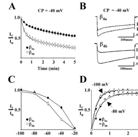
inactivation for Cav2.1 complexes containing either!4aor!4bat &40 mV is shown in Figure 1A. For comparison purposes, wefit the data points for both!4aand!4bto two exponentials (smooth
curves in thefigure). The time constants for the fast component of entry ('1) for!4aand!4bwere 28.6)2.6 and 18.9)1.2 sec,
respectively, and the time constants for the slow component of entry ('2) were 769 ) 23.6 and 384 ) 14.8 sec, respectively.
Overall, the IT/IR ratio for Cav2.1 complexes containing !4b
decreased to 0.5 in "70 sec, whereas those containing !4a
re-quired"380 sec (data not shown). This indicated that!4bcaused
a more thanfivefold acceleration of the kinetics of slow inactiva-tion. Representative current traces for reference and 5 min test
pulses from a conditioning potential of &40 mV for Cav2.1
complexes containing !4a (top) and !4b (bottom) are shown in
Figure 1B. As seen in thefigure and as described in our previous study, Cav2.1 complexes containing!4aunderwent open-state fast
inactivation faster than did complexes containing!4b. After 5 min
at&40 mV, the rate of fast inactivation was unaltered for com-plexes containing!4a, and slowed only somewhat for complexes
containing!4b. The absence of any appreciable tail-current
indi-cated that deactivation was not affected by prolonged depolariza-tion. TheI5/IRratio is plotted against the range of conditioning
potentials (&100 to&20 mV) in Figure 1C. Thefigure illustrates that the voltage dependence of Cav2.1 slow inactivation is shifted
to the left for complexes containing!4brelative to those
contain-ing !4a. Half-maximal inactivation occurred at approximately Figure 1. Effects of!4aand!4bon slow inactivation and recovery from slow inactivation of Cav2.1 Ca2!channels. Studies were performed with
Xenopusoocytes expressing"1A,"2/$-1, and either!4aor!4b.Reference (IR) and test current (IT) traces were generated by 300 msec step depo-larizations from various holding potentials to either 0 mV (!4b) or!10 mV (!4a). Maximum values from 300 msecIRandITcurrent traces were used to calculateIT/IRwhere indicated. Barium (5 mM) was used as the charge carrier.A, Influence of!4aand!4bon the development of slow inactivation at a conditioning potential (CP) of&40 mV. After a refer-ence pulse (IR) measured from a holding potential of&80 mV, oocytes were held at&40 mV for 5 min. During this time, 300 msec test pulses (IT) were applied every 15 sec. Eachpointrepresents the mean value of
&50 mV for complexes containing!4band&35 mV for!4a. These
values are"10 mV (!4b) and 5 mV (!4a) more negative than were
determined for inactivation in response to 20 sec conditioning prepulses (Helton and Horne, 2002). Figure 1D shows that re-covery from 5 min of slow inactivation at &30 mV is nearly complete when the membrane potential is stepped back to &80 mV, and that there is no difference in the time course of recovery for Cav2.1 complexes containing either!4aor!4b. Recovery was
somewhat faster and more complete when the membrane poten-tial was stepped back to &100 mV. The recovery data at both potentialsfit well to single exponentials. The time constants for recovery for!4aand!4bat&80 mV were 28.6)2.0 and 27.8)
1.6 sec, respectively, and at&100 mV, 18.9)0.5 and 17.2)0.4 sec, respectively.
Alternative splicing of the
!
4subunit affects
#
-CTx-MVIIC block of Ca
v2.1 Ca
2"channels
The results to this point indicate that changes in the structure of the!4subunit N terminus impact"1Asubunit structures that are
important for many aspects of gating, including activation, open-state inactivation, and fast and slow closed-open-state inactivation. Given that recent evidence indicates that cytosolic determinants of two-membrane-spanning K! channel gating are coupled to
changes in outer vestibule structure (Perozo et al., 1999; Jiang et al., 2002a,b), we next sought to determine whether alternative splicing of the!4subunit would affect the block of Cav2.1
chan-nels by a marine snail peptide conotoxin,#-CTx-MVIIC.
Cono-toxin interactions with voltage-gated Ca2!channels are entirely
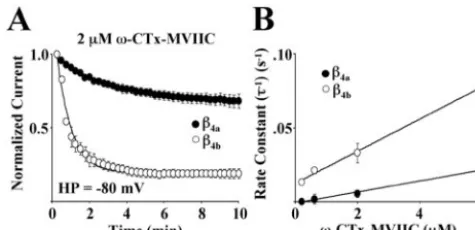
extracellular and occur through binding sites located near H5 (P) helices in several of the six helix transmembrane-spanning motifs (Ellinor et al., 1994). Figure 2Ashows the effects of 2&M#
-CTx-MVIIC on Cav2.1 Ca2!channel complexes expressed in Xeno-pusoocytes in the presence of either!4aor!4b. The oocytes were
held at&80 mV for 10 min and stimulated every 15 sec. Under these conditions, #-CTx-MVIIC associated with Cav2.1
com-plexes containing !4bat a faster rate ('' 50)0.75 sec) than
complexes containing!4a(''200)16 sec). The loss of Ca2!
current resulting from slow inactivation over 10 min at&80 mV
(+15%) was subtracted from the data plotted in thefigure. Figure 2Bdemonstrates that as expected for a first-order reaction, the rate constants ('&1) for toxin block were linearly dependent on
toxin concentration as described by the equation (')&1'k on[Tx] ! koff. Slopes of linear fits to the data for Cav2.1 complexes
containing either!4aand!4bwere 3.7*10&6M&1!sec&1and
1.1 * 10&5 M&1 ! sec&1, respectively. This indicated that the
on-rate (kon) for toxin block was approximately threefold faster
for Cav2.1 complexes containing !4b than for those containing
!4a.
The molecular determinants of alternatively spliced
!
4subunit differential effects on gating and
pharmacology are located within amino acids 10–20
of
!
4bHaving characterized many of the functional consequences of alternative splicing of the !4 A domain, we focused next on
identifying the key structural determinants underlying the ob-served differences in effects. It was of particular interest to deter-mine whether or not the effects of alternative splicing on gating and pharmacology could be assigned to separate structural enti-ties. To accomplish this, wefirst created a series of!4bdeletion
mutants in which the N terminus was shortened by multiples of 10 aa (!4b#1–10 through!4b#1–49) and characterized their effects
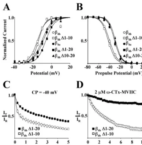
on gating and pharmacology of Cav2.1 complexes. Figure 3A–D
shows that, relative to full-length!4b, deletion of thefirst 10 aa
(!4b#1–10) had no effect on the voltage dependence of activation
(Fig. 3A), isochronal (20 sec prepulse) inactivation (Fig. 3B), onset into slow inactivation (Fig. 3C), or susceptibility to block by 2&M#-CTx-MVIIC (Fig. 3D). However, when amino acids 1–20
were removed (!4b#1–20), both the voltage dependence of
acti-vation (Fig. 3A) and inactivation (Fig. 3B) of Cav2.1 complexes
shifted to more depolarized potentials. As shown in thefigure, the acquired gating properties were essentially identical those for Cav2.1 complexes containing!4a. Cav2.1 complexes containing
!4b#1–20 also had a slower onset into slow inactivation (Fig. 3C)
and were less susceptible to block by 2&M#-CTx-MVIIC (Fig. 3D). The effects of constructs !4b #1–30, !4b #1–40, and !4b #1–49 were identical to those of!4b#1–20 (data not shown). As
afirst attempt at determining whether the effects of!4b#1–20
were simply the result of a decreased size of the!4bN terminus,
we reintroduced amino acids 1–10 to the N terminus of!4b#1–20
to create the construct!4b#10–20. As shown in Figure 3A,B, this
did not restore theV1/2of either activation or inactivation to the
hyperpolarized potentials characteristic of Cav2.1 complexes
con-taining!4b. Together, these results indicated that the molecular
determinants responsible for the observed differences between Cav2.1 complexes containing!4aversus!4bwere located in amino
acids 10–20 of!4b.Moreover, it was apparent that their influence
extended to changes in both gating and pharmacology.
The
!
4A domain is a distant homolog of the third PDZ
domain of PSD-95
With the results of the deletion experiments highlighting a spe-cific location for the molecular determinants of !4b gating and
pharmacology effects, and with the observation that the !1b A
domain resembles PDZ domains (Hanlon et al., 1999), we began a systematic comparison of the!4bsequence with similar regions
of a number of PDZ domains. Unexpectedly, we found that the entire!4bA domain was weakly homologous to the third PDZ
domain of PSD-95 (Fig. 4A). Of the 87 aa that have been shown by x-ray crystallography to form the modular PDZ structure of PSD-95 (Doyle et al., 1996), 27 of these ("31%) are conserved in
Figure 2. Effects of!4aand!4bon the blockade of Cav2.1 channels by
#-CTx-MVIIC. Studies were performed withXenopusoocytes express-ing"1A,"2/$-1, and either!4aor!4b.A, Onset and degree of block by a 10 min exposure to 2&M#-CTx-MVIIC for Cav2.1 subunit combinations at a holding potential (HP) of&80 mV. Eachpointrepresents the mean of seven (!4a) or eight (!4b) different recordings. The SEM is shown for eachdata pointunless smaller than thesymbol. Onset of block for both subunit combinations wasfit (line) to a single-exponential time course plus a constant.B, The rate constants for the time course of the onset of toxin block were determined from steady-state degree of block from single exponentialfits at four different toxin concentrations (0.2, 0.6, 2, and 6 &M) for Cav2.1 complexes containing either!4aor!4b. The averaged rate constants were plotted as a function of toxin concentration (minimum of
the !4b sequence. Most importantly, these identities are
con-served within key secondary structural elements, such as!-strand
C and"-helix 2 of PSD-95. Also of note is the conservation of an RG(S/T)T motif in what would be the equivalent of the carbox-ylate binding loop (CBL) between!-strands A and B of PSD-95 and the loss of the GLGF motif that is extremely common among PDZ domain subtypes (Bezprozvanny and Maximov, 2001; Har-ris and Lim, 2001). Four of!4bamino acids 10–20 (G10, D13,
P15, and P18) were found in PSD-95. We used these as a starting point for further defining key !4b residues involved in setting
Cav2.1 gating parameters.
Figure 4B lists a series of site-directed mutants (left), along with their effects on the voltage dependence (V1/2) of activation
(middle) and inactivation (right) of Cav2.1 complexes. TheV1/2
values for complexes containing !4a and !4b are included for
comparison. Interestingly, the first site-directed !4b mutant
tested, G10A, D13A, P15A, P18A, in which all four of the amino
acids in common with PSD-95 were altered, displayed activation and inactivation properties similar to that of!4a. To determine
whether this was a specific effect, we altered four different amino acids in the !4b 10–20 sequence to create the mutant, T11A,
S17A, T19A, S20A. As shown in Figure 4B, Cav2.1 gating
prop-erties changed little in response to these mutations. Cav2.1
com-plexes containing the G10A, D13A, P15A, P18A mutant also had
!4a-like slow inactivation and pharmacological properties (data
not shown). This indicates that the conserved amino acids are playing a defining role in the gating motif. To delineate the structure in more detail, we subsequently characterized six of the possible G10, D13, P15, P18 amino acid pairs for their effects on gating. Surprisingly, none of the pairs were absolutely essential for maintaining wild-type!4bgating behavior, although small but
statistically significant hyperpolarizing effects on activation were noted forfive of the six pairs. To complete the alanine substitu-tion study, we created the mutant H16A, which had a small but statistically significant effect on inactivation but not activation.
One interpretation of these results is that !4b amino acids
10–20 form a ligand motif that interacts with a binding pocket located somewhere either on the"1Asubunit or on the!4subunit
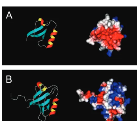
itself. The affinity of the ligand motif for its receptor site could be defined, for example, by the sum of the interactions of amino acids G10, D13, P15, and P18 with their individual targets. Any given pair may be capable of maintaining a binding interaction under the conditions of our experiments. As afirst step toward addressing this possibility, we created three-dimensional struc-tural models of the!4aand!4bA domains (Fig. 5A,B) using the
real-space optimization method used in the computer program MODELLER (Sali and Blundell, 1993). The models were initi-ated using the distance and dihedral angle restraints derived from alignments with portions of the sequence of the third PDZ domain of PSD-95. For!4b, amino acids 10–96 were aligned with
amino acids 303–390 of PSD-95. There is 31% sequence identity over this region, which is considered minimally acceptable for this type of comparative modeling (Martı´-Renom et al., 2000). For
!4a, amino acids 10–49 of!4bwere deleted from the alignment.
Thus, the models do not include thefirst 15 aa of!4aand thefirst
9 aa of!4b. Figure 5A(left) shows that!4amodels as a compact
structure containing three!-sheets and 2"-helices. Stereochem-ical quality assessment of the model using PROCHECK-NMR (Laskowski et al., 1993) identified 41 residues (87.7%) in most favored regions, 5 (10.6%) in additional and generously allowed regions, and 1 (2.1%) in a disallowed region. Calculation of the electrostatic surface potential using MOLMOL (Koradi et al., 1996) reveals that the face of the molecule as oriented in Figure
5A, left, contains a pocket of negative charge (red residues)
between the two"-helices (Fig. 5A, right). Figure 5B, rightand
left, illustrates that the overall effect of alternative splicing to form
!4b is to bury the charged pocket beneath three additional
!-sheets. Interestingly, the molecule acquires as the result of
splicing a positively charged binding pocket (blue residues) in what would be the equivalent of the CBL in PSD-95 (Fig. 4A). The stereochemical quality of the!4bmodel as shown is not quite
as good as that for!4a. PROCHECK-NMR identified 64 residues
(79%) in most favored regions, 11 residues (13.6%) in additional allowed regions, and 3 residues (3.7%) each in generously allowed and disallowed regions. Two of the three disallowed residues (R30 and K34) flank what would be the equivalent PSD-95
!-sheet B. Together with the loss of the highly conserved PDZ GLGF sequence, these results are consistent with the notion that through evolution this region of the !4b structure has evolved Figure 3. Localization of differential effects on Cav2.1 gating and
phar-macology to!4bN-terminal amino acids 10–20. Thefirst 10 (!4b#1–10),
first 20 (!4b#1–20), or second 10 (!4b#10–20) aa of the N terminus of the
away from the capacity to bind C-terminal peptide motifs. Of most importance to our present results, however, is the observa-tion that !4b amino acids 10–20 model as an extended arm
(pointing to the left in Fig. 5B, right, left) that may serve as a ligand motif. Interestingly, the orientation of the arm appears to be dictated by the isomerization state of proline18 (data not shown).
Differential distribution of alternatively spliced
!
4subunit mRNA
We noted in our previous study (Helton and Horne, 2002) that, based on extensive cDNA library screening,!4awas the
predom-inant alternatively spliced variant of the!4subunit expressed in
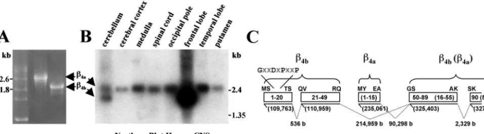
human spinal cord. To confirm this observation, we performed a comparative Northern blot analysis using a commercially avail-able multiple tissue Northern blot (Human Brain II; Clontech) and a!4cDNA probe containing sequence common to both!4a
and!4b. The mRNAs for!4aand!4bcan be readily distinguished
by their distinct migration pattern in agarose-formaldehyde gels (Fig. 6A). The results of the Northern blot analysis, shown in Figure 6B, were striking, revealing that not only was !4a the
predominant form of !4 subunit in the spinal cord, but also in
other“reptilian”portions of the human CNS such as the medulla and putamen. Moreover, !4a was the predominant form of !4
subunit expressed in evolutionarily older regions of the cerebrum, the temporal lobe, and occipital pole. In marked contrast,!4bwas
highly expressed in the most recent and most highly integrative region of the cerebrum, the frontal lobe. The two forms of the!4
subunit were equally expressed in the cerebellum.
A basic local alignment search tool search of the human ge-nome (Altschul et al., 1990) with!4sequences revealed that the
exons coding for alternatively spliced forms of the!4subunit A
domain are distributed widely on human chromosome 2. Figure 6C shows that, depending on the splice variant, the coding se-quence for the!4PDZ domain is contained within three (!4a) or
four (!4b) exons spread out over "218,000 bases. The coding
sequence for the GXXDXPXXP motif is included in the 5$-most exon of a pair of short exons that code for!4bamino acids 1–49.
Assembly of the !4bmRNA requires that three RNA segments
(536 bases, 214,959 bases, and 2329 bases) be spliced out. The short exon coding for thefirst 15 aa of!4a lies between the!4b
N-terminal exons and the exon coding amino acids 50–89 and 16–55 of!4band!4a, respectively. By comparison, the third PDZ
domain of PSD-95 is encoded by two exons separated by a 200 bp intron (data not shown).
DISCUSSION
We have identified an alternatively spliced proline-rich motif in the Ca2!channel!
4subunit that has considerable influence over
gating of neuronal Cav2.1 Ca2!channels. Given that the motif
also affects extracellular toxin binding, it is likely that the
inter-Figure 4. The !4subunit is a distant homolog of PSD-95. Identification of a conserved GXXDX-PXXP motif critical to Cav2.1 gating.A, Amino acid alignment of the A domains of the human spinal cord !4a(amino acids 1–63) and!4b(amino acids 1–97) subunits with the third PDZ domain (amino acids 294–391) of PSD-95. Vertical bars denote identical amino acids between !4b and PSD-95. Important amino acids involved in modulating the leftward shift in the voltage dependence of activation and inactiva-tion of!4b(GXXDXPXXP) are highlighted.Arrows and hatched bars represent predicted "-helices and !-strands of the third PDZ domain of PSD-95, respec-tively.B, Differences in theV1/2values of activation and inactivation of !4a, !4b, and !4b N-terminal amino acid mutants.Solid barsrepresent averageV1/2 values of a minimum of nine different recordings for each!4subunit variant. Positive or negative shifts in theV1/2values (in millivolts) of activation and inacti-vation of!4aand!4bmutants are relative to theV1/2 values of activation and inactivation of!4b. Currents were generated as described in Figure 3,AandB. The SEM for each bar is shown.Asterisksdenote statistical significance (p + 0.05) by a Student’s two-sample equal variancettest.
action of this motif with its binding site has wide-reaching impact on resting and open-state Ca2! channel conformations. This
notion is supported by recent images of the conformational changes that occur with gating of bacterial two-membrane-spanning K!channels (Liu et al., 2001; Jiang et al., 2002a,b). Like
eukaryotic six-membrane-spanning K! channels, KcsA and
MthK channels are tetramers that pack with fourfold symmetry around a central pore (Doyle et al., 1998; Jiang et al., 2002a). The principal structural elements of KcsA and MthK from N to C terminus include an outer transmembrane helix (M1), a pore helix (P), and an inner transmembrane helix (M2). These corre-spond to S5, H5, and S6 segments of voltage-gated Ca2!
chan-nels, respectively. In the closed conformation of the KcsA struc-ture, the four M2 helices are straight and arranged such that they form the walls of an inverted teepee that narrows from a 12 Å
diameter at its center to a 4Åpore at its tip (Doyle et al., 1998). After opening, the KcsA M2 helices tilt away from the perme-ation pathway and rotate about their helical axis (Liu et al., 2001). In MthK, bending and splaying of the inner helices after opening expands the diameter of the pore threefold (Jiang et al., 2002a,b). The nearly 30°bend occurs at a“gating hinge”corresponding to a glycine residue just below the selectivity filter. Applying a radial-outward force on the intracellular aspects of the inner helices places a torque on the gating hinge such that a conforma-tional change is transmitted the full length of the M2 helix. This results in a widening of the outer vestibule [Jiang et al. (2002b), see supplementary information (movie)]. It is has been hypothe-sized that similar mechanical forces are at work in the gating of voltage-gated Ca2!channels (Jiang et al., 2002b).
Considered in the context of this mechanical framework, our results suggest that an interaction of the!4bligand motif with an
inner aspect of the Cav2.1 complex either directly or indirectly fine-tunes the torque experienced by "1A S6 segments. In so
doing, the interaction alters the conformation of the voltage sensor or gate (or both) as well as the outer vestibule. This would explain why alternative splicing of the !4 subunit would affect
both gating and toxin sensitivity. Given the potential for the ligand motif interaction to occur over a wide range (Helton and Horne, 2002), it is not possible from our results to pinpoint which S6 helices might be most affected. And although our #
-CTx-MVIIC binding results might be providing some direction, a
previous study has shown that #-CTx-GVIA binding to"1B is
affected by alterations in any of the four P loops that make up the outer vestibule (Ellinor et al., 1994). A case can be made for an indirect effect on the IS6 helix, because the primary"1-!4subunit
interaction occurs on the intracellular loop between homology domains I and II (I-II loop) (Pragnell et al., 1994). This is consistent with a previous study showing that IS6 is a critical determinant of voltage-dependent inactivation in Cav2.1 and
Cav2.3 channels (Zhang et al., 1994). However, site-directed
mutagenesis and domain-swapping studies have highlighted the equal importance of IIS6, IIIS6, and IVS6 in Ca2!channel gating
(for review, see Stotz and Zamponi, 2001; Shi and Soldatov, 2002), making the case for direct effects on these S6 helices equally plausible. It is interesting that many of the proteins that have evolved to modulate Ca2!channel gating target"
1I-II (!
subunits, G!%subunits, protein kinase C) and II-III loops
(syn-taxin, synaptotagmin, and synaptosomal-associated protein-25). The IS6 and IIS6 (but not IIIS6 or IVS6) helices of Cav2.1, 2.1,
and 2.3 Ca2!channels have glycines in hinge positions
compa-rable with those present in KcsA and MthK (see alignments in Horne et al., 1993).
Despite extensive binding studies with !4 subunits, there is
currently no evidence to support the notion that the !4b N
terminus binds directly to "1A subunits (Walker et al., 1998,
1999). One possible explanation for this is that the interaction is too weak to be detected in solution binding assays. The other possibility is that the "1A subunit is not the primary binding
target. The sequence of the ligand motif (GXXDXPXXP) may provide an important clue as to the nature of its own binding site. Proline-rich motifs are common within the primary structures of many ligands important for protein–protein interactions (for re-view, see Kay et al., 2000; Macias et al., 2002). Src homology 3 (SH3) and WW domains, for example, recognize proline-rich sequences containing a core PXXP, where X denotes any amino acid. These sequences adopt a PPII helix conformation that presents a hydrophobic surface as well as backbone carbonyls that are ideal for hydrogen bonding. Proline-rich ligands bind with low affinity, allowing for rapid modulation and added versatility in signaling pathways. In many respects, the!4bligand motif
resem-bles the serine/threonine proline motifs recognized by group IV WW domains (Sudol and Hunter, 2000). As is the case for the!4b Figure 6. Differential distribution of!4aand!4bmRNA in the human CNS.A, Electrophoresis of full-length!4a(left) and!4b(right) cRNAs (includes 5$and 3$untranslated) in a 1% agarose formaldehyde denaturing gel. RNA markers (in kilobases) are indicated on theleft.B, Northern blot analysis performed with human multiple tissue blot (Human Brain II; Clontech) and a32P-labeled!
4subunit probe (coding nucleotides 215–1628 plus"300 bp 3$untranslated sequence). Molecular masses on therightcorrespond to labeled blot markers.C, A human!4subunit genome map depicting the lengths of intron sequences (b, bases) between alternatively spliced!4aand!4bN-terminal exons and the beginning of exon 2.Solid linesrepresent exons, and
dashed linesrepresent introns.Numbersinparenthesesbelowsolid linesindicate position on chromosome 2.Boxesindicate protein sequence (!4ain
motif, the structural basis for recognition of these motifs is based on the summed contributions of a series of side-chain interac-tions, none of which is absolutely essential for ligand binding (Verdecia et al., 2000). It is possible that the !4b proline-rich
motif binds to its own B domain, which is structurally similar to SH3 and WW domains (Hanlon et al., 1999). This possibility is supported by what is known about related membrane-associated guanylate kinase family proteins in which intramolecular interac-tions are a key aspect of their functional diversity (Dimitratos et al., 1999).
To date, no specific function has been assigned to the GXX-DXPXXP motif of the third PDZ domain of PSD-95. Most attention has been paid to the structure of the carboxylate binding loop to which it is immediately adjacent (Doyle et al., 1996) (Fig. 4,CBL). A partial list of the proteins that interact specifically with the third PDZ domain of PSD-95 include the cell-surface neu-roligins (Irie et al., 1997), the microtubule binding protein cysteine-rich interactor of PDZ 3 (Niethammer et al., 1998), the Rho effector protein citron (Zhang et al., 1999), and the !1
adrenergic receptor (Hu et al., 2000). It would be interesting to determine whether the GXXDXPXXP motif of PSD-95 plays a role maintaining the structure of the CBL. Such a role could also be considered for the GXXDXPXXP motif of!4b. As is
appar-ent in the !4b A domain model structure (Figs. 4A, 5B), the
sequences of !4b corresponding to the hydrophobic CBL and
!-strand B of PSD-95 are highly positively charged. It is possible, as is true for some PDZ domains (Cuppen et al., 1998), that this region binds to an internal (as opposed to a C-terminal) nega-tively charged domain. Interestingly, the immediate 5$sequence of the II-III loop of Cav2.1 and Cav2.2 Ca2!channels fits this
description, because it is densely packed with glutamate residues. Moreover, several lines of evidence point to the II-III loop as a critical determinant of channel gating and pharmacology. Similar to the effects of !4bon"1A, binding of syntaxin to a“synprint
site” just downstream from this region in the II-III loop of"1B
shifts theV1/2of inactivation to more hyperpolarized potentials
(Bezprozvanny et al., 2000) and accelerates entry into slow inac-tivation (Degtiar et al., 2000), and an"1Bsplice variant that lacks
a large portion of the II-III linker region (#1) (Kaneko et al., 2002) inactivates at more depolarized potentials and is less sen-sitive to#-CTx-MVIIA than is the full-length Cav2.2 variant.
Viewed from the perspective of changing Ca2!channel
func-tion, the differential distribution of !4aand !4bsubunit mRNA
displayed in Figure 6Bprovides an unexpected snapshot of the evolution of forebrain synapses. It raises the possibility that with the introduction of!4bto the genome, synapses acquired
prop-erties thatfit better with the overall demand to organize complex neural networks. It could be that with the advent of Cav2.1
complexes that enter more readily into closed inactivation states (Fig. 1A,C) without perceptible gain in time required for recov-ery (Fig. 1D), synapses inherited an enhanced mechanism for synaptic plasticity. In this regard, short-term synaptic depression has been linked to Ca2!channel inactivation (Forsythe et al.,
1998) through mechanisms shown to be ! subunit dependent (Patil et al., 1998). This form of short-term synaptic plasticity has been implicated in cortical gain control (Abbott et al., 1997) and low-pass temporalfiltering (Fortune and Rose, 2001). In addition, long-lasting Cav2.1 channel inhibition has been identified as the
mechanism underlying certain forms of long-term synaptic de-pression (Robbe et al., 2002). Accordingly, our future studies will be directed toward characterizing the responsiveness of Cav2
Ca2! channels containing alternatively spliced !
4 subunits to
changes in more dynamic regulatory inputs, such as neuronal
firing frequency and action potential waveform.
REFERENCES
Abbott LF, Varela JA, Sen K, Nelson SB (1997) Synaptic depression and cortical gain control. Science 275:220–224.
Altschul SF, Gish W, Miller W, Myers EW, Lipman DJ (1990) Basic local alignment search tool. J Mol Biol 215:403–410.
Berjukow S, Marksteiner R, Sokolov S, Weiss RG, Margreiter E, Hering S (2001) Amino acids in segment IVS6 and !-subunit interaction support distinct conformational changes during Cav2.1 inactivation. J Biol Chem 276:17076–17082.
Berrou L, Bernatchez G, Parent L (2001) Molecular determinants of inactivation within the I-II linker of"1E(CaV2.3) calcium channels. Biophys J 80:215–228.
Bezanilla F (2000) The voltage sensor in voltage-dependent ion chan-nels. Physiol Rev 80:555–592.
Bezprozvanny I, Maximov A (2001) PDZ domains: more than just a glue. Proc Natl Acad Sci USA 98:787–789.
Bezprozvanny I, Zhong P, Scheller RH, Tsien RW (2000) Molecular determinants of the functional interaction between syntaxin and N-type Ca2!channel gating. Proc Natl Acad Sci USA 97:13943–13948. Catterall WA (2000) Structure and regulation of voltage-gated Ca2!
channels. Annu Rev Cell Dev Biol 16:521–555.
Cha A, Snyder GE, Selvin PR, Bezanilla F (1999) Atomic scale move-ment of the voltage-sensing region in a potassium channel measured via spectroscopy. Nature 402:809–813.
Colovos C, Yeates TO (1993) Verification of protein structures: patterns of nonbonded atomic interactions. Protein Sci 2:1511–1519.
Cuppen E, Gerrits H, Pepers B, Wieringa B, Hendriks W (1998) PDZ motifs in PTP-BL and RIL bind to internal protein segments in the LIM domain protein RIL. Mol Biol Cell 9:671–683.
Degtiar VE, Scheller RH, Tsien RW (2000) Syntaxin modulation of slow inactivation of N-type calcium channels. J Neurosci 20:4355–4367. Dimitratos SD, Woods DF, Stathakis DG, Bryant PJ (1999) Signaling
pathways are focused at specialized regions of the plasma membrane by scaffolding proteins of the MAGUK family. BioEssays 21:912–921. Doyle DA, Lee A, Lewis J, Kim E, Sheng M, MacKinnon R (1996)
Crystal structures of a complexed and peptide-free membrane protein-binding domain: molecular basis of peptide recognition by PDZ. Cell 85:1067–1076.
Doyle DA, Morais-Cabral J, Pfuetzner RA, Kuo A, Gulbis JM, Cohen SL, Chait BT, MacKinnon R (1998) The structure of the potassium channel: molecular basis of K! conduction and selectivity. Science 280:69–77.
Ellinor PT, Zhang JF, Horne WA, Tsien RW (1994) Structural deter-minants of the blockade of N-type calcium channels by a peptide neurotoxin. Nature 372:272–275.
Forsythe ID, Tsujimoto T, Barnes-Davies M, Cuttle MF, Takahashi T (1998) Inactivation of presynaptic calcium current contributes to syn-aptic depression at a fast central synapse. Neuron 20:797–807. Fortune ES, Rose GJ (2001) Short-term synaptic plasticity as a temporal
filter. Trends Neurosci 24:381–385.
Glauner KS, Mannuzzu LM, Gandhi CS, Isacoff EY (1999) Spectro-scopic mapping of voltage sensor movement in the Shaker potassium channel. Nature 402:813–817.
Hanlon MR, Berrow NS, Dolphin AC, Wallace BA (1999) Modeling of a voltage-dependent Ca2!channel!subunit as a basis for understand-ing its functional properties. FEBS Lett 445:366–370.
Hans M, Urrutia A, Deal C, Brust PF, Stauderman K, Ellis SB, Harpold MM, Johnson EC, Williams ME (1999) Structural elements in domain IV that influence biophysical and pharmacological properties of human "1A-containing high-voltage-activated calcium channels. Biophys J 76:1384–1400.
Harris BZ, Lim WA (2001) Mechanism and role of PDZ domains in signaling complex assembly. J Cell Sci 114:3219–3231.
Helton TD, Horne WA (2002) Alternative splicing of the!4subunit has
"1subunit subtype-specific effects on Ca2!channel gating. J Neurosci 22:1573–1582.
Horn R (2000) A new twist in the saga of charge movement in voltage-dependent ion channels. Neuron 25:511–514.
Horne WA, Ellinor PT, Inman I, Zhou M, Tsien RW, Schwarz TL (1993) Molecular diversity of Ca2!channel"
1subunits from the marine ray
Discopyge ommata. Proc Natl Acad Sci USA 90:3787–3791.
Hu LA, Tang Y, Miller WE, Cong M, Lau AG, Lefkowitz RJ, Hall RA (2000) !1-adrenergic receptor association with PSD-95: inhibition of receptor internalization and facilitation of!1-adrenergic receptor in-teraction with N-methyl-D-aspartate receptors. J Biol Chem 275:38659–38666.
Irie M, Hata Y, Takeuchi M, Ichtchenko K, Toyoda A, Hirao K, Takai Y, Rosahl TW, Sudhof TC (1997) Binding of neuroligins to PSD-95. Science 277:1511–1515.
Crystal structure and mechanism of a calcium-gated potassium channel. Nature 417:515–522.
Jiang Y, Lee A, Chen J, Cadene M, Chait BT, MacKinnon R (2002b) The open pore conformation of potassium channels. Nature 417:523–526.
Kaneko S, Cooper CB, Nishioka N, Yamasaki H, Suzuki A, Jarvis SE, Akaike A, Satoh M, Zamponi GW (2002) Identification and charac-terization of novel human Cav2.2 (a1B) calcium channel variants lack-ing the synaptic protein interaction site. J Neurosci 22:82–92. Kay BK, Williamson MP, Sudol M (2000) The importance of being
proline: the interaction of proline-rich motifs in signaling proteins with their cognate domains. FASEB J 14:231–241.
Koradi R, Billeter M, Wuthrich K (1996) MOLMOL: a program for display and analysis of macromolecular structures. J Mol Graph 14:51–55:29–32.
Kozak M (1991) An analysis of vertebrate mRNA sequences: intima-tions of translational control. J Cell Biol 115:887–903.
Laskowski RA, Moss DS, Thornton JM (1993) Main-chain bond lengths and bond angles in protein structures. J Mol Biol 231:1049–1067. Lin Z, Haus S, Edgerton J, Lipscombe D (1997) Identification of
func-tionally distinct isoforms of the N-type Ca2! channel in rat sympa-thetic ganglia and brain. Neuron 18:153–166.
Liu YS, Sompornpisut P, Perozo E (2001) Structure of the KcsA channel intracellular gate in the open state. Nat Struct Biol 8:883–887. Macias MJ, Wiesner S, Sudol M (2002) WW and SH3 domains, two
different scaffolds to recognize proline-rich ligands. FEBS Lett 513:30–37.
Martı´-Renom MA, Stuart AC, Fiser A, Sanchez R, Melo F, Sali A (2000) Comparative protein structure modeling of genes and genomes. Annu Rev Biophys Biomol Struct 29:291–325.
Niethammer M, Valtschanoff JG, Kapoor TM, Allison DW, Weinberg TM, Craig AM, Sheng M (1998) CRIPT, a novel postsynaptic protein that binds to the third PDZ domain of PSD-95/SAP90. Neuron 20:693–707.
Patil PG, Brody DL, Yue DT (1998) Preferential closed-state inactiva-tion of neuronal calcium channels. Neuron 20:1027–1038.
Perozo E, Cortes DM, Cuello LG (1999) Structural rearrangements underlying K!-channel activation gating. Science 285:73–78. Pragnell M, De Waard M, Mori Y, Tanabe T, Snutch TP, Campbell KP
(1994) Calcium channel!-subunit binds to a conserved motif in the I-II cytoplasmic linker of the"1-subunit. Nature 368:67–70.
Robbe D, Alonso G, Chaumont S, Bockaert J, Manzoni AJ (2002) Role of P/Q-Ca2! channels in metabotropic glutamate receptor 2/3-dependent presynaptic long-term depression at nucleus accumbens synapses. J Neurosci 22:4346–4356.
Sali A, Blundell TL (1993) Comparative protein modelling by satisfac-tion of spatial restraints. J Mol Biol 234:779–815.
Shi C, Soldatov NM (2002) Molecular determinants of voltage-dependent slow inactivation of the Ca2! channel. J Biol Chem 277:6813–6821.
Stotz SC, Zamponi GW (2001) Identification of inactivation determi-nants in the domain IIS6 region of high voltage-activated calcium channels. J Biol Chem 276:33001–33010.
Stotz SC, Hamid J, Spaetgens RL, Jarvis SE, Zamponi GW (2000) Fast inactivation of voltage-dependent calcium channels. A hinged-lid mech-anism? J Biol Chem 275:24575–24582.
Sudol M, Hunter T (2000) New wrinkles for an old domain. Cell 103:1001–1004.
Verdecia MA, Bowman ME, Lu KP, Hunter T, Noel JP (2000) Struc-tural basis for phosphoserine-proline recognition by group IV WW domains. Nat Struct Biol 7:639–643.
Walker D, Bichet D, Campbell KP, De Waard M (1998) A!4 isoform-specific interaction site in the carboxyl-terminal region of the voltage-dependent Ca2!channel"
1Asubunit. J Biol Chem 273:2361–2367. Walker D, Bichet D, Geib S, Mori E, Cornet V, Snutch TP, Mori Y, De
Waard M (1999) A new!subtype-specific interaction in"1Asubunit controls P/Q-type Ca2! channel activation. J Biol Chem 274:12383–12390.
Zhang JF, Ellinor PT, Aldrich RW, Tsien RW (1994) Molecular deter-minants of voltage-dependent inactivation in calcium channels. Nature 372:97–100.
Zhang W, Vazquez L, Apperson M, Kennedy MB (1999) Citron binds to PSD-95 at glutamatergic synapses on inhibitory neurons in the hippocampus. J Neurosci 19:96–108.