University of South Carolina University of South Carolina
Scholar Commons
Scholar Commons
Theses and Dissertations
2016
Factors Influencing The Collagen Fiber Angle Distribution in The
Factors Influencing The Collagen Fiber Angle Distribution in The
Mouse Aorta
Mouse Aorta
Shana Roach Watson University of South Carolina
Follow this and additional works at: https://scholarcommons.sc.edu/etd
Part of the Other Medical Sciences Commons
Recommended Citation Recommended Citation
Watson, S. R.(2016). Factors Influencing The Collagen Fiber Angle Distribution in The Mouse Aorta. (Doctoral dissertation). Retrieved from https://scholarcommons.sc.edu/etd/3551
FACTORS INFLUENCING THE COLLAGEN FIBER ANGLE DISTRIBUTION IN THE MOUSE AORTA
by
Shana Roach Watson Bachelor of Science Clemson University, 2011
Submitted in Partial Fulfillment of the Requirements For the Degree of Doctor of Philosophy in
Biomedical Science School of Medicine University of South Carolina
2016 Accepted by:
Susan Lessner, Major Professor
John Eberth, Chairman Examining Committee Norma Frizzell, Committee Member Chandrashekhar Patel, Committee Member
Michael Sutton, Committee Member
DEDICATION
Mom, you have always been a major supporter of education. I give you all the credit for my accomplishments. I can assure you, greater is coming.
To my only and older sibling, Terrell: Your fulfillment as a father figure has been a treasure and I have never told you but, “Thank you”.
To the Roach family, you guys are the best support system anyone could ever even wish for. I have carried your prayers along the way.
The Bible says, “To write a vision and make it plain.”
-Habakkuk 2:2
ACKNOWLEDGEMENTS
I would like to thank my advisor, Dr. Susan Lessner, for her guidance throughout my graduate career. She has shown me that women can be leaders in STEM fields. In addition I would like to acknowledge the current and former members of the Lessner lab, John Johnson, Henry Bateman, Ying Wang, Nishant Karasala, Mohamed Gabr, and Lindsey Davis. Each one of you has taught me something different and I am appreciative. My undergraduate assistants, Marcella Marks and Savannah Han, were a major help.
Thanks to my collaborators, Dr. Edsel Pena and Piaomu Liu for providing statistical analysis of multiple projects. Further, I would like to thank the
members of my committee, Drs. John Eberth, Norma Frizzell, Chandrashekhar Patel, and Michael Sutton for their encouragement and professional advice. Sincerest thanks to Merissa Baxter and my PREP mentors Drs. Bert Ely and Richard Hunt for pushing me towards success from the very start.
And in memory of my ASBMB mentor, Dr. Marion B. Sewer who had a
ABSTRACT
The aortic extracellular matrix (ECM) consists of microstructural proteins, collagen and elastin, together with proteoglycans and other components. The matrix metalloproteinases (MMPs) are proteolytic enzymes that influence
morphological and structural changes in the ECM and can degrade the matrix as it responds to cellular behaviors such as angiogenesis, apoptosis, proliferation, and migration. Collagen is the most important component among the
extracellular proteins because it provides strength and stability to the tissue. Changes in collagen content play a major role in the development of
atherosclerosis. These changes can be induced by increased or decreased proteinase activity. Therefore, we studied the collagen fiber angle distribution in mouse models of atherosclerosis with or without a deficiency of a selected MMP. Quantification of the collagen fiber data is meaningful in providing insight into the mechanical behavior of the artery and leads to an understanding of how the diseased aorta maintains homeostasis. Furthermore, such data can be utilized to increase the understanding of disease progression, including but not limited to atherosclerosis and aortic aneurysm development.
We characterized collagen fiber angles in mouse models of
aorta using second-harmonic generation (SHG) microscopy. Angle
measurements were acquired using a well-established computer software
program, Continuity 6.4b. The angle measurements were exported into bivariate histograms. We then designed a multiple regression analysis to compare the distributions of absolute angles between two diet groups, controlling for diet, mouse strain, anatomical location, and radial position in the aortic wall. Data extracted from bivariate histograms were analyzed in R, a programming language and statistical software.
In trying to understand the changes seen between chow diet and Western diet fed mice, we began to study physiological variables such as blood pressure and blood flow velocity. While we did not find any differences in the
hemodynamics, we were able to determine that factors beyond atherogenesis, for example aging, influenced aortic collagen fiber angle distributions. With aging and atherosclerosis, the extracellular matrix may experience an increase in collagen content and fibrous tissue. We evaluated changes in fractional collagen amount, in particular, collagen type I. To understand if changes in collagen fibers are initiated by endothelial dysfunction (a pathological condition that often
accompanies atherosclerosis progression), we performed immunohistochemical studies of two endothelial cell-derived factors, intercellular adhesion molecule-1 (ICAM-1) and endothelial nitric oxide synthase (eNOS).
TABLE OF CONTENTS
DEDICATION ... iii
ACKNOWLEDGEMENTS ... iv
ABSTRACT ... v
LIST OF TABLES ... x
LIST OF FIGURES ... xi
LIST OF ABBREVIATIONS ... xiii
CHAPTER 1INTRODUCTION ... 1
1.1 COLLAGEN FIBERS ... 2
1.2 OPTICAL PROPERTIES ... 4
1.3 THE ARTERIAL WALL ... 6
1.4 ATHEROSCLEROSIS ... 9
1.5 ANIMAL MODELS OF ATHEROSCLEROSIS ... 11
1.6 MATRIX METALLOPROTEINASES ... 12
1.7 SPECIFIC AIMS ... 13
CHAPTER 2(SECOND)HARMONIC DISHARMONY:NONLINEAR MICROSCOPY SHINES NEW LIGHT ON THE PATHOLOGY OF ATHEROSCLEROSIS ... 16
2.1 ABSTRACT ... 17
2.2 INTRODUCTION ... 17
2.3 ANIMAL MODELS AND IMAGING TARGETS ... 22
2.5 SHGADVANTAGES AND DISADVANTAGES IN ASSESSING
ATHEROSCLEROSIS ... 33
2.6 CONCLUSION ... 34
CHAPTER 3COMPARISON OF AORTIC COLLAGEN FIBER ANGLE DISTRIBUTION IN MOUSE MODELS OF ATHEROSCLEROSIS USING SECOND-HARMONIC GENERATION MICROSCOPY ... 39
3.1 ABSTRACT ... 40
3.2 INTRODUCTION ... 41
3.3 MATERIALS AND METHODS ... 43
3.4 RESULTS ... 46
3.5 DISCUSSION... 48
3.6 LIMITATIONS OF THE STUDY ... 51
3.7 CONCLUSIONS ... 52
CHAPTER 4DIET ALTERS AGE-RELATED REMODELING OF AORTIC EXTRACELLULAR MATRIX IN MICE SUSCEPTIBLE TO ATHEROSCLEROSIS ... 58
4.1 ABSTRACT ... 58
4.2 INTRODUCTION ... 60
4.3 MATERIALS AND METHODS ... 63
4.4 RESULTS ... 70
4.5 DISCUSSION... 73
4.6 CONCLUSION ... 76
CHAPTER 5CONCLUSION ... 90
5.1 SUMMARY AND FUTURE DIRECTIONS ... 90
REFERENCES ... 95
LIST OF TABLES
LIST OF FIGURES
Figure 1.1 Arterial Wall Layers ... 9
Figure 1.2 Atherosclerosis Progression ... 10
Figure 2.1 TPEF-SHG Microscope ... 36
Figure 2.2 SHG and Histology Images ... 37
Figure 2.3 SRS and SHG Images of ChCs ... 38
Figure 3.1 Aortic Tree Sampling Areas ... 53
Figure 3.2 SHG Images from Mice on chow vs Western Diet ... 54
Figure 3.3 3D Histograms of Collagen Fiber Angle Distribution in apoE KO and apoE MMP12 DKO Mice ... 55
Figure 3.4 Boxplot for 6 months chow vs. Western Diet ... 56
Figure 4.1 Boxplot for Age Study………...………..80
Figure 4.2 3D Histograms of Aging apoE KO Mice ... 81
Figure 4.3 Collagen Content from PSR Staining ... 82
Figure 4.4 Collagen Type 1 Detection ... 83
Figure 4.5 ICAM-1 Expression ... .84
Figure 4.6 eNOS Expression ... 85
Figure 4.7 Increasing Body Weight Due to Aging and Diet ... 86
Figure 4.8 Assessment of Aortic Diameters at Systole and Diastole ... 87
Figure 4.9 Average BP for apoE KO ... 88
LIST OF ABBREVIATIONS
CHAPTER
1
INTRODUCTION
Cardiovascular disease is the leading cause of death worldwide (Pagidepati et al., 2013). While the underlying causes are multifactorial,
atherosclerosis is the most common (Frostegard, 2013; Pagidepati et al., 2013). Atherosclerosis is a chronic inflammatory disease characterized by endothelial dysfunction and lipid accumulation in the artery wall, which represents a
significant source of morbidity and mortality in the United States (Jamkhande et al., 2014). Arteries undergo long-lasting changes in arterial structure and
function, termed vascular remodeling, in response to atherosclerotic lesion development (Galis & Khatri, 2002). Vascular remodeling is defined as any lasting change in size and/or composition in an adult blood vessel. Examples of remodeling occur physiologically when there is an increase or decrease in blood flow or blood pressure, or when vascular injury occurs. As a result of one or more of these events, the distensibility of the artery is altered.
atherosclerotic plaques are covered by a cap rich in fibrillar collagens.
Information about the organization of collagen fibers can increase our knowledge regarding the mechanical behavior of the vessel and where the load is being carried.
The importance of collagen fiber angles on the mechanical behavior of the arterial wall has been well-established (Holzapfel et al., 2000; Wolinsky et al., 1969; Canham et al., 1996; Holzapfel et al., 2009), and research also shows that collagen plays a major role in disease (Hallaron et al., 1995; Lanne et al., 1992; Katsuda et al., 1992). However, there are limited published experimental studies accounting for changes in collagen fiber angles throughout the thickness of diseased (i.e. aged and/or atherosclerotic) aortas (Holzapfel, 2000; Collins et al., 2011; Hill et al., 2012). Because the detection of collagen in the three layers of the aortic wall typically requires labeling techniques such as the use of
antibodies, we used second harmonic generation microscopy (SHG) to visualize collagen fiber orientation. Collagen has birefringent properties and produces an SHG signal (Roth & Freund, 1981; Georgiou et al., 2000; Campagnola et al., 2002). This is of particular importance because, with aging and atherosclerosis, the extracellular matrix (ECM) experiences changes in the fibrous structure and content of collagen (Phillip et al., 2015).
1.1 Collagen Fibers
Collagen is the most abundant structural protein in mammals. It
generally involved in the formation of supramolecular networks (Richard-Blum, 2011). The native collagen molecule can be described as a triple helix
constituted by three polypeptide chains (α chains). The central collagen domain consists of the repeating amino acid sequence glycine - X - Y, where X and Y are frequently proline or hydroxyproline, with non-collagenous domains at the
terminal ends of the peptide chains (Vuorio & De Crombugghe, 1990; Van der Rest & Garrone, 1991). The secreted procollagen molecules have their non-collagenous domain removed by specific proteinases, leaving the characteristic collagen triple helices that aggregate in quarter-staggered fibrils (Schriefl, 2012).
The fibrous protein is found in extracellular matrices and connective tissues. There are at least 16 types of collagen, but 80-90% of the body’s
collagen consists of types I, II, and III (Lodish, 2000). These collagen molecules pack together to form long (300nm), thin (1.5nm-diameter) fibrils of similar structure (Lodish, 2000). Smooth muscle cells produce most collagens in the intima and media of vascular walls, and fibroblasts have a similar role in the adventitia (Fitzsimmons et. al., 2002). Fibrillar collagens in all layers of the vessel wall serve the same purpose, which is to help the tissue withstand mechanical loading (Lodish, 2000). Collagen fibers often align in the direction of preferred loading in the tissue (Rubbens et al., 2009). This is an example of form following function.
synthesize different fibrillar collagens at the same time, a specific molecular mechanism is required for the recognition and discrimination of α chains during assembly of the procollagen molecule (Exposito et al., 2011). While the major types found in the vasculature are the classical fibrillar collagen types I and III (Hulmes, 2008), arterial collagen content and distribution is thought to be species-dependent (Grant, 1967).
Within atherosclerotic lesions, collagen type I is the most abundant. It is readily available for studies because it can be easily isolated. Collagen type I was also the first to be characterized among the collagens. It has been observed to form large fibers, approximately 10-500nm in diameter and several
micrometers long (Lodish et al., 2000).
Assembly of these collagen fibers begins with a lag phase in which
collagen molecules associate to form metastable nuclei. This stage is followed by a rapid growth phase in which more molecules accumulate around the nuclei leading to growth in fiber length and width. The final step is a plateau region in which no further growth is observed (Ward, 1986). These three phases of self-assembly of collagen fibers can be influenced by entropy (Ward, 1986).
1.2 Optical properties
Collagen can be visualized by methods such as polarized light microscopy (Puchtler et al., 1973; Bennett, 1950; Junqueira et al., 1979; Eberth et. al., 2009), small angle light scattering (Sacks et al., 1997), scanning confocal laser
Tsamis et al., 2013a), and second harmonic generation microscopy (Williams et al., 2005; Doras et al., 2011; Arifler et al., 2007; Watson et. al., 2015). Imaging methodologies such as second harmonic generation (SHG) microscopy, third harmonic generation, and coherent anti-Stokes Raman spectroscopy are classically used for three-dimensional structural visualization of collagen fibers with high contrast(Jemal et al.; Mueller & Fusenig, 2002; American Cancer Society, 2006; Koss, 1992). In recent years, many second-harmonic generation (SHG) experiments have been devoted to the study of collagen structures in biological tissues (Zipfel et al., 2003; Doras et al., 2011). This technique exhibits promise for biomedical assessment of tissue structure, especially in processes where the ECM is being actively remodeled (Williams et al., 2005) .
Collagen has a highly crystalline triple-helix structure that is not centrosymmetric, and the molecules are organized on the scale of the
wavelength of light (Erikson et al., 2007). Because the SHG signal is intrinsic to specific structures, this technique allows for quantification of collagen fibers without physical or biochemical processing of the tissue. Individual
noncentrosymmetric molecules will generate a SHG signal, but molecules
arranged in a crystalline array will give a much stronger response. SHG is useful in providing information about the orientation, crystallinity, and morphology of collagen fibers (Zoumi et al, 2004; Schenke-Layland et al., 2005).
technique (Denk et al., 1990; Williams et al., 2005; Zipfel et al., 2003) to yield high contrast and optical sectioning capabilities through relatively thick tissues.
Characterizing collagen fiber orientation throughout the vessel wall was the main focus of this dissertation and will be discussed further in the following chapters.
1.3 The Arterial Wall
Arteries consist of three tissue layers: the tunica intima (innermost layer), tunica media (middle layer), and tunica adventitia (outermost layer). Arteries can be classified into three types based on their properties and sizes: elastic arteries, muscular arteries, and arterioles(Moore & Dalley, 1999). The group of elastic arteries consists of the larger vessels, which are able to expand freely to
maintain pulsatile blood pressures (systolic and diastolic). Among this group are the thoracic and abdominal aorta. These arteries store energy generated by the contraction of the heart; thus, this group also includes the aortic arch and its major branches and the pulmonary artery. The group of muscular arteries, also known as the distributing arteries, includes the renal, brachial, iliac, and coronary arteries. These vessels are medium in size, contain layers of smooth muscle, and serve to draw blood from the elastic arteries and branches into the arterioles. Arterioles are the smallest type of arteries and supply blood to the
Intimal Layer
In its simplest form, the tunica intima (‘intima’) of the normal artery is made of endothelial cells that are in direct contact with the blood flow. The endothelial cell layer is attached to an underlying basement membrane supported by the internal elastic lamina (an elastic connective tissue layer separating the intima and the media). This architecture allows for the exchange of nutrients and gases between the layers.
The intima is a very thin layer that exists to provide a non-clotting interface between the blood and the vessel wall (Labrosse, 2007). When the artery has not experienced any damage, the intimal layer has no structural importance to the vessel wall. It only becomes mechanically significant with arteriosclerosis and aging (Holzapfel, 2005).
Medial Layer
The tunica media (‘media’) is the muscular middle layer of the artery, positioned between the intimal layer and the adventitial layer. The media of arteries has more smooth muscle cells (SMCs) than the media of veins to control distribution of blood to the downstream tissues. SMCs play a role in both
vasoconstriction and vasodilation, which are normal physiological functions of arteries needed to distribute blood. Among the SMCs are collagen fiber bundles and elastin (Clark & Glagov, 1979), which are the primary load-bearing
proteoglycans, and fibronectin. In a normal vessel, SMCs have a contractile phenotype, but during atherosclerotic plaque development they transition to a synthetic phenotype and begin to deposit collagen types I and III (Adiguzel et al., 2009).
Adventitial Layer
The tunica adventitia (‘adventitia’) consists of a network of mostly collagen type I fiber bundles (von der Mark, 1981) that display varying degrees of
organization. This layer is composed of fibroblasts, connective tissue, small nerves, and vasa vasorum (a capillary network which provides the outer layers of the vessel with their blood supply) (Labrosse, 2007; Holzapfel, 2008). Recent studies suggest that the adventitia contains stem cells that may be able to
respond to arterial injury (AHA, 2011). In addition, the adventitia is in contact with surrounding tissue and may actively participate in exchange of signals and cells between the vessel wall and neighboring tissues (AHA, 2011). Thus, the
Figure 1.1: Schematic of a healthy elastic arterial wall depicting the intimal (I), medial (M), and adventitial (A) layers. Other major components of the artery wall have been labeled. Reprinted with permission from Gasser et. al., 2006.
1.4 Atherosclerosis
Atherosclerosis is a chronic inflammatory disease of the major arteries that develops over decades and typically worsens with aging. Its initiation is characterized by the deposition of low-density lipoprotein (LDL), infiltration of monocytes, and maturation of monocytes into LDL-engulfing macrophages in the subendothelial layer of the intima (Libby, 2015). The initial fatty streak lesions progress into plaques when smooth muscle cells penetrate the intima, leading to production of collagen fibrils and other extracellular matrix components.
vessel where it can lead to flow-limiting stenoses (Libby et al., 2003; Libby et al., 2011).
Figure 1.2: Atherosclerosis timeline showing progression of the vascular disease over a 4-decade time period. Reprinted with permission from Pepine, et. al., 1998.
From the onset of atherosclerosis throughout its progression, endothelial dysfunction remains a key problem among vascular events (Davignon, 2004). Endothelial dysfunction is a physiopathological mechanism that can be defined as an imbalance between vasoconstricting and vasodilating substances
Endothelial nitric oxide synthase (eNOS) and intercellular adhesion molecule 1 (ICAM-1) are important in understanding vascular inflammation and endothelial dysfunction (Wolf & Lawson, 2012). Reduced generation of nitric oxide (NO) is considered to be a hallmark of endothelial dysfunction (Rajendran et al., 2013; Lucas, 2015). Normally, NO is released in response to flow-induced shear stress and produces vasodilation of the vessel. When NO bioavailability decreases, NO has reduced ability to inhibit atherosclerosis. eNOS is an enzyme isoform that synthesizes NO in vascular endothelial cells (Fish & Marsden,
2006). NO produced by eNOS is essential for regulating vascular tone. ICAM-1 is a protein typically expressed on endothelial cells that plays a critical role in the firm attachment and transendothelial migration of leukocytes (Muller, 2011). ICAM-1 expression has been observed consistently in atherosclerotic plaque (Blankenburg, 2003), differing from eNOS, which is frequently reduced in atherosclerosis. Thus, the loss of eNOS is a marker of endothelial dysfunction.
1.5 Animal Models of Atherosclerosis
The apolipoprotein E-deficient (apoE-/-) mouse and LDL receptor
knockout (LDLR-/-) mouse are widely used preclinical models that reproducibly develop hypercholesterolemia and atherosclerosis (Sánchez et al., 2014). In comparison to other mouse models of atherosclerosis, the apoE -/- has arguably been one of the most critical advances in elucidating factors affecting
weeks of age, these mice have foam cell accumulations in the vessel wall and delayed clearance of large atherogenic particles from circulation (Nakashima et al., 1994). ApoE-/- mice develop lesions of all phases of atherosclerosis
throughout the aortic tree (Nakashima et al., 1994).
1.6Matrix Metalloproteinases
Matrix metalloproteinases are a family of zinc-dependent enzymes that are capable of degrading ECM proteins. MMPs were first discovered by Jerome Gross and Charles Lapiere who observed enzymatic activity of collagen
degradation in the study of tadpole tail metamorphosis (Gross, 1962). To date, many groups of MMPs have been discovered. Of interest are MMP9 and MMP12, which have different substrate specificities. MMP9 is categorized as a gelatinase, while MMP12 is a metalloelastase.
Gene Name Location Description
MMP9 Gelatinase-B Secreted Substrates
include gelatin, collagen IV, and collagen V.
MMP12 Macrophage
metalloelastase
Secreted Substrates include elastin, fibronectin, and collagen IV.
whether it is permitting plaque growth by facilitating cellular migration into the intima, or preventing accumulation of ECM. For example, deletion of MMP12 in the apoE KO mouse results in a decrease in the number of buried fibrous caps, while deletion of MMP9 increases the number of caps (Adiguzel et al., 2009). Both MMP9 and MMP12 mediate collagen degradation in the fibrous cap and therefore may facilitate plaque rupture. Mice with a deficiency of matrix
metalloproteinases (MMPs) 9 or 12 are viable and live to be roughly the same age as wild-type mice.
1.7 Specific Aims
The overall goal of this project is to evaluate changes in collagen fiber organization caused by aging and/or high fat diet in atherosclerotic mouse aorta and to examine potential driving forces for vascular remodeling in this context. With aging and atherosclerosis, the extracellular matrix (ECM) experiences changes in the fibrous structure and content of collagen and elastin (Bell, 1990; Bell, 2003, AHA, 2013; Benetos, 1998). There exists a relationship between the structural proteins and the normal function of the aorta. Therefore, investigating how the artery compensates for structural changes that occur during aging and
plaque progression will lead to a better understanding of how arteries respond to
mechanical loading.
presence of atherosclerosis. We hypothesize that changes in collagen fiber angle distribution in mouse models of atherosclerosis are initiated by endothelial
dysfunction and depend on genotype (apoE KO vs. apoE MMP9 DKO vs. apoE MMP12 DKO), diet (chow diet vs. Western diet), and age (6 weeks vs. 6
months).
The following specific aims will be addressed to determine the factors influencing changes in collagen fiber angle distribution:
Specific Aim 1: To characterize 3D collagen fiber angle distribution (en-face oriented and radially-oriented) in aortic tissue in a mouse model of
atherosclerosis on 6 months chow diet and 6 months Western diet using second harmonic generation microscopy. I will examine potential driving forces for vascular remodeling by comparing measurements of blood pressure and blood flow velocity to extracellular matrix composition and collagen fiber organization among groups.
Specific Aim 2: To evaluate the contribution of age-dependent changes in
collagen fiber distribution by comparing collagen fiber arrangement in 6 week old mice to 6 month old mice with or without diet-induced atherosclerosis.
CHAPTER 2
(SECOND) HARMONIC DISHARMONY: NONLINEAR
MICROSCOPY SHINES NEW LIGHT ON THE PATHOLOGY OF
ATHEROSCLEROSIS
1
1Watson, S.R. and S.M. Lessner. Accepted by Microscopy and Microanalysis.
2.1 Abstract
There has been an increasing interest in second harmonic generation (SHG) imaging approaches for the investigation of atherosclerosis due to the deep penetration and three-dimensional sectioning capabilities of the non-linear optical microscope. Atherosclerosis involves remodeling or alteration of the collagenous framework in the affected vessels. The disease is often
characterized by excessive collagen deposition and altered collagen organization. Second harmonic generation has the capability to accurately characterize collagen structure, which is an essential component in
understanding atherosclerotic lesion development and progression. Previous SHG studies have examined collagen morphology, atherosclerosis burden, and cholesterol crystals. However, very few studies have attempted to quantitate differences in control versus atherosclerotic states or to correlate the application to clinical situations. This review highlights the potential of SHG imaging to directly and indirectly describe atherosclerosis as a pathological condition.
2.2 Introduction
devoted to the study of collagen structures in biological tissues (Zipfel et al., 2003; Doras et al., 2011b). This light microscopy technique exhibits promise for biomedical assessment of tissue structure, especially in processes where the extracellular matrix (ECM) is being actively remodeled (Williams et al., 2005).
Due to intrinsic contrast generated by filamentous proteins, many animal tissue structures can be imaged via SHG microscopy. These tissue structures include collagen fibrils (Watson et al., 2016; Brown et al., 2003; Stoller et al., 2002; Cox et al., 2003), microtubules (Psilodimitrakopoulos et al., 2013),
interfaces between two media (Erikson et al., 2007) and the actomyosin lattice of muscle cells (Campagnola et al., 2002; Chu et al., 2004). When combined with two photon-excited fluorescence (TPEF) signals, SHG has the advantage of visualizing the presence of endogenous sources in untreated live specimens (Plotnikov et al., 2006). Most importantly, SHG imaging benefits from the use of long excitation wavelengths (near-IR), which allow for deeper penetration within a sample when compared to conventional single-photon excitation microscopy.
Historical Overview
field of the exciting light is strong enough to deform or polarize a molecule (Gauderon et al., 2001).If the molecule is noncentrosymmetric, the resulting anisotropy creates an oscillating field at twice the frequency of the incident light, which is known as the second harmonic (Gauderon et al., 2001). Essentially SHG can be described as a coherent, nonlinear optical process in which two photons of incident light interact to give rise to a single photon having twice the frequency of the incident photons (Erikson, et al., 2007). The physical basis of this phenomenon implies that the ability to generate second harmonics of the incident light is specific to molecules that are not symmetric. Structures that are highly ordered on the molecular level are particularly strong SHG emitters because of high second order susceptibility. SHG will also take place at
interfaces where there is a huge difference in refractive index, such as at metal surfaces (Gauderon et al., 2001).
SHG was first used in microscopy as long ago as 1974 but became more useful in practice in 1978 following innovations by Gannaway and Sheppard, who used a continuous-wave laser (Gannaway & Sheppard, 1978). Modern SHG imaging employs low spectral resolution, high peak power femtosecond lasers (Meyer et al., 2013). Figure 2.1 shows an example of a modular, custom-built SHG microscope for studying cutting-edge nonlinear imaging methods (Liu et al., 2011a). The microscope has the same basic requirements as a two-photon
fluorescence microscope that is coupled to a pulsed infrared laser. When imaging an SHG-active molecule, the resulting anisotropy results in transmitted (or
and traveling in phase with it, although it commonly has a different plane of polarization. While SHG microscopy is commonly performed with detectors in the forward direction, epi-SHG has also been reported with signal collection in the backward (incident light) direction (Chen et al., 2008). In 2007, SHG microscopy was first applied to the study of atherosclerosis (Le et al., 2007). Results from those pioneering experiments are shown in Figure 2.2, which demonstrate SHG imaging of significant components of both the normal and atherosclerotic arterial wall without labeling.
Pathology
Atherosclerosis is a chronic inflammatory disease of the major arteries that develops over decades. Atherosclerosis-related diseases are the most common underlying cause of cardiovascular deaths and disability worldwide. Estimates for the United States alone suggest that 16 million people have atherosclerotic heart disease. In 2015, an estimated 1.2 million Americans were at risk for a heart attack. Despite significant advances in medicine, heart attacks and strokes due to atherosclerosis are responsible for more deaths than all other diseases combined (Lam, 2015).
The structure of the atherosclerotic aorta has been described in detail (Buck et. al., 1958). While the location of atherosclerotic plaque initiation was initially thought to be random, current consensus in the field recognizes that arterial geometry and its corresponding blood flow patterns are the primary
1999). Atherosclerotic plaques are well known to develop in the carotid arteries and at aortic branch points in animal models, regions having complex flow or flow recirculation. Both thoracic and abdominal aortas from animal models of
atherosclerosis have been studied (Watson et al., 2016; Isselbacher, 2005; Benvenuti et al., 2005). Atherosclerosis in the abdominal aorta may precede aneurysm and, as an indicator of systemic cardiovascular disease, may be predictive of future heart attack or stroke (Still & Marriott, 1964).
Considerable evidence from human arterial specimens and experimental models of atherogenesis supports the early involvement of the
monocyte/macrophage, the most important cellular component of the innate immune response, during atherogenesis. Circulating mononuclear phagocytes attach to activated endothelial cells by leukocyte adhesion molecules. Then, specialized cytokines known as chemokines direct cell migration of monocytes into the subendothelial layer of the intima, where the monocytes later mature into low-density lipoprotein (LDL)–engulfing macrophages (Libby et al., 2009). The initial fatty streak lesions formed in this manner progress into plaques when smooth muscle cells penetrate the intima, leading to production of collagen fibrils and other extracellular matrix components. In advanced stages, lipid-rich necrotic cores, calcium deposits, and intraplaque hemorrhage are often observed.
Although the most obvious impact of advanced atherosclerotic lesions is stenosis, clinical complications generally arise from plaque rupture and
plaques depends not only on their size but also on their composition of collagen fibrils, lipid droplet deposits, and lipid-rich cells (Libby, 2002; Davies et al., 1993).
Fibrillar collagens are well-known matrix proteins involved in the formation of atherosclerotic lesions (Doras et al., 2011b). Collagen types I and III are the major collagen types found in atherosclerotic plaques, with type I collagen comprising approximately two-thirds of the total collagen (Mostaço-Guidolin et al., 2013). Collagen fibers are readily imaged using SHG microscopy, as described below under imaging targets. Importantly, SHG can be applied to visualize collagen fibers in vivo without staining.
2.3 Animal Models and Imaging Targets
Animal Models
The apolipoprotein E-deficient (apoE-/-) mouse and low density lipoprotein (LDL) receptor knockout (LDLR-/-) mouse are widely used preclinical models that reproducibly develop hypercholesterolemia and atherosclerosis (Ma et al., 2012; Sánchez et al., 2014; Jawien et al., 2012).In comparison to other mouse models of atherosclerosis,the apoE-/- has arguably been one of the most critical
advances in elucidating factors affecting atherogenesis (Meir & Leitersdorf, 2004). It is particularly valued for its ability to spontaneously develop
throughout the aortic tree (Whitman, 2004; Nakashima et al., 1994; Knowles & Maeda, 2000). When examining these lesions under the SHG microscope ex vivo, en face analysis is widely used because of its speed and simplicity.
Larger animal models are better suited for preclinical studies of novel therapeutic approaches to the treatment of atherosclerosis. Pigs have an advantage over rodents as animal models of atherosclerosis due to their
phylogenetic similarity to humans, comparable heart size, and large vasculature, allowing for preclinical trials of devices and interventions intended for human application (Hamamdzic & Wilensky, 2013). Anatomical similarity and
development of high-risk atherosclerotic lesions make them a well-favored model for understanding atherosclerosis. There are at least four models of porcine atherosclerosis, including the diabetic/hypercholesterolemic model, the Rapacz-familial hypercholesterolemia pig, the proprotein convertase subtilisin/kexin type 9 (PCSK9) gain-of-function mutant pig model, and the Ossabaw miniature pig model of metabolic syndrome (Hamamdzic & Wilensky, 2013). Using an Ossabaw swine model, Le et al. showed that multimodal nonlinear optical microscopy that incorporated CARS, SHG, and TPEF imaging on the same platform enables a detailed study of atherosclerotic plaques, including the visualization of extracellular lipids and lipid-rich cells associated with plaque lesions (Le et al., 2007).
(Kolodgie et al., 1996). The rabbit model is useful because it can develop advanced lesions, but usually only with a combination of injury and
hypercholesterolemia. Rabbits are well-suited for clinical imaging methods, which do not work well on the smaller mouse models of atherosclerosis. An earlier study conducted by Mostaço-Guidolin and colleagues used a NLO laser scanning microscope for TPEF, SHG, and CARS imaging of extracellular structural proteins and lipid-rich structures within intact aortic tissue obtained from Wantanabe heritable hyperlipidemic (WHHLMI) rabbits (2010). These rabbits have a hereditary defect in LDL processing equivalent to human familial hypercholesterolemia and spontaneously develop lesions without high-fat feeding. Clear differences were observed in the NLO microscopic images
between healthy arterial tissue and regions dominated by atherosclerotic lesions (Mostaço-Guidolin et. al., 2010). Based on intensity changes from the multi-channel NLO images, the authors were able to differentiate between healthy regions of the vessel and those with plaque, and to show that severity of atherosclerotic lesions correlated with age in these rabbits. .
The zebrafish, while not a good model for advanced atherosclerosis, is potentially interesting in the context of SHG due to its small size and
confocal microscopy. These zebrafish constitutively expressed green fluorescent protein in endothelial cells, which enabled visualization of the vasculature
(Stoletov, et al, 2009). Potentially, the entire fish could be imaged under the NLO microscope in vivo using SHG to visualize fibrillar collagen and muscle myosin, with MPEF imaging of intrinsic fluorophores. We have not yet seen a published description of such a study using SHG microscopy; however, the use of this transparent zebrafish model appears to be a promising method to screen for new drugs and cardiovascular imaging agents (Miller, 2009).
Imaging Targets
Thus far, SHG imaging studies have evaluated atherosclerotic lesion burden, collagen structure, and cholesterol crystals. Using multimodal NLO microscopy incorporating CARS, SHG, and TPEF imaging, significant
components of arterial walls and atherosclerotic lesions such as extracellular lipid droplets, lipid-rich cells, low-density lipoprotein aggregates, collagen, and elastin can be imaged without any labeling (Le et al., 2007). The ability to probe both the structural proteins of the arterial wall and the accumulation of lipid within
atherosclerotic regions makes nonlinear imaging suitable for quantitative analysis of disease progression (Lim et al., 2010). Wang et al. employed this quantitative capability to determine lipid concentration levels in various lesions of the
Atherosclerotic Lesions and Plaque Burden
Changes in atherosclerotic plaque burden provide an experimental measure of the effectiveness of therapeutic strategies. Very few methods used
ex vivo in mice can assess atherosclerosis burden on a volumetric (as opposed
to area) basis quickly or accurately enough to process the number of samples required in preclinical studies. Sánchez and colleagues designed an easy-to-implement and relatively fast method for accurate volumetric quantification of atheroma plaque burden in mice. To visualize the lesions, the vessels were opened longitudinally for an en face examination and each lesion was stained with Oil Red O (Sánchez et al., 2014). Optical z-sections are taken from the top of the plaque, through the center, and at the base. After imaging, computer generated image reconstruction of the vessel is used to represent the plaque in its entirety, allowing for volume estimation. This approach can be implemented using a single excitation wavelength of a two-photon instrument to generate three confocal signals simultaneously: SHG from collagen, and TPEF from elastin and from Oil Red O-stained lipids.
Lim et al. applied three non-linear optical microscopy contrast
mechanisms to detect early stage Type II/III atherosclerotic plaques in C57BL6 and apoE-/- mice (Lim et al., 2010). CARS and SHG/TPEF, respectively, were used for visualizing lipid-rich macrophages and structural organization of
allowed for quantitative measurements of the impact of diet on plaque and the arterial wall (Lim et al., 2010).
Collagen Fiber Morphology and Structural Organization
Since the SHG signal is intrinsic to specific structures, this technique allows for quantification of collagen fibers without physical or biochemical processing of the tissue while permitting determination of fiber angular distribution (Yu et al., 2007). Individual noncentrosymmetric molecules will generate a SHG signal, but molecules arranged in a crystalline array will give a much stronger response. SHG is therefore useful in providing information about the orientation, crystallinity, and morphology of collagen fibers.
type I is of most interest because it is very important as a structural component of soft tissues, including the fibrous caps of atherosclerotic plaques.
Traditionally, the most popular methods to investigate tissue collagen organization have included conventional histochemistry with imaging by brightfield or cross-polarized light, immunohistochemistry, and electron
microscopy. However, these techniques require tissue processing and sample preparation methods, which can lead to undesirable alterations of the
extracellular matrix and damage tissue structure permanently. Several other imaging modalities exist such as magnetic resonance imaging and small angle X ray scattering, but some of these alternatives suffer from low chemical specificity and/or low spatial resolution. In conventional histology, collagen matrix is often evaluated after staining tissue sections using Masson’s Trichrome or similar
combinations of stains to differentiate collagen from elastic fibers. However,
processing for histological examination can cause artifacts, such as increased or
decreased interstitial volume. As a result, the natural alignment and spatial
distribution of extracellular matrix proteins may be poorly defined.
The unusual structure of type I collagen makes it an effective generator of second harmonics, which has been appreciated for over 20 years (Roth &
Freund, 1981). The SHG signal is wavelength independent over a wide range of wavelengths in the infrared region (Georgiou et al., 2000; Cox et al., 2003) and there have been reports of differences between the signal of normal and
embedding specimens. Changes in collagen fibril morphology such as fibril length, fibril thickness, or fibril alignment can then be tracked by texture analysis of SHG images as described below (Mostaço-Guidolin et al., 2013).
Over the past decade, methods to accurately characterize the tissue composition and morphology (Meyer et al., 2013) using SHG have emerged. SHG has become a useful tool for direct visualization of extracellular collagen in bulk tissues without invasive tissue staining (Mostaço-Guidolin et al., 2013). For tissue imaging, SHG does not induce photo bleaching because there is no net energy absorption by the sample (Palero et al., 2007; Mostaço-Guidolin et al., 2013). SHG, unlike TPEF, is a nonlinear optical scattering process which does not require absorption of photons followed by fluorescence emission. The scattered photons are coherent with the incident ones. Thus, SHG provides an important tool for imaging tissue in vivo or in sections (Erikson et al., 2007) because it does not heat the sample and causes less damage to the tissue. The capabilities of this technique led Chen and colleagues to apply it to the tendon-muscle junction of chicken wing where it was discovered that different sources of SHG (for example, collagen and myosin) can be separated in an SHG image. The epi-illuminated microscope configuration shows potential in differentiating molecular species responsible for SHG signals during in vivo studies (Chen et al., 2010). Doras and associates have used polarization state studies of SHG to trace atherosclerotic lesions in mice (Doras et al., 2011b). Their results
brings structural damage reflected in the spatial coherence of the collagen but that it does not affect the triple helix structure of collagen; that is, only the spatial distribution of collagen in tissue is disordered (Doras et al., 2011b).
Despite the ease of imaging collagen fibers by SHG, most published works rely on SHG to describe collagen organization without using quantitative measures (Mostaço-Guidolin et al., 2013). However, Mostaço-Guidolin, Chu, and their associates provided quantitative studies that effectively describe aspects of collagen fibril architecture. Mostaço-Guidolin and colleagues provided a
Cholesterol Crystals
The accumulation of lipids in the arterial wall results in the formation of lipid-rich plaques, a prominent feature of atherosclerosis (Virmani et al., 2006). Lipid accumulation in plaques has motivated quantitative evaluation of
cholesterol and cholesteryl esters. Cholesterol crystals are important in atherosclerosis because studies suggest that these crystals are capable of
inducing inflammation, a proven hallmark of atherogenesis (Suhalim et al., 2012). Cholesterol crystals are capable of perforating cell membranes and the fibrous cap of the plaque, which could activate apoptosis and cause plaque rupture (Abela, 2010). While cholesterol crystals have a strong stimulated Raman scattering (SRS) signal, they also exhibit a strong SHG signal which indicates that these structures are non-centrosymmetric (Suhalim et al., 2012). Suhalim et. al. produced the first published study using SHG to image cholesterol crystals in atherosclerotic specimens (Figure 2.3).
2.4 Recent Advances and Future Research Goals
associates as described above. Our lab has recently used SHG imaging to compare collagen fiber organization in apoE knockout (KO) and apoE matrix metalloproteinase-12 (MMP12) double knockout (DKO) mice as a function of diet, mouse strain, location (abdominal vs. thoracic aorta), and radial position (depth) in the vessel wall. We performed multivariate statistical analysis using collagen fiber angle absolute value as a quantitative measure (Watson et al., 2016). Using this approach, we have characterized collagen fiber angle
distribution throughout the thickness of the mouse aorta. Our results indicate that the aortas of both apoE KO and apoE MMP12 DKO mice fed a Western diet for 6 months have significantly lower (more circumferential) absolute collagen fiber angles compared to mice on a chow diet at a similar age. This difference coincides with the development of extensive aortic atherosclerosis, suggesting that differences in collagen fiber angle are part of the vascular remodeling process (Watson et al., 2016).
The SHG and statistical approaches described in Watson et al., 2016 should also be useful in comparing collagen fiber organization in fibrous caps under different treatment conditions designed to reduce the risk of plaque
the effect of aging during atherosclerosis development. Changes in the biomechanical properties of arteries associated with both aging and
atherosclerosis have been well documented, but there remains much that is not understood regarding how the ECM remodels while maintaining normal vascular function during these states. It is expected that collagen fiber architecture will be reorganized during both processes, but possibly in different orientations based on the local stress state of the vessel wall. A quantitative study that provides data on changes in individual tissue layers due to aging with or without atherosclerosis is needed to better understand the pathology as well as the timeline of the
disease.
2.5 SHG Advantages and Disadvantages in Assessing Atherosclerosis
This paper has discussed SHG as a powerful tool for tracking alterations in normal arterial structure that take place during atherosclerotic lesion
progression. SHG microscopy provides a simple yet elegant approach to describe collagen morphology in atherosclerotic lesions. It can be used to
determine collagen fiber thickness (Chu et al., 2007), length (Mostaço-Guidolin et al., 2013), and alignment (Watson et al., 2016). Furthermore, it has been shown that SHG is useful in quantifying plaque composition (Le et al., 2007) and
identifying areas with plaques that are vulnerable to rupture risk. Currently, collagen is the most widely studied aspect of atherosclerosis using SHG imaging techniques, but this technique does not currently possess the ability to
supplement SHG studies when the specific molecular identity of collagen fibers is of interest.
One disadvantage of SHG microscopy is that the structures of interest must be noncentrosymmetric. Otherwise, SHG must be paired with additional nonlinear imaging microscopies such as MPEF or CARS, which have also been shown to be very effective. Noncentrosymmetric structures viewed using SHG do not require staining and will not photobleach. SHG is superior to conventional imaging techniques because the use of near-IR lasers allows for imaging at greater penetration depth in biological tissues; with appropriate z-axis control, this feature permits optical sectioning of thick samples. In a clinical setting, we look forward to catheter-based SHG technologies to aid assessment of fibrous cap structure and stability in human atherosclerotic lesions.
2.6 Conclusion
While interest in biomedical applications of SHG microscopy has
increased in recent years, multimodal nonlinear imaging approaches have not yet been incorporated into systems that are simple and robust enough for clinical use. Development of such systems is an exciting area for future directions of research. It would be beneficial to advance this technology to improve
healthcare, given that current studies suggest that changes in spatial coherence of collagen may provide valuable information about damage caused by diseases such as atherosclerosis. Multimodal nonlinear optical imaging of plaque
Figure 2.3: SRS (A & D) and SHG (B & E) identification of ChCs in atherosclerotic plaques of an apoE -/- mouse. Of interest, B and E images show that bright spots in the SHG images were indeed generated through
CHAPTER 3
COMPARISON OF AORTIC COLLAGEN FIBER ANGLE
DISTRIBUTION IN MOUSE MODELS OF ATHEROSCLEROSIS
USING SECOND-HARMONIC GENERATION (SHG)
MICROSCOPY
2
2Watson, S.R, P. Liu, E.A. Pena, M.A. Sutton, J.F. Eberth, S.M. Lessner. 2016.
Microscopy and Microanalysis, 22(1):55-62. Reprinted here with permission of
3.1 Abstract
3.2 Introduction
Acute cardiovascular events such as myocardial infarction and stroke are frequently the result of atherosclerotic plaque rupture, which has become a leading cause of death globally (Anderson & Chu, 2007; Tunstall-Pedoe, 2006). Atherosclerosis-related diseases accounted for 68% of cardiovascular disease deaths in 2010 (NIH and NHLBI, 2012). Development of atherosclerosis leads to well-documented molecular and structural changes within the intimal and medial layers of the artery. Studies evaluating differences in collagen fiber orientation associated with atherosclerosis are needed to better understand how the artery maintains mechanical homeostasis after plaque formation.
Collagen can be visualized by many methods. In recent years, second-harmonic generation (SHG) experiments oftentimes have been used to study collagen structures in biological tissues (Zipfel et al., 2003; Doras et al., 2011a). This technique offers many advantages for biomedical assessment of tissue structure, especially in processes where the ECM is actively remodeled (Williams et al., 2005). The use of long excitation wavelengths (near-IR) in SHG systems allows for deeper penetration within a sample when compared to conventional single-photon excitation microscopy. Thus, SHG microscopy permits optical sectioning through relatively thick tissue layers, including the mouse aortic wall. Because the SHG signal is intrinsic to specific structures, this technique allows for both quantification of collagen fibers without staining and determination of fiber angular distribution in mouse aortic specimens.
Previous work has been devoted to the study of fiber directions in different arteries subjected to unique environmental conditions(Finlay et al. 2002;
Holzapfel et al., 2002; Rhodin, 1980; Wicker et al., 2008; Gasser et al., 2006; Hill et al., 2012; Collins et al., 2011). Fiber reorganization is likely an adaptive
process optimized to restore tissue homeostasis, which is perturbed in animal models of atherosclerosis. However, analysis of the collagen fiber angle
orientation in non-lesioned areas to accommodate arterial wall injury. In addition, we hypothesized that collagen fiber orientation differs between the thoracic and abdominal aorta, since previous studies have reported differences in
biomechanical behavior of these two regions (Guo & Kassab, 2003; Wolinsky & Glagov, 1969). Biomechanical differences have implications for vascular disease localization and progression. For example, in humans the abdominal aorta is particularly susceptible to aneurysm development (Xu et. al. 2001).
In this paper, we report the use of a SHG imaging technique to quantify the collagen fiber distribution and alignment in a mouse model of atherosclerosis in lesion-free regions of the vessel wall. We employ a novel statistical analysis to study 3D fiber distribution and provide details on differences in collagen fiber orientation in mice on chow diet vs. Western diet.
3.3 Materials and Methods
To promote atherosclerotic lesion formation within the aorta,
Sample Preparation
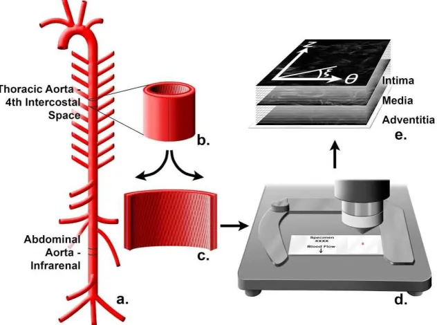
All mice were handled in compliance with protocols approved by the University of South Carolina Institutional Animal Care and Use Committee. Mice were ethically euthanized by carbon dioxide asphyxiation and then perfused through the left ventricle with heparinized normal saline for five minutes at physiological pressure, followed by pressure perfusion fixation for 10 minutes with 10% neutral buffered formalin. Aortas were harvested from apoE KO and apoE MMP12 DKO mice (n=5 each strain). Under a dissecting microscope, each aorta was opened longitudinally for en face examination of the collagen fibers. Non-lesioned areas were chosen in the thoracic aorta, which was sampled at the level of the fourth intercostal arteries, and the abdominal aorta, which was
sampled from the infrarenal area (Figure 3.1A). The samples were flattened to a slide and labeled according to the mean orientation of blood flow for each
specimen (Figure 3.1B-3.1D).
Second-harmonic generation microscopy
al., 2011; Liu H et al., 2013). The microscope's XYZ stage was controlled through three orthogonally mounted motors to obtain image stacks from four regions of each sample by moving the stage 100μm in each direction while optically slicing the vessel wall from top to bottom at 60X. The area in pixels for each image was 512x512 pixels with the actual calibrated image being 176x176 microns.
Semi-Automated Image Analysis
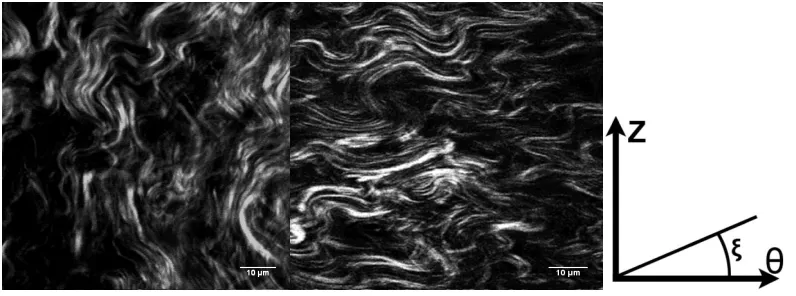
The individual, local fiber orientations were determined in the Z-θ plane (Figure 3.1E). To quantify collagen fiber orientation, we first define a coordinate system. The coordinate system used in this work is shown in Figure 3.1. Here, Z is along the axial/flow direction and θ is along the circumferential/hoop direction in the specimen. The fiber angle relative to the θ-direction is denoted by ξ, with +ξ defined as the CCW angle from θ towards Z. Thus ξ = 0o corresponds to the
circumferential direction (+θ) and ξ = 90o corresponds to the flow direction (+Z) in
the artery. To determine ξ for each specimen, we used an analytic protocol for automatic angle recognition as previously described (Wicker et al., 2008; Tang et al., 2014). Continuity software (version 6.4b,
Statistical Analysis
For this study, we used a mixed model to analyze fiber angle distribution as a function of four variables, using the R package “Ime4” (Bates et al., 2014).
The four variables were diet, strain, location, and through-thickness position. The average absolute angle was estimated by ξ = C
0 + C1(diet) + C2(anatomical
location) + C
3(mouse strain) + C4(through-thickness position), where the Cs are
the regression parameters. To account for the regional variability of the artery, we investigated through-thickness position as a categorical variable. Thus, ξ is a function of radial position in the artery, ξ = ξ(r), with the radius varying from the inside, ri, to the outside, ro. Thus, the three radial regions are ri < r1 < ri + t/3; ri +
t/3 ≤ r2 < ri + 2t/3; ri + 2t/3 ≤ r3 < ro, where t is the average thickness of the
vessel wall. Variables with P<0.05 were considered statistically significant.
3.4 Results
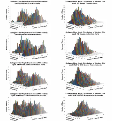
Collagen fiber organization in chow diet vs. Western diet mice
Constructed 3D histograms of collagen fiber distribution across aortic wall Figure 3.3 provides an illustration of representative 3D histograms of collagen fiber angle distribution for each mouse strain on chow diet and Western diet. Zero degrees is the circumferential direction. Figures 3.3 A and C present the distribution of the fiber angles in thoracic aorta specimens from each strain, and Figures 3.3 B and D present equivalent information for abdominal aorta specimens. The 3-D histograms reflect image stacks for one individual mouse on each diet. The relative radial position, β = (r – ri )/t, in each histogram represents
the position within the arterial wall, with β = 0 corresponding to the endothelial surface and β = 1 to the outer boundary of the adventitia. Note that β accounts for individual variations in tissue thickness among samples, since t can be varied to match the specimen geometry, with each β corresponding to one slice of the image stack.
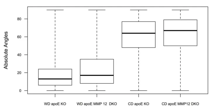
Statistical Analysis
Figure 3.4 shows a boxplot comparing the median, first, and third quartiles of collagen fiber angle observations for Western diet and chow diet fed mice of each strain. Here, ξ is the response variable, which is the absolute value of the collagen fiber angles. In the multiple regression model, we studied whether the four factors diet, anatomical location, strain, and β contributed to angle
multiple regression analysis showed a significant difference in average absolute fiber angle, |ξavg|, between mice on a chow diet vs. mice on a Western diet. A
summary of mean values and standard errors for each statistically significant regression parameter is provided in Table 3.1.
As shown in Table 3.1, there was no significant difference in |ξavg| between
mouse strains. Abdominal location within the aorta (thoracic vs. abdominal) and relative radial position within the specimen had small, but significant, effects on |ξavg|. Thoracic aorta specimens had average fiber angles, ξavg, that were 0.84°
lower (95% confidence interval [-1.00°, -0.67°] than abdominal specimens. In addition, statistical analysis showed that the outer third of the vessel (2/3 < β < 1) had slightly greater fiber angles (+0.3°, 95% confidence interval [+0.08°, +0.50°] compared to the inner two-thirds of the vessel (0 < β < 2/3). This observation is placed in context with previous work in the following section.
3.5 Discussion
Kassab, 2003), we are the first to report differences in collagen fiber angles due to a diet linked to atherosclerosis.
Changes in collagen fiber angle are important for understanding the capability of the artery to remodel after arterial lesion development. Structure-based material constitutive models incorporate the mean collagen fiber angle as a geometric parameter that can be identified based on mechanical behavior (Holzapfel, 2000). Originally these models assumed a single value of the angle for each fiber family, but more sophisticated recent material models take into account fiber dispersion (Holzapfel et al., 2015; Li & Robertson, 2009; Driessen et al., 2008).With regard to findings,our experimental results demonstrate a significant dispersion of collagen fibers around the mean fiber angle.
With regard to the mean fiber angle, the predicted angles of collagen fibers of a representative carotid artery from a rabbit using a two-fiber-family constitutive model are reported to be 29° in the media and 62° in the adventitia (Holzapfel, 2000). Our data for the adventitial layer (outer 1/3 of vessel) of the normal mouse thoracic aorta reveals a mean fiber angle (62.3°) similar to that predicted for the rabbit carotid adventitia. However, statistics reported in the previous section show that our measurements indicate little difference between the mean fiber angle in the medial layer (62.0°) and that in the outermost layer. We chose to compare the thoracic and abdominal portions of the mouse aorta because the abdominal aorta is more susceptible to aneurysm formation in humans. Aneurysm development can be regarded as an extreme case of
fiber arrangement. Using a structure-based constitutive model, Collins et al. (2011) reported that the predicted mean fiber angle for normal (wild type) mouse infrarenal abdominal aorta is 39° with respect to Z direction (51o relative to θ
direction) and Collins et al. (2012) show a mean collagen fiber angle in an aneurysmal aorta to be 41° with respect to the Z direction (49o relative to θ
direction). Our observed mean fiber angle for the infrarenal abdominal aortic media of 62.8° in chow diet-fed mice is higher than the predicted value based on the four fiber-family model. However, we examined collagen fiber angles in mice which were much older than those used by Collins, et al. (7-8 months vs. 2-3 months). These investigators found only subtle mechanical differences between the suprarenal and infrarenal abdominal aortas (Collins et al., 2011). We show that there are small but significant differences between the average absolute angles based on anatomical location (thoracic vs. abdominal aorta). On average, the mean collagen fiber angle in the thoracic aorta is 0.84° less than that in the abdominal aorta. Our data support the hypothesis that there is a small but significant difference in collagen organization between anatomical locations, even in mice with no significant vascular pathology.
In this study, we focused on differences due to atherosclerosis and
showed that Western diet feeding results in an apparent shift in average collagen fiber angles, with a mean difference of ≈ 43° (Figure 3.4). This result could
The Continuity image analysis protocol is effective for investigating
collagen fiber angle differences within the extracellular matrix. We show that this image analysis method can be used for atherosclerosis and vascular remodeling studies to compare differences in aortic collagen fiber organization in mice on a chow diet or Western diet. Collectively, SHG imaging techniques and Continuity provide a plethora of data. Since there was no standard approach to interpret three-dimensional fiber angle distributions, we developed a new statistical approach to analyze the data. Based on the statistical analysis, our data indicates that the aortas of both apoE KO and apoE MMP12 DKO mice fed a Western diet for 6 months have significantly lower mean collagen fiber angles than age-matched mice maintained on a chow diet.
3.6 Limitations of the Study
A limitation of this study is that we have not taken advantage of TPEF microscopy for viewing elastin in the same specimens. In addition, we report average local fiber orientations and do not account for the waviness of individual collagen fibers within this study.
(Hill et al., 2012). They noted that fiber orientation is the same in three
mechanical models where the investigators idealized collagen distribution as planar.
Our research interest was centered on differences in collagen fiber angle distribution that coincide with atherosclerotic lesion formation within the aorta. There is a need to understand differences between the thoracic and abdominal aorta because of the common association of atherosclerosis with abdominal aortic aneurysm. Finally, we focused on differences in mouse strains (apoE KO and apoE MMP12 DKO mice) to evaluate the contribution of matrix
metalloproteinases to collagen fiber alignment and we did not find any significant differences with the knockout of MMP12.
3.7 Conclusions
Our data indicate that the aortas of both apoE KO and apoE MMP12 DKO mice fed a Western diet for 6 months have significantly lower (more
Figure 3.2: Representative second-harmonic generation images of thoracic aorta of apoE KO mouse on a chow diet (left) versus thoracic aorta of apoE KO mouse on Western diet (right). Images were captured in the media layer. Bars = 10 µm. Circumferential direction is denoted by θ, with + ξ positive clockwise and
Figure 3.3 A: Representative three-dimensional (3D) histograms of fiber angle distributions as a function of relative distance from the lumen in the thoracic aorta of apolipoprotein E knockout (apoE KO) mice on a chow diet and on Western diet for 6 months. B: Representative 3D histograms of fiber angle distributions as a function of relative distance from the lumen in the abdominal aorta of apoE KO mice on a chow diet and on Western diet for 6 months. C: Representative 3D histograms of fiber angle distributions as a function of relative distance from the lumen in the thoracic aorta of apoE matrix metalloproteinase-12 double knockout (MMP12 DKO) mice on a chow diet and on Western diet for 6 months. D:
Table 3.1.Summary of mean effect and standard error for each independent variable. The coefficient of determination of the fitted model is R2 = 49.9%
Factor Estimate 95%
Confidence Interval
Standard Error
P-Value
Intercept, C0 62.83 2.03 <0.05
Diet (Western), C1 -43.35 (-37.73,
-48.96)
2.86 <0.05
Location (Thoracic), C2
-0.84 (1.00,
-0.67)
0.09 <0.05
Depth (Adventitia), C4
0.29
(0.08, 0.50)
CHAPTER
4
DIET ALTERS AGE-RELATED REMODELING OF AORTIC
EXTRACELLULAR MATRIX IN MICE SUSCEPTIBLE TO
ATHEROSCLEROSIS
4.1 Abstract
Collagen is an important extracellular matrix protein providing overall strength to many biological tissues. A major challenge in understanding how collagen contributes to the functional properties of the arterial wall is identifying how vascular cells respond to mechanical loading by restructuring the
surrounding extracellular matrix. Therefore, the objective of this study was to evaluate collagen architecture to understand the remodeling capabilities of the aorta during aging with or without atherosclerotic lesion development. We hypothesized that changes in collagen fiber orientation are initiated by
in apoE KO mice on chow diet versus those on Western diet. Compared to 6-week old apoE KO mice, multiple regression analysis showed a significant difference in average absolute fiber angle in response to age and high-fat diet. On average, absolute fiber angle increased by 26.4° in older mice on the same diet and decreased 22.4° in mice fed a Western diet for 6 months. Our data indicate that the aortas of 6-week old apoE KO mice have an absolute collagen fiber angle in the inner media of 35.2±1.9° but that fibers become more
distinct remodeling response in the presence of atherogenic stimuli, even in non-lesioned areas, as observed by a shift in collagen fiber orientation.
4.2 Introduction
Atherosclerosis is a chronic inflammatory disease of the arterial wall, constituting a significant source of morbidity and mortality in the United States. It can be classed as a disease of aging (Wang & Bennett et. al., 2012) or a result of unhealthy diet, such that increasing age or low-density lipoprotein cholesterol can be independent risk factors for the development of atherosclerosis (Wang & Bennett et al., 2012). A major pathological change often preceding the
development of atherosclerosis and associated with vascular aging is endothelial dysfunction, a diminished production/availability of nitric oxide and/or an
imbalance between vasoconstricting and vasodilating substances produced by or acting on the endothelium (Hadi et. al, 2005; Park & Park, 2015).
During the last 3 decades, it has been shown that the vascular