R E S E A R C H
Open Access
Asna1/TRC40 that mediates membrane
insertion of tail-anchored proteins is
required for efficient release of Herpes
simplex virus 1 virions
Melanie Ott
1, Débora Marques
2, Christina Funk
2and Susanne M. Bailer
1,2*Abstract
Background:Herpes simplex virus type 1 (HSV1), a member of the alphaherpesvirinae, can cause recurrent facial lesions and encephalitis. Two membrane envelopment processes, one at the inner nuclear membrane and a second at cytoplasmic membranes are crucial for a productive viral infection. Depending on the subfamily, herpesviruses encode more than 11 different transmembrane proteins including members of the tail-anchored protein family. HSV1 encodes three tail-anchored proteins pUL34, pUL56 and pUS9 characterized by a single hydrophobic region positioned at their C-terminal end that needs to be released from the ribosome prior to posttranslational membrane insertion. Asna1/TRC40 is an ATPase that targets tail-anchored proteins to the endoplasmic reticulum in a receptor-dependent manner. Cell biological data point to a critical and general role of Asna1/TRC40 in tail-anchored protein biogenesis. With this study, we aimed to determine the importance of the tail-anchored insertion machinery for HSV1 infection.
Methods:To determine protein-protein interactions, the yeast-two hybrid system was applied. Asna1/TRC40 was depleted using RNA interference. Transient transfection and virus infection experiments followed by indirect immunofluorescence analysis were applied to analyse the localization of viral proteins as well as the impact of Asna1/TRC40 depletion on virus infection.
Results:All HSV1 tail-anchored proteins specifically bound to Asna1/TRC40 but independently localized to their target membranes. While non-essential for cell viability, Asna1/TRC40 is required for efficient HSV1 replication. We show that early events of the replication cycle like virion entry and overall viral gene expression were unaffected by depletion of Asna1/TRC40. Furthermore, equal amounts of infectious virions were formed and remained cell-associated. This indicated that both nuclear egress of capsids that requires the essential tail-anchored protein pUL34, and secondary envelopment to form infectious virions were successfully completed. Despite large part of the virus life cycle proceeding normally, viral propagation was more than 10 fold reduced. We show that depletion of Asna1/TRC40 specifically affected a step late in infection during release of infectious virions to the extracellular milieu.
Conclusions:Asna1/TRC40 is required at a late step of herpesviral infection for efficient release of mature virions to the extracellular milieu. This study reveals novel tools to decipher exocytosis of newly formed virions as well as hitherto unknown cellular targets for antiviral therapy.
Keywords:Herpesvirus, HSV1, Integral membrane proteins, Tail-anchored protein biogenesis, Asna1/TRC40, pUL34, pUL56, pUS9, Nuclear egress, Exocytosis
* Correspondence:[email protected]
1Max von Pettenkofer-Institut, Ludwig-Maximilians-Universität München,
Pettenkoferstr. 9a, 80336 München, Germany
2Institute for Interfacial Engineering and Plasma Technology IGVP, University
of Stuttgart, Nobelstrasse 12, 70569 Stuttgart, Germany
Background
Herpesviruses have evolved a life cycle that strongly depends on two membrane-envelopment processes, one at the inner nuclear membrane (INM) called primary envelopment, and another at cytoplasmic membranes called secondary envelopment, both of which are crucial for a productive viral infection [1, 2]. Depending on the subfamily, herpesviruses encode more than 11 different transmembrane proteins involved in various aspects of the individual viral life cycle.
Tail-anchored (TA) proteins represent a specific class of transmembrane proteins characterized by a single transmembrane domain (TMD) positioned at its very C-terminal end. Thus, the hydrophobic region of a TA pro-tein remains associated with the ribosomal tunnel until translation is complete [3–6]. This requires that TA pro-teins are released from the ribosome prior to their post-translational insertion into various target membranes. The identification of the TMD recognition complex of 40 kDa (TRC40) also known as Asna1 provided a major breakthrough in understanding the TA protein biogen-esis. Asna1/TRC40 is an ATPase conserved in many species. It captures a TA protein following its ribosomal translation and together with several other components delivers it to a receptor of the endoplasmic reticulum (ER). Recent biochemical and structural analysis have further elucidated the mechanism of membrane inser-tion of TA proteins. The ATP-bound dimer of Asna1/ TRC40 or its orthologs form a hydrophobic groove that accommodates the TMD of TA proteins. The resulting Asna1/TRC40-TA protein complex is then recruited to the ER receptor resulting in release of the TA protein and membrane insertion, a process that may require ATP hydrolysis.
Like all TA proteins, the HSV1 TA proteins pUL34, pUL56 and pUS9 are characterized by a cytoplasmic do-main, a single C-terminal transmembrane domain (TMD) and a short luminal extension (Fig. 1a). HSV1 pUL34 is a protein conserved throughout the herpesvirus family ([7]; and references therein). Both its cyto-/nucleoplasmically exposed N-terminal domain (residues 1–252) and its C-terminal TMD (residues 252–272) are essential for viral replication [8–10]. Posttranslational membrane insertion of pUL34 occurs in the cytoplasm and thus prior to its targeting to the INM [7]. There, pUL34 associates with the nucleocapsid-bound pUL31 for subsequent primary envelopment and egress of capsids to the cytoplasm [7].
The other two HSV1 TA proteins, pUL56 and pUS9, are non-essential and specific for alpha-herpesviruses ([11, 12]; and references therein). pUL56 is composed of a cytoplasmic domain (residues 1–211) followed by a hydrophobic region (residues 211–231) and a short luminal domain (Fig. 1a). In pUS9, a short N-terminal domain (residues 1–69) is followed by a hydrophobic
domain between residues 69–89. Both pUL56 and pUS9 localize to the trans Golgi network (TGN) and are integrated into mature virions during secondary en-velopment [13].
Many herpesviral functions have been analysed in great detail while our knowledge of virus-host interactions and their importance for viral replication is far from complete. With this study we focus on the biogenesis of tail-anchored (TA) proteins and its importance for herpesviral infection. Upon knockdown of Asna1/TRC40, large part of the viral infection cycle proceeds normally and infec-tious virions are formed, their release to the extracellular milieu late in infection however is delayed. Together our data suggest that efficient transport of infectious virions along the secretory pathway requires Asna1 and thus the TA insertion machinery.
Methods
Cells, yeast 2-hybrid assay and general cloning
HeLa (ATCC CCL-2) and Vero cells (ATCC CRL-1587) were grown in DMEM containing 10 % FCS. Yeast 2-hy-brid (Y2H) analysis was done as described [14]. The UL34, UL45, UL56 and US9 genes previously cloned into the entry vector pDONR207 [15] were transferred
into the Gateway compatible Y2H bait vector
pGBKT7-DBD and/or the mammalian expression vec-tor pCR3-N-myc according to the manufacturer’s protocol (Invitrogen). The human Asna1/TRC40 gene previously cloned into the pDONR223 vector was transferred to the Gateway compatible Y2H prey vector pGADT7-AD according to the manufacturer’s protocol (Invitrogen).
Viruses
HSV1(F) (provided by B. Roizman, University of Chi-cago, USA) was used for infection experiments. The strain HSV1(17+)lox (provided by B. Sodeik, Hannover Medical School, Germany) was used as PCR template. HSV1 propagation and virus growth curves were performed as described [14]. To monitor infection, Vero cells were infected with HSV1(F) at the indicated MOI. Cell lysates were prepared at the indicated times post infection and analysed by Western blotting using pri-mary antibodies to the immediate early proteins ICP0 (anti-ICP0, Santa Cruz) and ICP27 (anti-ICP27, Virusys), to the early protein gB (anti-Glykoprotein B, Santa Cruz) and to the late proteins VP5 (anti-ICP5 (VP5), Abcam) and pUL34 [9] followed by secondary antibodies conju-gated to POX. Antibodies specific to β-actin (Abcam) were used as control.
Indirect immunofluorescence microscopy
transfection, the Effectene Transfection Reagent was used. For virus infection, HeLa cells were infected at the indicated MOI. In infected cells, binding of antibodies to the HSV1 Fc-receptor like proteins gE/gI was blocked overnight at 4 °C with human IgG (200μg/ml) and 10 % FCS in PBS [16]. The mouse monoclonal antibodies
anti-myc [9E10] (Santa Cruz), anti-ICP8 (provided by R. Heilbronn, Charité Universitätsmedizin Berlin CCM, Berlin, Germany), anti-Asna1/TRC40 ([M03], Klon 2H3 Abnova) and rabbit polyclonal antibodies anti-pUL34 [9], anti-Calreticulin (Sigma), and anti-Giantin (Abcam) were used as primary reagents. Goat rabbit or
anti-A
B
bait
prey
Asna1
Ctrl
1 234
211 231
TMD UL56-N
1 234
211 231
TMD pUL56 - N
1 275
252 272
TMD pUL34 - N
1 90
69 89
TMD Us9-N
1 90
69 89
TMD pUS9 - N
C
HeLa 12 h.p.i. DAPI
Mo
ck
Calreticulin
Asna1 merge
HSV1
(F)
Fig. 1HSV1 encodes three tail-anchored proteins that interact with Asna1/TRC40.aSchematic diagrams show the domain organization of
mouse antibodies coupled to Alexa488 or Alexa594 (all Invitrogen) were used as secondary reagents. Cells were examined using a Leica confocal laser scanning micro-scopes TCS SP5 and LSM710. Images were recorded using the Leica Application Suite AF6000 Software and processed using Adobe Photoshop.
SiRNA transfection
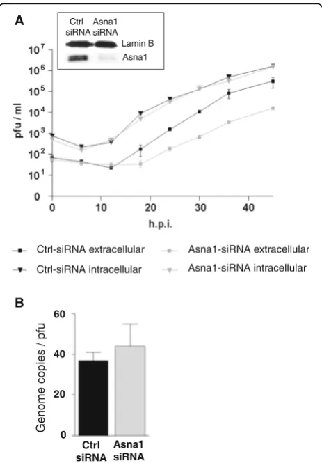
Gene silencing was essentially done as described [17]. Briefly, siRNAs (20 nM; GE Dharmacon), 150 μl of HBSS, and 1,5μl of transfection reagent were mixed and added to HeLa cells in DMEM with 5 % FCS seeded in 12-well plates. Efficiency of RNAi was monitored by Western blotting using mouse monoclonal antibodies to Asna1/TRC40 ([M03], Klon 2H3 Abnova) and poly-clonal goat anti-Lamin B (Santa Cruz) to control for loading. The siRNA duplexes used for Asna1/TRC40 knockdown and as controls (Ctrl) are shown in Table 1. Viral infection was generally performed 48 h (h) after siRNA treatment. Infectious virions were quantified by removing aliquots of medium and of infected cells at various time points followed by plaque assay on Vero cells [14]. To determine genome copy/pfu ratios of virions released from cells, HeLa cells were treated with Asna1/TRC40 specific or ctrl siRNA followed by infec-tion with HSV1(F) for 30 h. Realtime quantitative PCR using HSV1 specific primers were used to determine the genome copies, plaque assays were performed as described [14].
Results
HSV1 encodes three tail-anchored proteins that interact with Asna1/TRC40
HSV1 encodes three TA proteins, called pUL34, pUL56 and pUS9. Hydrophobicity plots show that pUL34, an essential protein conserved throughout the herpesviral family, contains a transmembrane domain (TMD) between residues 252–272 required for nuclear egress (Fig. 1a; [9]). Two other TA proteins, pUL56 and pUS9, that are non-essential and specific for alpha-herpesviruses, carry a TMD between residues 211–231 and 69–89, respectively (Fig. 1a). To determine whether pUL34, pUL56 and pUS9 inter-act with Asna1/TRC40, the yeast 2-hybrid (Y2H) system was applied. Asna1/TRC40 fused to the Gal4 activation
domain (AD) was tested for interaction with pUL34, pUL56 and pUS9 fused to the Gal4 DNA-binding domain (DBD). pUL45, that carries an N-terminal TMD co-translationally integrated into membranes by an Asna1/TRC40-independent mechanism, was used as control. Interaction of proteins was reported by growth of yeast cells on selective media. While DBD-pUL34, -pUL56 and -pUS9 co-expressed with AD-Asna1/TRC40 allowed for growth of yeast cells, this was not the case for co-ex-pression of DBD-pUL45 and AD-Asna1/TRC40 (Fig. 1b). We thus conclude that all three TA proteins of HSV1 spe-cifically interacted with Asna1/TRC40 supporting its func-tion in posttranslafunc-tional membrane inserfunc-tion of these viral proteins.
To analyse the subcellular distribution of Asna1/ TRC40 in presence and absence of HSV1 infection, HeLa cells were mock treated or infected with HSV1(F) for 12 h and subsequently processed for IF. Infected cells were readily identified based on their marginalized chro-matin as revealed by DAPI staining (Fig. 1c). Both in noninfected and infected cells, Asna1/TRC40 showed a pancellular distribution and significantly co-localized with the ER marker Calreticulin (Fig. 1c) suggesting that its distribution is essentially unaltered during HSV1 infection (Fig. 1c).
Targeting and membrane insertion of HSV1 pUL34, pUL56 and pUS9 occur independent of Asna1/TRC40
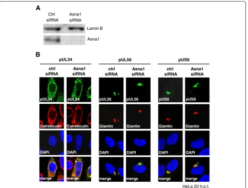
In absence of other viral proteins, pUL34 is targeted to the ER and the nuclear periphery, whereas pUL56 and pUS9 are located to the trans Golgi network (TGN). To determine whether Asna1/TRC40 is re-quired for proper membrane targeting of the HSV1 TA proteins, HeLa cells were transfected for 48 h with Asna1/TRC40 specific or control (ctrl) siRNA. Knockdown of Asna1/TRC40 was highly efficient as shown by Western blotting (Fig. 2a). Interestingly, de-pletion of Asna1/TRC40 did not affect cell viability of Hela cells (data not shown).
RNAi treated cells were then transfected with plas-mids encoding myc-tagged TA proteins and 20 h later analysed by IF using monoclonal anti-myc antibodies. Calreticulin or Giantin were used as markers of the ER and the TGN, respectively. pUL34 showed a reticular subcellular distribution and co-localized with the ER marker Calreticulin consistent with its localization to the ER and the nuclear periphery whether the cells were treated with Asna1/TRC40 specific or ctrl siRNA (Fig. 2b, left panel). pUL56 and pUS9 both located to the TGN as indicated by their co-localization with the TGN marker Giantin (Fig. 2b, middle and right panel). In Asna1 depleted cells, a certain amount of pUS9 was found in a perinuclear region suggesting that membrane insertion of pUS9 is influenced by the absence of Asna1. To summarize, Table 1SiRNAs used for gene silencing
all HSV1 TA proteins seemed to efficiently reach their tar-get membranes irrespective of the presence or absence of Asna1/TRC40.
Asna1/TRC40 is dispensable for virion entry and gene expression during HSV1 infection
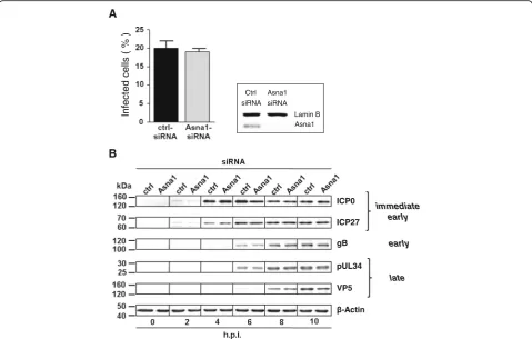
To determine whether Asna1/TRC40 is required for the herpesviral life cycle, Asna1/TRC40 knockdown was per-formed and monitored as shown before (Fig. 2a). Then, siRNA treated HeLa cells were infected with HSV1 at an MOI of 0.5 for 4 h (Fig. 3a). ICP0 expression was ana-lysed as indirect means of viral entry. IF analysis revealed that 20 % and 19 % of the cells treated with ctrl- and Asna1/TRC40-specific siRNAs, respectively, were infected by HSV1(F).
A time course experiment to detect viral proteins of all kinetic classes was performed. HeLa cells were first treated with Asna1/TRC40 or ctrl siRNA for 48 h and subsequently infected with HSV1 at an MOI of 1. Cell lysates were prepared at the indicated time-points and probed with antibodies specific to the immediate early regulators ICP0 and ICP27, to glycoprotein gB, to the nuclear egress protein pUL34 and the major capsid pro-tein ICP5 (VP5).β-Actin-specific antibodies were used to control for equal loading of cell samples (Fig. 3b). The transcriptional regulators ICP0 and ICP27 were detected 2 h post infection (h.p.i.), glycoprotein gB and the nuclear egress protein pUL34 appeared 6 h.p.i., while the major capsid protein ICP5 was detected 8 h.p.i.. Taken together, we found that Asna1/TRC40 is not required for virion entry and overall viral gene expression (Fig. 3b).
ctrl siRNA
Asna1 siRNA
pUL34
Calreticulin
pUL34
Calreticulin
DAPi
merge
pUL34
DAPi
merge
ctrl siRNA
Asna1 siRNA pUL56
pUL56
Giantin
DAPI
merge
pUL56
Giantin
DAPI
merge
ctrl siRNA
Asna1 siRNA pUS9
pUS9
Giantin
DAPI
merge
pUS9
Giantin
DAPI
merge
HeLa 20 h.p.t. Ctrl
siRNA
Asna1 siRNA
Lamin B
Asna1
A
B
Fig. 2Targeting and membrane insertion of HSV1 pUL34, pUL56 and pUS9 occur independent of Asna1/TRC40.aTo determine the effect of
Targeting of pUL34 to the nuclear envelope during infection is independent of Asna1/TRC40
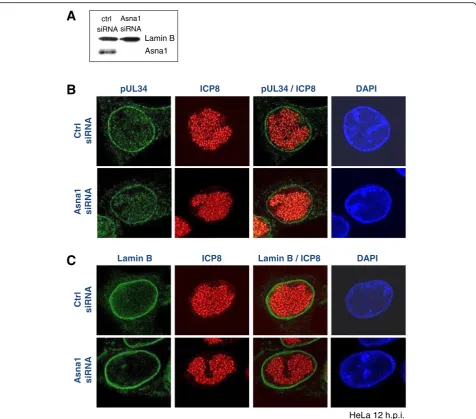
HSV1 pUL34 is essential with a conserved function in nuclear egress of capsids. To determine whether Asna1/ TRC40 is required for pUL34 biogenesis in the viral con-text, RNAi was performed as described (Figs. 2a and 4a). Subsequently, HeLa cells were infected with HSV1 at an MOI of 1 for 12 h and subsequently processed for IF (Fig. 4b and c). Antibodies specific to pUL34 (Fig. 4b) and Lamin B (Fig. 4c) showed that both proteins were exclu-sively located to the nuclear envelope. Furthermore, intra-nuclear replication centers formed normally, as revealed by ICP8-specific antibodies (Fig. 4b and c). Thus, we conclude that pUL34 membrane insertion and targeting to the INM, a prerequisite for NEC formation and capsid nuclear egress, proceeds normally in absence of Asna1/TRC40.
Asna1/TRC40 is required late in infection for efficient release of infectious virions from the cell
To determine the overall effect of Asna1/TRC40 knock-down on the outcome of an HSV1 infection, Asna1/TRC40
depleted HeLa cells or ctrl cells were infected with HSV1 at an MOI of 0.1. At the indicated time points, medium and infected cells were harvested separately and analysed for the presence of infectious virions using plaque assays. Equal amounts of infectious virions were formed and remained cell-associated whether cells were treated with Asna1/ TRC40 specific or ctrl siRNA (Fig. 5a). In contrast, about 10 times less infectious virions were released to the culture medium upon Asna1/TRC40 depletion. To determine the genome copy/pfu ratio of virus released 30 h.p.i. from the infected cells, realtime quantitative PCR was performed. Virions released from Asna1 siRNA treated cells showed reduced amounts of genomes as well as plaque forming units compared to the ctrl treated cells. In either case however, their genome copy/pfu ratio was comparable indi-cating that virions released to the extracellular milieu were similar in infectivity (Fig. 5b). Thus, although not essential for cellular growth and formation of infectious virions, Asna1/TRC40 is required for release of mature virions from the cells at a late step of virus infection and thus for efficient HSV1 propagation.
A
B
Ctrl siRNA
Asna1 siRNA
Lamin B Asna1
Infected
cells
( % )
immediate early
early
late
-Actin VP5 pUL34 gB ICP27 ICP0
Fig. 3Asna1/TRC40 is dispensable for virion entry and gene expression during HSV1 infection.aTo determine the importance of Asna1/TRC40
Discussion
Asna1/TRC40 plays a central role during TA protein biogenesis by binding to TMDs of newly synthesized TA proteins and targeting them to the ER receptor for subsequent membrane insertion [3–6]. While most data on Asna1/TRC40-mediated TA protein biogenesis are based on in vitro data, we show here that in vivo, a highly efficient knockdown of Asna1/TRC40 does not interfere with cell growth. Thus, Asna1/TRC40 seems to be non-essential for cell viability under normal condi-tions. This is surprising considering the many function-ally important TA host proteins [18], but consistent with the finding that components of the TA insertion machin-ery are non-essential in yeast cells unless additional stress
is present [5]. Pathways redundant with Asna1/TRC40 for posttranslational membrane insertion may involve the signal recognition particle SRP or the heat shock protein dimers Hsc70/Hsp40 [19–21].
Despite Asna1/TRC40 being non-essential for cellular life, our data show that it is important for efficient herpesviral propagation. Large part of the herpesviral life cycle including nuclear egress of capsids tolerates the absence of Asna1/TRC40; viral morphogenesis is com-parable to control treated cells giving rise to equal num-bers of intracellular infectious virions. Interestingly, however, a specific defect is observed at a very late step of the viral life cycle during release of mature virions to the extracellular milieu. Thus, our data reveal a novel
pUL34 ICP8
Ctrl
siRNA
A
s
na1
siRNA
pUL34 / ICP8 DAPI
HeLa 12 h.p.i.
ctrl siRNA
Asna1 siRNA
Lamin B Asna1
Ctrl
siR
N
A
As
n
a
1
siR
N
A
Lamin B ICP8 Lamin B / ICP8 DAPI
A
B
C
Fig. 4Targeting of pUL34 to the nuclear envelope during infection is independent of Asna1/TRC40. To determine the importance of Asna1/TRC40 for
role of Asna1/TRC40 late in HSV1 infection required for efficient cytoplasmic transport and/or release of infectious virions.
Recent evidence supports a role of the constitutive secretory pathway in delivering virus containing secretory vesicles to sites of exocytosis at the plasmamembrane [22]. A role for Asna1/TRC40 orthologs in transport and release/fusion of secretory vesicles is supported by studies in yeast [23] andCaenorhabditis elegans(C. elegans) [24]. In the herpesviral context, Asna1/TRC40 depletion could limit the amount of specific host TA proteins required at the site of virion exocytosis. In this respect, the SNARE proteins involved in various vesicular and membrane fusion processes represent an important group of TA host factors [18, 25, 26]. Indeed, depletion of the SNARE TA
protein Syntaxin 3 results in reduced release of infectious Human cytomegalovirus (HCMV) virions [27]. Alterna-tively, the highly productive virus infection may pose a general stress on the TA insertion machinery thereby overwhelming its capacity. We were unable to identify a specific compartment where the virions accumulated in absence of Asna1 (data not shown) suggesting that the overall dynamic of virion release is hampered.
Our data show that all HSV1 encoded TA proteins specifically bind to Asna1/TRC40 consistent with their TMD hydrophobicity index≥40 [4]. Despite its ability to bind pUL34, Asna1/TRC40 is redundant for pUL34 localization and function. Both pUL56 and pUS9 effect-ively reached their target membranes in absence of Asna1/TRC40. However, a certain amount of pUS9 was mislocalized upon isolated expression suggesting that Asna1 modulates pUS9 membrane insertion. Since indi-vidual TA proteins favor particular insertion factors [5], it is quite possible that pUL56 and pUS9 differ from pUL34 in their propensity to use Asna1/TRC40 for membrane targeting. Unfortunately, detailed analysis of pUL56 and pUS9 in the viral context is limited due to the lack of specific antibodies.
How could a reduced membrane insertion of the viral TA proteins pUL56 and pUS9 affect virion release to the extracellular milieu? Exposed on the cytoplasmic face of secretory vesicles, their N-terminal domains may inter-act with kinesin motor proteins [12, 28] to shuttle the vesicular content to sites of secondary envelopment. This way, pUL56 and pUS9 could modulate the trans-port of secretory vesicles containing infectious virions. Interestingly, pUS9 was recently reported to be neces-sary for anterograde transport of virions in neurons ([29]; and references therein). Thus, TA protein biogen-esis may have a particular impact on herpesviral neuro-pathology where long-distance axonal transport of virus containing secretory vesicles is likely to occur.
Biogenesis of TA membrane proteins destined for the INM is not well understood. Membrane insertion of the viral TA protein pUL34 that occurs prior to its transport to the INM [7] is essential for its function ([7, 9]; and references therein). Thus, pUL34 can serve as a viral reporter to gain insight into the biogenesis of TA pro-teins associated with the INM. Asna1/TRC40 specifically binds to pUL34 suggesting it supports membrane inser-tion of the INM protein pUL34. A role of Asna1/TRC40 in INM biogenesis is also provided by data on Emerin, a TA protein associated with Emery-Dreifuss muscular dys-trophy [30]. Together these data indicate that biogenesis of INM TA proteins engages Asna1/TRC40-dependent and -independent pathways [9, 30].
Taken together, our data reveal a role of the TA protein biogenesis during release of virions. Asna1/TRC40 deple-tion may provide a tool to study this poorly characterized
Ctrl
Fig. 5Asna1/TRC40 is required late in infection for efficient release of
process, a decisive step for virus spread. Since Asna1/ TRC40 knockdown preferentially perturbs virus replica-tion while cellular growth remains unaffected, analysis of TA protein biogenesis may reveal antiviral targets to in-hibit virus propagation.
Conclusions
■The TA protein insertion factor Asna1/TRC40 is nonessential.
■HSV1 encodes three tail-anchored proteins pUL34, pUL56 and pUS9.
■All HSV1 TA proteins specifically bind to Asna1/ TRC40.
■Asna1/TRC40 is required for efficient HSV1 replication.
■Asna1/TRC40 is redundant for nuclear egress of capsids.
■Depletion of Asna1/TRC40 results in a defect late in herpesviral infection during release of infectious virions.
Abbreviations
AD:Activation domain; Ctrl: Control; DBD: DNA binding domain;
ER: Endoplasmic reticulum; h: Hours; h.p.i.: Hours post infection; HSV1: Herpes simplex virus type 1; IF: Indirect immunofluorescence; INM: Inner nuclear membrane; NEC: Nuclear egress complex; RNAi: RNA interference; TA: Tail-anchor; TGN:transGolgi network; TMD: Transmembrane domain; TRC40: TMD recognition complex of 40 kDa; Y2H: Yeast 2-hybrid
Acknowledgements
The HSV1 strain F was provided by Bernard Roizman; we are grateful to Beate Sodeik for providing the BAC HSV1(17+)lox. We would like to thank Roger Everett, Beate Sodeik, Gary Cohen, Roselyn Eisenberg and Regine Heilbronn for the generous gift of antibodies.
Funding
This work was supported by grants of the Deutsche
Forschungsgemeinschaft. Funding formed the basis for the design of the study and collection, analysis and interpretation of data.
Availability of data and materials
All data and informations are available without restriction and included in the manuscript.
Authors’contributions
MO and SB conceived of the study, MO performed all experiments and analysis, SB participated in data interpretation and drafted the manuscript. All authors read and approved the final manuscript.
Competing interests
The authors declare that they have no competing interests.
Ethics approval and consent to participate Ethics approval was not required.
Received: 25 July 2016 Accepted: 17 October 2016
References
1. Johnson DC, Baines JD. Herpesviruses remodel host membranes for virus egress. Nat Rev Microbiol. 2011;9:382–94.
2. Mettenleiter TC, Klupp BG, Granzow H. Herpesvirus assembly: an update. Virus Res. 2009;143:222–34.
3. Stefanovic S, Hegde RS. Identification of a targeting factor for posttranslational membrane protein insertion into the ER. Cell. 2007;128:1147–59.
4. Rabu C, Wipf P, Brodsky JL, High S. A precursor-specific role for Hsp40/ Hsc70 during tail-anchored protein integration at the endoplasmic reticulum. J Biol Chem. 2008;283:27504–13.
5. Johnson N, Powis K, High S. Post-translational translocation into the endoplasmic reticulum. Biochim Biophys Acta. 2013;1833:2403–9. 6. Vilardi F, Lorenz H, Dobberstein B. WRB is the receptor for
TRC40/Asna1-mediated insertion of tail-anchored proteins into the ER membrane. J Cell Sci. 2011;124:1301–7.
7. Funk C, Ott M, Raschbichler V, Nagel CH, Binz A, et al. The Herpes Simplex Virus Protein pUL31 Escorts Nucleocapsids to Sites of Nuclear Egress, a Process Coordinated by Its N-Terminal Domain. PLoS Pathog. 2015;11:e1004957. 8. Bjerke SL, Cowan JM, Kerr JK, Reynolds AE, Baines JD, Roller RJ. Effects of
charged cluster mutations on the function of herpes simplex virus type 1 UL34 protein. J Virol. 2003;77:7601–10.
9. Ott M, Tascher G, Hassdenteufel S, Zimmermann R, Haas J, Bailer SM. Functional characterization of the essential tail anchor of the herpes simplex virus type 1 nuclear egress protein pUL34. J Gen Virol. 2011;92:2734–45. 10. Mettenleiter TC, Muller F, Granzow H, Klupp BG. The way out: what we know and
do not know about herpesvirus nuclear egress. Cell Microbiol. 2013;15:170–8. 11. Ushijima Y, Luo C, Kamakura M, Goshima F, Kimura H, Nishiyama Y. Herpes
simplex virus UL56 interacts with and regulates the Nedd4-family ubiquitin ligase Itch. Virol J. 2010;7:179.
12. Diefenbach RJ, Davis A, Miranda-Saksena M, Fernandez MA, Kelly BJ, et al. The Basic Domain of Herpes Simplex Virus 1 pUS9 Recruits Kinesin-1 To Facilitate Egress from Neurons. J Virol. 2016;90:2102–11.
13. Loret S, Guay G, Lippe R. Comprehensive characterization of extracellular herpes simplex virus type 1 virions. J Virol. 2008;82:8605–18.
14. Striebinger H, Zhang J, Ott M, Funk C, Radtke K, et al. Subcellular trafficking and functional importance of herpes simplex virus type 1 glycoprotein M domains. J Gen Virol. 2015;96:3313–25.
15. Fossum E, Friedel CC, Rajagopala SV, Titz B, Baiker A, et al. Evolutionarily conserved herpesviral protein interaction networks. PLoS Pathog. 2009;5:e1000570. 16. Dohner K, Wolfstein A, Prank U, Echeverri C, Dujardin D, et al. Function of
dynein and dynactin in herpes simplex virus capsid transport. Mol Biol Cell. 2002;13:2795–809.
17. Griffiths SJ. Screening for host proteins with pro- and antiviral activity using high-throughput RNAi. Methods Mol Biol. 2013;1064:71–90.
18. Kalbfleisch T, Cambon A, Wattenberg BW. A bioinformatics approach to identifying tail-anchored proteins in the human genome. Traffic. 2007;8:1687–94. 19. Abell BM, Pool MR, Schlenker O, Sinning I, High S. Signal recognition particle
mediates post-translational targeting in eukaryotes. EMBO J. 2004;23:2755–64. 20. Rabu C, Schmid V, Schwappach B, High S. Biogenesis of tail-anchored
proteins: the beginning for the end? J Cell Sci. 2009;122:3605–12. 21. Colombo SF, Longhi R, Borgese N. The role of cytosolic proteins in the insertion
of tail-anchored proteins into phospholipid bilayers. J Cell Sci. 2009;122:2383–92. 22. Hogue IB, Scherer J, Enquist LW. Exocytosis of Alphaherpesvirus Virions,
Light Particles, and Glycoproteins Uses Constitutive Secretory Mechanisms. MBio. 2016;7(3).
23. Schuldiner M, Collins SR, Thompson NJ, Denic V, Bhamidipati A, et al. Exploration of the function and organization of the yeast early secretory pathway through an epistatic miniarray profile. Cell. 2005;123:507–19. 24. Kao G, Nordenson C, Still M, Ronnlund A, Tuck S, Naredi P. ASNA-1
positively regulates insulin secretion in C. elegans and mammalian cells. Cell. 2007;128:577–87.
25. Salaun C, James DJ, Greaves J, Chamberlain LH. Plasma membrane targeting of exocytic SNARE proteins. Biochim Biophys Acta. 2004;1693:81–9. 26. Jahn R, Scheller RH. SNAREs–engines for membrane fusion. Nat Rev Mol Cell
Biol. 2006;7:631–43.
27. Cepeda V, Fraile-Ramos A. A role for the SNARE protein syntaxin 3 in human cytomegalovirus morphogenesis. Cell Microbiol. 2011;13:846–58. 28. Ushijima Y, Koshizuka T, Goshima F, Kimura H, Nishiyama Y. Herpes simplex
virus type 2 UL56 interacts with the ubiquitin ligase Nedd4 and increases its ubiquitination. J Virol. 2008;82:5220–33.
29. Kratchmarov R, Taylor MP, Enquist LW. Making the case: married versus separate models of alphaherpes virus anterograde transport in axons. Rev Med Virol. 2012;22:378–91.