International Journal of COPD
Muscle function in COPD: a complex interplay
Anna V Donaldson1,*
Matthew Maddocks2,*
Dario Martolini1,*
Michael I Polkey1
William D-C Man1,3
1NIHR Respiratory Biomedical
Research Unit, Royal Brompton and Harefield NHS Foundation Trust and Imperial College, London;
2King’s College London, Cicely
Saunders Institute; London; 3Harefield
Pulmonary Rehabilitation Unit, Harefield, United Kingdom
*These authors contributed equally to this review
Correspondence: William D-C Man Department of Respiratory Medicine, Harefield Hospital, Harefield UB9 6JH, United Kingdom
Tel/fax +44 1895 828 851 Email [email protected]
Abstract: The skeletal muscles play an essential role in life, providing the mechanical basis for respiration and movement. Skeletal muscle dysfunction is prevalent in all stages of chronic obstructive pulmonary disease (COPD), and significantly influences symptoms, functional capacity, health related quality of life, health resource usage and even mortality. Furthermore, in contrast to the lungs, the skeletal muscles are potentially remedial with existing therapy, namely exercise-training. This review summarizes clinical and laboratory observations of the respiratory and peripheral skeletal muscles (in particular the diaphragm and quadriceps), and current under-standing of the underlying etiological processes. As further progress is made in the elucidation of the molecular mechanisms of skeletal muscle dysfunction, new pharmacological therapies are likely to emerge to treat this important extra-pulmonary manifestation of COPD.
Keywords: skeletal muscle, pulmonary rehabilitation, exercise, quadriceps, diaphragm
Introduction
Chronic obstructive pulmonary disease (COPD) is major health problem. By 2020, COPD is predicted to be the third leading cause of death and fifth leading cause of chronic disability worldwide.1 Although a disease of the lungs, extra-pulmonary
fea-tures of COPD are increasingly recognized as important contributors to morbidity and mortality.2 Skeletal muscle dysfunction is of particular interest, as it directly influences
exercise performance,3 is associated with poor health status,4 and is an independent
predictor of health care utilization5 and mortality.6 Furthermore, respiratory muscle
function plays a key role in the pathogenesis of breathlessness7 and maximum
inspira-tory pressure is an independent predictor of survival in severe disease.8
The most commonly studied skeletal muscles are the quadriceps and the diaphragm. Cross-sectional studies, with careful matching of patients with controls, have revealed the complexity of muscle dysfunction in COPD. In turn, these have provided insight into the possible etiological factors and pathophysiological processes. This review describes the distribution and nature of changes to skeletal muscle function in COPD and how this relates to lung function. Possible etiological factors and mechanisms underpin-ning COPD muscle dysfunction will be discussed and how this may inform emerging non-pharmacological and pharmacological treatment approaches in this field.
The peripheral muscles
Compared with healthy controls matched for age and gender, isometric quadriceps strength – whether assessed by volitional9,10 or non-volitional measures11 – is reduced
Dove
press
R E V I E W
open access to scientific and medical research
Open Access Full Text Article
International Journal of Chronic Obstructive Pulmonary Disease downloaded from https://www.dovepress.com/ by 118.70.13.36 on 22-Aug-2020
For personal use only.
Number of times this article has been viewed
This article was published in the following Dove Press journal: International Journal of COPD
by about 20%–30% in patients with COPD. A marked increase in susceptibility to fatigue is also observed, with a more rapid decline in performance during continuous12,13 or
repeated bouts of exercise.14,15
The reduction in strength can largely be explained by a comparable reduction in quadriceps cross-sectional area (CSA) and mass, the latter being assessed by magnetic reso-nance imaging16 and the former by ultrasound17 or computed
tomography.9,18 Microscopically, atrophy of single muscle
fibers has been observed with a predilection for type IIX fibers.19 Quadriceps endurance is more likely to be related
to the relative loss of fatigue-resistant type I fibers20,21 and
subsequent reduction in oxidative capacity. Matched case-control pairs of vastus lateralis samples reveal a shift in fiber type expression away from type I and toward type II/IIX fibers.20 Concurrent structural changes include a reduction
in capillary density,22 number of capillary-muscle fiber
con-tacts21 and levels of oxidative enzyme activity.23 Samples from
patients with COPD consistently demonstrate reduced levels of aerobic enzyme activity – for example, citrate synthase and 3-hydroxyacyl CoA dehydrogenase23,24 – together with lower
concentrations of adenosine-5′-triphosphate and creatine phosphate.25 The resultant impaired capacity for oxidative
phosphorylation leads to a greater reliance on glycolysis dur-ing exercise and early accumulation of lactate that becomes limiting.26 Indeed, the oxidative:glycolytic enzyme activity
ratio correlates moderately with quadriceps endurance.27
Where other peripheral muscles have been studied, a preferential distribution of muscle wasting and weakness to the lower limbs is observed. Mathur et al found reduced volumes and CSAs in the hamstrings and adductors (21% and 30%, respectively) of patients with moderate to severe disease.16 In contrast, relatively preserved or maintained
strength has been found in upper-limb muscles, such as the adductor pollicis11 and elbow flexors,28 and grip strength.29
Structural and histochemical studies support this pattern of dysfunction. Biopsies of the biceps30 and deltoid muscles29
reveal no differences between patients and controls in fiber type profile, nor single-fiber CSA. Gea et al29 explored the
metabolism in the deltoid muscle, with findings suggesting a preserved oxidative capacity with raised lactate dehydro-genase and citrate synthase activity and no differences in levels of phosphofructokinase or creatine kinase. In summary, distribution of peripheral muscle dysfunction is not uniform in COPD. Changes are most marked in the lower limbs, which are necessary for locomotion, providing evidence that local factors such as disuse/immobility may be more influential than any systemic process.
The respiratory muscles
Although the capacity of the diaphragm to generate trans-diaphragmatic pressure is reduced in COPD, this is largely the product of hyperinflation, which places the muscle at a mechanical disadvantage. Indeed, when corrected for lung volume, the contractile strength of the diaphragm in COPD is not reduced compared with controls11 and may even be
enhanced in some cases.31 The maintenance of strength in
this muscle is probably due to persistent involuntary training secondary to the increased work of breathing. As a result, the diaphragm adapts by remodeling its fiber type profile toward a fatigue-resistant phenotype with a relative increase the proportion of type I fibers. Relative to controls, samples reveal increases 20%–50% in the overall proportion of type I fibers,32,33 matched by reductions in type IIX fibers.34
Less is known about change in single diaphragm fibers and debate exists as to whether their CSA or force-generating capacity is altered. Some studies report no change in fiber size,35 while others observe selective atrophy of type I
fibers.36 Similarly, lower isometric force-generating
capac-ity (normalized for CSA) has been reported among patient fibers tested in vitro,36,37 while others have found no difference
between patient and control fibers.38 More established is the
intrinsic resistance to fatigue that occurs via an increased concentration of mitochondria,38 capillary density,34 and
capacity to generate adenosine-5′-triphosphate through oxidative pathways, marked by an increased succinate dehydrogenase activity.39 In COPD patients, no fatigue of
the diaphragm is seen with maximum voluntary ventilation or exhaustive treadmill exercise.40,41 Diaphragm fibers from
patients are also more efficient than those from controls, with a lower adenosine-5′-triphosphate cost to maintain a similar isometric force.36 The reduced energy cost may be accounted
for by the number of cross-bridge formations within each fiber, with COPD diaphragm muscle fibers having fewer active cross-bridges and each exerting a greater force than in control muscle.35,36
Where other accessory respiratory muscles have been studied, these appear to adapt in the same manner in response to the increased work of breathing. The shift in fiber type from II to I observed in the diaphragm is also seen in the paraster-nal intercostal muscles of patients with severe disease.42
A contrasting shift in fiber type expression has been observed in the external intercostal muscles, which may reflect their postural role in this group.43 Functionally, pectoralis major
and latissimus dorsi strength are preserved relative to the quadriceps,9 as is abdominal strength, presumably due to the
additional activity of expiratory muscles in COPD.10
Dovepress Donaldson et al
International Journal of Chronic Obstructive Pulmonary Disease downloaded from https://www.dovepress.com/ by 118.70.13.36 on 22-Aug-2020
Table 1 Quadriceps and diaphragm structure and function in patients with COPD compared with controls
Quadriceps Diaphragm
Strength Reduced Unchanged
Endurance Reduced Increased
Overall CSA Reduced Unchanged
Single-fiber CSA Reduced in type IIX Reduced in type I Fiber type shift Type I to II Type II to I Capillary and
mitochondrial density
Reduced Increased
Metabolism – oxidative: glycolytic ratio
Reduced Increased
Abbreviation: CSA, cross-sectional area.
Chronic lung disease
Breathlessness
Muscle deconditioning
Inactivity Excess lactate/Co2
production
Figure 1 The downward spiral of disease.
In summary, the changes seen in the respiratory muscles are in stark contrast to those in quadriceps muscle in COPD. Whereas the quadriceps muscle is characterized by a reduced mass and loss of fatigue-resistant type I fibers and oxida-tive capacity, which impairs strength and endurance, the diaphragm remodels toward a fatigue-resistant profile, with a relative increase in type I fibers and resultant increase in oxidative capacity (Table 1), a pattern reflected in other accessory muscles of respiration. These observations sup-port muscle disuse being a major etiological factor for the differential adaptation of peripheral and respiratory muscles in COPD.
Muscle function and COPD severity
The traditional paradigm is that skeletal muscle dysfunction is a feature of severe or end-stage disease. Certainly, Bernard et al demonstrated a significant relationship between quadri-ceps strength and forced expiratory volume in 1 second (FEV1) percentage of predicted value, with the more flow-limited patients being weaker9 and the prevalence of quadriceps
weakness rises with increasing Global initiative for chronic Obstructive Lung Disease (GOLD) stage.44 Others have also
shown a moderate relationship between FEV1 and quadri-ceps endurance;45 whereas, in the diaphragm, muscle-fiber
proportion shift advances with increasing disease severity.37
However, the literature is far from being unequivocal and several studies have not found any correlations between airflow obstruction and muscle dysfunction.46,47 Very recent
data supports the presence of muscle dysfunction, even in the early stages of the disease. Seymour and colleagues showed that quadriceps weakness is common across all disease stages with a mean (95% confidence interval [CI]) prevalence of 31% (25%–38%) in GOLD stage I/II, rising (though not suffi-ciently to achieve statistical significance) to 38% (31%–46%) in GOLD stage IV.44 Endurance is compromised even in
patients with mild disease performing low-intensity tasks,15
while invasive evaluation of diaphragm contractile function, structure, and biochemistry demonstrated that cellular and molecular alterations occur, even in GOLD I/II patients.48
These data suggest that the relationship between airway obstruction and muscle dysfunction in COPD is modest at best and, certainly in some patients, muscle abnormalities may occur before any drop in FEV1 is detected.49 This could
be attributed to potential etiological factors such as smoking49
or reductions in physical activity.50,51
Muscle function and physical
inactivity in COPD
COPD patients often adopt sedentary lifestyles due to breath-lessness and, in some, this can precipitate a downward spiral of disease (Figure 1).52 Anaerobic quadriceps metabolism
results in lactate and carbon dioxide production, which stimu-lates ventilation and worsens breathlessness. Daily activity has been documented to be lower in COPD patients than in healthy controls and lower than recommended international guidelines for physical health maintenance.53 Recently,
Watz et al50 demonstrated that physical activity and steps
per day were reduced even in those with less severe disease. Individuals with early COPD were predominately (although not significantly) more sedentary than the control group of patients with “chronic bronchitis.”
Several lines of evidence support the important etiological role of physical inactivity in the development of COPD skel-etal muscle dysfunction. With advancing airway obstruction severity, physical activity declines and matches the loss of muscle mass observed in COPD patients,54 which correlates
with muscle force.55 Abnormalities in the quadriceps muscle
in COPD patients are similarly observed in patients with other chronic diseases, such as heart failure,56 suggesting a
common etiological factor like physical inactivity and resul-tant deconditioning. Furthermore, as previously discussed, muscle dysfunction is most marked in the lower limb muscles
Dovepress Muscle function in COPD: a complex interplay
International Journal of Chronic Obstructive Pulmonary Disease downloaded from https://www.dovepress.com/ by 118.70.13.36 on 22-Aug-2020
of locomotion, again supporting the role of disuse. During hospitalization for an exacerbation of COPD, physical inactiv-ity is marked and there is a corresponding reduction in quadri-ceps strength.57 Exercise interventions, during or shortly after
an exacerbation requiring hospitalization, result in significant improvements in quadriceps muscle strength.58,59
Large prospective population-based studies also support the relationships between physical inactivity and important clinical end-points in COPD. Physical activity is protective against hospital admissions60 and all-cause and respiratory
mortality61 in COPD; furthermore, physical activity can
also modify the smoking-related decline in lung function, therefore reducing the risk of COPD in those individuals.62
Other etiological factors
Low-grade systemic inflammation is thought to be reflected by higher levels of pro-inflammatory cytokines, such as tumor necrosis factor-alpha (TNF-α); interleukin (IL)-6, -8, -18; and acute-phase proteins in COPD patients. These are postulated to originate either from the peripheral lung or the respiratory muscles. Quadriceps strength has been related to IL-6 and TNF-α in a stable cohort of aged individuals63 and IL-8 in
those with COPD during an exacerbation.57 Elevated IL-6
levels are also associated with radiological evidence of quadriceps wasting in COPD64 and reduced lean body mass.65
A similar association was suggested in early studies for TNF-α; however, a possible confounding factor includes changes in assays used to quantify cytokine levels.66 Moreover,
quadriceps biopsy findings have not shown increased muscle levels of pro-inflammatory cytokines, including TNF-α, IL-6, IL-8, interferon-gamma, and transforming growth factor-beta, in COPD,67–69 and further work is required to determine the
contribution of local inflammation to muscle dysfunction. Hypoxemia and inflammation are thought to be the up-stream mediators of oxidative stress.70 An increase in
reactive oxygen species or reactive nitrogen species, and/or a reduction in antioxidant capacity, leads to local oxidative stress damaging cellular components. This can adversely affect muscle-fiber function via its contractile property71
and mitochondria respiration. Furthermore, oxidative stress can alter protein catabolism and anabolism and induce cell death.72 Peroxidation products from reactive oxygen
species-induced lipid membrane damage can be detected peripherally and have been shown to be elevated in COPD patients at rest and during an exacerbation.73 Studies of antioxidant
capac-ity in COPD are less consistent. Some investigators have reported elevated antioxidant enzymes in patients with severe COPD with muscle wasting,74,75 while others have shown no
differences in levels in the quadriceps muscle between COPD patients and controls.72,76,77 Antioxidant enzyme function may
be inadequate in the muscles of COPD patients and unable to respond to the increased oxidant stress after exercise.76
However, given that exercise generally improves muscle function in COPD, the observation that whole-body and localized-limb exercise induces increased oxidative stress in COPD patients76,78 questions whether oxidative stress is
indeed pathological or simply a physiological reflection of the muscle repair cycle.
As more than 50% of very severe COPD patients have preserved quadriceps strength,44 studies have sought to
demonstrate a genetic predisposition to either loss of muscle mass or muscle resistance to the effects of long-term physi-cal inactivity. The deletion (D) rather than the insertion (I) polymorphic variant of the angiotensin-converting enzyme (ACE) gene is associated with preserved quadriceps strength in COPD.79 This is associated with higher tissue ACE and
angiotensin II activity, which may affect muscle growth, and lower bradykinin levels. To establish whether the effects were mediated via increased ACE-related D allele kinin degrada-tion, bradykinin receptor polymorphisms were later studied.80
The +9/+9 (base pair repeat present) receptor polymorphism, which is associated with reduced gene transcription and lower mRNA, was more prevalent in COPD patients with low fat-free mass (FFM) index. However, this did not explain the previously identified ACE gene findings, as there was no interaction between the two genotypes on strength.80
Poly-morphisms of the vitamin D receptor are associated with reduced (FokI polymorphism) or greater quadriceps muscle strength (BsmI polymorphism) in COPD patients.81 However,
this association was not seen in healthy controls, suggesting a gene–environment interaction. Cachexia-associated polymor-phisms of inflammatory cytokines such as TNF-α and IL-6 remain to be discovered. A -511 polymorphism of the IL-1β
gene (the CC variant) has been shown to be associated with cachexia82 but functional implications are unknown.
Cachexia or loss of muscle mass is well described in COPD, even in the presence of retained weight and fat mass.83
Whether this process can be attributed directly to nutritional insufficiency or is secondary to a systemic inflammatory pro-cess remains unclear. Certainly, nutritional supplementation alone does not appear to improve measurements of FFM, lung function, or exercise capacity.84 However, in combination with
other anabolic stimuli, it can maintain or improve muscle mass but with undetermined effects on function.85
Imbalance between anabolic and catabolic hormones has also been suggested as contributing to muscle dysfunction
Dovepress Donaldson et al
International Journal of Chronic Obstructive Pulmonary Disease downloaded from https://www.dovepress.com/ by 118.70.13.36 on 22-Aug-2020
in COPD. Reduced circulating anabolic hormones such as testosterone and insulin-like growth factor-1 (IGF-1) have been reported in COPD patients.64 Trials of testosterone
have shown variable results; some have not been able to demonstrate an improvement in exercise capacity,86,87 but
others report an increase in muscle mass and strength with a combination of testosterone and resistance training in male individuals with low baseline testosterone levels.88
Insulin resistance and changes in glucose metabolism have also been found in COPD patients, but this remains poorly studied in the COPD population therefore the relationship with skeletal muscle dysfunction remains inconclusive.89
Similarly, although the effect of long-term, low-dose systemic corticosteroids on the proximal muscles is well described,90 short-term use of higher doses (prednisolone
30 mg for 2 weeks) does not cause significant skeletal muscle dysfunction nor alter metabolic parameters during exercise.91
Potential molecular mechanisms
and pathways
Cardinal molecular features of quadriceps muscle dysfunc-tion include muscle-fiber atrophy and muscle-fiber shift. Muscle atrophy results in loss of muscle mass and can result from an imbalance between muscle protein synthesis (MPS) and muscle protein breakdown (MPB), and/or individual fiber loss and gain. Muscle-fiber shift has been less exten-sively studied, but the loss of aerobic type I fibers classically results in a decrease in oxidative capacity, a reduction in mitochondria, and reduced muscle endurance. It is unclear at pres-ent whether these processes occur independpres-ently or are linked.
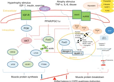
Animal models have identified key mediators in atro-phy and hypertroatro-phy signaling and therefore the control of muscle growth.92 These are summarized in Figure 2. The
anabolic hormone and growth factor IGF-1 stimulates the phosphoinositide 3-kinase/Akt (protein kinase B) pathway in skeletal muscle cells. Upon phosphorylation, Akt is able
Extracellular
Intracellular
AKT PI3K IGF-R
Hypertrophy stimulus Atrophy stimulus TNF-α, IL-6, disuse
PPAR/PGC1α
Ca2+
IGF-1, insulin, exercise
GSK3β
elF2B 4E-BP1 P70S6k
Muscle protein synthesis Muscle protein breakdown
Shifted balance in COPD quadriceps dysfunction
Proteosome
NFκB
MuRF-1 Nucleus FoxO
FoxO
?
Atrogin-1 Amino acids
Actomyosin Caspase 3
Calpains p38 or ERK1/2
Smad 7
Myostatin
Propeptide
Follistatin GASP-1
Nuclear
Gene targets include MyoD, myf5 + myogenin complex translocation of smad
FLRG
Alk4/5 ActRIIB
Smad 4 Smad
2/3
MAP
mTOR
Figure 2 Summary of pathways controlling muscle protein synthesis (MPS) and muscle protein breakdown (MPB). The role of myostatin in MPS and MPB has also been included. Myostatin is held in an inactive state by its pro-peptide, follistatin, and inhibitory binding proteins – growth and differentiation factor-associated serum protein-1 (GASP-1) and follistatin-like related gene, (FLRG) as shown. Upon activation, it binds to its transmembrane receptor activin receptor type IIB (ActRIIB), which then forms homodimers with activin receptor-like kinase 4 or 5 (Alk 4/5). The SMAD signaling pathway is then activated and translocation of this transcription factor complex to the nucleus occurs, where MyoD production and therefore myoblast proliferation and fusion are blocked. Myostatin is also proposed to increase proteosomal activity in a FoxO-dependent manner. Activation of MAP kinase is mediated via myostatin either via p38 or ERK1/2, which leads to the blocking of genes involved in myogenesis.
Notes:→ denotes stimulation; ⊣ indicates inhibition.
Abbreviations: 4E-BP1, eukaryotic translation initiation factor 4E binding protein-1; Akt, protein kinase B; ERK, extracellular signal-regulated kinase; eIF2B, eukaryotic initiation factor 2B; FoxO, forkhead box class O; GSK-3β, glycogen synthase kinase-3β; IGF-1, insulin-like growth factor-1; IL-6, interleukin-6; MAP, mitogen-activated protein; mTOR, mammalian target of rapamycin; MuRF, muscle-specific RING finger protein; p70S6k, 70-kD ribosomal S6 protein kinase; PGC1α, peroxisome proliferator-activated receptor gamma co-activator 1-alpha; PI3K, phosphoinositide 3-kinase; PPAR, peroxisome proliferator-activated receptor; SMAD, ; TNF-α, tumor necrosis factor-alpha; GASP-1, growth and differentiation factor-associated serum protein-1; IGF-R, insulin-like growth factor-1 receptor; Ca2+, calcium ion; NFκB, nuclear factor κB.
Dovepress Muscle function in COPD: a complex interplay
International Journal of Chronic Obstructive Pulmonary Disease downloaded from https://www.dovepress.com/ by 118.70.13.36 on 22-Aug-2020
to activate mammalian target of rapamycin (mTOR), which, in turn, activates 70-kD ribosomal S6 protein kinase93 and
inhibits eukaryotic translation initiation factor 4E bind-ing protein-1/ PHAS-1;94 these processes stimulate MPS.
Another downstream regulator of anabolism, which is mTOR independent, is the phosphorylation of glycogen synthase kinase-3β (GSK-3β) by phosphorylated Akt.95 This leads
to the release of eukaryotic initiation factor 2B (eIF2B) which upregulates MPS. Phosphorylated Akt also plays a role in MPB pathways. It downregulates two muscle-specific E3 ligases, atrogin-1 (muscle atrophy F-box or MAFbx), and muscle-specific RING finger protein (MuRF)-1 via inactivation of the forkhead box class O (FoxO) family of transcription factors.96 The muscle-specific ubiquitin
ligases contribute to protein degradation via the ubiquitin-proteasome pathway.97
Ubiquitin-mediated protein degradation may play an important role in COPD skeletal muscle dysfunction. Two small studies found elevated atrogin-198,99 and MuRF-198 in
quadriceps from COPD patients, but, in one of these, it was alongside elevated hypertrophy signaling. Although larger studies are required to confirm these observations, this may suggest COPD patients actually fail to restore muscle mass and therefore have some form of synthetic resistance. Indeed, tracer studies from immobilized individuals would support this theory of resistance to MPS to protein nutrition (termed “anabolic resistance”).100 MPS is greatly suppressed in the
immobilized post-absorptive state,101 while the contribution
from MPB is minimal. Therefore, the relative contribution of MPB in the process of disuse-induced muscle atrophy remains under question.102
Myostatin, or growth differentiation factor-8, is a member of the transforming growth factor-β (TGF-β) super-family and is a potent negative regulator of muscle mass, as demon-strated by naturally occurring mutations occurring in mice, cattle, and humans.103–105 Myostatin can influence muscle
wasting by affecting the number and size of muscle cells and by inducing muscle atrophy pathways. Myostatin upregulates p21 (cyclin-dependent kinase inhibitor), which negatively affects activated satellite cell/myoblast proliferation,106 and
downregulates myogenic differentiation factors (MyoD, myf5, and myogenin), which inhibit myoblast differentiation.107 It
can also exert an effect on muscle catabolism by activating the ubiquitin proteolytic system in a FoxO1-dependent manner108
and may possibly inactivate Akt, affecting MPS.108 Finally,
myostatin has also been shown to inhibit satellite cell activa-tion and self-renewal in a Pax7-dependent way.109
Several lines of evidence implicate a role for myostatin in COPD quadriceps dysfunction. Myostatin mRNA was elevated in weak COPD patients.99 Following an in-patient
resistance training program, hospitalized COPD patients had reduced myostatin transcripts, with a trend toward an increase in MyoD and myogenin.59 Following exercise training,
non-cachectic COPD patients demonstrated a reduction in myostatin protein, with a reduction in MuRF-1 and atrogin-1,110 while a modest reduction in myostatin was found
following resistance training with or without testosterone.111
Our group have also demonstrated a negative association between quadriceps muscle myostatin mRNA expression and quadriceps muscle strength in COPD patients.112
Animal studies have shown that skeletal muscle-fiber phenotype appears to be regulated by several independent signaling pathways. Fiber type switching in mice can be induced by changes in nerve activity from differing electrical stimulations,113 enabling the study of pathways that affect
myosin gene expression and metabolic profiles.
Myofiber gene activation occurs via calcium signaling through calcineurin (Cn) and various kinases – for example, Ca2+/calmodulin-dependent protein kinases II. Cn is a
calcium/calmodulin-regulated protein phosphatase that acts on transcription factors of the nuclear factor of activated T cells (NFAT) family. Ca2+/calmodulin-dependent protein
kinases II regulate myocyte enhancer factor 2, via histone deacetylase (HDAC), and has been suggested to interact with NFAT.114 Studies of transgenic mice115 and those treated
with Cn inhibitor116 have implicated Cn signaling in
activity-dependent maintenance of the slow gene program.113
Cn-NFAT signaling may also upregulate the transcription factor peroxisome proliferator-activated receptor (PPAR)-β/-γ
and the transcriptional co-activator PPAR-γ (PGC-1α),117 both
of which have been implicated in the muscle dysfunction of COPD patients. PPAR signaling can affect oxidative sig-naling and fiber type composition,118 in addition to having
inflammatory properties via effects on the nuclear factor kappa-light-chain-enhancer of activated B cells pathway.119
The three isoforms of PPARS (α, β/δ, and γ) are all present in skeletal muscle. PPAR-δ regulates fatty acid utilization and energy homeostasis120 and higher levels are expressed in
type I muscle fibers121 and can be induced by acute exercise in
healthy young men.122 A transgenic “marathon mouse” model,
in which PPAR-δ expression is increased, shows fiber shift opposite to that seen in COPD.121 PGC-1α is a co-activator of
PPAR-δ that can interact with transcription factors and basal transcriptional machinery123 and can stimulate mitochondrial
Dovepress Donaldson et al
International Journal of Chronic Obstructive Pulmonary Disease downloaded from https://www.dovepress.com/ by 118.70.13.36 on 22-Aug-2020
and oxidative enzymes.113 In one small study of 14 COPD
patients and nine control subjects, PPAR-α and -δ protein levels and PGC-1α mRNA were significantly lower in the quadriceps of moderate/severe COPD patients than in con-trols with a similar smoking history124 and PPAR-α mRNA
expression was lower still in cachectic patients. These findings suggest that PPAR-γ or -α content and/or function may in some way be involved in the change in oxidative gene program and mitochondrial dysfunction.125 However, as type I fibers
have higher expression of PPARs, these results may simply represent an association rather causation.
The mitogen-activated protein kinase (MAPK) pathway may have an influence on fiber shift via extracellular signal-regulated kinase (ERK) signaling. In muscle cell lines, a type I phenotype is induced when the ERK pathway is inhibited and a shift toward type I/IIa from IIx myosin heavy chain (MHC) results from MAPK phosphatase-1.126 Recent
data from COPD patients have been conflicting. Lemire and colleagues showed elevated ratios of phosphorylated to total level of p38 MAPK and ERK 1/2 in the quadriceps muscle compared with controls.127 These ratios were negatively
associated with mid-thigh muscle CSA, supporting the hypothesis that MAPK may contribute to the development of skeletal muscle dysfunction in COPD.127 In contrast, data
from a much larger cross-sectional study failed to show a role for p38 MAPK signaling.128
Emerging data suggest that microRNAs (miRNAs), small polynucleotides that can decrease mRNA translation or directly destabilize mRNA, may also be implicated in the control of skeletal muscle phenotype.129 Muscle-specific
miRNAs include those which affect myocyte proliferation and differentiation, for example, miR-1 and miR-206,130 and
others, such as miR-208b and miR-499,130,131 that modulate
the expression of slow MHC genes through regulation of tran-scriptional repressors.132,133 In COPD, we have recently shown
that the miRNA profile of the quadriceps muscle in COPD patients differs from that of controls, with a downregulation in the myocardin-related transcription-serum response factor axis and reduced expression of muscle-specific miRNAs, particularly miR-1.134 Reduction in miR-1 has been reported
in other models of inactivity resulting from denervation, nerve entrapment, or space flight and targets include myostatin135
and IGF-1.136 We found IGF-1 was elevated in the COPD
group consistent with previous reports of the overexpres-sion of muscle hypertrophy pathways.98 MiR-1 may also
contribute toward reduction in MHC I and fiber shift, via an increase of HDAC4. HDAC4 inhibits serum response
factor, an important regulator of MHC1 expression, and the expression of follistatin,137 which may activate the myostatin
pathway.
Non-pharmacological treatments
for muscle dysfunction in COPD
Exercise training remains the only known intervention to reverse some of the underlying skeletal muscle abnor-malities seen in COPD, further supporting the notion that reduced daily physical activity is the major etiological factor. Exercise training, in the form of pulmonary rehabilitation (PR), has emerged as the most effective non-pharmacological intervention in improving exercise capacity, dyspnea, and health status in COPD patients, as evidenced by numerous randomized controlled trials and meta-analyses.138 Given
that PR does not directly improve lung mechanics or gas exchange,139 it is likely that the main area of improvement
with exercise lies in the skeletal muscle. Dysfunction of the locomotor muscles may limit exercise performance because of leg discomfort,13 but also because early anaerobic
metabo-lism leads to lactic acid production. Lactic acid, buffered by bicarbonate, causes production of carbon dioxide and an increased ventilatory stimulus. As expiratory flow limitation is commonly present in COPD, increased ventilation can exacerbate dynamic hyperinflation and promote premature exercise termination and dyspnea.140
Quadriceps strength, endurance, and fatigability all improve significantly following exercise training.58,141,142 Even in the
acute setting, resistance training during an exacerbation can prevent muscle function deterioration,59 while PR shortly
fol-lowing hospital discharge can significantly accelerate recovery of quadriceps muscle strength.58 Debate remains as to the most
effective mode of exercise to induce not only different skeletal muscle adaptations but also long-term improvements in clini-cally relevant health outcomes. Typiclini-cally, chronic endurance training enhances the fatigue resistance of skeletal muscle by promoting a muscle-fiber type shift from fast-twitch fatigable type II fibers to slow-twitch fatigue-resistant type I fibers, increasing mitochondrial content and activity and improving skeletal muscle glucose transportation. However, resistance training reduces sarcopenia and promotes hypertrophy of muscle fibers, especially of type IIx.143
Intensity of exercise training is an important determinant of the physiological training effect.144 However, in patients
with severe COPD, intolerable sensations of breathlessness may prevent sufficiently long periods of high-intensity train-ing levels.145 Strategies to augment exercise tolerance by
Dovepress Muscle function in COPD: a complex interplay
International Journal of Chronic Obstructive Pulmonary Disease downloaded from https://www.dovepress.com/ by 118.70.13.36 on 22-Aug-2020
reducing dyspnea sensation or ventilatory limitation have included noninvasive mechanical ventilation,146 oxygen,147
and/or heliox supplementation,148 all of which have been
demonstrated to increase exercise tolerance in the laboratory setting. However, these are rarely systematically used as part of clinical PR programs. An alternative approach, which may be particularly suitable for patients with more severe COPD, is interval training, which allows patients to complete short periods of high-intensity exercise not possible with classical aerobic exercise training.149
Although the emphasis has so far been on the muscles of the lower limbs, there have been studies examining the effects of training the upper limbs or the respiratory muscles in COPD. A systematic review of upper-limb exercise-training studies in COPD showed improvements in arm exercise capacity, but the effects on symptoms, overall exercise capacity, and health-related quality of life were inconsistent.150 Similarly, debate continues with regard to
the role of inspiratory muscle training in the context of PR. Although most studies have demonstrated a positive effect on voluntary inspiratory muscle strength,151 it remains
unclear whether this is as a result of a genuine physiological improvement in the inspiratory muscles or a learning effect in performing the voluntary maneuver. Furthermore, the added benefit of inspiratory muscle training over a general exercise-training program seems relatively limited.151
In patients unable or unwilling to adhere to existing forms of exercise, neuromuscular electrical stimulation (NMES) may offer an alternative way of enhancing leg muscle strength.152
NMES uses a battery-powered stimulator unit to produce a con-trolled contraction of the muscles via skin electrodes. A typical program consists of 30–60 minutes of quadriceps stimulation, 3–5 times weekly for 4–6 weeks. NMES can lead to improve-ments in muscle strength and exercise performance, with pooled data revealing mean between-group differences in peak quadriceps torque and 6-minute walking distance of 9.7 Nm (95% CI 1.2, 18.1) and 48 m (95% CI 9, 86), respectively.153
Recent studies have also demonstrated favorable changes in markers of anabolism/catabolism154 and the quadriceps fiber
type profile following NMES.155 However, studies remain
small, follow-up data are lacking, and the patient phenotypes most likely to benefit have yet to be identified.
Pharmacological treatments
for muscle dysfunction in COPD
Despite the many benefits of PR, there are limitations. Firstly, exercise training does not fully reverse all of the abnormalities observed in the quadriceps muscle. Secondly, a proportion of
patients either has limited accessibility to PR or has issues with uptake and completion. Thirdly, improvements following PR decline toward baseline level within 12–18 months.156
Hence, there is interest in pharmacologically augmenting (or even replacing) exercise training to bring about structural and functional improvements in the skeletal muscles. However, as well as the technical problems involved in creating a drug that specifically benefits the muscles, there are regulatory hurdles to be overcome before such a compound can reach the market.157
As previously discussed, systemic inflammation, oxida-tive stress, and anabolic/catabolic hormone imbalance have been postulated as etiological factors for muscle dysfunc-tion in COPD. An early trial of infliximab, an anti-TNF-α
therapy, found that it was largely ineffective in improving lung function, exercise capacity, or health-related quality of life,158 although there did appear to be a trend for benefit in
cachectic patients. Furthermore, antioxidant therapy with N-acetylcysteine led to a 25% increase in quadriceps endur-ance compared with placebo.70 However, this was a very small
study of nine COPD patients in a controlled laboratory set-ting. Studies of anabolic hormones have had mixed results. Anabolic steroids increase body weight and FFM in COPD, either alone159 or in conjunction with exercise training,87 but
not muscle strength or exercise capacity. However, the addi-tion of testosterone to resistance training in hypogonadal COPD patients promotes anabolic pathways that can result in improved quadriceps strength and endurance.88 Recombinant
growth hormone (GH) improves FFM compared with placebo but does not improve muscle strength or exercise capacity.86
Ghrelin is a novel GH-releasing peptide that induces a posi-tive energy balance by decreasing fat utility and stimulating feeding through GH-independent mechanisms. In a small open-label study, ghrelin increased FFM, muscle strength, and 6-minute walk distance in cachectic COPD patients.160
Systemic side effects with hormonal drugs are a concern, hence there is current interest in the development of ana-bolic drugs without the unwanted side effects. An example is the selective androgen-receptor modulator class of drugs that have the benefits of anabolic/androgenic steroids with a hypothetically reduced risk of prostate cancer in men and virilizing effects in women.161
Another therapeutic approach is to use existing drugs for new indications. As previously discussed, common variations in the gene for the vitamin D receptor81 and deletion of the
allele of the ACE79 have been demonstrated to influence
mus-cle strength. Vitamin D supplementation or the administration of ACE inhibitors may have a future role in treating muscle
Dovepress Donaldson et al
International Journal of Chronic Obstructive Pulmonary Disease downloaded from https://www.dovepress.com/ by 118.70.13.36 on 22-Aug-2020
dysfunction in COPD, or at least augmenting the benefits of exercise training. Certainly, this approach has been used with positive results in elderly people with functional impair-ment.162 Similarly, levosimendan, a calcium sensitizer used
as a cardiac inotrope, has recently been shown to improve neuro-mechanical efficiency and contractile function of the human diaphragm in healthy subjects,163 and conceivably
it may also improve skeletal muscle dysfunction in COPD. However, with increasing understanding of the underlying molecular mechanisms, there is also hope that a number of novel pharmacological agents that address cachexia and skeletal muscle dysfunction in COPD will become available for clinical use. Already, prototype inhibitors of ubiquitin ligases and neutralizing antibodies to myostatin have been developed for cancer-related cachexia and muscle dystrophies.164 PPAR-δ agonists have recently been shown
to mimic and enhance exercise training and AICAR, an 5′
adenosine monophosphate-activated-kinase agonist, was sufficient to improve exercise endurance in mice alone.165
Thus, there is much future promise that novel therapeutic agents will become available to address important extra-pulmonary manifestations of COPD such as skeletal muscle dysfunction.
Future directions
A greater understanding of the etiology and basic mecha-nisms of skeletal muscle dysfunction should continue to underpin developments in the field, informing the iden-tification and testing of new pharmacological agents and strategies to augment or optimize PR across community and in-patient settings. Phenotyping of patients according to skeletal muscle dysfunction will also enhance the identifica-tion of those most likely to response to specific treatments, thus should be embraced in clinical trial design and practice, where possible.
Acknowledgments
This work is funded in part by the National Institute for Health Research (NIHR) Respiratory Biomedical Research Unit, Royal Brompton and Harefield Foundation Trust and Imperial College. AVD and MIP are either fully or partly sup-ported by the Biomedical Research Unit. MM is funded by an NIHR postdoctoral fellowship. WD-CM is supported by an NIHR Clinician Scientist Award and a Medical Research Council New Investigator Award. The views expressed in this publication are those of the authors and not necessarily those of the National Health Service, the NIHR, nor the Department of Health.
Disclosure
The authors declare no conflicts of interest in this work.
References
1. Lopez AD, Murray CC. The global burden of disease, 1990–2020. Nat
Med. 1998;4(11):1241–1243.
2. Man WD, Kemp P, Moxham J, Polkey MI. Skeletal muscle dysfunction
in COPD: clinical and laboratory observations. Clin Sci (Lond). 2009;
117(7):251–264.
3. Gosselink R, Troosters T, Decramer M. Peripheral muscle weakness contributes to exercise limitation in COPD. Am J Respir Crit Care Med. 1996;153(3):976–980.
4. Simpson K, Killian K, McCartney N, Stubbing DG, Jones NL. Randomised controlled trial of weightlifting exercise in patients with
chronic airflow limitation. Thorax. 1992;47(2):70–75.
5. Decramer M, Gosselink R, Troosters T, Verschueren M, Evers G. Muscle weakness is related to utilization of health care resources in COPD
patients. Eur Respir J. 1997;10(2):417–423.
6. Swallow EB, Reyes D, Hopkinson NS, et al. Quadriceps strength pre-dicts mortality in patients with moderate to severe chronic obstructive
pulmonary disease. Thorax. 2007;62(2):115–120.
7. Man W, Mustfa N, Nikoletou D, et al. Effect of salmeterol on respira-tory muscle activity during exercise in poorly reversible COPD. Thorax. 2004;59(6):471–476.
8. Gray-Donald K, Gibbons L, Shapiro SH, Macklem PT, Martin JG. Nutritional status and mortality in chronic obstructive pulmonary
disease. Am J Respir Crit Care Med. 1996;153(3):961–966.
9. Bernard S, LeBlanc P, Whittom F, et al. Peripheral muscle weakness in
patients with chronic obstructive pulmonary disease. Am J Respir Crit
Care Med. 1998;158(2):629–634.
10. Man WD, Hopkinson NS, Harraf F, Nikoletou D, Polkey MI, Moxham J. Abdominal muscle and quadriceps strength in chronic
obstructive pulmonary disease. Thorax. 2005;60(9):718–722.
11. Man W, Soliman M, Nikoletou D, et al. Non-volitional assessment of skeletal muscle strength in patients with chronic obstructive pulmonary
disease. Thorax. 2003;58(8):665–669.
12. Allaire J, Maltais F, Doyon JF, et al. Peripheral muscle endurance and the oxidative profile of the quadriceps in patients with COPD. Thorax. 2004;59(8):673–678.
13. Man WD, Soliman MG, Gearing J, et al. Symptoms and quadriceps fatigability after walking and cycling in chronic obstructive pulmonary
disease. Am J Respir Crit Care Med. 2003;168(5):562–567.
14. Mador MJ, Deniz O, Aggarwal A, Kufel TJ. Quadriceps fatigability after single muscle exercise in patients with chronic obstructive
pulmonary disease. Am J Respir Crit Care Med. 2003;168(1):102–108.
15. Coronell C, Orozco-Levi M, Mendez R, Ramirez-Sarmiento A, Gáldiz JB, Gea J. Relevance of assessing quadriceps endurance in patients with
COPD. Eur Respir J. 2004;24(1):129–136.
16. Mathur S, Takai KP, Macintyre DL, Reid D. Estimation of thigh muscle mass with magnetic resonance imaging in older adults and people with
chronic obstructive pulmonary disease. Phys Ther. 2008;88(2):219–230.
17. Seymour JM, Ward K, Sidhu PS, et al. Ultrasound measurement of rectus femoris cross-sectional area and the relationship with quadriceps
strength in COPD. Thorax. 2009;64(5):418–423.
18. Marquis K, Debigaré R, Lacasse Y, et al. Midthigh muscle cross-sectional area is a better predictor of mortality than body mass index
in patients with chronic obstructive pulmonary disease. Am J Respir
Crit Care Med. 2002;166(6):809–813.
19. Gosker HR, Engelen MP, van Mameren H, et al. Muscle fiber type IIX atrophy is involved in the loss of fat-free mass in chronic obstructive
pulmonary disease. Am J Clin Nutr. 2002;76(1):113–119.
20. Gosker HR, Zeegers MP, Wouters EF, Schols AM. Muscle fibre type shifting in the vastus lateralis of patients with COPD is associated
with disease severity: a systematic review and meta-analysis. Thorax.
2007;62(11):944–949.
Dovepress Muscle function in COPD: a complex interplay
International Journal of Chronic Obstructive Pulmonary Disease downloaded from https://www.dovepress.com/ by 118.70.13.36 on 22-Aug-2020
21. Whittom F, Jobin J, Simard PM, et al. Histochemical and mor-phological characteristics of the vastus lateralis muscle in patients with chronic obstructive pulmonary disease. Med Sci Sports Exerc. 1998;30(10):1467–1474.
22. Jobin J, Maltais F, Doyon JF, et al. Chronic obstructive pulmonary disease: capillarity and fiber type characteristics of skeletal muscle.
J Cardiopulm Rehabil. 1998;18(6):432–437.
23. Maltais F, Simard AA, Simard C, Jobin J, Desgagnés P, LeBlanc P. Oxidative capacity of the skeletal muscle and lactic acid kinetics during
exercise in normal subjects and in patients with COPD. Am J Respir
Crit Care Med. 1996;153(1):288–293.
24. Jakobsson P, Jorfeldt L, Henriksson J. Metabolic enzyme activity in the quadriceps femoris muscle in patients with severe chronic obstructive pulmonary disease. Am J Respir Crit Care Med. 1995;151(2 Pt 1): 374–377.
25. Kutsuzawa T, Shioya S, Kurita D, Haida M, Ohta Y, Yamabayashi H. Muscle energy metabolism and nutritional status in patients with chronic
obstructive pulmonary disease. A 31P magnetic resonance study. Am J
Respir Crit Care Med. 1995;152(2):647–652.
26. Maltais F, Jobin J, Sullivan MJ, et al. Metabolic and hemodynamic
responses of lower limb during exercise in patients with COPD. J Appl
Physiol. 1998;84(5):1573–1580.
27. Swallow EB, Gosker HR, Ward KA, et al. A novel technique for
nonvo-litional assessment of quadriceps muscle endurance in humans. J Appl
Physiol. 2007;103(3):739–746.
28. Newell SZ, McKenzie DK, Gandevia SC. Inspiratory and skeletal muscle strength and endurance and diaphragmatic activation in patients
with chronic airflow limitation. Thorax. 1989;44(11):903–912.
29. Gea JG, Pasto M, Carmona MA, Orozco-Levi M, Palomeque J, Broquetas J. Metabolic characteristics of the deltoid muscle in patients with chronic obstructive pulmonary disease. Eur Respir J. 2001;17(5):939–945.
30. Sato Y, Asoh T, Honda Y, Fujimatsu Y, Higuchi I, Oizumi K. Morphologic and histochemical evaluation of muscle in patients with chronic pulmonary emphysema manifesting generalized emaciation.
Eur Neurol. 1997;37(2):116–121.
31. Similowski T, Yan S, Gauthier AP, Macklem PT, Bellemare F. Contractile
properties of the human diaphragm during chronic hyperinflation. N
Engl J Med. 1991;325(13):917–923.
32. Levine S, Kaiser L, Leferovich J, Tikunov B. Cellular adaptations in the diaphragm in chronic obstructive pulmonary disease. N Engl J Med. 1997;337(25):1799–1806.
33. Mercadier JJ, Schwartz K, Schiaffino S, et al. Myosin heavy chain gene expression changes in the diaphragm of patients with chronic lung
hyperinflation. Am J Physiol. 1998;274(4 Pt 1):L527–L534.
34. Doucet M, Debigaré R, Joanisse DR, et al. Adaptation of the diaphragm and the vastus lateralis in mild-to-moderate COPD. Eur Respir J. 2004;24(6):971–979.
35. Ottenheijm CA, Heunks LM, Sieck GC, et al. Diaphragm dysfunction
in chronic obstructive pulmonary disease. Am J Respir Crit Care Med.
2005;172(2):200–205.
36. Stubbings AK, Moore AJ, Dusmet M, et al. Physiological properties of human diaphragm muscle fibres and the effect of chronic obstructive
pulmonary disease. J Physiol. 2008;586(Pt 10):2637–2650.
37. Levine S, Nguyen T, Kaiser LR, et al. Human diaphragm remodeling associated with chronic obstructive pulmonary disease: clinical
implica-tions. Am J Respir Crit Care Med. 2003;168(6):706–713.
38. Orozco-Levi M, Gea J, Lloreta JL, et al. Subcellular adaptation of
the human diaphragm in chronic obstructive pulmonary disease. Eur
Respir J. 1999;13(2):371–378.
39. Levine S, Gregory C, Nguyen T, et al. Bioenergetic adaptation of
individual human diaphragmatic myofibers to severe COPD. J Appl
Physiol. 2002;92(3):1205–1213.
40. Polkey MI, Kyroussis D, Hamnegard CH, et al. Diaphragm perfor-mance during maximal voluntary ventilation in chronic obstruc-tive pulmonary disease. Am J Respir Crit Care Med. 1997;155(2): 642–648.
41. Polkey MI, Kyroussis D, Keilty SE, et al. Exhaustive treadmill exercise does not reduce twitch transdiaphragmatic pressure in
patients with COPD. Am J Respir Crit Care Med. 1995;152(3):
959–964.
42. Levine S, Nguyen T, Friscia M, et al. Parasternal intercostal muscle
remodeling in severe chronic obstructive pulmonary disease. J Appl
Physiol. 2006;101(5):1297–1302.
43. Gea J, Orozco-Levi M, Aguar MC, et al. Adaptive changes concerning the types of fibres and isoforms of myosin in the external intercostal
muscle of COPD patients. Eur Respir J. 1996;9:160S.
44. Seymour JM, Spruit MA, Hopkinson NS, et al. The prevalence of quad-riceps weakness in COPD and the relationship with disease severity.
Eur Respir J. 2010;36(1):81–88.
45. Serres I, Gautier V, Varray A, Préfaut C. Impaired skeletal muscle endurance related to physical inactivity and altered lung function in
COPD patients. Chest. 1998;113(4):900–905.
46. Gosker HR, Kubat B, Schaart G, van der Vusse GJ, Wouters EF, Schols AM. Myopathological features in skeletal muscle of patients
with chronic obstructive pulmonary disease. Eur Respir J. 2003;22(2):
280–285.
47. Degens H, Sanchez Horneros JM, Heijdra YF, Dekhuijzen PN, Hopman MT. Skeletal muscle contractility is preserved in COPD patients with normal fat-free mass. Acta Physiol Scand. 2005;184(3): 235–242.
48. Ottenheijm CA, Heunks LM, Dekhuijzen RP. Diaphragm adaptations
in patients with COPD. Respir Res. 2008;9(1):12.
49. van den Borst B, Koster A, Yu B, et al. Is age-related decline in lean mass and physical function accelerated by obstructive lung disease or
smoking? Thorax. 2011;66(11):961–969.
50. Watz H, Waschki B, Meyer T, Magnussen H. Physical activity in patients
with COPD. Eur Respir J. 2009;33(2):262–272.
51. Kon SS, Man WD. Muscle mass and strength in obstructive lung disease:
a smoking gun? Thorax. 2011;66(11):933–935.
52. Polkey MI, Moxham J. Attacking the disease spiral in chronic
obstruc-tive pulmonary disease. Clin Med. 2006;6(2):190–196.
53. Bossenbroek L, de Greef MH, Wempe JB, Krijnen WP, Ten Hacken NH. Daily physical activity in patients with chronic obstructive pulmonary
disease: a systematic review. COPD. 2011;8(4):306–319.
54. Decramer M, Rennard S, Troosters T, et al. COPD as a lung disease with systemic consequences – clinical impact, mechanisms, and potential
for early intervention. COPD. 2008;5(4):235–256.
55. Pitta F, Troosters T, Spruit MA, Probst VS, Decramer M, Gosselink R. Characteristics of physical activities in daily life in chronic obstruc-tive pulmonary disease. Am J Respir Crit Care Med. 2005;171(9): 972–977.
56. Gosker HR, Lencer NH, Franssen FM, van der Vusse GJ, Wouters EF, Schols AM. Striking similarities in systemic factors contributing to decreased exercise capacity in patients with severe chronic heart failure
or COPD. Chest. 2003;123(5):1416–1424.
57. Spruit M, Gosselink R, Troosters T, et al. Muscle force during an acute exacerbation in hospitalised patients with COPD and its relationship
with CXCL8 and IGF-I. Thorax. 2003;58(9):752–756.
58. Seymour JM, Moore L, Jolley CJ, et al. Outpatient pulmonary
reha-bilitation following acute exacerbations of COPD. Thorax. 2010;65(5):
423–428.
59. Troosters T, Probst VS, Crul T, et al. Resistance training prevents dete-rioration in quadriceps muscle function during acute exacerbations of
chronic obstructive pulmonary disease. Am J Respir Crit Care Med.
2010;181(10):1072–1077.
60. Garcia-Aymerich J, Farrero E, Félez MA, Izquierdo J, Marrades RM, Antó JM; Estudi del Factors de Risc d’Agudització de la MPOC investi-gators. Risk factors of readmission to hospital for a COPD exacerbation:
a prospective study. Thorax. 2003;58(2):100–105.
61. Garcia-Aymerich J, Lange P, Benet M, Schnohr P, Anto JM. Regular physical activity reduces hospital admission and mortality in chronic obstructive pulmonary disease: a population based cohort study. Thorax. 2006;61(9):772–778.
Dovepress Donaldson et al
International Journal of Chronic Obstructive Pulmonary Disease downloaded from https://www.dovepress.com/ by 118.70.13.36 on 22-Aug-2020
62. Garcia-Aymerich J, Lange P, Benet M, Schnohr P, Antó JM. Regular physical activity modifies smoking-related lung function decline and reduces risk of chronic obstructive pulmonary disease: a
population-based cohort study. Am J Respir Crit Care Med. 2007;175(5):
458–463.
63. Yende S, Waterer GW, Tolley EA, et al. Inflammatory markers are associated with ventilatory limitation and muscle dysfunction in obstructive lung disease in well functioning elderly subjects. Thorax. 2006;61(1):10–16.
64. Debigaré R, Marquis K, Côté CH, et al. Catabolic/anabolic balance and
muscle wasting in patients with COPD. Chest. 2003;124(1):83–89.
65. Eid AA, Ionescu AA, Nixon LS, et al. Inflammatory response and body
composition in chronic obstructive pulmonary disease. Am J Respir Crit
Care Med. 2001;164(8 Pt 1):1414–1418.
66. Wagner PD. Possible mechanisms underlying the development of
cachexia in COPD. Eur Respir J. 2008;31(3):492–501.
67. Barreiro E, Schols AM, Polkey MI, et al; ENIGMA in COPD project. Cytokine profile in quadriceps muscles of patients with severe COPD.
Thorax. 2008;63(2):100–107.
68. Petersen AM, Penkowa M, Iversen M, et al. Elevated levels of IL-18 in plasma and skeletal muscle in chronic obstructive pulmonary disease.
Lung. 2007;185(3):161–171.
69. Crul T, Spruit MA, Gayan-Ramirez G, et al. Markers of inflammation and disuse in vastus lateralis of chronic obstructive pulmonary disease
patients. Eur J Clin Invest. 2007;37(11):897–904.
70. Koechlin C, Couillard A, Simar D, et al. Does oxidative stress alter
quadriceps endurance in chronic obstructive pulmonary disease? Am J
Respir Crit Care Med. 2004;169(9):1022–1027.
71. Reid MB. Nitric oxide, reactive oxygen species, and skeletal muscle
contraction. Med Sci Sports Exerc. 2001;33(3):371–376.
72. Barreiro E, de la Puente B, Minguella J, et al. Oxidative stress and respiratory muscle dysfunction in severe chronic obstructive pulmonary
disease. Am J Respir Crit Care Med. 2005;171(10):1116–1124.
73. Supinski GS, Callahan LA. Free radical-mediated skeletal muscle
dysfunction in inflammatory conditions. J Appl Physiol. 2007;102(5):
2056–2063.
74. Barreiro E, Rabinovich R, Marin-Corral J, Barberà JA, Gea J, Roca J. Chronic endurance exercise induces quadriceps nitrosative stress in
patients with severe COPD. Thorax. 2009;64(1):13–19.
75. Gosker HR, Bast A, Haenen GR, et al. Altered antioxidant status in peripheral skeletal muscle of patients with COPD. Respir Med. 2005;99(1):118–125.
76. Couillard A, Maltais F, Saey D, et al. Exercise-induced quadriceps oxidative stress and peripheral muscle dysfunction in patients with
chronic obstructive pulmonary disease. Am J Respir Crit Care Med.
2003;167(12):1664–1669.
77. Barreiro E, Gea J, Corominas JM, Hussain SN. Nitric oxide synthases and protein oxidation in the quadriceps femoris of patients with chronic
obstructive pulmonary disease. Am J Respir Cell Mol Biol. 2003;29(6):
771–778.
78. Couillard A, Koechlin C, Cristol JP, Varray A, Prefaut C. Evidence of local exercise-induced systemic oxidative stress in chronic obstructive
pulmonary disease patients. Eur Respir J. 2002;20(5):1123–1129.
79. Hopkinson NS, Nickol AH, Payne J, et al. Angiotensin converting enzyme genotype and strength in chronic obstructive pulmonary disease.
Am J Respir Crit Care Med. 2004;170(4):395–399.
80. Hopkinson NS, Eleftheriou KI, Payne J, et al. +9/+9 Homozygosity of the bradykinin receptor gene polymorphism is associated with reduced fat-free mass in chronic obstructive pulmonary disease. Am J Clin Nutr. 2006;83(4):912–917.
81. Hopkinson NS, Li KW, Kehoe A, et al. Vitamin D receptor genotypes influence quadriceps strength in chronic obstructive pulmonary disease.
Am J Clin Nutr. 2008;87(2):385–390.
82. Broekhuizen R, Grimble RF, Howell WM, et al. Pulmonary cachexia, systemic inflammatory profile, and the interleukin 1beta-511 single nucleotide polymorphism. Am J Clin Nutr. 2005;82(5): 1059–1064.
83. Engelen MP, Schols AM, Baken WC, Wesseling GJ, Wouters EF. Nutritional depletion in relation to respiratory and peripheral skeletal
muscle function in out-patients with COPD. Eur Respir J. 1994;7(10):
1793–1797.
84. Ferreira IM, Brooks D, Lacasse Y, Goldstein RS. Nutritional support for
individuals with COPD: a meta-analysis. Chest. 2000;117(3):672–678.
85. Op den Kamp CM, Langen RC, Haegens A, Schols AM. Muscle
atrophy in cachexia: can dietary protein tip the balance? Curr Opin
Clin Nutr Metab Care. 2009;12(6):611–616.
86. Burdet L, de Muralt B, Schutz Y, Pichard C, Fitting JW. Administration of growth hormone to underweight patients with chronic obstructive
pulmonary disease. A prospective, randomized, controlled study. Am
J Respir Crit Care Med. 1997;156(6):1800–1806.
87. Creutzberg EC, Wouters EF, Mostert R, Pluymers RJ, Schols AM. A role for anabolic steroids in the rehabilitation of patients with
COPD? A double-blind, placebo-controlled, randomized trial. Chest.
2003;124(5):1733–1742.
88. Casaburi R, Bhasin S, Cosentino L, et al. Effects of testosterone and resistance training in men with chronic obstructive pulmonary disease.
Am J Respir Crit Care Med. 2004;170(8):870–878.
89. Schols AM. Pulmonary cachexia. Int J Cardiol. 2002;85(1):101–110.
90. Decramer M, Lacquet LM, Fagard R, Rogiers P. Corticosteroids
contribute to muscle weakness in chronic airflow obstruction. Am J
Respir Crit Care Med. 1994;150(1):11–16.
91. Hopkinson NS, Man WD, Dayer MJ, et al. Acute effect of oral steroids on muscle function in chronic obstructive pulmonary disease. Eur Respir J. 2004;24(1):137–142.
92. Nader GA. Molecular determinants of skeletal muscle mass:
getting the “AKT” together. Int J Biochem Cell Biol. 2005;37(10):
1985–1996.
93. Rommel C, Bodine SC, Clarke BA, et al. Mediation of IGF-1-induced skeletal myotube hypertrophy by PI(3)K/Akt/mTOR and PI(3)K/Akt/
GSK3 pathways. Nat Cell Biol. 2001;3(11):1009–1013.
94. Hara K, Yonezawa K, Kozlowski MT, et al. Regulation of eIF-4E
BP1 phosphorylation by mTOR. J Biol Chem. 1997;272(42):
26457–26463.
95. Cross DA, Alessi DR, Cohen P, Andjelkovich M, Hemmings BA. Inhibition of glycogen synthase kinase-3 by insulin mediated by protein
kinase B. Nature. 1995;378(6559):785–789.
96. Sandri M, Sandri C, Gilbert A, et al. Foxo transcription factors induce the atrophy-related ubiquitin ligase atrogin-1 and cause skeletal muscle
atrophy. Cell. 2004;117(3):399–412.
97. Bodine SC, Latres E, Baumhueter S, et al. Identification of ubiquitin ligases required for skeletal muscle atrophy. Science. 2001;294(5547): 1704–1708.
98. Doucet M, Russell AP, Léger B, et al. Muscle atrophy and hypertrophy
signaling in patients with chronic obstructive pulmonary disease. Am
J Respir Crit Care Med. 2007;176(3):261–269.
99. Plant PJ, Brooks D, Faughnan M, et al. Cellular markers of muscle
atrophy in chronic obstructive pulmonary disease. Am J Respir Cell
Mol Biol. 2010;42(4):461–471.
100. Rennie MJ. Anabolic resistance: the effects of aging, sexual
dimor-phism, and immobilization on human muscle protein turnover. Appl
Physiol Nutr Metab. 2009;34(3):377–381.
101. de Boer MD, Selby A, Atherton P, et al. The temporal responses of protein synthesis, gene expression and cell signalling in human quadriceps muscle and patellar tendon to disuse. J Physiol. 2007; 585(Pt 1):241–251.
102. Marimuthu K, Murton AJ, Greenhaff PL. Mechanisms regulating muscle mass during disuse atrophy and rehabilitation in humans.
J Appl Physiol. 2011;110(2):555–560.
103. McPherron AC, Lawler AM, Lee SJ. Regulation of skeletal muscle
mass in mice by a new TGF-beta superfamily member. Nature. 1997;
387(6628):83–90.
104. McPherron AC, Lee SJ. Double muscling in cattle due to
muta-tions in the myostatin gene. Proc Natl Acad Sci U S A. 1997;94(23):
12457–12461.
Dovepress Muscle function in COPD: a complex interplay
International Journal of Chronic Obstructive Pulmonary Disease downloaded from https://www.dovepress.com/ by 118.70.13.36 on 22-Aug-2020