R E S E A R C H
Open Access
Transcriptomic analysis comparing mouse
strains with extreme total lung capacities
identifies novel candidate genes for
pulmonary function
Leema George
1†, Ankita Mitra
1†, Tania A. Thimraj
1†, Martin Irmler
2†, Sangeetha Vishweswaraiah
1, Lars Lunding
3,
Dorothea Hühn
4,15, Alicia Madurga
5, Johannes Beckers
2,6,7, Heinz Fehrenbach
8, Swapna Upadhyay
9,10,
Holger Schulz
11,12†, George D. Leikauf
13†and Koustav Ganguly
1,9,10,14*†Abstract
Background:
Failure to attain peak lung function by early adulthood is a risk factor for chronic lung diseases. Previously,
we reported that C3H/HeJ mice have about twice total lung capacity (TLC) compared to JF1/MsJ mice. We identified
seven lung function quantitative trait loci (QTL: Lfnq1-Lfnq7) in backcross/intercross mice derived from these
inbred strains. We further demonstrated, superoxide dismutase 3, extracellular (
Sod3
),
Kit
oncogene (
Kit
) and
secreted phosphoprotein 1 (
Spp1
) located on these Lfnqs as lung function determinants. Emanating from the
concept of early origin of lung disease, we sought to identify novel candidate genes for pulmonary function
by investigating lung transcriptome in C3H/HeJ and JF1/MsJ mice at the completion of embryonic development, bulk
alveolar formation and maturity.
Methods:
Design-based stereological analysis was performed to study lung structure in C3H/HeJ and JF1/MsJ mice.
Microarray was used for lung transcriptomic analysis [embryonic day 18, postnatal days 28, 70]. Quantitative real time
polymerase chain reaction (qRT-PCR), western blot and immunohistochemical analysis were used to confirm selected
differences.
Results:
Stereological analysis revealed decreased alveolar number density, elastin to collagen ratio and increased
mean alveolar volume in C3H/HeJ mice compared to JF1/MsJ. Gene ontology term
“
extracellular region
”
was enriched
among the decreased JF1/MsJ transcripts. Candidate genes identified using the expression-QTL strategy include:
ATP-binding cassette, sub-family G (WHITE), member 1 (
Abcg1),
formyl peptide receptor 1 (
Fpr1),
gamma-aminobutyric acid
(GABA) B receptor, 1 (
Gabbr1)
; histocompatibility 2 genes: class II antigen E beta (
H2-Eb1
), D region locus 1 (
H2-D1
), and
Q region locus 4 (
H2-Q4
); leucine rich repeat containing 6 (testis)
(Lrrc6),
radial spoke head 1 homolog (
Rsph1),
and
surfactant associated 2 (
Sfta2).
Noteworthy genes selected as candidates for their consistent expression include: Wnt
inhibitor factor 1 (
Wif1
)
,
follistatin
(Fst),
chitinase-like 1 (
Chil1
), and
Chil3
.
Conclusions:
Comparison of late embryonic, adolescent and adult lung transcript profiles between mouse strains with
extreme TLCs lead to the identification of candidate genes for pulmonary function that has not been reported earlier.
Further mechanistic investigations are warranted to elucidate their mode of action in determining lung function.
Keywords:
Transcriptomics, Lung development, Chronic obstructive pulmonary disease, Asthma, WNT Signaling
* Correspondence:[email protected] †Equal contributors
1SRM Research Institute, SRM University, Chennai 603203, India
9Lung and Airway Research, Institute of Environmental Medicine, Karolinska
Institutet, Box 287, SE-171 77 Stockholm, Sweden
Full list of author information is available at the end of the article
Background
The origin of many chronic lung diseases may be rooted
early in life because of failure to achieve optimal peak
lung function by early adulthood [1
–
3]. Lung
dysfunc-tion may develop in utero and during early life
suggest-ing an underlysuggest-ing impairment of lung development,
growth and maturation [4]. For example, persistent
wheezing in the early years and lower lung function by
age 6 years among children is associated with later onset
of asthma. Similarly, following a cohort over 22 years,
Lange et al. [5] reported that low forced expiratory
vol-ume in 1 s (FEV1) before age 40 as a risk factor for
chronic obstructive pulmonary disease (COPD).
Dimin-ished lung function and hindered lung development is
likely due to multiple environmental (e.g., maternal
smoking) and genetic factors [6
–
18]. Thus, accumulating
evidence suggests that early life determinants of lung
function are possible determinants of obstructive airways
diseases later in life. Therefore, in this study we sought
to identify genetic determinants of lung function
devel-opment that could provide important insight into the
predisposition of chronic lung disease.
Evolving from the mouse phenome project, we
previ-ously assessed lung function in several inbred mouse
strains [19
–
21]. The greatest differences were noted in
C3H/HeJ as compared to JF1/MsJ mice. In male C3H/
HeJ mice, total lung capacity (TLC) and specific TLC
(TLC/body weight) = 1837
μ
l and 64
μ
l/g, respectively.
In male JF1/MsJ mice, TLC = 904
μ
l and specific
TLC = 40
μ
l/g. A similar difference was observed in
female mice (C3H/HeJ: TLC = 1443
μ
l and specific TLC
65
μ
l/g and JF1/MsJ: TLC = 874
μ
l and specific TLC
53
μ
l/g). Contrasting C3H/HeJ and JF1/MsJ back- and
inter-cross mice, lung function phenotyping was followed
by quantitative trait loci (QTL) analysis identifying lung
function QTLs (Lfnq1
–
Lfnq7) [20]. Consistent with the
early life impairment concept, JF1/MsJ mice were more
susceptible to lung injury following exposure to carbon
nanoparticle (CNP) and acrolein (a toxic component of
tobacco smoke) compared to C3H/HeJ [22
–
24].
Examining the mouse lung function QTL regions Lfnq1
and Lfnq2 located on mouse chromosome (mCh) 5 [20],
we previously demonstrated that superoxide dismutase 3
extracellular (
Sod3
), Kit oncogene (
Kit
) and secreted
phosphoprotein 1 (
Spp1
) as determinants of lung function
in mice [21, 25
–
27].
SOD3
variants were also associated
with declined lung function in children [25]. When
compared to strain-matched C57BL/6 J control mice,
gene-targeted
Sod3
(−/−)mice exhibited poor ventilation
efficiency [21];
Kit
mutant mice (
Kit
W-sh/
W-sh) exhibited
increased TLC, residual volume, lung compliance (CL),
and alveolar size [27]; and gene-targeted
Spp1
(−/−)mice
exhibited decreased TLC and increased specific CL
and
alveolar size [26].
In this study, we examined differences in lung
archi-tecture and lung transcripts in these highly divergent
mouse strains. Transcriptomic analysis was performed at
three strategic times covering completion of embryonic
lung development, bulk alveolar formation and growth.
Morphometric studies in the adult mouse lung were
per-formed to identify any structural alterations arising from
developmental differences between C3H/HeJ and JF1/
MsJ mice. Differential transcript expression was also
used to identify genes associated with TLC QTLs located
within Lfnq3 on mCh15 (40.3
–
72.5 Mbp) and Lfnq4 on
mCh17 (10.9
–
39.0 Mbp) associated with TLC located
within Lfnq3 on mCh15 (40
–
73 Mbp) and Lfnq4 on
mCh17 (10
–
39 Mbp).
Methods
Mice
All procedures were approved by the Bavarian Animal
Research Authority, Animal Research Authority of
Schleswig-Holstein, and University of Pittsburgh, PA.
Frozen and paraffin embedded tissues were procured to
carry out experiments at SRM University, India
accord-ing to the Institutional Animal Ethics Committee (IAEC)
permission. Mouse strains C3H/HeJ (#000659) and JF1/
MsJ (#003720) were purchased from Jackson laboratory
(Bar Harbor, ME, USA) and housed in specific pathogen
free conditions. Food and water were available ad
libi-tum. Three developmental stages, namely, embryonic
day (E) 18 (completion of embryonic lung development),
postnatal day (P) 28 (completion of bulk alveolar
forma-tion) and P70 (completion of lung growth and maturity)
were selected for the microarray studies. E18 embryos
(days post coitum) were stage matched. Mice were
anes-thetized (4 mg/kg xylene and 188 mg/kg ketamine i.p.)
and the posterior aorta was severed. To obtain tissue for
mRNA and protein analysis (
n
= 5 mice/strain/stage,
female), the diaphragm was punctured, and the chest
cavity opened. Lungs were excised, frozen in liquid
nitrogen, and stored (
−
80 °C). Whole lung was used for
RNA extraction in case of stage E18. The left lobe of the
lung was used for RNA extraction and the right lobe
was used for protein extraction for P28 and P70.
Design-based stereological analysis of lung structures and
lung function measurements
(20 cm H20) in situ by intra-tracheal instillation of a
mixture of 1% glutaraldehyde (Serva, Germany), 1%
paraformaldehyde in 0.1 M sodium cacodylate solution
(Merck, Germany) [31]. After 20 min, the trachea was
ligated and the lungs were fixed overnight (4 °C). Lung
volume was determined by fluid displacement. To obtain
systematic uniform random (SUR) samples, lungs were
embedded (2% agar-agar) and sectioned (2 mm). Tissues
were
randomly
embedded
into
glycolmethacrylate
(GMA, Technovit 7100, HeraeusKulzer, Germany) for
light microscopy or epoxy resin (Araldite, Serva,
Germany) for transmission electron microscopy. For
light microscopy, samples were stained with 1% OsO4
in
0.1 M sodium cacodylate solution (Merck, Germany),
followed by half-saturated aqueous uranyl acetate (Agar
Scientific, UK), acetone dehydrated GMA embedded,
and sectioned (2
μ
m). For TEM analysis, SUR sampling
of tissue blocks (1x1x1 mm
3) was performed. Tissue
blocks were post-fixed with 1% OsO4
in 0.1 M sodium
cacodylate solution followed by 3% aqueous tannic acid
(Mallinckrodt, USA) staining for elastin [32].
Lung function measurements
Lung functions were measured twice with air or twice
with saline filled lung as described previously [33]. Saline
enables exclusion of surface tension. Lungs were filled to
TLC (30 cm H2O), emptied in 0.1 ml steps, and volume
and corresponding plateau pressures were determined
after each step. CL
was determined as the linear slope of
the pressure-volume curve.
RNA and Protein extraction
Lung RNA was isolated (RNeasy #74104; Qiagen, Hilden,
Germany), quantified (Thermofisher nanodrop 2100), and
quality assessed (Agilent Bioanalyzer). For protein analysis,
lung was homogenized using 50 mM TrisHCl, 150 nM
NaCl, NP-40-1%; sodium desoxycholate
–
0.5%
W
/
V
;
SDS-0.1%, radio immunoprecipitation solution (RIPA buffer
#RB4476 Biobasic; Ontario, Canada) in 1:100 ratio
supple-mented with protease inhibitors (# ab6562 Abcam,
Cambridge, UK) and centrifuged (10,000×g, 15 min, 4 °C).
The supernatant was aspirated. Total protein content was
determined spectrophotometrically (620 nm Protein Assay
Dye Reagent # 500
–
0006; BioRad, USA).
Transcript and protein analyses
Transcriptome-wide analysis of steady state mRNA levels
was analyzed by microarray (
n
= 5/strain/stage,
GeneChip®-MouseExon 1.0 ST Array Cat. #902473, Affymetrix, Santa
Clara, CA). Selected candidates were confirmed by
quanti-tative real time - polymerase chain reaction (qRT-PCR) and
western blot as described previously [21, 25]. Total RNA
was amplified (GeneChip®Whole Transcript (WT) Sense
Target Labeling Assay) without rRNA depletion. Expression
console (v.1.3.0.187, Affymetrix) was used for quality
con-trol and to obtain annotated normalized RMA data (setting:
Gene Level
–
Extended: RMA-Sketch). The dataset was
filtered for probesets with an annotation for gene symbols
and only the probeset with the highest number of probes
per gene was used in statistical analysis. Array data has
been submitted to the Genome Expression Omnibus
(GEO) database at National Center for Biotechnology
Infor-mation NCBI (GSE80078). For qRT-PCR, Quantitech
primers (Qiagen) for Wnt inhibitory factor 1 (WIF1 Cat. #
QT01065848),
frizzled
homolog
6
(FZD6
Cat.
#
QT00109998), and beta actin (ACTB Cat. # QT00095242)
were used. ACTB was used as the reference control. For
western blot analysis primary antibody for chitinase-like 3
(CHI3L3, Cat. # ab192029, Abcam) and horse radish
perox-idase conjugated goat anti-rabbit antibody (Cat. # ab97051,
Abcam) was used as the secondary antibody. Clarity
West-ern ECL substrate (Cat. # 170
–
50,605; Biorad) was used for
immunodetection (Syngene G: Box). The blots were
stripped using Restore western blot stripping buffer (Cat #
21059, Thermofisher, Waltham, MA) for reprobing with
ACTB primary antibody (Cat. # ab8227, Abcam).
Immunohistochemistry
Lungs were fixed (20 cm H2O) in situ with 4%
phos-phate buffered formaldehyde, embedded into agar-agar
blocks, and sectioned (2
μ
m) for SUR sampling.
Depar-affinized sections were subjected to antigen retrieval by
microwaving slides (3 X 5 min, 20% power) in 1 mM
cit-rate solution (pH 6.0). Slides were rehydcit-rated in
phosphate-buffered saline, followed by immersion into a
mixture of methanol and H2O2
(Cat. # 1.08597.1000;
Merck, Darmstadt, Germany) (30 min) to inhibit
en-dogenous peroxidases. Tissues were treated with 5%
horse serum or 4% bovine serum albumin (BSA) in PBS
(45 min, 20 °C) or goat serum in 1:10 ratio (20 min).
Pri-mary antibody omission was used as a negative staining
control. Mouse WIF1 rabbit polyclonal primary antibody
(Cat. #ab186845, Abcam), mouse FZD6 polyclonal goat
antibody (Cat. # AF1526; R&D Systems, McKinley, MN),
or follistatin (FST) polyclonal goat antibody (Cat. #
AF669; R&D Systems) were used. Biotinylated horse
anti-goat antibody (Cat. # BA9500; Vector Laboratories,
Bur-lingame, CA) was used as secondary antibody. Vectastain
elite ABC kit (Cat. # PK-6100; Vector Laboratories) was
used with diaminobenzidine (DAB) reagent (Merck
KGaA).
Tissues
were
counterstained
with
Meyer
’
s
hematoxylin (Cat. # 1.09249.1000; Merck KGaA).
Statistics
cases. For microarray data, statistical analyses were
per-formed by utilizing the statistical programming
environ-ment
R
[34]
implemented
in
CARMAweb
[35].
Genewise testing for differential expression used
t
-test
and Benjamini-Hochberg multiple testing correction
[false discovery rate (FDR)
≤
0.10]. Gene ontology (GO)
term and pathway enrichment analyses were done with
Database for Annotation, Visualization and Integrated
Discovery (DAVID) v6.7 (
p
< 0.05, FDR
≤
0.05) [36].
Results
Design-based stereological analysis of lung architecture
Previously, we noted that mice C3H/HeJ mice had
in-creased TLC and TLC/Bw than age and sex-matched
JF1/MsJ mice [20]. To assess these differences further,
we employed design-based stereological analysis to
examine the lung architecture of each strain. The light
and TEM comparison of age- and sex-matched lungs of
JF1/MsJ and C3H/HeJ mice revealed significantly
differ-ent alveolar architecture at an age of 12
–
14 weeks
(Table 1, Additional file 1: Figure S1). Interestingly,
C3H/HeJ mice had increased mean chord length and
mean alveolar volume but decreased alveolar number
density and elastin to collagen ratio compared to JF1/
MsJ mice. These structural differences are consistent
with the functional measures of increased CLair, CLsaline,
and specific CLsaline
and ratio of CL-saline/CL-air
in C3H/
HeJ compared to JF1/MsJ mice. Increased compliance
could result from decreased elasticity and surface
ten-sion of the larger alveoli of C3H/HeJ lung. Because
al-veolar number and size were complementary, the lungs
of both mouse strains exhibit near equal total alveolar
surface area. To achieve higher alveolar surface area for
adequate gas exchange, the smaller lung volume of JF1/
MsJ mice would require more alveolar septa.
Transcript analysis in the lung (E18, P28 and P70)
The number of increased and decreased transcripts (fold
change
≥
|2|-fold and FDR < 0.10) in JF1/MsJ mice
com-pared to C3H/HeJ was: (i) E18: 45 increased and 102
de-creased; (ii) P28: 65 increased and 109 dede-creased; and (iii)
P70: 46 increased and 127 decreased (Additional file 1:
Table S1-S6). Using DAVID, decreased transcripts were
found to be enriched in the gene ontology category
“
Extracellular region
”
(GO: 00005576) (Table 2) at E18
and P70. This was consistent with the observed strain
spe-cific differences in elastin to collagen ratio and lung
archi-tecture. Collagen type XXVIII, alpha 1 (COL28A1)
transcript was decreased by
−
2.8 fold in JF1/MsJ lungs
compared to C3H/HeJ at E18 and P70. At E18, P28, and
P70, 12 transcripts increased and 22 decreased in JF1/MsJ
compared to C3H/HeJ (Table 3). At stages E18 and P28,
16 transcripts increased and 28 decreased in JF1/MsJ
compared to C3H/HeJ (Additional file 1: Table S7). At
stages P28 and P70, 32 transcripts increased and 70
de-creased lung transcripts in JF1/MsJ compared to C3H/HeJ
(Additional file 1: Table S8).
Evaluation of selected transcripts
Noteworthy transcripts altered in the later stages include
several members of the wingless-type MMTV integration
site family, member 1 (WNT1) protein family including
in-creased Wnt inhibitor factor 1 (WIF1) and follistatin (FST)
and decreased frizzled homolog 6 (FZD6) in JF1/MsJ
com-pared to C3H/HeJ lungs. Decreased transcripts also
in-cluded chitinase-like 1 (CHIL1), CHIL3, and hemolytic
component (HC). qRT-PCR analysis of lung WIF1 was
consistent with the microarray results (Fig. 1a, b). Western
blot analysis for CHIL3 in the total lung protein
homogen-ate was also consistent with the microarray results
(Fig. 1c). Expression of the WNT pathway proteins
(WIF1, FST and FZD6) were assessed by
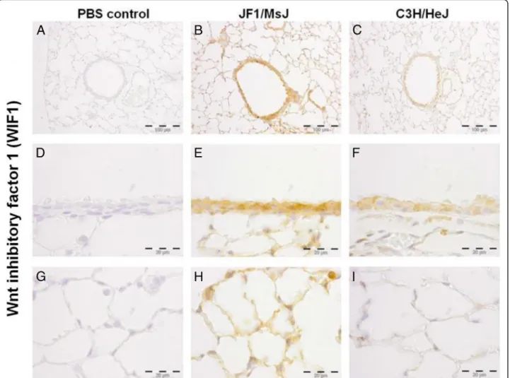
immunohisto-chemistry (Figs. 2, 3, 4). Increased immunoreactive WIF1
(Fig. 2) and FST (Fig. 3) were detected in the alveolar
epi-thelial type I and II cells, bronchial epiepi-thelial cells, and
macrophages of JF1/MsJ compared to C3H/HeJ mice.
De-creased immunoreactive FZD6 was detected in the
bron-chial epithelial cells and endothelial cells of JF1/MsJ
compared to C3H/HeJ mice (Fig. 4).
Expression quantitative trait loci (eQTL) analysis
To further assess candidate genes for Lfnq3 and Lfnq4
associated with TLC, we compared JF1/MsJ to C3H/HeJ
lung transcript levels at any of the three investigated
stages. Employing this strategy, we identified 9 eQTLs
located on mCh15 (Lfnq3) and mCh17 (Lfnq4) (Table 4)
not previously reported for lung function development
in mice. The candidate for Lfnq3 was leucine rich repeat
containing 6 (testis) (
Lrrc6
). Several candidates for Lfnq4
were identified that include radial spoke head 1 homolog
(Chlamydomonas) (
Rsph1
), formyl peptide receptor 1
(
Fpr1
), ATP-binding cassette, sub-family G (WHITE),
member 1 (
Abcg1
), 3 histocompatibility 2 genes
includ-ing class II antigen E beta (
H2-Eb1
), D region locus 1
(
H2-D1
), Q region locus 4 (
H2-Q4
), surfactant associated
2 (
Sfta2
), and gamma-aminobutyric acid (GABA) B
re-ceptor, 1 (
Gabbr1
).
Discussion
vascular genes in mice. The processes controlling lung
de-velopment and growth are believed to be recapitulated
fol-lowing lung injury as genetic subroutines for repair and
remodeling processes [40, 41]. Therefore, it is plausible that
individuals having impaired lung development and growth
that do not result in clinically significant symptom may
have inefficient repair processes thereby predisposing them
to subsequent chronic lung diseases. In this context,
elucidating the genomics of lung function development
through dissecting the genetics of lung development and
growth is a promising approach to identify the
predispos-ing factors. Thus, in this study, we sought to identify
candi-date genes for TLC by comparing the lung transcript
profile of JF1/MsJ and C3H/HeJ strains at 3 critical times
involving late embryonic, adolescent, and adult lung
development.
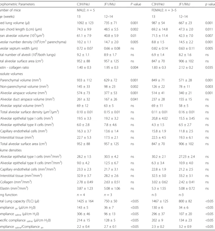
Table 1
Morphometric evaluation of C3H/HeJ and JF1/MsJ mouse lung
Morphometric Parameters C3H/HeJ JF1/MsJ Pvalue C3H/HeJ JF1/MsJ pvalue
Number of mice MALE:n= 5 FEMALE:n= 3–5
Age (weeks) 13 12–14 13 12–14
Fixed lung volume (μl) 1092 ± 123 735 ± 71 0.001 987 ± 54 667 ± 23 0.001
Mean chord length (Lcm) (μm) 74.3 ± 9.9 48.5 ± 5.5 0.002 69.2 ± 14.8 47.3 ± 2.0 0.011
Mean alveolar volume (103/μm3) 61.1 ± 7.9 40.8 ± 5.9 0.01 71.5 ± 11.4 42.3 ± 7.0 0.007
Alveolar number density (106/cm3parenchyma) 10.2 ± 1.1 14.7 ± 2.0 0.005 8.8 ± 1.5 14.5 ± 2.2 0.001
Alveolar septum width (μm) 0.72 ± 0.07 0.66 ± 0.08 ns 0.82 ± 0.14 0.63 ± 0.11 0.005
Total number of alveoli (106/both lungs) 9.2 ± 1.1 8.9 ± 1.7 ns 6.9 ± 1.4 8.2 ± 1.6 ns
Total alveolar surface area (cm2) 952 ± 88 957 ± 125 ns 847 ± 70 906 ± 102 ns
Elastin - collagen ratio 1.40 ± 0.3 1.95 ± 0.3 0.004 1.83 ± 0.3 2.12 ± 0.2 0.035
Absolute volumes
Parenchymal volume (mm3
) 933 ± 112 629 ± 72 0.001 849 ± 71 571 ± 28 0.001
Non-parenchymal volume (mm3) 145 ± 33 98 ± 23 0.002 126 ± 22 78 ± 11 0.003
Alveolar airspace volume (mm3) 574 ± 73 377 ± 53 0.001 514 ± 41 340 ± 21 0.001
Alveolar duct airspace volume (mm3) 261 ± 32 167 ± 26 0.041 237 ± 28 155 ± 15 ns
Alveolar septal volume (mm3) 69 ± 12 63 ± 5 ns 69 ± 11 58 ± 5 ns
Total alveolar surface density (μm2/μm3) 0.10 ± 0.01 0.15 ± 0.01 ns 0.11 ± 0.01 0.16 ± 0.01 ns
Alveolar epithelial type I cells (mm3) 19.5 ± 3.3 19.2 ± 3.2 ns 20,8 ± 4.02 15.5 ± 3.45 ns
Alveolar epithelial type II cells (mm3) 6.0 ± 2.8 7.8 ± 4.6 ns 4.3 ± 1.5 6.5 ± 2.7 ns
Capillary endothelial cells (mm3) 16.3 ± 3.7 13.6 ± 1.4 ns 15.8 ± 1.9 11.8 ± 2.5 ns
Interstitial tissue (mm3) 22.7 ± 5.3 17.5 ± 2.1 ns 22.5 ± 4.5 19.3 ± 6.1 ns
Total alveolar surface area (cm2) 952 ± 88 957 ± 125 ns 847 ± 70 906 ± 102 ns
Volume densities
Alveolar epithelial type I cells (mm3/mm3) 28.2 ± 1.5 30.5 ± 4.2 ns 30.2 ± 2.1 27.23 ± 2.4 ns
Alveolar epithelial type II cells (mm3/mm3) 9.0 ± 4.2 12.5 ± 6.7 ns 6.3 ± 3.4 10.9 ± 4.0 ns
Capillary endothelial cells (mm3/mm3) 23.3 ± 2.3 21.7 ± 3.1 ns 22.8 ± 1.9 21.2 ± 2.5 ns
Interstitial tissue (mm3/mm3) 32.9 ± 3.7 28.2 ± 2.6 ns 32.5 ± 3.0 33.2 ± 3.1 ns
Collagen (mm3/mm3) 2.78 ± 0.49 2.63 ± 0.51 ns 3.02 ± 0.62 2.42 ± 0.41 ns
Elastin (mm3/mm3) 3.87 ± 1.23 5.08 ± 1.06 ns 5.3 ± 1.55 5.08 ± 0.72 ns
Lung function n= 4 n= 3 n-3 n-3
Total Lung capacity (TLC) (μl) 1425 ± 164 750 ± 50 <0.05 1467 ± 125 800 ± 82 <0.05
Complianceair(μl/cm H20) 143 ± 5 36 ± 7 <0.05 130 ± 6 34 ± 6 <0.05
Compliancesaline(μl/cm H20) 306 ± 46 96 ± 13 <0.05 296 ± 37 107 ± 20 <0.05
Specific compliancesaline(μl/cm H20) 214 ± 15 128 ± 5 <0.05 202 ± 9 134 ± 23 <0.05
Compliancesaline/Complianceair 2.2 ± 0.4 2.7 ± 0.1 <0.05 2.3 ± 0.2 3.2 ± 0.9 <0.05
nsnot significant
The lung architectural differences detected in JF1/MsJ
and C3H/HeJ mice are suggestive of their difference in
development, growth and maturation events. JF1/MsJ
mice have decreased mean chord length and mean
al-veolar volume but increased alal-veolar number density
compared to C3H/HeJ. These findings suggest that JF1/
MsJ mice with smaller alveoli (decreased mean chord
length and alveolar volume) compensated by increased
alveolar number density. Thus, the lungs of both strains
exhibit near equal total alveolar surface area for gas
ex-change. Lung architectural differences detected between
these mouse strains are consistent with their functional
characteristics. Increased specific CL-saline/air
ratio in JF1/
MsJ mice compared to C3H/HeJ suggests that surface
tension contributes more than the elastic recoiling
prop-erties of JF1/MsJ lung in determining the lung
compli-ance. This is in agreement with the smaller alveoli in
JF1/MsJ mice. Increased elastin to collagen ratio and the
decreased CL-Air/TLC and CL-Saline/TLC in JF1/MsJ mice
indicates that elastic recoil due to tissue properties is
higher in JF1/MsJ mice compared to C3H/HeJ.
In this study, we compared the lung transcript
expres-sion contrasting JF1/MsJ and C3H/HeJ mice at 3 stages,
(I) embryonic day 18 (completion of embryonic lung
de-velopment), (II) P28 (adolescent lung, bulk alveolar
forma-tion completed); and (III) P70 (mature lung). Noteworthy
transcripts with consistent expression across the stages
ex-amined included those encoded by Wnt pathway and
Table 2
Extracellular region is an enriched gene ontology category (GO: 0005576) associated with the significantly decreased
transcripts in JF1/MsJ vs C3H/HeJ mouse lung at embryonic day 18 (E18) and postnatal day 70 (P70)
Gene Gene symbol Entrez ID Fold Difference
E18 decreased transcripts of GO term extracellular region; Enrichment Score = 2.83;p= 0.0005
Chemokine (C-X-C motif) ligand 15 Cxcl15 20,309 −14.6
Hemolytic complement Hc 15,139 −8.61
Kallikrein 1-related peptidase b21 Klk1b21 16,616 −8.25
Surfactant associated protein A1 Sftpa1 20,387 −3.80
Collagen, type XXVIII, alpha 1 Col28a1 213,945 −3.15
Paraoxonase 3 Pon3 269,823 −2.96
Ceruloplasmin Cp 12,870 −2.95
Neuron-derived neurotrophic factor Ndnf 68,169 −2.41
Fibroblast growth factor 1 Fgf1 14,164 −2.21
EGF-like, fibronectin type III and laminin G domains Egflam 268,780 −2.13
Transferrin Trf 22,041 −2.12
Interleukin 33 Il33 77,125 −2.04
P70 decreased transcripts of GO term extracellular region; Enrichment Score = 4.18;p= 0.04
Hemolytic complement Hc 15,139 −27.88
Chitinase 3-like 3 Chi3l3 12,655 −15.93
Kallikrein 1-related peptidase b21 Klk1b21 16,616 −11.92
Hepcidin antimicrobial peptide Hamp 84,506 −4.87
Chemokine (C-C motif) ligand 6 Ccl6 20,305 −3.92
Matrix metallopeptidase 3 Mmp3 17,392 −3.62
Insulin-like growth factor binding protein 6 Igfbp6 16,012 −2.83
Collagen, type XXVIII, alpha 1 Col28a1 213,945 −2.76
Angiopoietin-like 7 Angptl7 654,812 −2.72
Leptin receptor Lepr 16,847 −2.28
Chitinase, acidic Chia 81,600 −2.26
Oxidized low density lipoprotein (lectin-like) receptor 1 Olr1 108,078 −2.21
Coagulation factor VII F7 14,068 −2.18
Paraoxonase 3 Pon3 269,823 −2.18
Plasminogen activator, tissue Plat 18,791 −2.10
Secreted Ly6/Plaur domain containing 1 Slurp1 57,277 −2.02
chitinase-like genes. Gene ontology cellular component
“
extracellular region
”
was enriched among decreased JF1/
MsJ transcripts. We employed an e-QTL strategy and
consistent expression to identify novel candidate genes for
lung function development. e-QTL genes previously not
reported with TLC development include
Lrrc6, Fpr1
,
Abcg1
,
Rsph1
,
H2-Eb1
,
H2-
D1,
H2-Q4
,
Sfta2
, and
Gabbr1
.
WIF1 inhibits extracellular WNT signaling that plays a
crucial role during the lung development. Loss of
SMAD1 transcriptional activation of
Wif1
has been
asso-ciated with its decreased expression and increased Wnt/
β
-catenin signalling, resulting in abnormal distal lung
epithelial cell differentiation and branching
morphogen-esis [42]. Reduced SMAD1 and WIF1 expression in
Table 3
Lung transcripts (34 genes) consistently increased (12) or decreased (22) across embryonic stage 18 (E18), postnatal stage
(P) 28 and P70 in JF1/MsJ (JF1) mice compared to C3H/HeJ (C3H)
Gene Name Gene symbol Entrez ID Probeset Fold Difference
E18 P28 P70
Glutaredoxin 3 Glrx3 30,926 6,923,313 12.22 6.08 3.27
RIKEN cDNA G730007D18 gene G730007D18Rik 100,038,502 7,006,004 3.66 2.87 2.41
Holliday junction recognition protein Hjurp 381,280 6,760,490 3.58 2.09 2.20
Synaptonemal complex central element protein 1 Syce1 74,075 6,972,143 3.00 2.06 2.13
Predicted gene 14,403 Gm14403 433,520 6,883,977 2.98 2.79 2.68
RIKEN cDNA 2,610,028 J07 gene 2610028J07Rik 71,813 6,859,453 2.94 2.74 2.82
Microspherule protein 1 Mcrs1 51,812 6,918,337 2.62 2.03 2.30
RIKEN cDNA 2610507I01 gene 2610507I01Rik 72,203 6,788,659 2.60 5.35 3.74
Olfactory receptor 1061 Olfr1061 259,022 6,888,451 2.51 2.81 3.77
Olfactory receptor 170 Olfr170 258,959 6,844,403 2.47 4.67 3.43
Uncharacterized protein C130090J04 C130090J04 328,049 6,860,959 2.36 2.12 2.20
RIKEN cDNA 6,330,403 K07 gene 6330403K07Rik 103,712 6,789,483 2.32 7.15 6.56
Glucagon-like peptide 1 receptor Glp1r 14,652 6,849,762 −2.02 −2.33 −2.06
Solute carrier family 39 (metal ion transporter), member 8 Slc39a8 67,547 6,901,634 −2.07 −2.43 −2.51
Sodium channel, voltage-gated, type III, alpha Scn3a 20,269 6,887,324 −2.08 −5.31 −4.49
Sterile alpha motif domain containing 9-like Samd9l 209,086 6,951,281 −2.08 −2.74 −3.05
Interferon-induced protein 44 Ifi44 99,899 6,910,592 −2.10 −2.86 −2.19
Transmembrane protein 181A Tmem181a 77,106 6,848,520 −2.13 −2.60 −2.40
NME/NM23 family member 7 Nme7 171,567 6,754,701 −2.13 −2.49 −2.70
Protein phosphatase 1, regulatory (inhibitor) subunit 3A Ppp1r3a 140,491 6,951,783 −2.19 −2.10 −3.06
Megalencephalic leukoencephalopathy with subcortical cysts 1 homolog (human) Mlc1 170,790 6,837,773 −2.24 −2.78 −3.13
Cytochrome P450, family 2, subfamily J, polypeptide 6 Cyp2j6 13,110 6,923,520 −2.29 −2.27 −2.39
RIKEN cDNA A330076H08 gene A330076H08Rik 320,026 6,967,799 −2.35 −2.53 −2.06
Aminolevulinate, delta-, dehydratase Alad 17,025 6,922,241 −2.82 −2.54 −2.67
RIKEN cDNA D230046O15 gene D230046O15Rik 106,824 6,856,766 −3.00 −3.63 −2.54
Nuclear paraspeckle assembly transcript 1 (non-protein coding) Neat1 66,961 6,867,774 −3.54 −3.45 −2.70
Surfactant associated 2 Sfta2 433,102 6,850,183 −4.02 −2.19 −2.23
Transmembrane protein 181A Tmem181a 77,106 6,812,918 −4.26 −4.08 −3.40
RIKEN cDNA B930063I24 gene B930063I24Rik 319,330 6,756,745 −5.06 −4.33 −6.19
G protein-coupled receptor 137B Gpr137b 83,924 7,003,081 −5.39 −5.38 −3.06
Zinc finger, X-linked, duplicated B Zxdb 668,166 7,021,109 −6.18 −7.68 −7.23
Kallikrein 1-related peptidase B21 Klk1b21 16,616 6,960,239 −8.25 −19.87 −11.92
Hemolytic complement Hc 15,139 6,886,022 −8.61 −32.16 −27.88
RIKEN cDNA A730017L22 gene A730017L22Rik 613,258 6,890,967 −13.13 −15.62 −14.60
nitrofen-induced hypoplastic lung resulted in
develop-mental retardation during the saccular stage [43]. Our
findings of increased lung WIF1 (>5-fold) transcript and
protein levels in alveolar epithelial cells type I and II, as
well as in bronchial epithelial cells during alveolar
devel-opment in JF1/MsJ mice compared to C3H/HeJ strongly
supports it to be a candidate for pulmonary function
development.
Consistent with WIF1 expression, FST is also increased
(>4-fold) in JF1/MsJ lungs compared to C3H/HeJ.
Canon-ical Wnt/
β
-catenin signalling controls FST signalling in
satellite cell derived myoblasts [44]. FST-deficient mice
exhibit severely retarded overall growth, breathing failure,
and perinatal death with fluid filled lungs and poorly
ex-panded alveolar spaces [45]. FST binds and inhibits
acti-vins, which are members of the transforming growth
factor beta superfamily that plays a significant role in the
lung developmental processes [46]. Therapeutic
interven-tion with FST markedly reduced the number of infiltrating
cells and ameliorated the destruction of lung architecture
in bleomycin-treated rats [47].
FZD6 also functions as a negative regulator of the
ca-nonical Wnt/
β
-catenin signaling cascade. FZD6 controls
macroscopic hair patterning in mice and studies show
epithelial cells as the source of FZD6-dependent
signal-ing [48]. Wnt signalsignal-ing pathway was enriched for
anno-tation in the murine Developing Lung Characteristic
Subtranscriptome (mDLCS) involving C57BL/6 J, A/J
and C3H/HeJ inbred strains. WIF1 and FZD6 transcripts
also expressed significant strain dependent modulation
during alveolar development [39].
The proteins encoded by
Chil1
(aka
Chi3i1
or
YKL40
)
and
Chil3
(aka
Chi3l3
or
Ym1
) are similar to bacterial
chitinases but lacks chitinase activity. JF1/MsJ mice with
lower basal lung function have decreased lung CHIL1
(>2 fold) and CHIL3 (>15 fold) transcripts. In humans,
the gene homolog for mouse
Chil1
is
CHIL1
. Variations
in
CHIL1
have been associated with circulating and
air-way CHIL1 protein levels along with asthma prevalence,
severity, hospital admissions, and lung function in
chil-dren and young adults [49
–
53]. Variations in
CHIL1
are
also considered to modulate age-adjusted lung function
in cystic fibrosis patients [54]. Elevated serum CHIL1
protein is associated with severe persistent asthma
among adults but not in children [55]. However,
in-creased CHIL1 levels are detected among children with
severe, steroid resistant asthma [56]. Serum CHIL1
levels are negatively correlated with percent FEV
1while
A
B
C
Fig. 1Comparative expression of lung Wnt inhibitor factor 1 (Wif1) and frizzled homolog 6 (Fzd6) and chitinase 3 like 3 (CHIL3) in JF1/MsJ and
C3H/HeJ mice.aExpression of lungWif1mRNA in JF1/Msf was increased compared with C3H/HeJ mice at postnatal (P) days P28 and P70.b
Expression of lungFzd6mRNA in JF1/Msf was decreased compared to C3H/HeJ mice at P28 and P70. Quantitative real-time polymerase chain reaction was performed, and the comparative cycle number threshold (CT) method (ΔΔCT) was used [ΔCT= CT(gene)-CT(Actb)]. Data are presented as expression
positively correlated with low attenuation area/total lung
area percent in COPD patients [57]. Elevated CHIL1
protein has been detected in bronchoalveolar lavage
fluid and sputum, with higher numbers of
CHIL1-expressing cells in bronchial biopsies of smokers with
COPD [58, 59]. CHIL1 can activate Wnt/
β
-catenin
sig-naling in alveolar macrophage [60].
Amino acid sequence homology of CHIL3 to proteins
associated with tissue remodeling together with their
binding capacity with heparin sulfate and GlcN oligomers
suggests their role in airway wall remodeling in the allergic
lung [61]. CHIL3 protein is abundantly expressed in the
allergic mouse lung and enhances Th2 cytokine
produc-tion by inhibiting 12/15(S)-lipoxygenase which generates
lipid metabolites that interferes with T cell proliferation
[62]. However, a homologous sequence to
Chil3
has not
been found in the human genome.
The evaluation of eQTL located in Lfnq3 and Lfnq4
provided 9 candidate genes worthy of further analysis
for their possible role in lung development and growth.
Located in Lfnq3 region linked to TLC on mCh15,
Lrrc6
is
a gene expressed in the respiratory epithelium that is
essential for proper cilia axonemal assembly of inner
and outer dynein arms. Kott and colleagues identified an
early frameshift in
LRRC6
that is associated with primary
ciliary dyskinesia (PCD) [63]. PCD is a group of
autosomal-recessive developmental disorders resulting
from cilia and sperm-flagella defects, which can lead to
respiratory infections, situs inversus, and male infertility
[64]. In zebrafish,
lrrc6 (aka seahorse)
mutants produce
ventral body curvature and kidney cysts [65, 66] and
LRRC6 protein associates with disheveled, which
con-strains the canonical Wnt/
β
-catenin pathway and
pro-motes the non-canonical Wnt pathway during gastrulation
[67]. In gene-targeted mice lacking functional LRRC6, cilia
microtubules remained normal but the outer dynein arms
(ODAs), the structures essential for the ciliary beating, are
absent. In the absence of LRRC6, ODA proteins that
Fig. 2Localization of Wnt inhibitor factor 1 (WIF1) protein in the lungs of four weeks old female C3H/HeJ and JF1/MsJ mice (n= 5). Increased
WIF1 immunostaining was detected in theb, eairway epithelium andhalveolar type I and type II cells of JF1/MsJ lungs compared with those of
normally are assembled in the cytoplasm and transported
to the ciliary axoneme, remain in the cytoplasm and are
not transported to the ciliary axoneme [68, 69].
Of the eight differentially expressed transcripts located
within the Lfnq4 region in mCh17 that has been linked
to TLC,
Rsph1
,
Fpr1
,
Abcg1
, and
Sfta2
are most
note-worthy. Like
LRRC6
variants, splice site (275-2A > C)
and non-sense (433 C > T) polymorphisms in
RSPH1
are associated with PCD [64]. Although persons with
these variants can have unexplained neonatal respiratory
distress [70], the splice site variation in RSPH1 is
associ-ated with a lower prevalence of neonatal respiratory
dis-tress, later onset of daily wet cough, and better adult
lung function than other forms of PCD [71].
Mitochondrial damage-associated molecular patterns
(DAMPs) include formyl peptides that activate human
neutrophils through FPR1. On human neutrophils, FPR1
interacts with annexin A1 (aka lipocortin I), which
medi-ated anti-inflammatory activities of glucocorticoids and
can regulate epidermal growth factor receptor (EGFR)
localization and activity [72, 73]. Gene-targeted
Fpr1
−/−mice have a slight, but significant decrease in mean
chord length compared to strain-matched (C57BL/6 J)
control mice [74]. In addition,
Fpr1
−/−mice have
de-creased inflammation and emphysema compared to
control mice after cigarette smoke exposure. In
humans, cigarette smoking increases the number of
FPRs on the surface of neutrophils, which is greater
in smokers with COPD than smokers without COPD
[75, 76]. In addition, a variant rs867228 (minor allele
frequency = 0.21) producing a loss-of-function allele
in
FPR1
is associated with poor metastasis-free and
overall survival in breast and colorectal cancer
pa-tients receiving adjuvant chemotherapy [77].
Patients with pulmonary alveolar proteinosis (PAP)
display impaired surfactant clearance, foamy, lipid-filled
alveolar macrophages, and increased cholesterol
metabo-lites within the lung. Also regulated by Wnt/
β
-catenin
signalling [78], ABCG1 lipid transporter is considered to
be a key downstream target of colony stimulating factor
Fig. 3Localization of follistatin (FST) protein in the lungs of four weeks old female C3H/HeJ and JF1/MsJ mice (n= 5). Increased of FST
immunostaining was detected in theb,eairways andhalveolar type I and type II cells of JF1/MsJ lungs compared with those ofc,f,i
2 (aka GM-CSF) and is necessary for proper surfactant
catabolism [79, 80]. Mice lacking ABCG1 develop
alveo-lar type II cell hypertrophy with lipid deposition,
in-creased levels of surfactant [81], and develop more
severe lung fibrosis after bleomycin [82]. Compared with
strain-matched control mice, the lungs of ABCG1 null
mice are inflamed with macrophage accumulation,
lymphocytic infiltration, hemorrhage, eosinophilic
crys-tals, and elevated levels of cytokines and cytokine
recep-tors. BALF samples obtained from
Abcg1
−/−mice are
marked by increased foamy macrophages and leukocytes
and the presence of multiple markers of inflammation
including crystals of CHIL3 protein [83]. In mice,
ABCG1 is also required for pulmonary B-1 B cell and
natural antibody homeostasis [84]. Variations in
ABCG1
(rs13050646) and
HLA-A
(rs3823343, rs2517725) have
been associated to IgE dysregulation and atopy [85].
SFTA2 is a secretory protein predominantly expressed
by alveolar type II cells and non-ciliated bronchiolar
cells [86]. Similar to hydrophobic surfactant proteins B
(SFTB) and SFTPC, SFTA2 protein may share particular
physicochemical properties [87] and lung SFTA2 mRNA
deceases in mice treated with lipopolysaccharide [86].
However, unlike SFTFB and SFTPC, SFTA2 does not
co-localize to lamellar bodies, but is found to co-localize
to the Golgi and clathrin vesicles like hydrophilic
SFTPA1 and SFTPD [86]. Little is known about the
dis-ease phenotypes associated with SFTA2 variants, but
hu-man
SFTA2
is located in a chromosomal locus
associated with diffused panbronchiolitis.
Recent studies have implicated Wnt/
β
-catenin
path-way to play an important role in alveolar development
[39]. In an attempt to summarize the plausible effect of
altered expression of the identified candidate genes
based on the above reviewed information, our data
sug-gest that Wnt/
β
-catenin signaling may be altered during
postnatal lung development and growth of JF1/MsJ as
compared to C3H/HeJ mice. This is supported by the
Fig. 4Localization of frizzled homolog 6 (FZD6) protein in the lungs of four weeks old female C3H/HeJ and JF1/MsJ mice (n= 5). Decreased
FZD6 immunostaining was detected in theb, eairways andhalveolar type I and type II cells of JF1/MsJ lungs compared with those ofc,f,i
increased WIF1 transcript and protein levels in JF1/MsJ
lungs. Primary ciliary proteins, like LRRC6 and RSPH,
are required not only for regulation of Wnt/
β
-catenin
signaling, but also act as downstream effectors of the
Wnt/
β
-catenin pathway. [88] Alterations in a
chitinase-like protein, CHIL1, an activin-binding protein, FST, and
a lipid transporter, ABCG1, also implicates this pathway.
However, elucidation of the precise mode of action of
genetic variants in these genes in determining lung
func-tion development warrants funcfunc-tional and mechanistic
investigations.
Conclusions
This study demonstrates that the divergent pulmonary
function between C3H/HeJ and JF1/MsJ mice is
associ-ated with differences in transcription expression profiles
in lung. We further dissected the altered genetic
signa-ture of divergent lung function development among
C3H/HeJ and JF1/MsJ mice and identified several novel
genes, especially those with roles in Wnt/
β
-catenin
sig-nalling, not previously associated to lung function. The
study also provides a reference of transcripts altered
between mouse strains with divergent TLC at the
com-pletion of embryonic lung development, bulk alveolar
formation and lung growth. The generated data will
provide a point base for future association, functional
and mechanistic studies for pulmonary function.
Additional file
Additional file 1: Table S1.Increased lung transcripts in JF1/MsJ (JF1)
compared to C3H/HeJ (C3H) mice at embryonic day 18.Table S2.
Decreased lung transcripts in JF1/MsJ (JF1) compared to C3H/HeJ (C3H) mice at embryonic stage 18.Table S3.Increased lung transcripts in JF1/ MsJ (JF1) compared to C3H/HeJ (C3H) mice at postnatal day 28.Table S4.
Decreased lung transcripts in JF1/MsJ (JF1) compared to C3H/HeJ (C3H) mice at postnatal day 28.Table S5.Increased lung transcripts in JF1/MsJ (JF1) compared to C3H/HeJ (C3H) mice at postnatal day 70.Table S6.
Decreased lung transcripts in JF1/MsJ (JF1) compared to C3H/HeJ (C3H) mice at postnatal day 70.Table S7.Transcripts showing consistent pattern of expression lung transcripts in JF1/MsJ (JF1) compared to C3H/HeJ (C3H) mice across E18/ P28 stages.Table S8.Transcripts showing consistent pattern of expression lung transcripts in JF1/MsJ (JF1) compared to C3H/HeJ (C3H) mice across P28/ P70 stages.Figure S1.Representative lung sections showing smaller alveoli in JF1/Msf (JF1) mice compared to C3H/HeJ (C3H) in both males and females. (DOCX 216 kb)
Abbreviations
Abcg1:ATP-binding cassette, sub-family G (WHITE), member 1; ACTB: Beta actin; BALF: Bronchoalveolar lavage fluid;Chil: Chitinase-like 1;Chil3: Chitinase-like 3; CL: Compliance; COPD: Chronic obstructive pulmonary disease; E: Embryonic
day; e-QTL: Expression-quantitative trait loci; FEV1: Forced expiratory volume in
1 s;Fpr1: Formyl peptide receptor 1;Fst: Follistatin;Fzd6: Frizzled homolog 6; Gabbr1: Gamma-aminobutyric acid (GABA) B receptor, 1; GO: Gene ontology; H2-D1: Histocompatibility 2, D region locus 1;H2-Eb1: Histocompatibility 2, class II antigen E beta;H2-Q4: Histocompatibility 2, Q region locus 4; IAEC: Institutional Animal Ethics Committee; ip: Intra-peritoneal;Kit:Kitoncogene; Lfnq: Lung
Table 4
Candidate genes for total lung capacity (TLC) in mice identified through the expression quantitative trait loci (e-QTL)
strategy comparing C3H/HeJ (C3H) and JF1/MsJ (JF1). [cut off for fold change
≥
2.0 fold at least in one stage among embryonic
stage 18, postnatal stage (P) 28 and P70; false discovery rate = 0.10]
Trait QTL Region QTL Location
(Basepair)
Gene Description Gene symbol/ location (basepair)
EntrezID Fold Difference
E18 P28 P70
Lfnq3 Chromosome: 15
TLC, TLC/Bw D15Mit85 to D15Mit105
40,313,626–72,500,731 Leucine rich repeat containing 6 (testis)
Lrrc6
(66379858–66,500,910)
54,562 −2.03 ns ns
Lfnq4 Chromosome: 17
TLC D17Mit156 to D17Mit234
10,987,597–39,053,168 Radial spoke head 1 homolog (Chlamydomonas)
Rsph1
(31255019–31,277,356)
22,092 ns 2.81 2.63
Formyl peptide receptor 1 Fpr1
(17876471–17,883,939)
14,293 −2.05 −1.75 −2.26
ATP-binding cassette, sub-family G (WHITE), member 1
Abcg1
(31057694–31,117,984)
11,307 ns −1.76 −2.44
Surfactant associated 2 Sfta2
(35649708–35,650,569)
433,102 −4.02 −2.19 −2.23
Histocompatibility 2, class II antigen E beta
H2-Eb1
(34305867–34,316,674)
14,969 ns −2.23 −2.21
Histocompatibility 2, D region locus 1
H2-D1
(35263094–35,267,497)
14,964 ns 2.21 2.11
Histocompatibility 2, Q region locus 4
H2-Q4
(35379555–35,384,674)
15,015 −1.52 −3.19 −2.86
Gamma-aminobutyric acid (GABA) B receptor, 1
Gabbr1
(37045966–37,074,305)
54,393 −2.20 −1.68 −1.62
function quantitative trait loci;Lrrc6: Leucine rich repeat containing 6 (testis); mCh: Mouse chromosome; mDLCS: Developing lung characteristic
subtranscriptome; P: Postnatal day; qRT-PCR: Quantitative real time polymerase chain reaction; QTL: Quantitative trait loci;Rsph1: Radial spoke head 1 homolog (Chlamydomonas);Sfta2: Surfactant associated 2;Sod3: Superoxide dismutase 3, extracellular;Spp1: Secreted phosphoprotein 1; SUR: Systematic uniform random; TEM: Transmission electron microscope; TLC: Total lung capacity;Wif1: Wnt inhibitor factor 1
Acknowledgements
not applicable.
Funding
This study was supported by DST SERB: SB/SO/AS-026/2013; Department of Biotechnology, Government of India: BT/PR12987/INF/22/205/2015, VINNOVA (2016–01951) (K.G.); National Institutes of Health grants ES015675, HL077763, and HL085655 (G.D.L.); European Respiratory Society (ERS): ERS-LTRF-2015-3567 (SU); ERS-STRF-2014-7156 (AM); CSIR-SRF: [9/1045(0007) 2 K14-EMR-1], Fulbright Nehru Doctoral Research Fellowship (IIE grantee id 15,151,382 (TAT); Helmholtz Portfolio Theme‘Metabolic Dysfunction and Common Disease’, The Helmholtz Alliance‘Imaging and Curing Environmental Metabolic Diseases, ICEMED’(J.B.); German Center of Lung Research (DZL) H.F., L.L.
Availability of data and materials
Microarray data has been submitted to the Genome Expression Omnibus (GEO) database at National Center for Biotechnology Information NCBI (GSE80078).
Authors’contributions
KG, MI, JB, HF, HS and GDL conceived the project and designed the experiments; LG, AM, TAT, MI, LL, DH, AMH, SU and KG performed the experiments; LG, TAT, AM, MI, SV,HF, SU, HS, GDL and KG analyzed the data; LG, MI, HF, HS, GDL and KG wrote the manuscript. All authors have read and approved the manuscript.
Ethics approval and consent to participate
Human participants: human data or human tissue: not applicable.
Mice: The use of animals was in accordance with the German Law of Animal Protection and approved by the Bavarian Animal Research Authority and the Animal Research Authority of Schleswig-Holstein (reference number V312– 72241.123-3. All procedures were also approved by IACUC of the University of Pittsburgh, PA, USA. Frozen and paraffin embedded tissues were procured to carry out experiments at SRM University, India according to the Institutional Animal Ethics Committee (IAEC) permission [79/IAEC/2013].
Consent for publication
not applicable.
Competing interests
The authors declare that they have no competing interests.
Publisher
’
s Note
Springer Nature remains neutral with regard to jurisdictional claims in published maps and institutional affiliations.
Author details
1
SRM Research Institute, SRM University, Chennai 603203, India.2Institute of Experimental Genetics, Helmholtz Zentrum Muenchen, German Research Center for Environmental Health, 85764 Neuherberg, Munich, Germany.
3Priority Area Asthma & Allergy, Division of Asthma Exacerbation &
Regulation, Research Center Borstel, Airway Research Center North (ARCN), 23845 Borstel, Germany.4Department of Medicine, Pulmonary and Critical
Care Medicine, University Medical Centre Giessen and Marburg, Philipps-University Marburg, Marburg, Germany.5Department of Internal
Medicine (Pulmonology), University of Giessen and Marburg Lung Center (UGMLC), 35392, Giessen, Germany.6German Center for Diabetes Research
(DZD), 85764 Neuherberg, Germany.7Experimental Genetics, Technische
Universität München, 85354 Freising, Germany.8Priority Area Asthma &
Allergy, Division of Experimental Pneumology, Research Center Borstel, Airway Research Center North (ARCN), 23845 Borstel, Germany.9Lung and
Airway Research, Institute of Environmental Medicine, Karolinska Institutet, Box 287, SE-171 77 Stockholm, Sweden.10Institute of Lung Biology and
Disease, Helmholtz Zentrum Muenchen, German Research Center for Environmental Health, 85764 Neuherberg, Munich, Germany.11Institute of
Epidemiology I, Helmholtz Zentrum Muenchen, German Research Center for Environmental Health, 85764 Neuherberg, Munich, Germany.
12Comprehensive Pneumology Center Munich (CPC-M), Munich, Germany. 13Department of Environmental and Occupational Health, Graduate School
of Public Health, University of Pittsburgh, Pittsburgh, PA 15219, USA.14Work
Environment Toxicology; Institute of Environmental Medicine, Karolinska Institutet, Box 287, SE-171 77 Stockholm, Sweden.15Present address:
Lahn-Dill-Kliniken, Klinikum Wetzlar, Medizinische Klinik II, Forsthausstraße 1, D-35578 Wetzlar, Germany.
Received: 25 April 2017 Accepted: 25 July 2017
References
1. Krauss-Etschmann S, Bush A, Bellusci S, Brusselle GG, Dahlen SE, Dehmel S, Eickelberg O, Gibson G, Hylkema MN, Knaus P, Konigshoff M, Lloyd CM, Macciarini P, Mailleux A, Marsland BJ, Postma DS, Roberts G, Samakovlis C, Stocks J, Vandesompele J, Wjst M, Holloway J. Of flies, mice and men: a systematic approach to understanding the early life origins of chronic lung disease. Thorax. 2013;68:380–4.
2. Stocks J, Hislop A, Sonnappa S. Early lung development: lifelong effect on respiratory health and disease. Lancet Respir Med. 2013;1:728–42. 3. Stocks J, Sonnappa S. Early life influences on the development of chronic
obstructive pulmonary disease. Ther Adv Respir Dis. 2013;7:161–73. 4. Martinez FD. The origins of asthma and chronic obstructive pulmonary
disease in early life. Proc Am Thorac Soc. 2009;6:272–7.
5. Lange P, Celli B, Agusti A, Boje Jensen G, Divo M, Faner R, Guerra S, Marott JL, Martinez FD, Martinez-Camblor P, Meek P, Owen CA, Petersen H, Pinto-Plata V, Schnohr P, Sood A, Soriano JB, Tesfaigzi Y, Vestbo J. Lung-Function Trajectories Leading to Chronic Obstructive Pulmonary Disease. N Engl J Med. 2015;373:111–22.
6. Repapi E, Sayers I, Wain LV, Burton PR, Johnson T, Obeidat M, Zhao JH, Ramasamy A, Zhai G, Vitart V, Huffman JE, Igl W, Albrecht E, Deloukas P, Henderson J, Granell R, WL MA, Rudnicka AR, Wellcome Trust Case Control C, Barroso I, Loos RJ, Wareham NJ, Mustelin L, Rantanen T, Surakka I, Imboden M, Wichmann HE, Grkovic I, Jankovic S, Zgaga L, Hartikainen AL, Peltonen L, Gyllensten U, Johansson A, Zaboli G, Campbell H, Wild SH, Wilson JF, Glaser S, Homuth G, Volzke H, Mangino M, Soranzo N, Spector TD, Polasek O, Rudan I, Wright AF, Heliovaara M, Ripatti S, Pouta A, Naluai AT, Olin AC, Toren K, Cooper MN, James AL, Palmer LJ, Hingorani AD, Wannamethee SG, Whincup PH, Smith GD, Ebrahim S, TM MK, Pavord ID, AK ML, Morris AD, Porteous DJ, Cooper C, Dennison E, Shaheen S, Karrasch S, Schnabel E, Schulz H, Grallert H, Bouatia-Naji N, Delplanque J, Froguel P, Blakey JD, NRS T, Britton JR, Morris RW, Holloway JW, Lawlor DA, Hui J, Nyberg F, Jarvelin MR, Jackson C, Kahonen M, Kaprio J, Probst-Hensch NM, Koch B, Hayward C, Evans DM, Elliott P, Strachan DP, Hall IP, Tobin MD. Genome-wide association study identifies five loci associated with lung function. Nat Genet. 2010;42:36–44.
7. Yao TC, Du G, Han L, Sun Y, Hu D, Yang JJ, Mathias R, Roth LA, Rafaels N, Thompson EE, Loisel DA, Anderson R, Eng C, Arruabarrena Orbegozo M, Young M, Klocksieben JM, Anderson E, Shanovich K, Lester LA, Williams LK, Barnes KC, Burchard EG, Nicolae DL, Abney M, Ober C. Genome-wide association study of lung function phenotypes in a founder population. J Allergy Clin Immunol. 2014;133:248–55.e241-210.
8. Hancock DB, Eijgelsheim M, Wilk JB, Gharib SA, Loehr LR, Marciante KD, Franceschini N, van Durme YM, Chen TH, Barr RG, Schabath MB, Couper DJ, Brusselle GG, Psaty BM, van Duijn CM, Rotter JI, Uitterlinden AG, Hofman A, Punjabi NM, Rivadeneira F, Morrison AC, Enright PL, North KE, Heckbert SR, Lumley T, Stricker BH, O'Connor GT, London SJ. Meta-analyses of genome-wide association studies identify multiple loci associated with pulmonary function. Nat Genet. 2010;42:45–52.
Barr RG, Beilby J, Blakey JD, Boban M, Boraska V, Brisman J, Britton JR, Brusselle GG, Cooper C, Curjuric I, Dahgam S, Deary IJ, Ebrahim S, Eijgelsheim M, Francks C, Gaysina D, Granell R, Gu X, Hankinson JL, Hardy R, Harris SE, Henderson J, Henry A, Hingorani AD, Hofman A, Holt PG, Hui J, Hunter ML, Imboden M, Jameson KA, Kerr SM, Kolcic I, Kronenberg F, Liu JZ, Marchini J, McKeever T, Morris AD, Olin AC, Porteous DJ, Postma DS, Rich SS, Ring SM, Rivadeneira F, Rochat T, Sayer AA, Sayers I, Sly PD, Smith GD, Sood A, Starr JM, Uitterlinden AG, Vonk JM, Wannamethee SG, Whincup PH, Wijmenga C, Williams OD, Wong A, Mangino M, Marciante KD, WL MA, Meibohm B, Morrison AC, North KE, Omenaas E, Palmer LJ, Pietilainen KH, Pin I, Pola Sbreve Ek O, Pouta A, Psaty BM, Hartikainen AL, Rantanen T, Ripatti S, Rotter JI, Rudan I, Rudnicka AR, Schulz H, Shin SY, Spector TD, Surakka I, Vitart V, Volzke H, Wareham NJ, Warrington NM, Wichmann HE, Wild SH, Wilk JB, Wjst M, Wright AF, Zgaga L, Zemunik T, Pennell CE, Nyberg F, Kuh D, Holloway JW, Boezen HM, Lawlor DA, Morris RW, Probst-Hensch N, International Lung Cancer C, Consortium G, Kaprio J, Wilson JF, Hayward C, Kahonen M, Heinrich J, Musk AW, Jarvis DL, Glaser S, Jarvelin MR, Ch Stricker BH, Elliott P, O'Connor GT, Strachan DP, London SJ, Hall IP, Gudnason V, Tobin MD. Genome-wide association and large-scale follow up identifies 16 new loci influencing lung function. Nat Genet. 2011;43:1082–90.
10. Soler Artigas M, Wain LV, Repapi E, Obeidat M, Sayers I, Burton PR, Johnson T, Zhao JH, Albrecht E, Dominiczak AF, Kerr SM, Smith BH, Cadby G, Hui J, Palmer LJ, Hingorani AD, Wannamethee SG, Whincup PH, Ebrahim S, Smith GD, Barroso I, Loos RJ, Wareham NJ, Cooper C, Dennison E, Shaheen SO, Liu JZ, Marchini J, Medical Research Council National Survey of H, Development Respiratory Study T, Dahgam S, Naluai AT, Olin AC, Karrasch S, Heinrich J, Schulz H, TM MK, Pavord ID, Heliovaara M, Ripatti S, Surakka I, Blakey JD, Kahonen M, Britton JR, Nyberg F, Holloway JW, Lawlor DA, Morris RW, James AL, Jackson CM, Hall IP, Tobin MD, SpiroMeta C. Effect of five genetic variants associated with lung function on the risk of chronic obstructive lung disease, and their joint effects on lung function. Am J Respir Crit Care Med. 2011;184:786–95.
11. Obeidat M, Hao K, Bosse Y, Nickle DC, Nie Y, Postma DS, Laviolette M, Sandford AJ, Daley DD, Hogg JC, Elliott WM, Fishbane N, Timens W, Hysi PG, Kaprio J, Wilson JF, Hui J, Rawal R, Schulz H, Stubbe B, Hayward C, Polasek O, Jarvelin MR, Zhao JH, Jarvis D, Kahonen M, Franceschini N, North KE, Loth DW, Brusselle GG, Smith AV, Gudnason V, Bartz TM, Wilk JB, O'Connor GT, Cassano PA, Tang W, Wain LV, Soler Artigas M, Gharib SA, Strachan DP, Sin DD, Tobin MD, London SJ, Hall IP, Pare PD. Molecular mechanisms underlying variations in lung function: a systems genetics analysis. Lancet Respir Med. 2015;3:782–95. 12. Tang W, Kowgier M, Loth DW, Soler Artigas M, Joubert BR, Hodge E, Gharib
SA, Smith AV, Ruczinski I, Gudnason V, Mathias RA, Harris TB, Hansel NN, Launer LJ, Barnes KC, Hansen JG, Albrecht E, Aldrich MC, Allerhand M, Barr RG, Brusselle GG, Couper DJ, Curjuric I, Davies G, Deary IJ, Dupuis J, Fall T, Foy M, Franceschini N, Gao W, Glaser S, Gu X, Hancock DB, Heinrich J, Hofman A, Imboden M, Ingelsson E, James A, Karrasch S, Koch B, Kritchevsky SB, Kumar A, Lahousse L, Li G, Lind L, Lindgren C, Liu Y, Lohman K, Lumley T, WL MA, Meibohm B, Morris AP, Morrison AC, Musk B, North KE, Palmer LJ, Probst-Hensch NM, Psaty BM, Rivadeneira F, Rotter JI, Schulz H, Smith LJ, Sood A, Starr JM, Strachan DP, Teumer A, Uitterlinden AG, Volzke H, Voorman A, Wain LV, Wells MT, Wilk JB, Williams OD, Heckbert SR, Stricker BH, London SJ, Fornage M, Tobin MD, O'Connor GT, Hall IP, Cassano PA. Large-scale genome-wide association studies and meta-analyses of longitudinal change in adult lung function. PLoS One. 2014;9:e100776. 13. Loth DW, Soler Artigas M, Gharib SA, Wain LV, Franceschini N, Koch B,
Pottinger TD, Smith AV, Duan Q, Oldmeadow C, Lee MK, Strachan DP, James AL, Huffman JE, Vitart V, Ramasamy A, Wareham NJ, Kaprio J, Wang XQ, Trochet H, Kahonen M, Flexeder C, Albrecht E, Lopez LM, de Jong K, Thyagarajan B, Alves AC, Enroth S, Omenaas E, Joshi PK, Fall T, Vinuela A, Launer LJ, Loehr LR, Fornage M, Li G, Wilk JB, Tang W, Manichaikul A, Lahousse L, Harris TB, North KE, Rudnicka AR, Hui J, Gu X, Lumley T, Wright AF, Hastie ND, Campbell S, Kumar R, Pin I, Scott RA, Pietilainen KH, Surakka I, Liu Y, Holliday EG, Schulz H, Heinrich J, Davies G, Vonk JM, Wojczynski M, Pouta A, Johansson A, Wild SH, Ingelsson E, Rivadeneira F, Volzke H, Hysi PG, Eiriksdottir G, Morrison AC, Rotter JI, Gao W, Postma DS, White WB, Rich SS, Hofman A, Aspelund T, Couper D, Smith LJ, Psaty BM, Lohman K, Burchard EG, Uitterlinden AG, Garcia M, Joubert BR, WL MA, Musk AB, Hansel N, Heckbert SR, Zgaga L, van Meurs JB, Navarro P, Rudan I, Oh YM, Redline S, Jarvis DL, Zhao JH, Rantanen T, O'Connor GT, Ripatti S, Scott RJ, Karrasch S, Grallert H, Gaddis NC, Starr JM, Wijmenga C, Minster RL, Lederer DJ, Pekkanen J, Gyllensten U, Campbell H, Morris AP, Glaser S, Hammond CJ,
Burkart KM, Beilby J, Kritchevsky SB, Gudnason V, Hancock DB, Williams OD, Polasek O, Zemunik T, Kolcic I, Petrini MF, Wjst M, Kim WJ, Porteous DJ, Scotland G, Smith BH, Viljanen A, Heliovaara M, Attia JR, Sayers I, Hampel R, Gieger C, Deary IJ, Boezen HM, Newman A, Jarvelin MR, Wilson JF, Lind L, Stricker BH, Teumer A, Spector TD, Melen E, Peters MJ, Lange LA, Barr RG, Bracke KR, Verhamme FM, Sung J, Hiemstra PS, Cassano PA, Sood A, Hayward C, Dupuis J, Hall IP, Brusselle GG, Tobin MD, London SJ. Genome-wide association analysis identifies six new loci associated with forced vital capacity. Nat Genet. 2014;46:669–77.
14. Young S, Sherrill DL, Arnott J, Diepeveen D, LeSouef PN, Landau LI. Parental factors affecting respiratory function during the first year of life. Pediatr Pulmonol. 2000;29:331–40.
15. Murray CS, Pipis SD, McArdle EC, Lowe LA, Custovic A, Woodcock A, National Asthma Campaign-Manchester A, Allergy Study G. Lung function at one month of age as a risk factor for infant respiratory symptoms in a high risk population. Thorax 2002;57: 388–392.
16. Mullane D, Turner SW, Cox DW, Goldblatt J, Landau LI, le Souef PN. Reduced infant lung function, active smoking, and wheeze in 18-year-old individuals. JAMA Pediatr. 2013;167:368–73.
17. Turner S, Fielding S, Mullane D, Cox DW, Goldblatt J, Landau L, le Souef P. A longitudinal study of lung function from 1 month to 18 years of age. Thorax. 2014;69:1015–20.
18. Skripak JM. Persistent effects of maternal smoking during pregnancy on lung function and asthma in adolescents. Pediatrics. 2014;134(Suppl 3):S146. 19. Reinhard C, Eder G, Fuchs H, Ziesenis A, Heyder J, Schulz H. Inbred strain
variation in lung function. Mamm Genome. 2002;13:429–37.
20. Reinhard C, Meyer B, Fuchs H, Stoeger T, Eder G, Ruschendorf F, Heyder J, Nurnberg P, de Angelis MH, Schulz H. Genomewide linkage analysis identifies novel genetic Loci for lung function in mice. Am J Respir Crit Care Med. 2005; 171:880–8.
21. Ganguly K, Stoeger T, Wesselkamper SC, Reinhard C, Sartor MA, Medvedovic M, Tomlinson CR, Bolle I, Mason JM, Leikauf GD, Schulz H. Candidate genes controlling pulmonary function in mice: transcript profiling and predicted protein structure. Physiol Genomics. 2007;31:410–21.
22. Ganguly K, Upadhyay S, Irmler M, Takenaka S, Pukelsheim K, Beckers J, De Angelis MH, Hamelmann E, Stoeger T, Schulz H. Impaired resolution of inflammatory response in the lungs of JF1/Msf mice following carbon nanoparticle instillation. Respir Res. 2011;12:94.
23. Leikauf GD, Concel VJ, Liu P, Bein K, Berndt A, Ganguly K, Jang AS, Brant KA, Dietsch M, Pope-Varsalona H, Dopico RA Jr, Di YP, Li Q, Vuga LJ, Medvedovic M, Kaminski N, You M, Prows DR. Haplotype association mapping of acute lung injury in mice implicates activin a receptor, type 1. Am J Respir Crit Care Med. 2011;183:1499–509.
24. Ganguly K, Upadhyay S, Irmler M, Takenaka S, Pukelsheim K, Beckers J, Hamelmann E, Schulz H, Stoeger T. Pathway focused protein profiling indicates differential function for IL-1B,−18 and VEGF during initiation and resolution of lung inflammation evoked by carbon nanoparticle exposure in mice. Part Fibre Toxicol. 2009;6:31.
25. Ganguly K, Depner M, Fattman C, Bein K, Oury TD, Wesselkamper SC, Borchers MT, Schreiber M, Gao F, von Mutius E, Kabesch M, Leikauf GD, Schulz H. Superoxide dismutase 3, extracellular (SOD3) variants and lung function. Physiol Genomics. 2009;37:260–7.
26. Ganguly K, Martin TM, Concel VJ, Upadhyay S, Bein K, Brant KA, George L, Mitra A, Thimraj TA, Fabisiak JP, Vuga LJ, Fattman C, Kaminski N, Schulz H, Leikauf GD. Secreted phosphoprotein 1 is a determinant of lung function development in mice. Am J Respir Cell Mol Biol. 2014;51:637–51. 27. Lindsey JY, Ganguly K, Brass DM, Li Z, Potts EN, Degan S, Chen H, Brockway
B, Abraham SN, Berndt A, Stripp BR, Foster WM, Leikauf GD, Schulz H, Hollingsworth JW. c-Kit is essential for alveolar maintenance and protection from emphysema-like disease in mice. Am J Respir Crit Care Med. 2011;183: 1644–52.
28. Hsia CC, Hyde DM, Ochs M, Weibel ER, Structure AEJTFoQAoL. An official research policy statement of the American Thoracic Society/European Respiratory Society: standards for quantitative assessment of lung structure. Am J Respir Crit Care Med. 2010;181:394–418.
29. Fehrenbach H, Zimmermann G, Starke E, Bratu VA, Conrad D, Yildirim AO, Fehrenbach A. Nitrogen dioxide induces apoptosis and proliferation but not emphysema in rat lungs. Thorax. 2007;62:438–46.