Preferential Vasodilator Effects of
Levosimendan in Resistance Pulmonary
Arteries in a Rodent Pulmonary Embolism Model
Camila Bedo and Juan C Grignola
Department of Pathophysiology, School of Medicine, Hospital de Clínicas, Universidad de la República, Montevideo, Uruguay
Corresponding author: Juan C Grignola
Department of Pathophysiology, School of Medicine, Hospital de Clínicas, Universidad de la República Avda Italia 2870, PC 11600, Montevideo, Uruguay
E-mail: [email protected]
Introduction
The pulmonary vasculature consists of large, elastic, extra-parenchymal conduit pulmonary arteries (CPA, order 1 to 2) that arise from the sixth aortic arch and small, muscular resistance intrapulmonary arteries (RPA, ≥ 4th order), that originate from the mesenchymal lung bud by capillary plexus expansion [1]. This subdivision is associated with different response to several stimuli. While CPA dilates or fails to constrict to hypoxia, RPA is responsible for hypoxic pulmonary vasoconstriction, control the regional distribution of blood flow and largely determine pulmonary vascular resistance. This functional difference mainly depends on the distribution of electrophysiologically distinct myocytes in CPAs and RPAs arteries [1, 2].
Levosimendan is a positive inotropic agent (by increasing the sensitivity of troponin C to calcium) with vasodilating properties (by lowering of intracellular free Ca++, opening of different potassium channels and the inhibition of phosphodiesterase type III), also termed inodilator [3, 4]. There are several animals studies in different acute pulmonary hypertension (PH) models secondary to thromboxane A2 infusion [5], endotoxemia [6], acute pulmonary embolism (PE) [7, 8], and hypoxia [9] and some clinical studies that demonstrated the vasodilator effect of levosimendan on the pulmonary circulation, restoring right ventricular-arterial coupling as it increased right ventricular contractility concomitantly [10, 11]. Acute PE-induced PH results from two main mechanisms:
Highlights
Background
We compared the vasoactive effects of levosimendan on isolated conduit (CPA) and resistance (RPA) pulmonary arteries versus mesenteric arteries and we assessed the PA vascular function and the PA vasoactive effects of levosimendan in a rodent PE model. Methods
One group of male Wistar rats (200-300 g) was killed by decapitation to obtain pulmonary and mesenteric rings. Another group was assigned to a massive PE or saline solution infusion. After euthanasia mesenteric arteries and CPA (i.d. 1-2 mm) and RPA (≤ 0.5 mm) were dissected and cut into 2-3 mm wide rings recording contractile tension. We obtained the concentration-response curves of cumulative doses of levosimendan on pre-contracted arterial rings from decapitated and sham/embolized animals. A set of RPA rings was exposed to acute hypoxia. The effect of PE on the pulmonary vasoactive function was assessed by dose-response curves of acetylcholine (ACh) and endothelin-1 (ET-1) of PA rings from sham/embolized animals.
Results
Levosimendan relaxant potency of RPA was similar to mesenteric arteries and higher than CPA, while mesenteric rings showed the maximal relaxant effect, followed by RPA and CPA, respectively. PE did not affect the vasoactive response of PA rings either to ACh or to ET-1, and the relaxant effects of CPA and RPA to levosimendan were also preserved. Acute hypoxia reduced (P<0.05) but did not avoid the RPA relaxant effect of levosimendan.
Conclusions
Levosimendan is a more specific vasodilator of RPA with a similar relaxant potency as mesenteric arteries, which is preserved after PE but significantly reduced during hypoxia.
Keywords: levosimendan; pulmonary embolism; pulmonary arteries; vasodilation; hypoxia
Citation: Bedo C, Grignola JC. Preferential Vasodilator Effects of Levosimendan in Resistance Pulmonary Arteries in a Rodent Pulmonary Embolism Model. International Cardiovascular Forum Journal. 2017; 11: 16-22. DOI: 10.17987/icfj.v11i0.433
* Corresponding author. E-mail: [email protected] ISSN: 2410-2636
the mechanical obstruction of pulmonary vessels (passive mechanism) and the arterial vasoconstriction secondary to pulmonary neurohumoral activation (e.g. thromboxane A2, serotonin), neurogenic reflex, increased oxidative stress and hypoxemia (active mechanism) [12]. There is experimental evidence that secondary pulmonary vasoconstriction is the major contributor to the potentially fatal increase in pulmonary dynamic afterload in PE. Therefore the treatment should not only focus on removing the obstructing blood clot but also on reducing this vasoconstrictor response [13]. We have previously reported a significant decrease of the dynamic afterload despite the persistence of mechanical obstruction by clots during levosimendan infusion in a PE-induced PH ovine model [14]. The concomitant decrease of total pulmonary vascular resistance with the preservation of the pulmonary arterial characteristic impedance suggests a predominant vasodilator effect on distal arterial vessels [14].
We hypothesized that levosimendan would have a different vasodilator potency between CPA and RPA, with a predominant distal vasodilatation effect. The aims of the present work were: to compare the vasoactive effects of levosimendan on isolated CPA and RPA versus mesenteric arteries of rats; to assess the pulmonary arterial vasoconstriction and vasodilatation response and the pulmonary arteries vasoactive effects of levosimendan in a rodent PE model and to assess the effects of hypoxia on the RPA relaxant responses to levosimendan.
Methods
This study was approved by the Institutional Animal Care and Use Committee (CHEA, Facultad de Medicina, Universidad de la República. N° 070153-000611-13. http://www.chea.udelar.edu. uy/). We rigidly performed all institutional protocols to handle animals under experimentation according to Guide for Care and Use of Laboratory Animals (NIH Publication N° 85–23, revised 1996), prepared by the National Academy of Sciences’ Institute for Laboratory Animal Research.
Male Wistar rats (200-300 g) were used in this study. Rats fasted overnight with free access to water. One group of animals was killed by decapitation, and the lungs and mesenteric beds were rapidly immersed in Krebs solution to obtain pulmonary and mesenteric rings. Another group of animals was anesthetized with pentobarbital (40 mg/kg, i.p.) and fentanyl (50 mg/kg, i.p.). Both were maintained with pentobarbital (10 mg/kg/h), and fentanyl (1-2 mg/kg/h) administered intravenously throughout an infusion pump (Syringe pump, GRASEBY 3400, Smiths-Medical, Ohio, USA). Normothermia was kept using a heating pad. The animals were tracheotomized and mechanically ventilated (ServoVentilator model 300, SIEMENS AG, Munich, Germany). The tidal volume and the fraction of inspired oxygen were set in 8 mL/Kg and 60%, respectively. Respiratory rate was adjusted to maintain a baseline physiologic arterial oxygen and carbon dioxide tension. Blood samples were taken regularly (every 30 min) to analyze arterial oxygen and carbon dioxide tension (Blood gas analyzer, Radiometer, ABL520, Denmark).
We placed two 22G fluid-filled catheters into both external jugular veins for blood withdrawal and drug infusion, respectively. Another fluid-filled catheter was positioned into the common carotid artery to monitor systemic arterial pressure. We performed
a sternotomy and placed a 22G fluid-filled catheter into the PA through a minimal stab in the right ventricular outflow. The distal tip of the catheter was positioned in the main PA before its bifurcation. All pressure transducers (P23Db Gould Statham) were zeroed to atmospheric pressure at the mid-axillary level. Once we completed the instrumentation, the animals allowed stabilizing for 15 minutes. Baseline hemodynamic data were obtained. Animals were divided into two groups:
a) Sham group (saline solution, n = 8): infusion of 1 ml of saline infusion. Animals were euthanized once reached 60 minutes. b) Embolized group (PE, n = 8): one milliliter of blood was collected
and allowed to clot at room temperature for five minutes. Clots were mechanically disaggregated achieving a diameter ~ 0.5 mm, and venous embolization was carried out progressively through the right jugular vein every 15 minutes over 60 minutes until systemic hypotension was reached, ensuring to produce a massive PE. Once hypotension developed, the animal was euthanized with an overdose of pentobarbital.
After euthanasia, heart and lungs were removed, and lungs were rapidly placed in Krebs solution.
Tissue preparation and contractile tension recording
Mesenteric arteries (i.d. 1-2 mm), CPA (i.d. 1-2 mm, second order) and RPA (i.d. ≤ 0.5 mm, fifth order) were carefully dissected from surrounding tissue and cut into 2-3 mm wide rings for studies on intact preparations [15]. During manipulation of the arteries, care was taken not to touch their intimal surface to preserve the endothelium layer.
The rings were suspended between two wire hooks in 5 ml organ baths for contractile tension recording with an isometric transducer (Myograph model 610M, Danish Myo Technology, Aarhus-Denmark for mesenteric arteries and RPA rings and KG-20 force transducers, World Precision Instruments, Sarasota-USA for CPA rings) [16]. The organ baths contained Krebs solution maintained at 37°C and continuously bubbled with a 95% O2-5% CO2 mixture as previously described. Tissues were stretched to a predetermined optimal resting tension of 0.5 g for pulmonary rings and 2.0 g for mesenteric rings [15]. The presence of functional endothelium was tested by the assessment of the relaxant response to ACh (10-6 M) in rings pre-contracted with 5HT (10-5 M). The ability of ACh to induce relaxation was taken as an indicator of the presence of functional endothelium. We discarded those rings with a relaxant response ≤ 20% of maximal tension. Total time of the experiments never exceeded 120 minutes. Based on previous experiments there was no spontaneous relaxation in pre-contracted rings during this period.
Experimental protocol
Six to eight mesenteric and PA rings were obtained from each animal. The rings were first stimulated by raising the K+ concentration of the buffer to 80 mM and then washing three times, allowing to recover the resting tension.
This mixture allowed us to avoid possible vasoactive response differences among arteries depending on the vasoconstrictor used [15], and mimics several forms of PH including PE, achieving a high vascular tone that presumably reflects what happens in massive PE [17]. Once equilibration (about 25 min), a concentration-response curve to levosimendan (10-9 M to 3´10-5 M) was carried out by cumulative addition of drug after a steady-state relaxant response was reached after each increment. The range of levosimendan doses and the interval between doses (7 min) are based on previous experiments.
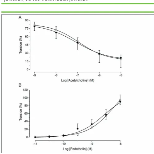
Assessment of the PA vascular vasoconstriction and vasodilatation properties: the analysis of the endothelium function was performed through concentration-response curves of cumulative doses of acetylcholine (ACh, 10-9 to 10-5 M) in rings previously contracted by serotonin (5HT, 10-5 M). The vascular smooth muscle function was assessed by concentration-response curves of cumulative doses of endothelin-1 (ET-1, 10-11 to 10-8 M).
Assessment of the effects of hypoxia on levosimendan RPA relaxant responses: a set of RPA rings extracted from euthanized rats by decapitation was exposed to acute hypoxia. Hypoxia was induced by aerating the organ bath with 95% N2-5% CO2 (PO2 24 ± 1 mm Hg) [18] and then we obtained the dose-response curve to levosimendan.
Drugs and reagents
Acetylcholine chloride, serotonin hydrochloride, endothelin-1 and thromboxane A2 mimetic U46619 and Dimethyl Sulphoxide (DMSO) were obtained from Sigma-Aldrich, SPAIN. Levosimendan was obtained from Sigma Chemical Co, USA. They were dissolved in distilled deionized water except for levosimendan which was dissolved in DMSO. The final concentration of DMSO in the organ bath was less than 0.1 % and had no effect on the vessel reactivity. The concentration of drugs was expressed as a final molar concentration in the tissue chamber.
Data analysis
The maximal vasoactive effect (Emax, expressed as a percentage of the initial contractile response), which is an index of the efficacy of the vasoactive drug, and the drug concentration exhibiting 50% of the Emax (EC50, expressed as negative logarithmic concentration, -log EC50: pD2), which is an index of the potency of the vasoactive drug, were calculated from the fitted concentration-response curves for each ring. Data were averaged for each animal in all experiments. Relaxation responses to levosimendan are expressed as percentages of tension developed with KCl 80 mM.
Statistical analysis
Results are expressed as mean ± SEM, with n equal to the number of animals. Individual cumulative concentration-response curves were fitted assuming the sigmoid dose-response curves (Levenberg-Marquardt algorithm) by using Origin Pro Software (version 9.1, San Diego, CA, USA). For multiple comparisons (e.g. the vasoactive effects of levosimendan on the mesenteric arteries, CPA and RPA), statistical analysis was performed using a one-way ANOVA followed by a Bonferroni post hoc test, otherwise (e.g. rings from sham versus PE rats, CPA versus RPA rings and normoxic versus hypoxic RPA rings) we used a
two-tailed unpaired Student’s t-test. Differences were considered statistically significant when P < 0.05.
Results
Vasodilator effects of levosimendan
Levosimendan induced a concentration-dependent relaxation, but was unable to fully relax 3´10-9 M ET-1, 3´10-8 M thromboxane A2 mimetic U46619 and 3´10-6 M 5HT-induced contractions in mesenteric as pulmonary rings. The highest concentration tested (3´10-4) produced a maximal relaxation of 85 ± 6% of the mesenteric arteries.
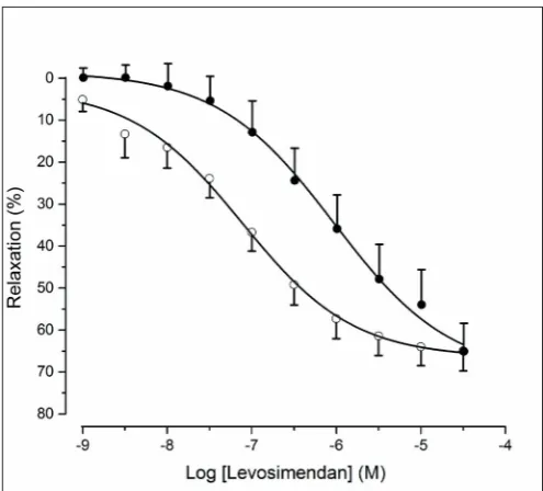
RPA and mesenteric rings showed the maximal relaxant potency, while the highest maximal relaxant effect was obtained in mesenteric arteries followed by RPA and CPA, respectively (P < 0.05) (Table 1, Figure 1).
Effects of pulmonary embolism on pulmonary artery
vasodilatation and vasoconstriction response
Table 2 shows systemic and pulmonary arterial pressures of sham and embolized animals. Neither the endothelium function (Sham: pD2 6.9 ± 0.07 and Emax 20 ± 7% vs. PE: pD2 7.1 ± 0.09 and Emax 21 ± 17%) nor the vascular smooth muscle function (Sham: pD2 8.4 ± 0.2 and Emax 94 ± 11% vs. PE: pD2 8.6 ± 0.2 and Emax 90 ± 17%) of the PA rings from embolized rats was changed in comparison of PA rings from sham animals (Figure 2A, B).
Effects of levosimendan on conduit and resistance
PAs from embolized rats
The relaxant effect of levosimendan was not significantly affected by PE. Like in the sham group, rings from embolized rats showed a significant higher levosimendan potency in RPA rings in comparison to CPA rings (Table 3, Figure 3).
Effects of hypoxia on the RPA relaxant responses to
levosimendan
Figure 4 shows the effects of hypoxia on the RPA relaxant responses to levosimendan. Exposure to hypoxia induced a significant decrease of relaxant potency of levosimendan in RPA rings with similar efficacy (Emax) (normoxia: pD2 7.11 ± 0.06 and Emax 65 ± 5% vs. hypoxia: pD2 6.03 ± 0.09 and Emax 65 ± 7%).
Discussion
smooth muscle function), and the relaxant effects of CPA and RPA to levosimendan were also preserved. 3) Hypoxia reduced but did not avoid the RPA relaxant effect of levosimendan.
The higher vasodilatation potency of levosimendan on RPA versus CPA is consistent with our previously results in an ovine model of blood clot PE-induced PH. Briefly, we have demonstrated that levosimendan reduced the dynamic afterload increase in PE-induced PH with a decrease in total pulmonary vascular resistance (input impedance) and relative preservation of main PA impedance (characteristic impedance), suggesting a predominant vasodilator effect over distal resistive pulmonary vessels [14].
The fact that PA rings without functional endothelium (≤ 20% of tension decrease during ACh exposition) showed similar dose-response curves of an accumulative addition of levosimendan, would make the levosimendan relaxant effects endothelium-independent, like in others vascular beds [19, 20]. Mesenteric rings relaxation to levosimendan showed similar dose-response
parameters of other systemic vascular beds as the human internal mammary arteries (pD2 6.8 ± 0.1 and Emax 75 ± 5%) [20].
PE is associated with the release of vasoconstrictors and secondary pulmonary vasoconstriction, which add to the mechanical obstruction. This vasoconstriction is at least partially reversed by different vasodilator therapy that attenuates PE-induced PH [21]. De Witt et al. [5] showed that levosimendan induces vasodilatation in the pulmonary vascular bed of the cat in thromboxane A2-induced PH. Furthermore, levosimendan has been shown to prevent endotoxin-induced PH [6]. Wiklund et al. [9] showed that levosimendan attenuated hypoxic pulmonary vasoconstriction in a porcine model, suggesting that the pulmonary vasodilatation effect of the drug is apparent when PH is present. To the best of our knowledge, this is the first study on the comparative effects of levosimendan in isolated CPA and RPA rings in a rodent PE model.
The predominant distal vasodilator effect (RPA) of levosimendan with mild vasodilatory effect on CPA in the vessels from the PE animals could preserve the ventricular-arterial coupling and the
Table 1.
Parameters (pD2 and Emax) of theconcentration-response curves of mesenteric arteries, conduit and resistance pulmonary arteries (PA) to levosimendan calculated from Figure 1.
Conduit PA Resistance PA Mesenteric artery
Emax (%) 55 ± 5 70 ± 6* 85 ± 6*°
pD2 5.71 ± 0.22 7.00 ± 0.21* 6.57 ± 0.13*
n 8 8 3
pD2 is the negative logarithm of concentration which relaxed 50% and Emax is the maximal relaxant effect achieved with the highest concentration of levosimendan tested. *P < 0.05 vs. Conduit PA; °P < 0.05 vs. Resistance PA.
Table 2.
Hemodynamic data of sham and pulmonary embolized ratsSham
(n = 8) Pulmonary Embolism (n = 8)
mPAP, mm Hg 13 ± 2 26 ± 3*
mPAo, mm Hg 96 ± 12 43 ± 11*
Mean ± SEM.*P< 0.05 vs sham. mPAP: mean pulmonary arterial pressure; mPAo: mean aortic pressure.
Mean ± SEM.*P< 0.05 vs sham. mPAP: mean pulmonary arterial pressure; mPAo: mean aortic pressure.
Figure 1. Relaxant effects of cumulative doses of levosimendan on mesenteric arteries (triangles), conduit (squares) and resistance (circles) pulmonary arteries pre-contracted with a mixture of 3x10⁻⁹ M endothelin-1, 3x10⁻⁸ M thromboxane A2 mimetic U46619 and 3x10⁻⁶ M serotonin. Results are expressed as the mean ± SEM (n = 3 for mesenteric rings, and n = 8 for pulmonary arteries).
proximal-distal PA coupling during the treatment of PE-induced PH through avoiding the increase of proximal arterial stiffness [7, 22]. We have previously shown that proximal PA vasoconstriction induced by vascular smooth muscle activation improves both buffering and conduit function of the PA during acute PH mainly due to the increase in wall viscosity, preventing increased wall stiffness secondary to the recruitment of collagen fibers [23]. This different response to levosimendan could be linked to the differential distribution of electrophysiologically distinct smooth muscle cells in CPAs and RPAs arteries [1]. It is well known the role of smooth muscle contractile phenotypic diversity in the vascular system in determining the unique properties of selected regional circulations and its potential influence on drug targeting in disease [24].
It is noteworthy that neither endothelium nor vascular smooth muscle dysfunction was observed in PA rings from rats embolized since both, the increased oxidative stress and alterations in the availability of nitric oxide are implicated in the pathogenesis of PH associated with PE [25, 26]. The lack of vascular dysfunction
observed would be attributable to at least two considerations. One explanation would be that vascular dysfunction is observed in vivo and once PA rings are isolated their response in vitro to the diverse stimulus was similar to vessels from sham animals. Another explanation would be related with the lower time spent to the PE-induced PH on other PE models (from 180 min to several hours) [27, 28]. Anyway, if the activation of the inflammatory response with the release of vasoactive mediators and increase of oxidative stress play some role in the pathogenesis of PH associated with PE [21, 29], levosimendan would also have the advantage of attenuating the PE-induced PH through mechanisms involving antioxidant effects [30, 31].
The fact that the exposure of hypoxia reduces the relaxant potency without avoiding completely the RPA relaxant effect of levosimendan, would be an in vivo advantage since it would allow decreasing RV afterload (high relaxant potency in normoxic alveolar-capillaries units) without completely preventing the hypoxic vasoconstriction (main mechanism for distributing the pulmonary arterial flow to ventilated alveoli and preserving an adequate ventilation/perfusion ratio).
Lowering pulmonary arterial pressure while maintaining systemic vascular resistance and adequate cardiac output is crucial for several clinical scenarios like submassive and massive PE. Although levosimendan showed similar relaxant potency of both mesenteric and resistance pulmonary arteries which it could be disadvantageous in clinical settings, there are different strategies to ameliorate the appearance of hypotension as to not use the loading dose [14] and use in combination with other vasopressor agents [32].
Limitations
Some limitations of our study should be taken into consideration. We used the same concentrations of the mixture of vasoconstrictors
Table 3.
Parameters (pD2 and Emax) of theconcentration-response curves of conduit and resistance pulmonary arteries (PA) from sham and embolized animals to levosimendan calculated from figure 3.
SHAM Pulmonary Embolism
Conduit PA Resistance PA Conduit PA Resistance PA
Emax (%) 50 ± 4 66 ± 5 52 ± 7 69 ± 9
pD2 5.83 ± 0.2 6.85 ± 0.19* 6.02 ± 0.13 6.82 ± 0.12*
n 8 8 8 8
pD2 is the negative logarithm of concentration which relaxed 50% and Emax is the maximal relaxant effect achieved with the highest concentration of levosimendan tested. *P < 0.05 vs. Conduit PA.
Figure 3. Relaxant effects of cumulative addition of levosimendan on conduit (squares) and resistance (circles) pulmonary rings pre-contracted with a mixture of 3x10⁻⁹ M endothelin-1, 3x10⁻⁸ M thromboxane A2 mimetic U46619 and 3x10⁻⁶ M serotonin from sham (open symbols) and embolized (filled symbols) animals. Results are expressed as the mean ± SEM (n = 8).
in pulmonary and mesenteric arteries, and therefore, the vasodilator response of levosimendan was not evaluated under equi-effective concentrations of endothelin-1, U46619, and serotonin for a given artery. The effect of levosimendan on the whole systemic arterial tree is the sum of the effects in all vascular beds and, thus, extrapolation of mesenteric arteries to the whole systemic circulation has to be done with caution. We have not measured any oxidative stress parameters like plasma nitrite/ nitrate concentrations and plasma lipid peroxide concentrations to demonstrate the presence of an increase in oxidative stress after lung embolization in PE group. Although we employed a relatively short period to embolize the lungs, a significant increase in plasma nitrite/nitrate concentrations were observed after 60 min of lung embolization in a canine PE model [29]. We were careful to analyze several PA rings from different lobs of both lungs of each animal to obtain a representative sample beyond the final distribution of blood clots.
Conclusions
The results of the present study suggest that levosimendan is a more specific vasodilator of resistance PA with a similar relaxant potency as mesenteric arteries, which is preserved after PE but significantly reduced during hypoxia.
Thus, levosimendan could reduce pulmonary dynamic afterload with lesser effects on conduit PA and the hypoxic vasoconstriction, improving the right ventricular-arterial coupling and proximal-distal vascular coupling, and preserve an adequate ventilation/perfusion ratio, respectively, during PH treatment.
Further studies are required to demonstrate the mechanisms of action of levosimendan in conduit and resistance pulmonary arteries.
Declarations of interest
The authors declare no conflicts of interest.
Acknowledgments
The authors gratefully acknowledge Lic Bianca Barreira for her technical assistance and Dr Francisco Perez-Vizcaíno for his support in the development of the experiments (Department of Pharmacology, Universidad Complutense, Madrid, Spain).
Camila Bedo is supported by a scholarship of the Scientific Research Sectorial Committee (CSIC) and the Postgraduate Academic Committee (CAP) of the Universidad de la República. The authors state that they abide by the “Requirements for Ethical Publishing in Biomedical Journals” [33].
This study was presented in part in the XVIII World Congress of Cardiology, Melbourne, Australia, May 2014 (Global Heart 2014; 9(1S): e182).
References
[1] S.L. Archer, J.M. Huang, H.L. Reeve, V. Hampl, S. Tolarova, E. Michelakis, et al. Differential distribution of electrophysiologically distinct myocytes in conduit and resistance arteries determines their response to nitric oxide and hypoxia, Circ Res. 78 (1996) 431-442.
[2] S.L. Archer, X.C. Wu, B. Thebaud, A. Nsair, S. Bonnet, B. Tyrrell, et al. Preferential expression and function of voltage-gated, O2-sensitive k+ channels in resistance pulmonary arteries explains regional heterogeneity in hypoxic pulmonary vasoconstriction: Ionic diversity in smooth muscle cells, Circ Res. 95 (2004) 308-318.
[3] D. Farmakis, J. Alvarez, T.B. Gal, D. Brito, F. Fedele, C. Fonseca, et al. Levosimendan beyond inotropy and acute heart failure: Evidence of pleiotropic effects on the heart and other organs: An expert panel position paper, Int J Cardiol. 222 (2016) 303-312.
[4] H. Yokoshiki, N. Sperelakis. Vasodilating mechanisms of levosimendan, Cardiovasc Drugs Ther. 17 (2003) 111-113.
[5] B.J. De Witt, I.N. Ibrahim, E. Bayer, A.M. Fields, T.A. Richards, R.E. Banister, et al. An analysis of responses to levosimendan in the pulmonary vascular bed of the cat, Anesth Analg. 94 (2002) 1427-1433.
[6] A. Oldner, D. Konrad, E. Weitzberg, A. Rudehill, P. Rossi, M. Wanecek. Effects of levosimendan, a novel inotropic calcium-sensitizing drug, in experimental septic shock, Crit Care Med. 29 (2001) 2185-2193.
[7] F. Kerbaul, V. Gariboldi, R. Giorgi, C. Mekkaoui, R. Guieu, P. Fesler, et al. Effects of levosimendan on acute pulmonary embolism-induced right ventricular failure, Crit Care Med. 35 (2007) 1948-1954.
[8] Malacrida L, Taranto E, Angulo M, Alvez Cruz I, G. JC. Levosimendan improves right ventricular function and energy metabolism in a sheep model of submasive pulmonary embolism, Eur Heart J: Acute Cardiovasc Care. 1 (2012) 10.
[9] A. Wiklund, D. Kylhammar, G. Radegran. Levosimendan attenuates hypoxia-induced pulmonary hypertension in a porcine model, J Cardiovasc Pharmacol. 59 (2012) 441-449.
[10] F.X. Kleber, T. Bollmann, M.M. Borst, A. Costard-Jackle, R. Ewert, M. Kivikko, et al. Repetitive dosing of intravenous levosimendan improves pulmonary hemodynamics in patients with pulmonary hypertension: Results of a pilot study, J Clin Pharmacol. 49 (2009) 109-115.
[11] A. Morelli, J.L. Teboul, S.M. Maggiore, A. Vieillard-Baron, M. Rocco, G. Conti, et al. Effects of levosimendan on right ventricular afterload in patients with acute respiratory distress syndrome: A pilot study, Crit Care Med. 34 (2006) 2287-2293.
[12] G. Stratmann, G.A. Gregory. Neurogenic and humoral vasoconstriction in acute pulmonary thromboembolism, Anesth Analg. 97 (2003) 341-354. [13] Y.M. Smulders. Contribution of pulmonary vasoconstriction to
haemodynamic instability after acute pulmonary embolism. Implications for treatment?, Neth J Med. 58 (2001) 241-247.
[14] L. Devera, Malacrida L, Taranto E, Angulo M, Alvez I, G. JC. [efectos del levosimendan sobre la función arterial y la poscarga dinámica pulmonares durante el tromboembolismo submasivo], Rev Esp Cardiol. 61 (2008) 140. [15] F. Perez-Vizcaino, E. Villamor, M. Moro, J. Tamargo. Pulmonary versus
systemic effects of vasodilator drugs: An in vitro study in isolated intrapulmonary and mesenteric arteries of neonatal piglets, Eur J Pharmacol. 314 (1996) 91-98.
[16] M.J. Mulvany, W. Halpern. Contractile properties of small arterial resistance vessels in spontaneously hypertensive and normotensive rats, Circ Res. 41 (1977) 19-26.
[17] K.E. Wood. Major pulmonary embolism: Review of a pathophysiologic approach to the golden hour of hemodynamically significant pulmonary embolism, Chest. 121 (2002) 877-905.
[18] A. Cogolludo, L. Moreno, G. Frazziano, J. Moral-Sanz, C. Menendez, J. Castaneda, et al. Activation of neutral sphingomyelinase is involved in acute hypoxic pulmonary vasoconstriction, Cardiovasc Res. 82 (2009) 296-302. [19] O. Yildiz, C. Nacitarhan, M. Seyrek. Potassium channels in the vasodilating
action of levosimendan on the human umbilical artery, J Soc Gynecol Investig. 13 (2006) 312-315.
[20] O. Yildiz, M. Seyrek, V. Yildirim, U. Demirkilic, C. Nacitarhan. Potassium channel-related relaxation by levosimendan in the human internal mammary artery, Ann Thorac Surg. 81 (2006) 1715-1719.
[21] C.A. Dias-Junior, D.C. Souza-Costa, T. Zerbini, J.B. da Rocha, R.F. Gerlach, J.E. Tanus-Santos. The effect of sildenafil on pulmonary embolism-induced oxidative stress and pulmonary hypertension, Anesth Analg. 101 (2005) 115-120.
[22] W. Tan, K. Madhavan, K.S. Hunter, D. Park, K.R. Stenmark. Vascular stiffening in pulmonary hypertension: Cause or consequence? (2013 grover conference series), Pulm Circ. 4 (2014) 560-580.
[23] D.B. Santana, J.G. Barra, J.C. Grignola, F.F. Gines, R.L. Armentano. Pulmonary artery smooth muscle activation attenuates arterial dysfunction during acute pulmonary hypertension, J Appl Physiol (1985). 98 (2005) 605-613.
[24] J.J. Reho, X. Zheng, S.A. Fisher. Smooth muscle contractile diversity in the control of regional circulations, Am J Physiol Heart Circ Physiol. 306 (2014) H163-172.
[25] D.C. Souza-Costa, T. Zerbini, A.C. Palei, R.F. Gerlach, J.E. Tanus-Santos. L-arginine attenuates acute pulmonary embolism-induced increases in lung matrix metalloproteinase-2 and matrix metalloproteinase-9, Chest. 128 (2005) 3705-3710.
[26] S.J. Kao, H.I. Chen. Nitric oxide mediates acute lung injury caused by fat embolism in isolated rat’s lungs, J Trauma. 64 (2008) 462-469.
[27] J.E. Tanus-Santos, W.M. Gordo, M. Cittadino, H. Moreno, Jr. Plasma cGMP levels in air embolism-induced acute lung injury, J Crit Care. 15 (2000) 137-141. [28] M. Toba, T. Nagaoka, Y. Morio, K. Sato, K. Uchida, N. Homma, et al.
[29] C.A. Dias-Junior, J.E. Tanus-Santos. Hemodynamic effects of sildenafil interaction with a nitric oxide donor compound in a dog model of acute pulmonary embolism, Life Sci. 79 (2006) 469-474.
[30] Z. Papp, I. Edes, S. Fruhwald, S.G. De Hert, M. Salmenpera, H. Leppikangas, et al. Levosimendan: Molecular mechanisms and clinical implications: Consensus of experts on the mechanisms of action of levosimendan, Int J Cardiol. 159 (2012) 82-87.
[31] J.T. Parissis, I. Andreadou, V. Bistola, I. Paraskevaidis, G. Filippatos, D.T. Kremastinos. Novel biologic mechanisms of levosimendan and its effect on the failing heart, Expert Opin Investig Drugs. 17 (2008) 1143-1150. [32] M.S. Nieminen, M. Buerke, A. Cohen-Solal, S. Costa, I. Edes, A. Erlikh,
et al. The role of levosimendan in acute heart failure complicating acute coronary syndrome: A review and expert consensus opinion, Int J Cardiol. 218 (2016) 150-157.