Acute Interventional Management of
Spontaneous Coronary Artery Dissection:
Case Series and Literature Review.
Enrico Cerrato
1, Ilaria Meynet
2, Giorgio Quadri
1, Federico Giacobbe
4, Cristina Rolfo
1,
Francesco Tomassini
1, Fabio Ferrari
1, Fabio Mariani
1, Luca Lo Savio
2, Matteo Bianco
3,
Paola Destefanis
3, Alessia Luciano
3, Carol Gravinese
4, Emanuele Tizzani
2, Sara Giolitto
2, Antonella
Corleto
2, Fabrizio D’Ascenzo
4, Umberto Barbero
5, Fernando Macaya
6, Javier Escaned
6, Roberto
Pozzi
3, Ferdinando Varbella
11. Interventional Cardiology, San Luigi Gonzaga University Hospital, Orbassano and Infermi Hospital, Rivoli, Turin, Italy 2. Division of Cardiology, Rivoli Infermi Hospital, Rivoli, (Turin), Italy
3. Division of Cardiology, San Luigi Gonzaga University Hospital, Orbassano, Italy
4. University of Turin, Città della Salute e della Scienza di Torino’, Division of Cardiology, Turin, Italy 5. Division of Cardiology, SS. Annunziata Savigliano - ASL CN1, Savigliano (CN), Italy
6. Interventional Cardiology, Hospital Clinico San Carlos, Madrid, Spain
Corresponding author: Enrico Cerrato,
Interventional Cardiology
San Luigi Gonzaga University Hospital, Orbassano and Infermi Hospital, Rivoli, Turin, Italy. E-mail: [email protected]
Introduction
Spontaneous Coronary Artery Dissection (SCAD) is an acute spontaneous separation between the layers of the coronary artery wall, causing the formation of a false lumen with or without intimal rupture. By definition, this should not be related to external trauma, direct instrumentation (iatrogenesis) nor complicated atherosclerosis[1,2]. The dissection can both act as an obstacle for the blood flow and as a path for clot activation.
SCAD is an important cause of myocardial infarction, though probably under-diagnosed. The incidence of SCAD in consecutive angiographic case series ranges between 0.07 and 0.2%[1,2] and rises up to 2-4%[2,3] in the coronary angiographies performed during acute coronary syndromes. Importantly, it has
been reported to underlie 35% of myocardial infarctions in young female populations[3]. Generally, patients suffering SCAD show a smaller burden of coronary risk factors and are younger in age than the typical patients affected by atherosclerotic acute coronary syndromes[4,5].
There is still lack of consensus concerning the best treatment for SCAD; a couple of studies have reported outcomes of patients conservatively managed, treated with coronary angioplasty or with surgical coronary by-pass graft. However, no randomized trial is available, and the predictors of success for each of the therapeutic approaches are currently under investigation.
Abstract
Spontaneous coronary artery dissection (SCAD) treatment is currently a matter of debate as scarce data are available for the interventional cardiologists. In the present review we introduce four representative clinical scenarios in which different interventional strategies were carried out. Subsequently, we discuss different tools and useful techniques for the treatment of SCAD, presenting the advantages and drawbacks of the conservative approach versus percutaneous coronary intervention (PCI) with Drug Eluting Stent (DES) or bioresorbable scaffolds implantation, and/or cutting balloon angioplasty.
Keywords: Interventional cardiology; Coronary artery disease; Vascular disease; Spontaneous coronary artery dissection; Drug Eluting Stent; Bio-resorbable scaffolds.
Citation: Cerrato E, Meynet I, Quadri G, Giacobbe F, Rolfo C, Tomassini F, Ferrari F, Mariani F, Lo Savio L, Bianco M, Destefanis P, Luciano A, Gravinese C, Tizzani E, Giolitto S, Corleto A, D’Ascenzo F, Barbero U, Macaya F, Escaned J, Pozzi R, Varbella F. Acute Interventional Management of Spontaneous Coronary Artery Dissection: Case Series and Literature Review: International Cardiovascular Forum Journal. 2018;15:19-24. DOI: 10.17987/icfj.v15i0.544
ISSN: 2410-2636 © Barcaray Publishing © 2018 Author(s). This is an Open Access article distributed under the terms of the Creative Commons Attribution CC-BY-4.0 license CC-BY-4.0 (http://creativecommons.org/ licenses/by/4.0/), which permits use, distribution and reproduction, provided the original work is properly cited. Published by Barcaray (International) Publishing.
In the present review we reported the experience with four SCAD cases treated with different approaches that illustrate the array of strategies that are currently available.
A case conservatively managed
A 45-year-old woman with no cardiovascular risk factors and a familial history of Ehler-Danlos syndrome was admitted for chest pain with transient Electrocardiogram (EKG) inferior ST-Elevation. Coronary angiogram showed a severe and long narrowing from the proximal to the mid-distal segments of the right coronary artery (RCA), compatible with an angiographic type 2b SCAD pattern (according to the latest classification proposed[1], with TIMI 3 FLOW (Figure 1, Panel A). Given her clinical stability, she was deemed suitable for conservative management. The patient developed EKG and echocardiographic signs of limited inferior necrosis, but was asymptomatic and had preserved a normal Ejection Fraction (EF). A surveillance scheduled coronary angiogram performed 1 week later showed a normal coronary artery suggesting the complete reabsorption of intramural haematoma (Figure 1, Panel B). Single antiplatelet therapy with acetylsalicylic acid was prescribed. Patient remains asymptomatic after more than 2 years of clinical follow-up.
A case treated with drug eluting stents.
A 42-year-old woman on estrogenic hormonal therapy and a history of multiple sclerosis treated with interferon presented with anterior ST-elevation myocardial infarction (STEMI). Emergent coronary angiogram showed spontaneous dissection of distal left anterior descending (LAD) (Figure 2, Panel A, left), which was subsequently complicated by a superimposed iatrogenic dissection of left main, proximal LAD and proximal Cx artery with severe hemodynamic impairment (Figure 2, Panel A, right). Percutaneous coronary intervention (PCI) was performed with implantation of 6 drug eluting stents (total stent length = 140mm) involving Left Main (LM), LAD and proximal Circumflex (LCX) (Figure 2, Panel B). The patient was discharged asymptomatic, with a moderate reduction of EF. After few months a myocardial Single-Photon Emission Computed Tomography (SPECT) demonstrated no inducible ischemia. Three years later, the patient was hospitalized with a diagnosis of non ST-Elevation Myocardial Infarction (NSTEMI) due to a diffuse spontaneous dissection of LCX and obtuse marginal (OM) branch downstream from the previous stenting (Figure 2, Panel C). The patient was stable and therefore was conservatively managed. Long-term dual antiplatelet therapy (DAPT) was prescribed and interrupted
2 years after because of menorrhagia, metrorrhagia causing anaemia. She is to date asymptomatic after approximately 3 years from the latest dissection.
A case treated with bio-reabsorbable scaffolds.
A 45-year-old hypertensive woman was admitted for NSTEMI. The coronary angiography showed type 2-3 spontaneous coronary dissection in mid-LAD with TIMI flow grade 3 (Figure 3, Panel A). Although an initial conservative approach was adopted, the patient experienced recurrent chest pain and developed EKG signs of acute asymptomatic anterior ischemia in the fifth day of hospitalisation. Coronary angiography was repeated, showing significant worsening of the LAD narrowing, causing sub-occlusion of true lumen (Figure 3, Panel B). PCI was performed under Optical Coherence Tomography (OCT) imaging guidance with successful implantation of two overlapped magnesium-made bio-reabsorbable scaffold (Figure 3, Panel C). After a few days asymptomatic, the patient was discharged and prescribed at least three years of DAPT. Surveillance angiogram showed a good angiographic and imaging outcome.
Figure 1.
Panel A: baseline RCA angiography showed type 2b SCAD. A conservative strategy was chosen.
Panel B: RCA angiography performed 1 week after sponteneous SCAD healing showed
Figure 2.
Panel A: Coronary angiography showed SCAD involving LAD. Note also left main catheter induced iatrogenic dissection
Panel B: Final angiographic result after multiple drug eluting stents implantation
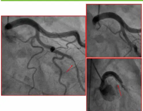
A case treated with cutting balloon.
A 50-year-old woman with no cardiovascular risk factors was admitted with an anterior STEMI. The coronary angiography showed spontaneous dissection of distal LAD. After successfully wiring the distal vessel, several dilatations with a 3.0/10mm cutting balloon at 12 atmospheres were performed (Figure 4, Panel A). Because of the absence of symptoms and TIMI 3 flow after dilatation, the operator opted to avoid stenting. The good result achieved in the acute phase (persistence of dissection flap but preserved flow) was confirmed with elective coronary angiography after both one week and one year (Figure 4, Panel B). The patient was discharged with EF 40% and with indication for one year of Dual Antiplatelet Therapy DAPT.
Treatment options in SCAD management
Conservative strategy
No randomized trials comparing conservative versus interventional strategies have ever been carried out so far in SCAD. The challenge of conducting such studies is due to the low incidence of the disease plus the varied severity of clinical presentations.
Today the available literature shows that the therapeutic strategy is conditioned by clinical presentation and stability, coupled with angiographic characteristics (site and extension of dissection or TIMI flow)[6]. The fact that PCI in SCAD is burdened with a high complication rate favours a conservative approach over a revascularisation strategy (either PCI or Coronary Artery Bypass Grafting) whenever clinically possible (i.e. stable patient without ongoing ischaemia).
Furthermore, there is a general understanding that, with a conservative treatment, dissections tend to heal completely over a certain period of time: with angiographic evidence from about 1 month after the acute episode[2,3,7]. Therefore, when revascularization is not required (i.e. in hemodynamically stable patient) the conservative management should be the first choice for these patients.
As mentioned above, revascularization is associated with high rates of failure and complications, likely due to an exaggerated vessel fragility[1,2,8]. This includes the risk of catheter-induced iatrogenic dissection of proximal-ostial locations, which may lead to serious clinical consequences and complex interventions (Figure 5).
Additionally, patients receiving a conservative strategy showed better in-hospital outcomes compared to the ones receiving revascularization, though similar long-term outcomes [4,6}. Distal location of the dissection and TIMI flow II or III may be considered predictive factors to favour the adoption of a conservative approach[6].
Nevertheless, our group recently highlighted the unpredictability of SCAD and the importance of a close clinical surveillance following an initial conservative strategy. In this case series[9], among four patients with similar angiographic and clinical presentation, two cases experienced a malignant evolution with need of emergent PCI and extensive stenting (including left main) while in the other two cases complete angiographic healing was demonstrated in follow-up angiograms.
Revascularisation should be reserved for high-risk patients with ongoing ischemia, left main artery dissection, ventricular arrhythmias or hemodynamic instability
Percutaneous Coronary Intervention
Procedural success of PCI in SCAD is dramatically low compared to that in the atherosclerotic population. The main studies assessing the role of PCI have shown a high rate of complications and unfavourable outcomes[10]. For an optimal procedural planification, careful assessment of the involved segment (proximal vs distal), lesion length and vessel sizing is mandatory in order to consider all potential challenges and feasibility.
Figure 3.
Panel A: coronary angiography showed SCAD of mid-LAD. Conservative treatment was chosen
Panel B: because of rest angina, a coronary angiography was repeated and showed SCAD progression
Since SCAD is a disease related to the weakening of the arterial wall, entering the true lumen with a guidewire is quite challenging. Consequently, PCI may easily lead to iatrogenic dissection or may provoke propagation of the existing one. Furthermore, even when true lumen is appropriately wired, the subsequent implantation of stent may determine the “squeezing” of the intramural hematoma (with possible extension of the dissection itself) and increase the risk of in-stent restenosis and stent thrombosis.
In an Italian series of 134 SCAD patients where successful PCI was achieved in 72.5%, patients treated conservatively had lower in-hospital major cardiac adverse events (MACEs) compared with those treated with revascularization (3.8% vs. 16.1%)[6]. Likewise, in the Mayo Clinic series of 189 patients, PCI failure occurred in 53%, and emergency CABG was required in 13%[8,10].
Wiring of the true lumen distal to the dissection site is probably the most challenging part of the procedure. As a first-line approach, we suggest using floppy non-plastic wires to navigate in the true lumen compressed by the intramural hematoma. If the wire fails to advance into the true lumen, especially in the case of complete occlusion of the vessel, hydrophilic wires could be used in order to facilitate distal re-entry in the true lumen. A distal tip microcatheter injection is often necessary to confirm correct position in the true lumen before any balloon dilatation (Figure 6). IVUS guidance may be employed to identify the false lumen and ensure correct wire placement within the true lumen. Likewise, IVUS will be useful for stent sizing.
In fact, choosing the right length and dimension of the stent represents the next challenge. The distal coronary segments are the most frequently affected and these may be too small for stenting. Moreover, haematoma resorption could lead to late stent malapposition (stent under-sizing).
Drug Eluting Stents
Today Drug Eluting Stents (DES) are considered the standard of care for the invasive treatment of the atherosclerotic disease. However, in the past years, doubts about DES use in patients with SCAD were raised[11]. As a matter of fact, DES reduce the risk of neointimal growth, but on the other side they may potentially delay the healing of the dissected vessel. Recently, a large observational study compared the use of DES against Bare Metal Stent (BMS) and demonstrated the same advantages
of DES observed in atherosclerotic disease over BMS even in patients with SCAD [12].
In any case, when a stent is to be implanted, less conventional interventional approaches can be considered to reduce the number of adverse outcomes[1–3,13]:
• Extended stent lengths to cover the intramural haematoma (IMH) borders to reduce the chances of proximal or distal propagation. Some authors[2] suggested to exceed at least 5 mm the dissection edges
• Sealing firstly the proximal and distal extremes of the affected segments with short stents to constrain haematoma before stenting the middle segment (sandwich technique)[14].
• Targeting an intimal tear for focal stenting or stenting just the proximal segment of the dissection to prevent proximal propagation.
• High-pressure dilation or post-dilation pursuing optimal stent deployment should be avoided to prevent iatrogenic propagation
• In cases of highly-compressive IMH, consider the use of cutting balloon prior to stent deployment (hybrid PCI)
• Consider optimising stent implantation in a staged procedure (allowing vessel remodelling/healing)
Bio-resorbable Scaffolds
When approaching a long dissected segment, operators usually prefer to implant long/multiple overlapped stents in order to cover the entire dissected segment. However, this exposes these generally young patients to the lifelong risk of restenosis and thrombosis[15].
For this reason, Bio-resorbable scaffolds (BRS) were proposed for the treatment of SCAD[16,17] in several case series in which bioreabsorbable (AbsorbTM) device was used[18,19] (Abbott
Vascular, Abbott Park, Illinois). Recently, the first case of use of BRS Magnesium Made Magmaris (BIOTRONIK, Buelach, Switzerland) Scaffold for SCAD treatment was also reported by our group[20,21].
The use of bioresorbable coronary scaffolds could be advantageous as there is general evidence that the temporary presence of the scaffold could reduce the risk of late maladjustment and thrombosis after resorption of IMH[20]. BRS provide a temporary support to seal the dissection and would potentially allow complete vessel healing over time. Coronary arteries affected by spontaneous dissection are usually free from hard plaque or heavy calcification which makes easier a full expansion of the scaffold avoiding the need for aggressive pre- and post-dilatation, strongly recommended for BRS in atherosclerotic vessels. Moreover, BRS compared to permanent metallic DES could potentially reduce the risk of malapposition after IMH reabsorption. BRS are also useful when a long scaffolding is demanding, especially considering the young age of these patients.
Nevertheless, some drawbacks must be underscored. Firstly, the initial enthusiasm kept on these devices has been tempered by clinical trials showing an increased risk of target lesion failure, both early and late after implantation of first-generation AbsorbTM
in comparison to DES[22]. Newer generation of scaffolds with faster reabsorption process and improved platforms will probably overcome these issues in the future even avoiding the risk of
Figure 4.
Panel A: coronary angiography showed distal LAD closure (type 4 SCAD)
prolonging DAPT over time in such patients. Moreover, most of the failure burden of BRS derives from the use of it in small-calibre vessels (2.5mm)[23]. As a matter of fact, such vessels are usually managed conservatively in SCAD. Finally, intracoronary imaging meticulous guidance should be central in any BRS implantation to achieve a satisfactory deployment and avoid complications.
Cutting Balloon
An alternative to deploy a stent/scaffold is to use cutting balloon only leaving the vessel prosthesis-free. The experience with such device is still limited to case reports, but the concept is very intriguing[24]. The basis derives from the potential to fenestrate the intimal-medial layer and depressurize the false lumen, restoring flow and alleviating acute ischaemia. Conversely, there is a theoretical potential risk of coronary rupture, thus the use of an undersized balloon is probably a reasonable option.
Less conventional interventional approaches
Among other specific devices, a rationale for the use of self-expandable stent (SES) has been recently proposed. Basically, SES could potentially overcome the risk of late malapposition of the struts (due to the underestimation of the vessel caliber) finalizing the expansion of the stent once the IMH reabsorption is completed. However the role of SES is limited to the best of our knowledge to a single case report[25].
Role of intravascular imaging in SCAD treatment
Use of intravascular imaging techniques (IntraVascular Ultra Sound (IVUS) or Optical Coherent Tomography (OCT)) plays a relevant role in confirming SCAD diagnosis when angiographic appearance is ambiguous. But more importantly, it probably should be an essential tool to guide PCI[2]. As discussed previously, imaging is helpful to prove the correct position of the intracoronary wire; to accurately size the lumen in order to choose the right dimensions of the stent;, to verify the complete stent apposition and the whole coverage of the disease and potentially to show coexistent atherosclerosis or other vascular disorder. On the other hand, extraordinary costs and availability may limit the use of intravascular imaging techniques. Moreover, requirement of anticoagulation could provoke haematoma extension. Last but not least, vessel instrumentation can produce iatrogenic damage and superimposed dissections, as a consequence of the wire, the guiding catheter or the contrast infusion in case of OCT. In a single-centre study, some degree of iatrogenic damage was seen in a fifth of the cases were intracoronary imaging was used for diagnosing SCAD, which led to unplanned PCI in most of them[26].Conclusions
SCAD represents today a coronary condition with unique pathophysiological characteristics clearly different from the common atherosclerosis. Therefore, when a PCI strategy is preferred, a tailored approached is required taking advantages from extensive use of imaging whenever it is feasible. Finally new technologies answer generations of scaffold has to keep in mind in order to avoid a permanent extensive stenting of an artery usually free from atherosclerosis and with a good chance of complete healing over time. However, dedicated trial should be conducted to define their safety and effectiveness in long term.
Declarations of interest
Enrico Cerrato received speaker fee in educational events supported by BIOTRONIK, ABBOTT, VOLCANO and a research grant from ASTRAZENECA.
Giorgio Quadri, Francesco Tomassini, Cristina Rolfo received speaker fee in educational events supported by BIOTRONIK, ABBOTT
Fabio Ferrari and Fabio Mariani received speaker fee in educational events supported by BIOTRONIK and BAYER. Javier Escaned received speaker fee at educational events and a consultant for BOSTON SCIENTIFIC, PHILIPS VOLCANO. Ferdinando Varbella joined advisory board from ORBUSNEICH, BIOSENSORS, ABBOTT VASCULAR, CID, TELEFLEX, STENTYS. Fabrizio D’Ascenzo received fee from ASTRAZENECA, BOSTON, CHIESI, MEDTRONIC, CROS NT and research grant from BIOSENSOR.
Figure 5.
Panel A: Catheter induced coronary dissection risk. Left 50 years-old woman presenting with ACS-NSTE. Diagnostic angiogram performed with a 4F right radial access showed a Type 3 SCAD on the Obtusal Marginal Branch. Because of the absence of symptoms and/or high-risk features at the time of catheterization, it was medically treated.
Panel B: Left Main at the end of the diagnostic procedure.
Panel C: Left Main dissection with abrupt closure of the left coronary artery.
Distal tip microcatheter injection of medium contrast to check the position of the wire in the coronary vessel.
Panel D: microcatheter tip is in the wrong position, medium contrast fill the false lumen.
Fernando Macaya received a grant from Fundación Interhospitalaria Investigación Cardiovascular and Roberto Pozzi declares no conflicts of interest.
Acknowledgements
The authors state that they abide by the authors’ responsibilities and ethical publishing guidelines of the International Cardiovascular Forum Journal [27].
References
1. Adlam, D. et al. European Society of Cardiology, acute cardiovascular care association, SCAD study group: a position paper on spontaneous coronary artery dissection. Eur Heart J doi:10.1093/eurheartj/ehy080
2. Saw, J., Mancini, G. B. J. & Humphries, K. H. Contemporary Review on Spontaneous Coronary Artery Dissection. J. Am. Coll. Cardiol. 68, 297–312 (2016). DOI: 10.1016/j.jacc.2016.05.034
3. Hayes, S. N. et al. Spontaneous Coronary Artery Dissection: Current State of the Science: A Scientific Statement From the American Heart Association. Circulation CIR.0000000000000564 (2018). doi:10.1161/ CIR.0000000000000564
4. Alfonso, F. et al. Spontaneous coronary artery dissection: novel insights on diagnosis and management. Cardiovasc Diagn Ther 5, 133–140 (2015). DOI: 10.3978/j.issn.2223-3652.2015.03.05
5. Adams, H. et al. Different patients, different outcomes: A case-control study of spontaneous coronary artery dissection versus acute coronary syndrome. J Interv Cardiol 31, 41–47 (2018). DOI: 10.1111/joic.12447
6. Lettieri, C. et al. Management and Long-Term Prognosis of Spontaneous Coronary Artery Dissection. Am. J. Cardiol. 116, 66–73 (2015). DOI: 10.1016/j.amjcard.2015.03.039
7. Vanzetto, G. et al. Prevalence, therapeutic management and medium-term prognosis of spontaneous coronary artery dissection: results from a database of 11,605 patients. Eur J Cardiothorac Surg 35, 250–254 (2009). DOI: 10.1016/j.ejcts.2008.10.023
8. Tweet, M. S. et al. Clinical features, management, and prognosis of spontaneous coronary artery dissection. Circulation 126, 579–588 (2012). DOI:10.1161/CIRCULATIONAHA.112.105718
9. Quadri, G. et al. Importance of Close Surveillance of Patients With Conservatively Managed Spontaneous Coronary Artery Dissection. JACC Cardiovasc Interv 11, e87–e89 (2018). DOI: 10.1016/j.jcin.2018.03.020 10. Tweet, M. S. et al. Spontaneous coronary artery dissection: revascularization
versus conservative therapy. Circ Cardiovasc Interv 7, 777–786 (2014). DOI:10.1161/CIRCINTERVENTIONS.114.001659
11. Imai, M. et al. Incidence, risk factors, and clinical sequelae of angiographic peri-stent contrast staining after sirolimus-eluting peri-stent implantation. Circulation 123, 2382–2391 (2011). DOI: 10.1161/CIRCULATIONAHA.110.003459 12. Conrotto, F. et al. Safety and efficacy of drug eluting stents in patients
with spontaneous coronary artery dissection. Int. J. Cardiol. 238, 105–109 (2017). DOI: 10.1016/j.ijcard.2017.03.027
13. Macaya, F. et al. Spontaneous coronary artery dissection: contemporary aspects of diagnosis and patient management. Open Heart 5, e000884 (2018). DOI: 10.1136/openhrt-2018-000884
14. Dashwood, A. M., Saw, J., Dhillon, P. & Murdoch, D. Use of a Three-Stent Technique for a Case of Spontaneous Coronary Artery Dissection. Canadian Journal of Cardiology 33, 830.e13-830.e15 (2017). DOI: 10.1016/j. cjca.2017.02.007
15. Lempereur, M., Fung, A. & Saw, J. Stent mal-apposition with resorption of intramural hematoma with spontaneous coronary artery dissection. Cardiovasc Diagn Ther 5, 323–329 (2015). DOI: 10.3978/j.issn.2223-3652.2015.04.05
16. Macaya, F. et al. Bioresorbable Scaffolds to Treat Spontaneous Coronary Artery Dissection. Circ Cardiovasc Interv 9, e003133 (2016). DOI: 10.1161/ CIRCINTERVENTIONS.115.003133
17. Macaya, F. et al. Long-term follow-up of spontaneous coronary artery dissection treated with bioresorbable scaffolds. EuroIntervention (2018). doi:10.4244/EIJ-D-18-00519
18. Cockburn, J., Yan, W., Bhindi, R. & Hansen, P. Spontaneous Coronary Artery Dissection Treated With Bioresorbable Vascular Scaffolds Guided by Optical Coherence Tomography. Canadian Journal of Cardiology 30, 1461. e1-1461.e3 (2014).DOI: 10.1016/j.cjca.2014.06.025
19. Cerrato, E., Tomassini, F., Rolfo, C., Gagnor, A. & Varbella, F. Spontaneous coronary artery dissection treated with biovascular scaffolds guided by intravascular ultrasounds imaging. Cardiovasc Interv Ther 32, 186–189 (2017). DOI: 10.1007/s12928-016-0391-3
20. Quadri, G., Tomassini, F., Cerrato, E. & Varbella, F. First reported case of magnesium-made bioresorbable scaffold to treat spontaneous left anterior descending coronary artery dissection. Catheter Cardiovasc Interv 90, 768– 772 (2017). DOI: 10.1002/ccd.27214
21. Quadri, G., Cerrato, E., Rolfo, C. & Varbella, F. Spontaneous coronary artery dissection treated with magnesium-made bioresorbable scaffold:
1-Year angiographic and optical coherence tomography follow-up. Catheter Cardiovasc Interv (2018). doi:10.1002/ccd.27971
22. Sorrentino, S. et al. Everolimus-Eluting Bioresorbable Scaffolds Versus Everolimus-Eluting Metallic Stents. J. Am. Coll. Cardiol. 69, 3055–3066 (2017). DOI: 10.1016/j.jacc.2017.04.011
23. Stone, G. W. & Granada, J. F. Very Late Thrombosis After Bioresorbable Scaffolds: Cause for Concern? J. Am. Coll. Cardiol. 66, 1915–1917 (2015). DOI: 10.1016/j.jacc.2015.08.863
24. Ito, T., Shintani, Y., Ichihashi, T., Fujita, H. & Ohte, N. Non-atherosclerotic spontaneous coronary artery dissection revascularized by intravascular ultrasonography-guided fenestration with cutting balloon angioplasty. Cardiovasc Interv Ther 32, 241–243 (2017). DOI: 10.1007/s12928-016-0397-x 25. Mele, M., Langialonga, T., Maggi, A., Villella, M. & Villella, A. Self-expanding
stent for spontaneous coronary artery dissection: a rational choice. J Cardiovasc Med (Hagerstown) 17 Suppl 2, e254–e256 (2016). DOI: 10.2459/JCM.0000000000000371
26. Macaya, F. et al. Feasibility and safety of intracoronary imaging for diagnosing spontaneous coronary artery dissection. JACC Cardiovasc Imaging, (2018). DOI: 0.1016/j.jcmg.2018.09.023