STRUCTURAL CHARACTERIZATION AND HYDROLYSIS
STUDIES OF REBAUDIOSIDE E, A MINOR SWEET
COMPONENT OF STEVIA REBAUDIANA
Venkata Sai Prakash Chaturvedula
[a]* and Indra Prakash
[a]Keywords:stevia rebaudiana, compositae, asteraceae, rebaudioside A, spectral data, chemical studies.
From the commercial extract of the leaves of Stevia rebaudiana, a diterpene glycoside was isolated which was characterized as 13-[(2-O -β-D-glucopyranosyl-β-D-glucopyranosyl)oxy] ent-kaur-16-en-19-oic acid-(2-O-β-D-glucopyranosyl-β-D-glucopyranosyl) ester (1); also known as rebaudioside E. The complete 1H and 13C NMR assignments of rebaudioside E was achieved by the extensive 1D and 2D NMR
(1H and 13C, COSY, HMQC, HMBC) as well as mass spectral data. Further, hydrolysis studies were performed on rebaudioside E using
acid and enzymatic studies to identify aglycone and sugar residues in its structure.
* Corresponding Authors
E-Mail: [email protected]
[a] Organic Chemistry Department, The Coca-Cola Company, Global Research and Development, One Coca-Cola Plaza, Atlanta, GA 30313, USA Chapters
Introduction
Stevia rebaudiana (Bertoni) is a perennial shrub belonging to the family of Asteraceae (Compositae) native to Brazil and Paraguay, but now grown commercially in a number of countries, particularly in Japan, Taiwan, Korea, Thailand and Indonesia.1-2 Extracts of the leaves of S. rebaudiana
have been used for decades to sweeten food and beverages in Japan, South America and China. The major constituents in the leaves of S. rebaudiana are the potently sweet glycosides namely steviolbioside, stevioside, rebaudiosides A and E, dulcoside A and rubusoside; which are glycosides of the diterpene steviol, ent-13-hydroxykaur-16-en-19-oic acid.3-4 These compounds are also known as Stevia
sweeteners. Rebaudioside E is minor component of S. rebaudiana and tastes about 150-200 times sweeter than sucrose and is non-caloric.
In our continuing research to discover natural sweeteners, we have collected commercial extracts of S. rebaudiana
from various suppliers all over the World and isolated several novel diterpene glycosides.5-12 Apart from isolating
novel compounds from S. rebaudiana and utilizing them as possible natural sweeteners or sweetness enhancers, we are also engaged in understanding the stability of the steviol glycosides in various systems of interest and identification of degradation products using various spectroscopic techniques as well as synthesis using naturally occurring starting materials and their taste evaluation.13-18 In this
article, we are describing the isolation, characterization and complete 1H and 13C NMR spectral assignments for the
diterpene glycoside 13-[(2-O -β-D-glucopyranosyl-β-D-glucopyranosyl)oxy] ent-kaur-16-en-19-oic acid-(2-O -β-D-glucopyranosyl-β-D-glucopyranosyl) ester (1) which is also known as rebaudioside E (Figure 1). The complete NMR assignments were achieved on the basis of 1D (1H and 13C)
and 2D (COSY, HMQC and HMBC) NMR as well as high resolution mass spectroscopic data. Acid, and enzymatic
hydrolysis studies on compound 1 were carried out to identify aglycone and sugar residues.
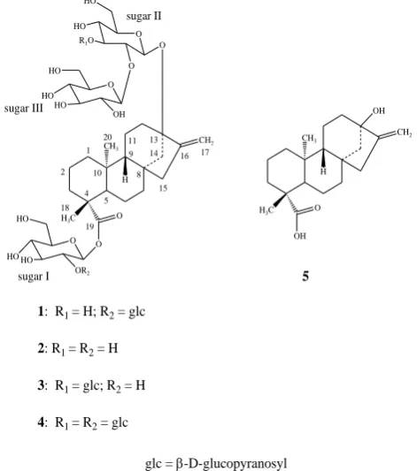
Figure 1. Structure of rebaudioside E (1) and other compounds
EXPERIMENTAL
General Instrumentation Procedures
Melting points were measured using a SRS Optimelt MPA 100 instrument and are uncorrected. Optical rotation was performed using Rudolph Autopol V at 25 oC and IR
spectral data was acquired using a Perkin Elmer 400 Fourier Transform Infrared (FT-IR) Spectrometer with Universal attenuated total reflectance (UATR) polarization accessory. HPLC analysis was performed using an Agilent (Wilmington, DE) 1200 system, including a quaternary
CH2
H CH3
H3C O
O 1
2
4 5
8 9 10
11 13 14
15 16 17
18 19
20 O
O O HO
HO HO
HO
HO R1O
O HO
HO HO
1: R1 = H; R2 = glc
2: R1 = R2 = H
3: R1 = glc; R2 = H
4: R1 = R2 = glc
CH2
H CH3
OH
H3C O
OH
5
sugar I
sugar II
sugar III OH
O
OR2
pump, a temperature controlled column compartment with additional 6-port switching valve, an autosampler and a UV absorbance detector. Phenomenex Prodigy (Torrance, CA) ODS (3) with a Phenomenex guard column, 250 x 21.2 mm, 5 m (p/n 00G-4097-P0); and Phenomenex Synergi (Torrance, CA) Hydro RP, 250 x 10 mm, 4 m (p/n 00G-4336-N0) were used for the purification of rebaudioside E (1).
Analytical HPLC was carried out with a Waters 600E multisolvent delivery system using a Phenomenex Luna C18
(150 x 4.6 mm, 5 m) column for sugar identification.
NMR spectra were acquired on a Bruker DRX 500 MHz instrument with a 5 mm inverse detection probe using standard pulse sequences. The NMR spectra were performed in CD3OD and C5D5N; chemical shifts are given in (ppm),
and coupling constants are reported in Hz. The spectral data was referenced to the residual solvent signal (H 3.30, and
C 49.0 for CD3OD or H 7.19, and C 123.5 for pyridine-d5).
MS and MS/MS data were generated with a Waters Premier QTof mass spectrometer equipped with an electrospray ionization source. Samples were diluted with H2O:acetonitrile (1:1) containing 0.1% formic acid and
introduced via infusion using the onboard syringe pump. The samples were diluted to yield good signal to noise (s/n) which occurred at an approximate concentration of 0.01 mg mL-1.
Plant Material
SG95, the commercial aqueous extract consisting of a mixture of diterpenoid glycosides of the leaves of S. rebaudiana was obtained from the Pure Circle (Kuala Lumpur, Malaysia). The authenticity of the crude extract was confirmed by performing its retention time (tR) comparison with the internal standard compounds of known steviol glycosides isolated from S. rebaudiana using the preparative HPLC method as reported earlier.19 A voucher
specimen is deposited at The Coca-Cola Company, No. VSPC-3166-002.
Isolation and Characterization
Compound 1 was purified in two rounds by using an Agilent HPLC 1200 system equipped with a reversed phase (RP) HPLC. The initial round of purification using reversed phase HPLC is summarized below:
Column: Phenomenex Prodigy (Torrance, CA) ODS (3) with a Phenomenex guard column, 250 x 21.2 mm, 5 m (p/n 00G-4097-P0); Mobile Phase A: H2O; Mobile Phase B:
Acetonitrile; Flow Rate: 20 mL min-1; Injection volume:
1000 μL at 77 mg/mL, prepared in Acetonitrile / H2O (1:1).
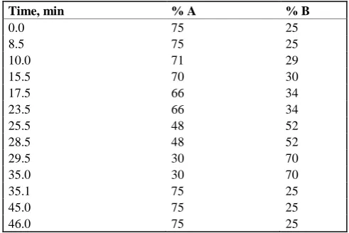
UV detection at 210 nm was used for obtaining a fraction rich of the steviol glycoside 1 with a total run time of 46 min (Table 1).
All of the baseline material eluting between retention time (tR) 11 and 18 min was collected and dried by rotary
evaporation under reduced pressure for second round of purification.
Table 1. RP-HPLC method utilized for the purification of fraction rich in Rebaudioside E (1)
A second round of purification was used using an Agilent HPLC 1200 system equipped with a reversed phase (RP) HPLC using the method given below: Column: Phenomenex Synergi Hydro RP, 250 x 10 mm, 4 m (p/n 00G-4336-N0); UV Detection: 210 nm; Mobile Phase A: H2O (0.01156%
HOAc, 0.02844% NH4OAc); Mobile Phase B: Acetonitrile;
Flow Rate: 5.0 mL/min; Injection volume: 250 μL, prepared in H2O.
Table 2. RP-HPLC method utilized for the purification of pure rebaudioside E (1)
Using the above mentioned HPLC method shown in Table 2, collected the peak eluting at tR 7.05 min; and dried the
corresponding solution under nitrogen yielded 1.
13-[(2-O-β-D-glucopyranosyl-β-D-glucopyranosyl)oxy] ent-kaur-16-en-19-oic acid-(2-O-β-D-glucopyranosyl-β-D-gluco-pyranosyl) ester (Rebaudioside E, 1)
White powder; IR νmax: 3315, 2943, 1722, 1055, 910 cm-1; 1H-NMR (600 MHz, CD
3OD, δ ppm) and 13C-NMR (150
MHz, CD3OD, δ ppm) spectroscopic data see Table 3;
HRMS (M+H)+ m/z 967.4418 (calcd. for C
44H71O23:
967.4386); (M+NH4)+m/z 984.4690 (calcd. for C44H74O23N:
984.4652); (M+Na)+m/z 989.4237 (calcd. for C
44H70O23Na:
989.4206).
Acid hydrolysis of 1
To a solution of compound 1 (2 mg) in MeOH (3 ml) was added 3 ml of 5% H2SO4 and the mixture was refluxed for 8
hours. The reaction mixture was then neutralized with saturated sodium carbonate and extracted with ethyl acetate (EtOAc) (2 x 25 ml) to give an aqueous fraction containing sugars and an EtOAc fraction containing the aglycone part.
Time, min % A % B
0.0 75 25
8.5 75 25
10.0 71 29
15.5 70 30
17.5 66 34
23.5 66 34
25.5 48 52
28.5 48 52
29.5 30 70
35.0 30 70
35.1 75 25
45.0 75 25
46.0 75 25
Time, min %A %B
0.0 75 25
8.5 75 25
10.0 71 29 16.5 70 30 16.51 0 100
21 0 100
21.1 75 25
The aqueous phase was concentrated and compared with standard sugars using the TLC systems EtOAc/n -butanol/water (2:7:1) and CH2Cl2/MeOH/water (10:6:1),20-22
the sugar was identified as glucose.
Determination of sugar configuration in 1
Compound 1 (1 mg) was hydrolyzed with 0.5 M HCl (2 mL) for 1.5 h. After cooling, the mixture was passed through an Amberlite IRA400 column and the eluate was lyophilized. The residue was dissolved in pyridine (1 mL) and heated with L-cysteine methyl ester HCl (5 mg) at 60ºC for 1.5 h, and then O-tolyl isothiocyanate (25 L) was added to the mixture and heated at 60 ºC for an additional 1.5 h. The reaction mixture was analyzed by HPLC: column Phenomenex Luna C18, 150 x 4.6 mm (5 u); 25% acetonitrile-0.2% TFA water, 1 mL min-1; UV detection at
250 nm. The sugar was identified as D-glucose (tR, 12.32 min) [authentic samples, D-glucose (tR, 12.38) and
L-glucose (tR, 11.16 min)].23
Enzymatic hydrolysis of 1
Compound 1 (1 mg) was dissolved in 10 ml of 0.1 M sodium acetate buffer, pH 4.5 and crude pectinase from
Aspergillus niger (50 uL, Sigma-Aldrich, P2736) was added. The mixture was stirred at 50 oC for 48 hr. The product
precipitated out during the reaction and was filtered and then crystallized. The resulting product obtained from the hydrolysis of 1 was identified as steviol (5) by comparison of its co-TLC with standard compound and 1H NMR
spectral data.24
Results and Discussion
Compound 1 was isolated as a colorless powder and its positive mode of ESI Tof mass spectrum indicated an [M+H]+ ion at m/z 967.4418 together with [M+NH
4]+ and
[M+Na]+ adducts at m/z 984.4690 and 989.4237,
respectively. The mass of the [M+H]+ ion was in good
agreement with the molecular formula C44H70O23 (calcd for
C44H71O23: 967.4386) for rebaudioside E (1) and this
composition was supported by 13C NMR spectral data. The
negative mode of ESI mass spectrum gave an [M-H]- ion at
m/z 965.4263 which was in good agreement with the molecular formula C44H70O23 (calcd for C44H69O23:
965.4230) supported further its molecular weight. The +ESI and -ESI data indicated that compound 1 has a nominal mass of 966 Daltons with the molecular formula C44H70O23
and is, therefore, an isomer of rebaudioside A (3). The MS/MS spectrum of 1, fragmenting on the [M+H]+ ion at
m/z 967 indicated the sequential loss of four hexose moieties at m/z 805.3868, 643.3336, 481.2813, and 319.2299.
The 1H NMR spectrum of 1 showed the presence of two
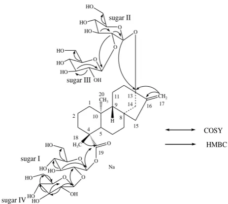
methyl singlets at δ 1.08 and 1.40, two olefinic protons as singlets at δ 5.06 and 5.73 of an exocyclic double bond, nine methylene and two methine protons between δ 0.71-2.52 characteristic for the ent-kaurane diterpenoids isolated earlier from the genus Stevia.3-10 The basic skeleton of
ent-kaurane diterpenoids was supported by COSY (1/2; H-2/H-3; H-5/H-6; H-6/H-7; H-9/H-11; H-11/H-12) and HMBC (H-1/2, 10; H-3/1, 2, 4, 5, 18, C-19; H-5/C-4, C-6, C-7, C-9, C-10, C-18, C-19, C-20; H-9/8, 10, 11, 12, 14, 15; H-14/H-9/8, 9, 13,
C-15, C-16 and H-17/C-13, C-C-15, C-16) correlations. The presence of four sugar units in its structure was supported by the 1H NMR spectrum of 1 which showed the anomeric
protons at δ 5.14, 5.29, 5.45, and 6.28. Enzymatic hydrolysis of 1 furnished an aglycone which was identified as steviol (5) by comparison of 1H NMR 24 and co-TLC with
standard compound. Acid hydrolysis of 1 with 5% H2SO4
afforded glucose which was identified by direct comparison with authentic samples by TLC.20-22 The stereochemistry of
the sugar was identified as D-glucose by preparing its corresponding thiocarbamoyl-thiazolidine carboxylate derivatives with L-cysteine methyl ester and O-tolyl isothiocyanate, and in comparison of their retention times with the standard sugars as described in the literature.23 The 1H and 13C NMR values for all the carbons in 1 were
assigned on the basis of COSY, HMQC and HMBC correlations (Table 3).
Figure 2. Key COSY and HMBC correlations of 1
Based on the results from NMR spectral data and hydrolysis experiments of 1, it was concluded that there are four β-D-glucosyl units in its structure similar to rebaudioside A (3). A close comparison of the 1H and 13C
NMR values of 1 with stevioside (2) and rebaudioside D (4) suggested the presence of a 2-substituted β-D-glucosyl unit at C-13 in the form of ether and another 2-substituted β-D-glucosyl unit at C-19 position in the form of an ester. This was confirmed by the key HMBC correlations: H-2′′/C-1′′, C-3′′, C-1′′′; H-1′′′/C-2′′, C-2′′′, C-3′′′; H-2′/C-1′, C-3′, C-1′′′′ and H-1′′′′/C-2′, C-2′′′′, C-3′′′′. The large coupling constants observed for the four anomeric protons of the glucose moieties at δ 5.14 (d, J=7.6 Hz), 5.29 (d, J=7.7 Hz), 5.45 (d,
J=7.8 Hz), and 6.28 (d, J=7.9 Hz), suggested their
β-orientation as reported for steviol glycosides.5-12 Based on
the results from chemical and spectral studies, 1 was assigned as 13-[(2-O -β-D-glucopyranosyl-β-D-glucopyranosyl)oxy] ent-kaur-16-en-19-oic acid-(2-O -β-D-glucopyranosyl-β-D-glucopyranosyl) ester. The structure was further supported by the key COSY and HMBC correlations as shown in Figure 2.
COSY
HMBC
CH2
H CH3
H3C O
O 1 2
4 5
8 9 10
11 13 14
15 16 17
18 19 20
O
O O HO
HO HO
OH HO HO
HO
O HO
HO HO
sugar I
sugar II
sugar III
O
sugar IV
O O HO
HO
HO OH
Table 3. 1H and 13C NMR spectral data (chemical shifts and
coupling constants) for rebaudioside E (1).a-c
Position 1H NMR 13C NMR
1 0.71 t (13.4), 1.68 m 40.3 2 1.42 m, 2.11 m 19.7 3 1.08 m, 2.75 d (13.1) 37.4
4 --- 44.3
5 0.96 d (12.2) 57.1 6 1.84 d (12.1), 2.09 m 21.7 7 1.26 m, 1.32 m 41.3
8 --- 42.5
9 0.86 m 53.7
10 --- 39.2
11 1.64 m 20.3 12 1.94 m, 2.14 m 37.0
13 --- 86.1
14 1.73 d (11.1), 2.52 d (11.1) 44.3 15 2.03 m, 2.07 m 47.6 16 --- 154.5 17 5.06 s, 5.73 s 104.6 18 1.40 s 28.9 19 --- 175.6 20 1.08 s 16.3 1′ 6.28 d (7.9) 93.2 2′ 4.38 m 80.9 3′ 4.28 m 77.9 4′ 4.24 m 71.4 5′ 3.90 m 78.6 6′ 4.33 m, 4.43 m 61.8 1′′ 5.14 d (7.6) 97.5 2′′ 4.17 m 84.2 3′′ 4.32 m 77.8 4′′ 4.20 m 71.1 5′′ 3.71 m 77.3 6′′ 4.24 m, 4.35 m 62.0 1′′′ 5.45 d (7.8) 105.4 2′′′ 4.04 t (8.2) 76.0 3′′′ 4.23 m 77.7 4′′′ 4.28 m 70.5 5′′′ 3.96 m 78.1 6′′′ 4.44 m, 4.52 m 62.4 1′′′′ 5.29 d (7.7) 106.3 2′′′′ 4.13 t (8.4) 76.9 3′′′′ 4.23 m 77.7 4′′′′ 4.32 m 71.3 5′′′′ 3.96 m 78.1 6′′′′ 4.44 m, 4.52 m 62.4
a assignments made on the basis of COSY, HMQC and HMBC
correlations; b Chemical shift values are in δ (ppm); c Coupling
constants are in Hz.
Though partial NMR spectral data has been reported earlier for rebaudioside E (1) by Ohta et al25, this is the first
report of the complete 1H and 13C NMR spectral
assignments based on 1D (1H and 13C) and 2D (COSY,
HMQC and HMBC) NMR as well as high resolution mass spectroscopic data which was supported by hydrolysis studies.
Conclusions
We are herewith reporting the complete 1H and 13C NMR
spectral assignments for 13-[(2-O -β-D-glucopyranosyl-β-D-glucopyranosyl)oxy] ent-kaur-16-en-19-oic acid-(2-O
-β-D-glucopyranosyl-β-D-glucopyranosyl) ester (Rebaudioside E,
1) that were made on the basis of extensive 1D and 2D NMR as well as high resolution mass spectral data. Further, acid hydrolysis furnished D-glucose suggesting the presence only sugar unit and enzymatic hydrolysis furnished steviol.
Acknowledgements
We wish to thank AMRI, Bothell, WA, USA for obtaining some selected spectral data and Chris Mubarak, Analytical Sciences Department, The Coca-Cola Company, Atlanta, GA for providing IR spectral data.
References
1Mosettig. E., Beglinger, U., Dolder, F., Lichiti, H., Quitt, P.,
Waters, J. A. J. Am. Chem. Soc.,1963, 85, 2305.
2Mosettig. E., Beglinger, U., Dolder, F., Lichiti, H., Quitt, P.,
Waters, J. A. J. Org. Chem.,1955, 20, 884-899.
3Wayne, E. S., Lin, L. Carbohydr. Res.,2009, 344, 2533-2538. 4Brandle, J. E., Starrratt, A.N., Gijen, M. Can. J. Plant Sci.,1998,
78, 527–536.
5Chaturvedula, V. S. P., Prakash, I. J. Med. Plants Res.,2011, 5,
4838-4842.
6Chaturvedula, V. S. P., Mani, U.; Prakash, I. Molecules,2011, 16,
3552–3562.
7Chaturvedula, V. S. P., Prakash, I. Molecules,2011, 16, 2937–
2943.
8Chaturvedula, V. S. P., Prakash, I. Carbohydr. Res., 2011, 346,
1057–1060.
9Chaturvedula, V. S. P., Rhea, J., Milanowski, D., Mocek, U.,
Prakash, I. Nat. Prod. Commun.,2011, 6, 175–178.
10Chaturvedula, V. S. P., Prakash, I. Nat. Prod. Commun.,2011, 6,
1059–1062.
11Chaturvedula, V. S. P., Clos, J. F., Rhea, J., Milanowski, D.,
Mocek, U., DuBois, G. E., Prakash, I. Phytochemistry Lett., 2011, 4, 209–212.
12Chaturvedula, V. S. P., Mani, U., Prakash, I. Carbohydr. Res.,
2011, 346, 2034–2038
13Chaturvedula, V. S. P., Klucik, J., Mani, U., Prakash, I.
Molecules,2011, 16, 8402–8409.
14Chaturvedula, V. S. P., Clos, J. F., Prakash, I. Int. J. Pharm.
Pharm. Sci.,2011, 3, 316–323.
15Chaturvedula, V. S. P., Clos, J. F., Prakash, I. Int. J. Pharm.
Pharm. Sci.,2011, 3 (Suppl. 5), 421–425.
16Prakash, I., Clos, J. F., Chaturvedula, V. S. P. Food Res. Int.
2012, 48, 65–75.
17Chaturvedula, V. S. P., Campbell, M., Miguel, R. I. S., Prakash, I.
Molecules,2012, 17, 8908–8916.
18Chaturvedula, V. S. P., Campbell, M., Prakash, I. Int. J. Mol. Sci.,
2012, 13, 15126-15136.
19Clos, J. F., DuBois, G. E., Prakash, I. J. Agric. Food Chem.2008,
56, 8507-8513.
20Bedir, E., Toyang, N. J., Khan, I. A., Walker, L. A., Clark, A. M.,
J. Nat. Prod.,2001, 64, 95-97.
21Chaturvedula, V. S. P., Schilling, J. K., Miller, J. S.,
Andriantsiferana, R., Rasamison, V. E., Kingston, D. G. I.
Planta Med.,2003, 69, 440-444.
22Huan, V. D., Yamamura, S., Ohtani, K., Kasai, R., Yamasaki, K.,
23Tanaka, T., Nakashima, T., Ueda, T., Tomii, K., Kouno, I. Chem.
Pharm. Bull.,2007, 55, 899-901.
24Ohtani, K., Aikawa, Y., Kasai, R., Chou, W., Yamasaki, K.,
Tanaka, O. Phytochemistry,1992, 31, 1553-1559.
25Ohta, M., Sasa, S., Inoue, A., Tamai, T., Fujita, I., Morita, K.,