© 2019 by the Serbian Biological Society How to cite this article: Kim D, Park NH, Hwang JA, Kim J, Na YJ, Hwang JS, Lee 483 CS, Yang DC. Camellia sinensis leaf extracts lacking catechins exert depigmentary
effects through ERK-dependent, MiTF-mediated tyrosinase downregulationin melan-a cells and a human skin equivalent. Arch boil Sci. 2019;71(3):483-8.
Camellia sinensis
leaf extracts lacking catechins exert depigmentary effects through
ERK-dependent, MiTF-mediated tyrosinase downregulation in melan-a cells and a
human skin equivalent
Donghyun Kim1, Nok Hyun Park2, Jeong Ah Hwang3, Jeongkee Kim2, Yong-Joo Na3, Jae Sung Hwang2, Chang
Seok Lee4,* and Deok-Chun Yang1,2,#
1Department of Oriental Medicinal Biotechnology, College of Life Science, Kyung Hee University, Yongin-si, Gyeonggi
do 17104, Republic of Korea
2Department of Genetic Engineering and Graduate School of Biotechnology, College of Life Sciences, Kyung Hee University,
Yongin-si, Republic of Korea
3Amorepacific R&D Center, Yongin-si, Republic of Korea
4Department of Beauty and Cosmetic Science, Eulji University, Seongnam-si, Republic of Korea
Corresponding authors: #[email protected]; *[email protected]
Received: May 10, 2019; Revised: May 23, 2019; Accepted: May 24, 2019; Published online: May 28, 2019
Abstract: Green tea from Camellia sinensis, a popular beverage worldwide, is also considered a herbal medicine. The
bioactive compounds of green tea include polyphenols, polysaccharides, amino acids, and vitamins. Tea polyphenols are composed of various catechins such as epigallocatechin-3-gallate (EGCG), whereas polysaccharides include complex pectic substances and glycoproteins. Unfractionated C. sinensis leaf extracts show various pharmacological effects that are attrib-utable to catechins, which include antimelanogenic properties. Most studies have focused on the biological function of catechins in green tea, and the effects of C. sinensis leaf extracts lacking catechins (CSLE-LC) have received little research attention. We examined the skin-whitening properties of the fraction lacking catechins. The melanin content in melan-a cells was significantly reduced, as has been shown for unfractionated C. sinensis extracts and catechins. We also elucidated the molecular mechanism underlying the antimelanogenic effects of CSLE-LC on skin melanocytes. Our results show that CSLE-LC acts through inhibition of MiTF and subsequent activation of extracellular-signal regulated kinase (ERK) to reduce tyrosinase protein levels. We confirmed the whitening ability of CSLE-LC using a human skin equivalent. Our findings provide the first evidence that CSLE-LC could exert efficient antimelanogenesis activity, and suggest that polysaccharides as well as catechins contribute to the whitening efficacy of C. sinensis leaf extracts.
Keywords: Camellia sinensis;catechins; melanogenesis; tyrosinase
INTRODUCTION
Green tea produced from the leaves of Camellia
sin-ensis is one of the most popular beverages consumed
worldwide. The potential health benefits of green tea, produced mainly in Asian countries from the leaves of the C. sinensis plant, have been widely studied [1]. Among the important components in green tea are polyphenolic compounds known as catechins. Green tea contains four main catechins: epicatechin (EC), epicatechin gallate (ECG), epigallocatechin (EGC) and epigallocatechin-3-gallate (EGCG) [1-2]. The benefi-cial effects of green tea catechins include antioxidant
from green tea have been shown to possess many health benefits, including antioxidant, anti-aging, antitumor and antibacterial properties [8-10], few studies have investigated the efficacy of C. sinensis leaf extracts that lack catechins.
Melanogenesis, the process of melanin production, serves to protect the skin against deleterious ultraviolet (UV) irradiation [11]. However, overexpression of melanin causes freckles, senile lentigines and other forms of melanin hyperpigmentation, which are seri-ous human aesthetic problems [12]. However, despite the development of numerous depigmentary agents, none has proved to be clinically satisfactory in terms of safety or efficacy. Tyrosinase is an enzyme that cata-lyzes the rate-limiting step in melanogenesis, and its expression is regulated by microphthalmia-associated transcription factor (MiTF) [13]. Thus, inhibition of tyrosinase upregulation or activation could be a useful targeting strategy for the development of whitening compounds against hyperpigmentation. EGCG and extracts from C. sinensis inhibit melanin synthesis in melanocytes; however, their mechanism of action is not completely understood [14-16]. To date, there are no reports on the antimelanogenic efficacy of extracts of the leaves of C. sinensis lacking catechins (CSLE-LC), which mostly consist of polysaccharides. In this study, we examined whether CSLE-LC, obtained by removal of catechins, inhibits melanin synthesis. In addition, we demonstrated the mechanism of action underlying the antimelanogenesis effects of CSLE-LC.
MATERIALS AND METHODS Preparation of CSLE-LC
Extracts were prepared from C. sinensis leaves collected from the Jangwon Green Tea Tree Garden (Jeju, Repub-lic of Korea). Briefly, green tea leaves were extracted with distilled water for 8 h at room temperature, filtered through sterilized gauze, and then centrifuged to re-move water-insoluble materials. The aqueous extract was concentrated and then mixed with five volumes of 95% ethanol for 24 h to isolate crude polysaccharides. After centrifugation, C. sinensis leaf extracts lacking chlorophylls were obtained. The obtained extracts were filtered (at 30 kDa molecular weight cut-off (MWCO)), and the solvent was evaporated.
2,2-Diphenyl-1-picrylhydrazyl (DPPH) assay
A DPPH discoloration assay was carried out to exam-ine the scavenging effect of CSLE-LC and gallic acid (positive control for antioxidative activity). Briefly, a mixture of test materials and DPPH solution was incubated at 37°C for 30 min. After incubation, the absorbance of each sample at 517 nm was measured by spectrophotometry. The DPPH-scavenging effect was expressed as percent inhibition, calculating as DPPH-scavenging effect as follows: (%)=[(A0-A1)/A0] ×100, where A0 is the absorbance of DPPH and A1 is the absorbance of the test materials.
Cell culture
The melan-a cell line was kindly provided by Dr. Doro-thy C. Bennett (St. George’s Hospital Medical School, London, UK) and cultured as a monolayer at 37°C in a humidified 10% CO2 incubator (Thermo Scientific, Waltham, MA, USA). The culture media consisted of RPMI-1640 (Lonza Ltd., Basel, Switzerland) contain-ing 10% fetal bovine serum (FBS; GIBCO), 100 U/mL potassium penicillin, 100 mg/mL streptomycin sulfate (Lonza Ltd., Basel, Switzerland).
Cell viability assay
Cell viability was determined using the 3-(4,5-di-methylthiazol-2-yl)-2,5-diphenyltetrazolium bromide (MTT) assay according to the manufacturer’s instruc-tions. Briefly, cells were seeded in 96-well plates and cultured for 24 h. One hundred uL of MTT solution were added to each well containing melan-a cells and the cells were incubated for 1 h. The dark blue formazan crystals that formed in intact cells were dissolved in dimethyl sulfoxide (DMSO) and the absorbance at 570 nm was measured using a microplate reader.
Determination of the melanin content
Melan-a cells were seeded in 48-well plates at 1.5×10⁴
at 405 nm was measured, and the resulting values were normalized to the total protein content in each well.
Western blotting
Cells were lysed with RIPA buffer (Sigma-Aldrich, MO, USA) containing protease and phosphatase inhibitors. Insoluble debris was removed by centrifugation at 10000 x g for 20 min, and the protein concentration was determined using the Lowry method (Bio-Rad, Hercules, CA, USA). Equal amounts of protein were resolved by sodium dodecyl sulfate polyacrylamide gel electrophoresis (SDS-PAGE) and transferred to nitrocellulose membranes (Invitrogen, Carlsbad, CA, USA). Membranes were subsequently blocked with 5% skimmed milk in Tris-buffered saline contain-ing 0.1% Tween-20 (TBS⁄T) and incubated with the indicated antibodies. The antibody against MiTF was purchased from Proteintech (Rosemont, IL, USA). Antibodies against tyrosinase were a kind gift from Dr. V. J. Hearing (National Institute of Health, Bethesda, MD, USA). The antibodies against actin, p-ERK and tERK were purchased from cell signaling technology (MA, USA). Immunoreactive proteins were visualized using enhanced chemiluminescence (ECL) reagents (Amersham Bioscience, Piscataway, NJ, USA) and a LAS 3000 imager (Fuji film, Japan).
Skin equivalent (MelanoDerm) assay
MelanoDerm, an African-American human epidermal equivalent (MatTek Corp., Ashland, MA, USA) was grown at 37°C in a humidified 5% CO2 incubator, with replacement of media every 2 days. The skin equivalent was treated with different concentrations of CSLE-LC, diluted in DPBS; 1% kojic acid was used as a positive control. The pigmentation level in the skin equivalent was calculated by comparing variations in L* values (a lightness/darkness index) on days 0 and 9, and calculating the difference between them (ΔL).
Statistical analysis
Data are expressed as means±SD (standard devia-tion), and a Student’s t-test was used for statistical comparisons. A p-value<0.05 was considered statis-tically significant (individual p-values are given in figure legends).
RESULTS
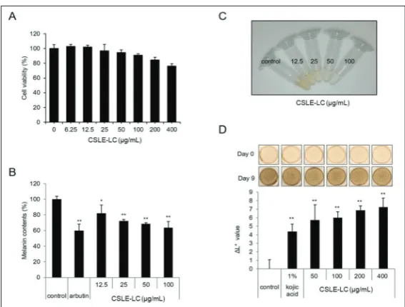
Prior to assessing the antimelanogenic efficacy of CSLE-LC, we tested whether CSLE-LC exerts antioxidative activity, which would indicate the presence of residual catechins, such as EGCG, a representative antioxidant. As shown in Fig. 1, CSLE-LC showed no antioxidant activity, indicating that CSLE-LC, isolated as described here, has few catechins. Next, melan-a cells were treated with the indicated concentrations of CSLE-LC and cell viability was assessed. CSLE-LC showed no cytotoxicity towards melan-a cells at concentrations up to 200 µg/ mL (Fig. 2A); thus, a concentration of CSLE-LC less than 200 µg/mL was chosen for subsequent experi-ments in melan-a cells. We next treated melan-a cells with CSLE-LC and measured total melanin 6 days later. Quantitative analysis showed that CSLE-LC treatment induced a concentration-dependent decrease in melanin content in melan-a cells (Fig. 2B), decreasing melanin production by 27.6% at 25 µg/mL, 31.5% at 50 µg/mL and 36.2% at 100 µg/mL. Notably, the color of lysates was clearly brightened by the treatment with CSLE-LC, as shown in the upper-right square box in Fig. 2B. Because melan-a cells originate from mouse melanocytes, we examined the whitening efficacy of CLSE in human cells. For this, we used a human skin equivalent preparation as an epidermis model called MelanoDerm, which consists of well-differentiated human keratinocytes and melanocytes. Treatment of the MelanoDerm preparation with CSLE-LC for 9 days caused brightening of skin tissue at all concentrations tested (Fig. 2C), with no evidence of cytotoxicity (data not shown). Kojic acid was used as a reference
rial for whitening experiments. Analysis of the results showed that CSLE-LC increased the ΔL value of skin equivalents in a concentration-dependent manner compared with the control. These results indicate that CSLE-LC decreased the melanin content in melanocytes and skin equivalents.
To elucidate the mechanism by which CSLE-LC depigments melanocytes, we incubated melan-a cells in the presence or absence of CSLE-LC and then deter-mined the expression levels of key mol-ecules involved in melanogenesis. Mela-nin synthesis is mainly regulated by the rate-limiting enzyme tyrosinase. Hence, we determined whether the inhibitory efficacy of CSLE-LC was related to the expression of tyrosinase. As shown in Fig. 3A, CSLE-LC suppressed tyrosinase expression in a concentration-dependent manner, suggesting that CSLE-LC sup-presses melanogenesis by reducing tyrosi-nase expression. We found that CSLE-LC treatment also reduced the expression of MiTF protein, the master transcriptional regulator of the gene encoding for tyrosi-nase (Fig. 3B). These results suggest that CSLE-LC inhibited tyrosinase expression by reducing MiTF expression.
Activation of the ERK (extracellular-signal regulated kinase) pathway induces MiTF degradation, ultimately causing mel-anogenic stimuli to dissipate, and therefore represents a major negative feedback mechanism in melanogenesis [17-18]. Thus, we examined whether CSLE-LC affects the ERK pathway. As shown in Fig. 4A, CSLE-LC strongly induced ERK phosphoryla-tion (activaphosphoryla-tion) within 30 min of treatment. Notably, PD98059, a specific inhibitor of ERK pathway, reversed the inhibitory effect of CSLE-LC on tyrosinase and MiTF expression (Fig. 4B). These results suggest that CSLE-LC likely inhibits melanogenesis by acceler-ating MiTF degradation through activation of the ERK signaling pathway, and subsequently reducing tyrosinase expression.
DISCUSSION
In the present study, we observed that C. sinensis
leaf extracts lacking catechins (CSLE-LC) decreased melanin content by reducing tyrosinase protein levels, resulting in a whitening effect in melanocytes and skin equivalents. We further found that CSLE-LC suppressed tyrosinase protein levels by promoting
Fig. 2. CSLE-LC has antimelanogenic activity. A – Melan-a cells were plated and treated with CSLE-LC for 24 h and the MTT assay was performed. B – Melan-a cells were plated and treated with CSLE-LC for 6 days and the melanin content was determined. C –The photograph shows the color of the lysate after CSLE-LC treatment. D – The skin equivalent was treated with CSLE-LC for 9 days, after which the pigmentation levels were assessed photographically (upper) and calculated by comparing variations in L* values (the lightness/darkness index) on days 0 and 9, and calculating the difference between them (ΔL). Kojic acid was used as a reference compound for whitening experiments. (*p<0.05, **p<0.01, ***p<0.001 vs. control group).
ERK-dependent degradation of MiTF, the transcription factor responsible for expression of the gene encoding for tyrosinase. These results suggest for the first time that bioactive components besides catechins in C.
sinensis leaf extracts, presumably polysaccharides, can
affect depigmentation in melanocytes. The leaves of
C. sinensis contain a number of bioactive components,
including catechins and polysaccharides. In this study, we removed catechins from C. sinensis leaf extracts, resulting in an extract (CSLE-LC) that presumably retained almost all polysaccharide constituents, which in tea leaves represent a group of heteropolysaccharides. Polysaccharides are high-molecular-weight polymers that consist of at least ten monosaccharides mutually joined by glycosidic linkages [20] and are generally recognized as a safe and non-toxic food additive. How-ever, polysaccharide composition varies according to the starting tea material and place where the teas were produced, leading to differences in bioactivity [19-20].
Preparation methods also affect the polysaccharide composition, further adding to differences in biological efficacy. Therefore, the preparation method, chemical composition and location where plants are cultured should be considered in evaluating the contribution of polysaccharides to physiological activities. In our study, we used leaves of C. sinensis from Korea. The leaves were extracted using distilled water and the catechins were removed. The extracts obtained ex-hibited whitening efficacy. On the bases of our re-sults, we speculate that the antimelanogenic efficacy of CSLE-LC is not attributable to catechins but to polysaccharides, although more experiments will be
needed to confirm this, for example, by using specific purified polysaccharides extracted from C. sinensis leaves. There are a few experimental limitations to our study. First, although we used a standard manufacturing approach for removing catechins from C. sinensis leaf extracts, we did not perform component analyses of CSLE-LC by chromatography.
To confirm that CSLE-LC contains no catechins, we instead tested its anti-oxidant activity based on the premise that catechins are potent antioxidants [19]. In this study, CSLE-LC had no antioxidative activity, whereas gallic acid, one of the main products derived from EGCG, showed strong antioxidant activ-ity. These results suggest that the CSLE-LC used in this study contained few, if any, bioactive catechins. Second, we did not determine whether CSLE-LC af-fected the transcriptional level of tyrosinase or MiTF in melanocytes. Instead, we confirmed that CSLE-LC reduced tyrosinase protein levels through phospho-ERK-induced MiTF degradation, which is consistent with the known role of ERK signaling in the degrada-tion of MiTF protein.
In conclusion, this study demonstrated that C.
sinensis leaf extracts lacking catechins regulate
mela-nin synthesis through ERK-dependent degradation of MiTF and subsequent downregulation of tyrosinase expression. Therefore, C. sinensis leaf extracts, even without catechins, could be developed as a new skin-lightening agent.
Funding: This research was supported and funded by the Amore-pacific R&D Center.
Author contributions: DH Kim and NH Park have contributed to this work equally. DH Kim, NH Park, CS Lee, and DC Yang designed the study. DH Kim and NH Park prepared the CSLE-LC and performed most of the experiments. JA Hwang, JK Kim and YJ Na performed the experiments, in part. JS Hwang, CS Lee and DC Yang analyzed the data and contributed to the writing of the manuscript. DH Kim and NH Park contributed equally.
Conflicts of interest disclosure: The authors have no conflicts of interest to declare.
REFERENCES
1. Reygaert WC.Green Tea Catechins: Their Use in Treat-ing and PreventTreat-ing Infectious Diseases. Biomed Res Int. 2018;2018:9105261.
2. Janssens PL, Hursel R and Westerterp-Plantenga MS. Nutra-ceuticals for body-weight management: The role of green tea catechins. Physiol Behav. 2016;162:83-7.
3. Afaq F, Adhami VM, Ahmad N, Mukhtar H. Inhibition of ultra-violet B-mediated activation of nuclear factor kappaB in nor-mal human epidernor-mal keratinocytes by green tea Constituent (-)-epigallocatechin-3-gallate. Oncogene. 2003;22(7):1035-44. 4. Broadhurst CL, Polansky MM, Anderson RA. Insulin-like
biological activity of culinary and medicinal plant aqueous extracts in vitro. J Agric Food Chem. 2000;48(3):849-52. 5. Cho SY, Park PJ, Shin HJ, Kim YK, Shin DW, Shin ES, Lee HH,
Lee BG, Baik JH, Lee TR. (-)-Catechin suppresses expres-sion of Kruppel-like factor 7 and increases expresexpres-sion and secretion of adiponectin protein in 3T3-L1 cells. Am J Physiol Endocrinol Metab. 2007;292(4):E1166-72.
6. Kao YH, Hiipakka RA, Liao S. Modulation of endocrine sys-tems and food intake by green tea epigallocatechin gallate. Endocrinology. 2000;141(3):980-7.
7. A Waheed, FS Hasid, N Ahmad, B Mand Khan. An Over View of Tea Plantation in Pakistan. Asian J Plant Sci. 2002:1(4):495-8. 8. Camila TS, Lauro MD, Yanna DR, Nessana D, Simone MMP,
Guilherme L, Philip AJG, Marcello I. Polysaccharides from green and black teas and their protective effect against murine sepsis. Food Res Int. 2013;53(2):780-5.
9. Xu P, Wu J, Zhang Y, Chen H, Wang YF. Physicochemical characterization of puerh tea polysaccharides and their anti-oxidant and alpha-glycosidase inhibition. J Funct Foods. 2014;6:545-54.
10. Wang YF, Shao SH, Xu P, Chen H, Lin-Shiau SY, Deng YT, Lin JK. Fermentation process enhanced production and bioactivities of oolong tea polysaccharides. Food Res Int. 2012;46(1):158-66.
11. Swalwell H, Latimer J, Haywood RM, Birch-Machin MA. Investigating the role of melanin in UVA/UVB- and hydrogen peroxide-induced cellular and mitochondrial ROS produc-tion and mitochondrial DNA damage in human melanoma cells. Free Radic Biol Med. 2012;52:626-34.
12. Briganti S, Camera E, Picardo M. Chemical and instrumental approaches to treat hyperpigmentation. Pigment Cell Res. 2003;16:101-10.
13. Hearing VJ, Tsukamoto K. Enzymatic control of pigmentation in mammals. FASEB J. 1991;5(14):2902-9.
14. Kim YC, Choi SY, Park EY. Anti-melanogenic effects of black, green, and white tea extracts on immortalized melanocytes. J Vet Sci. 2015;16(2):135-43.
15. Kim DS, Park SH, Kwon SB, Li K, Youn SW, Park KC. (-)-Epi-gallocatechin-3-gallate and hinokitiol reduce melanin syn-thesis via decreased MITF production. Arch Pharm Res. 2004;27(3):334-9.
16. Sato K, Toriyama M. Depigmenting effect of catechins. Mol-ecules. 2009;14(11):4425-32.
17. Wu M, Hemesath TJ, Takemoto CM, Horstmann MA, Wells AG, Price ER, Fisher DZ, Fisher DE. c-Kit triggers dual phos-phorylations, which couple activation and degradation of the essential melanocyte factor Mi. Genes Dev. 2000;14(3):301-12.
18. Xu W, Gong L, Haddad MM, Bischof O, Campisi J, Yeh ET, Medrano EE. Regulation of microphthalmia-associated transcription factor MiTF protein levels by association with the ubiquitin-conjugating enzyme hUBC9. Exp Cell Res. 2000;15;255(2):135-43.
19. Kim HS, Quon MJ, Kim JA. New insights into the mechanisms of polyphenols beyond antioxidant properties; lessons from the green tea polyphenol, epigallocatechin 3-gallate. Redox Biol. 2014;2:187-95.