© 2018 by the Serbian Biological Society How to cite this article: Chen L, Han H, Amin A, Zhang L, Ma S. Hydrolysis product of Nigella A obtained from Nigella glandulifera Freyn seeds promotes apoptosis and AMPK-mediated autophagy in human colon cancer SW620 cells. Arch Biol Sci. 2018;70(4):603-12.
Hydrolysis product of Nigella A obtained from
Nigella glandulifera
Freyn seeds promotes
apoptosis and AMPK-mediated autophagy in human colon cancer SW620 cells
Lili Chen1,2, Haote Han3,4, Awais Amin3,4, Lin Zhang3,4,# and Shenglin Ma1,5,*
1Wenzhou Medical University, Wenzhou 325035, P.R. China
2Huangyan Hospital of Wenzhou Medical University, Wenzhou 318020, P.R. China
3The Key Laboratory of Biomedical Engineering, Ministry of Education, Department of Biomedical Engineering, Zhejiang
University, Hangzhou 310028, P.R. China
4Zhejiang-Malaysia Joint Research Center for Traditional Medicine, Zhejiang University, Hangzhou 310028, P.R. China 5Department of Oncology, the Hangzhou First People’s Hospital, Hangzhou 310006, P.R. China
Corresponding authors: *[email protected]; #[email protected]
Received: November 8, 2017; Revised: March 30, 2018; Accepted: March 30, 2018; Published online: May 15, 2018
Abstract: Nigella B (NB) is the hydrolysis product of Nigella A (NA), which is extracted from the seeds of Nigella
glandu-lifera Freyn. NB has several beneficial characteristics, including antiproliferative activity against several cancer cell lines. In this study, we analyzed the in vitro and in vivo anticancer activity of both NA and NB as well as the potential molecular mechanisms behind the actions of NB. We found that NB treatment led to autophagy and soft apoptosis in colon cancer cells (SW620). NA treatment had no effect on either. Further study showed that NB treatment in SW620 cells led to inhib-ited phosphorylated mammalian target of rapamycin (p-mTOR) expression but increased phosphorylated-5’ adenosine monophosphate protein kinase (AMPK) expression, a key regulator of autophagy. This suggests that the AMPK-mTOR pathway plays a crucial role in autophagy induction. Separate in vivo studies using NA (40 mg/kg, intragastric administra-tion (i.g.)) and NB (40 mg/kg, i.g.) resulted in inhibited tumor growth in nude mice by 42.82% and 37.20% respectively, when compared with vehicle-administered animals. In vitro tumor protein expression was consistent with its expression in vitro. Taken together, our results reveal an anticancer function for NA and NB in colon cancer and support the use of NA as an antitumor prodrug, and NB as a novel therapeutic drug.
Key words: nigella A; nigella B; apoptosis; autophagy; AMPK; mTOR
INTRODUCTION
Colon cancer is one of the most common causes of cancer-related deaths worldwide. The current treat-ment for colon cancer patients is primarily based on surgical resection and may be followed by chemo-therapy [1,2]. Despite these interventions as well as advances in surgical approaches and early detection, overall prognosis is poor [3]. 5-Fluorouracil (5-FU) is the gold standard for colon cancer chemotherapy. However, 5-FU resistance is common and is recog-nized as a key reason for colon cancer therapy failure [2]. Given this, finding new treatment strategies for colon cancer has become an important focus over the last decade.
Defects in the execution of programmed cell death (PCD) are considered to be major factors in tumori-genesis. Clinical investigators have long exploited cell death mechanisms in the treatment of various types of cancer. Type I PCD, also known as apoptosis, is medi-ated by a cascade of proteins from the caspase family. This initiation and mediation ultimately leads to a va-riety of morphological changes in the cell. Apoptosis is divided into the intrinsic and extrinsic pathways, which depends on the particular death program trigger [1]. Type II PCD, also known as autophagy-related cell death, is presumed to result from excess autophagy. This program is important to a wide variety of process-es, including cellular homeostasis, genome protection,
development, anti-aging, and the regulation of cell size. Autophagy is characterized by the cytosolic presence of acidic vascular organelles (AVOs), leading to disrup-tion of cytoplasmic organelles and subsequent nuclear collapse [4]. This process also involves the sequestra-tion of bulk cytoplasm and organelles into autophago-somes, which later fuse with lysosomes. This produces an autolysosome that is subsequently degraded by the cells’ own lysosomal system [5].
Various signaling pathways have been implicated in the up- and downregulation of autophagy. For in-stance, Akt/mTOR pathway inhibition stimulates the nucleation and elongation of the phagophore mem-brane. In addition, class III phosphatidylinositol-4,5-bisphosphate 3-kinase (PI3K) activity is important for the formation of the autophagosomal membrane, along with a multiprotein complex surrounding the mammalian orthologue of yeast Atg6 (Beclin)-1 [6]. Autophagy-related genes (Atgs) are necessary in early autophagy for the formation of the phagophore mem-brane. An important Atg is light chain 3 (LC3), which exists in two forms: cytoplasmic LC3-I and its pro-teolytic derivative LC3-II, which binds to autophago-somes and is subjected to lysosomal degradation [7].
Despite the importance of PCD in the development of colon cancer, studies are limited and controversial. Past work has simultaneously and quantitatively ana-lyzed both apoptotic and autophagic pathways in clini-cal colorectal cancer tissue, and it was found that both were notably downregulated in colorectal tumor tissues when compared with noncancerous tissues [1]. Previ-ous studies showed that autophagy inhibition resulting from 3-methyladenine (3-MA) or small interference RNA targeting Atg7 enhanced the 5-FU-induced ap-optosis and cytotoxicity in human colon cancer [2]. Panitumumab is a human monoclonal antibody raised against the epidermal growth factor receptor for the treatment of metastatic colorectal carcinoma and it has also been reported to exert its cytotoxic effects by in-ducing autophagy [8]. Recent studies have reported that autophagy and apoptosis are both involved in chemo-therapeutic agent-induced cancer cell death, indicating that autophagy and apoptosis are the primary target mechanisms for new therapeutic agents [9].
Nigella seeds are widely used for medicinal pur-poses, which takes into account their
antiinflammato-ry, antioxidant, antimicrobial and anticancer proper-ties [10-12]. Saponins have the highest content and are the characteristic compound found in Nigella seeds. However, the biological activities of saponins derived from this plant have been rarely studied. To this end, Nigella A (3-O-[β-D-xylopyranosyl(1lopyranorhamno syl(1→2)-α-L-arabinosyl)28-O-[α-L-rhamnosyl-(1a4)-β-D-glucosyl-(1→6)-β-D-glucosyl]-hederagenin, NA] (Fig. 1A) was extracted from the seeds of Nigella glandulifera Freyn and has been reported to have a broad spectrum of biological activities against several diseases [13-15]. However, its antitumor effects have been seldom reported.
We found that while NA is unable to inhibit the colon cancer cell line SW620, it does possess in vivo antitumor effects. However, the main hydrolysis prod-uct of NA, NB (3-O-[β-D-xylopyranosyl(1→3)-α-L-rhamnosyl(1→2)-α-L-arabinosyl]-hederagenin) (Fig. 1A) has both in vivo and in vitro antitumor effects. Moreover, these effects were observed to be mediated through an AMPK-activated autophagy and apoptosis induction pathway. To our knowledge, this is the first report describing the anticancer effects of NA and the mechanisms of NB on both autophagy induction and the apoptotic pathway in a human colon cancer cell line.
MATERIALS AND METHODS Reagents
York, USA) were also used. Cell incubation medium was purchased from Gino Biomedical Technology Co., Ltd (Hangzhou, China) and fetal bovine serum (FBS) was purchased from Gemini Bio Products (Gemini, CA, USA). The following kits were used in this study: DNA content quantitation assay kit (KeyGEN Nan-jing, China), Annexin V/PI apoptosis kit (KeyGEN Nanjing, China), Giemsa Stain kit (Solarbio, Beijing, China), monodansylcadaverine (MDC) Stain kit (Key-GEN Nanjing, China), BCA protein concentration de-termination kit (Beyotime, Shanghai, China) and the enhanced chemiluminescence (ECL) analysis system (Beyotime, Shanghai, China).
Cell lines and culture conditions
The human cancer cell lines HepG2, SKOV-3, and SW620 were all obtained from the Chinese Academy of Sciences (Shanghai, China). Cells were either cultured in Dulbecco’s modified Eagle’s medium (HepG2, SW620) or McCoy’s 5a medium (SKOV-3), supplemented with 10% FBS and 1% penicillin/streptomycin solution (100 IU/mL penicillin, 100 mg/mL streptomycin) at 37°C at
5% CO2. Experiments were conducted after cells were
incubated for 24 h. NA and NB were separately dissolved in DMSO and diluted into culture medium containing 5% FBS, with a final concentration of 0.05% DMSO in each well. The control group received the same volume of DMSO without added NA or NB.
Cell viability assay
Cell viability was determined using a 3-(4, 5-dimethyl-thiazol-2-yl)-2,5-diphenyltetrazolium bromide (MTT) assay and was determined using six replicates per ex-periment. Briefly, cells were seeded in 96-well plates at 5000 cells/well for a final volume of 100 μL. After 24 h
incubation at 37°C in 5% CO2, cells were treated with
either NA or NB across a range of final concentrations (12.5, 25, 50, or 100 μM) for 24 h, 48 h and 72 h, or pretreated with CQ (10 μM) or 3-MA (5 mM) for 48 h. They were then treated with 20 μL of the MTT stock solution (5 mg/mL) in each well and incubated for another 2 h. The medium was carefully removed from each well and replaced with 200 μL DMSO. Ab-sorbance was measured at 570 nm using a plate reader (Multiskcan Go, Thermo Scientific, USA).
Giemsa staining
SW620 cells were seeded into 12-well plates at 1×10⁵
cells/well for a final volume of 1 mL. After 24 h, cells were treated with NB to one of three final concentra-tions (12.5, 25 or 50 μM) in 2 mL of medium. All
treated cells were incubated at 37°C in 5% CO2 for
another 48 h. The medium was carefully removed from each well and the cells were washed three times with phosphate buffered saline (PBS), fixed for 3 min with methanol (chilled at -20°C), then stained with Giemsa for 0.5 h. After washing with water, 1 mL PBS was added to each well before imaging using optical microscopy (CKX31, Olympus, Japan).
Flow cytometry analysis of the cell cycle
The DNA content quantitation assay kit was used to conduct a cell cycle analysis. Briefly, 48 h after drug
treatment, cells were harvested in ice-cold PBS (3×10⁵
cells in 300 μL), and 700 μL 100% ethanol was added for cellular fixation. After incubation at 4°C overnight, fixed cells were resuspended in 100 μL RNase A and incubated at 37°C for 30 min, followed by incuba-tion in 400 μL propidium iodide (PI) for 30 min in darkness. The distribution of cells in different phases of the cell cycle was determined using a flow cytom-eter (Cytomics FC 500, Beckman Coulter, USA) and analyzed using Multicycle AV for Windows advanced DNA Cell Cycle Analysis software.
Apoptosis analysis
Visualization of autophagic vacuoles
Cancer cells were seeded into six-well plates and cul-tured for 24 h, followed by NB treatment at one of three concentrations (0, 12.5 or 25 μM) for 48 h. Cells were then stained with 0.05 mM MDC at 37°C for 1 h. After washing three times using 1x wash buffer, cells were incubated using the collection buffer. Changes to cellular fluorescence were then imaged using fluo-rescence microscopy (Zeiss Axioplan).
Western blotting analysis
Cells were either treated with NB at one of four con-centrations (0, 12.5, 25 or 50 μM) for 48 h or pre-treated with CQ (10 μM) for 1 h. Cells were then scraped and collected from 6-cm dishes and subjected to centrifugation. Cells were washed once with ice-cold PBS and subsequently incubated in 75 μL lysis buffer for 30 min. Lysates were centrifuged for 15 min at 4°C at 13000×g. Protein concentrations in the re-sulting supernatants were determined using a BCA protein concentration determination kit according to the manufacturer’s instructions. Equal amounts of proteins were separated in a 7.5% sodium dodecyl sulfate (SDS) polyacrylamide gel and then transferred to a polyvinylidene fluoride (PVDF) membranes. Membranes were blocked using 5% non-fat milk in 0.1% Tween-20 in tris-buffered saline (TBST) for 2 h and incubated in the appropriate antibody (1:500-1:1000) at 4°C overnight. After washing three times with TBST, the membrane was incubated for 2 h at room temperature with the appropriate secondary antibody (1:5000). Immunoblots were visualized us-ing an ECL system.
Transient transfection
Cells were transfected with the siAMPK or siControl using Lipofectamine® 2000 transfection reagent ac-cording to the manufacturer’s instructions. Briefly, 3 μg of plasmid siAMPK or siControl was added to wells with 6 μL of Lipofectamine 2000. Treatments were initiated 48 h after the end of a 24-h transfec-tion. Expression of each plasmid was confirmed by immunoblotting as described above.
SW620 xenograft tumor model
All animal research was conducted in accordance with the Declaration of Helsinki and with the Guide for the Care and Use of Laboratory Animals as adopted and promulgated by the United National Institutes of Health. All experimental protocols were approved by the Review Committee for the Use of Animal Subjects of Changshu Realistic Technology Co., LTD (Suzhou, China). We established a xenograft tumor model by transplanting SW620 cells into nude mice as previously described [16]. BALB/C male mice (5-7 weeks of age) were purchased from HuaFuKang Biotechnology Co., LTD (Beijing, China). Briefly, xenograft tumors were
established by subcutaneous injection of 1×10⁷ SW620
cells in a total volume of 0.1 mL of serum-free
me-dium. Once tumors were measurable (100-200 mm3),
the animals were randomized into three groups (n=5) and the orally administered vehicle (normal saline) group, as follows: NA (40 mg/kg, 80 mg/kg, 120mg/ kg), or NB (40 mg/kg), and intraperitoneally admin-istered chemotherapeutic cyclophosphamide (30 mg/ kg, as a positive control) groups, which were treated daily for three weeks. Mouse body weight was meas-ured daily. Tumor volumes were calculated by caliper measurements using the following standard formula:
V=0.5×(width2×length). After treatment with the drug,
the mice were killed. Tumors were harvested, snap-frozen in liquid nitrogen, and stored at -80°C until subsequent Western blot biomarker analysis.
Statistical analyses
Data are presented as means±standard deviation (SD). One-way analysis of variance (ANOVA) and least sig-nificant difference (LSD) tests were used for all ex-periments. Differences were considered significant at p<0.05. Statistical analyses were performed using SPSS Statistics 17.0 software.
RESULTS
NB, not NA, inhibited cell growth and induced vacuolization in SW620 cells
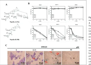
to evaluate the separate antitumor activities of NA and NB in human cancer cell lines HepG2, SKOV-3, and SW620. NB induced dose- and time-dependent reductions in cell viability in all examined cell lines. Of the examined cancer cell lines, SW620 cells were found to be the most sensitive to NB treatment (Fig.
1B). The IC50 values for NB ranged between 25 μM
and 35 μM at 48 h (Table 1). However, NA treatment had no effect on any of the cell lines (Fig. 1B) at the concentration of 100 μM. Therefore, the mechanism behind the effects of NB on SW620 cells was further examined. Optical microscopy revealed morphologi-cal changes after 48 h of treatment with different con-centrations of NB. More specifically, Giemsa staining revealed extensive cytoplasmic vacuolization in NB-treated SW620 cells (Fig. 1C). Rather than observing detached and shrunken apoptotic morphology, this finding indicated that NB mainly activated a non-apoptotic pathway. Since 50 μM concentration of NB produces serious damage to the cells (Fig. 1C), in sub-sequent experiments this concentration was excluded.
NB induced mild apoptosis in SW620 cells
An annexin V-/PI staining assay was used to quan-tify the percentage of apoptotic cells. Flow cytometry analysis showed that NB induced both early and late
apoptosis in SW620 cells in a concentration-depend-ent manner. As shown in Fig. 2A, a 48-h treatmconcentration-depend-ent with either 0, 12.5 or 25 μM of NB slightly enhanced the percentage of apoptotic cells from 0.2% (control) to 8.5% and 12.3%, respectively. Cleaved caspase-9 and cleaved PARP-1 were also significantly increased after NB treatment (Fig. 2B and C). Next, we inves-tigated changes in the cell cycle after NB treatment. We did not find significant accumulation of cells in the apoptotic cell population. Results indicated that the sub-G1 phase was slightly decreased to 2.535% after the treatment with 25 μM NB by PI (Fig. 2D). Taken together, our data suggested that NB reduced cell viability by inducing mild apoptosis in human colon cancer cells.
NB induced autophagy by mTOR inhibition in SW620 cells
The vacuolization that was observed pointed to au-tophagy, leading us to next check the pathway of ex-pression of autophagic proteins. This was performed after 48 h of NB treatment and using Western blotting. Analysis of different drug concentrations revealed a change in autophagic protein expression of LC3-II and Atg13, further supporting a role for autophagy. Analysis of upstream autophagy regulation revealed inhibition of
Fig. 1. The influence of NA and NB on cell proliferation and morphology.
A – Structures of Nigella A (NA) and
Nigella B (NB). B – Effects of NA and concentration-dependent effects of NB on cell viability of the hepatoma (HepG2), ovarian cancer (SKOV-3) and colon cancer (SW620) cell lines as detected using the MTT assay after 24 and 72 h of NA and NB incuba-tion. The experiment was repeated in
triplicate. C – NB-induced
Fig. 2. NB induced mild apoptosis in
SW620. A – Apoptosis analysis after
48 h of exposure to NB in SW620 cells. Dose-dependent effect of NB as evaluated by annexin V/PI staining of
SW620 cells at 48 h. B – SW620 cells
were treated with NB (0, 12.5, 25 or 50 μM) for 48 h and then were lysed to determinate the levels of caspase-9
and PARP-1 proteins. C – The gray
values of bands were analyzed using Image J software. Data are presented as relative expression to β-actin and as the mean±SD of three independ-ent experimindepend-ents; *p<0.05, **p<0.01.
D – Cells were treated with NB (0 or
25 μM) for 48 h and were then sub-jected to cell cycle analysis using flow cytometry.
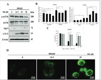
Fig. 3. NB induced autophagy-related cell death through mTOR inhibition.
A – Western blot analysis of NB-treat-ed cells and subsequent expression of several proteins related to NB-induced autophagy (mTOR, p-mTOR, LC3-II, and Atg13). SW620 cells were treated
with NB for 48 h. B – The gray
val-ues of mTOR, p-mTOR, LC3-II, and Atg13 were analyzed using Image J software. Data are presented as rela-tive expression to β-actin and as the mean±SD of three independent ex-periments; *p<0.05, **p<0.01. C – SW620 cells were pre-treated with CQ (10 μM) and 3-MA (5 mM) for 1 h, and co-treated with NB for another 48 h. Cell viability was detected by the MTT assay. The experiment was re-peated in triplicate; *p<0.05, **p<0.01.
(D) SW620 cells exposed to the
p-mTOR expression, indicating the involvement of the mTOR pathway (Fig. 3A and B). After CQ and 3-MA treatment, the NB-mediated inhibition on SW620 cells was attenuated (Fig. 3C). This finding underscored the role of autophagy in the effect of NB on SW620 cells. Furthermore, MDC cellular staining after 48 h of NB treatment showed autophagolysosomes, again indicat-ing that NB induced autophagy (Fig. 3D).
NB promoted autophagic flux and inhibited p-mTOR by AMPK activation
The role of NB on autophagic flux was detected by adding the autophagy inhibitor CQ. We found that after co-treatment with NB and CQ, LC3-II expres-sion was increased more than after separate treat-ments with either CQ or NB (Fig. 4A). This result indicated that NB promoted autophagic flux. We further found that NB inhibited p-mTOR expression through AMPK activation. This was concluded after lysing SW620 cells that had received 48 h of treat-ment with different concentrations of NB. Western blot analysis showed increased p-AMPK expression (Fig. 4B). To further confirm the cause of cell death, siRNA-mediated knockdown of AMPK and Western blotting revealed changes in LC3-II expression. More specifically, we found that siAMPK reduced NB-me-diated upregulation of LC3-II protein expression. This showed that autophagy was the critical mechanism induced by AMPK activation and subsequent mTOR inhibition (Fig. 4C). As shown in Fig. 4D-G, there were significant differences in the aforementioned proteins. When analyzed, these results showed bind-ing consistency, which further supported that NB both promoted autophagic flux and inhibited p-mTOR by AMPK activation.
NA and NB inhibited tumor growth and induced
in vivo autophagy
We next attempted to better evaluate the in vivo ef-fects of NA and NB on tumors transplanted into nude mice. When compared with vehicle-treatment, treat-ment with 40 mg/kg NB or 40, 80 or 120 mg/kg NA resulted in tumor growth inhibition (Fig. 5A and B). There were no significant differences in weight loss compared with the vehicle-treated animals that were
given NA (Fig. 5C). NB-treated mice had significantly lower weights during the early stages of treatment, but later body weight returned to normal (Fig. 5C). Since NB is a metabolite of NA, in vivo NA treatment may result in increased NB levels. This could result in increased in vivo efficacy. The tumor inhibition rates of 40 mg/kg NB and 40, 80, and 120 mg/kg NA were 37.2%, 40.78%, 47.38%, and 53.04%, respectively.
Western blotting revealed that both NB and NA treatments led to Atg13 and LC3-II upregulation in tumors. This finding indicated the induction of au-tophagy. Furthermore, p-mTOR was downregulated and AMPK was activated, which also indicated au-tophagy (Fig. 5D and E). These findings were con-sistent with the effects of treatment with NB on in vitro protein expression. Taken together, these results underscore the antitumor role played by NA by me-tabolizing into NB and inducing autophagy.
DISCUSSION
Several forms of nonapoptotic cell death have been described, including oncosis [17], autophagy [18], entosis [19] and necroptosis [20]. However, the role of autophagy in decisions of cell fate remain contro-versial. Recent attention has been paid to the role of autophagy in connection with PCD-autophagic death. From a functional perspective, autophagic death eliminates damaged and/or harmful cells, including cancer cells treated with anticancer reagents or cells infected with pathogenic microorganisms [21,22]. Autophagy is likely to be a mechanism for supplying amino acids to other, uninfected or non-cancer cells. To this end, levels of the membrane-bound protein LC3-II have been taken as a marker for the formation of autophagosomes [23,24]. Several lines of evidence have shown the role that AMPK-mediated signaling pathways play in sensing intracellular energy status and regulating cellular metabolism to maintain home-ostasis. Recent research also reported that AMPK positively regulates autophagy [25].
research from our lab, we obtained NB from the hy-drolysate of NA in vitro. In this study, we demon-strated that both compounds possess anticancer in vivo effects against colon cancer. However, NA was also effective in vitro. We speculated that NA func-tions as a prodrug, subsequently being metabolized to NB and showing its inherent antitumor activity.
Further study verified that the molecular mechanism of NB action in SW620 cells affected apoptotic and autophagic pathways. Initially, we found that cleaved caspase-9 and cleaved PARP-1 were both significantly increased, thereby enhancing the cellular apoptotic rate. This suggested the activation of the apoptotic pathway in NB-treated SW620 cells. Furthermore,
cy-Fig. 4. NB-promoted autophagic flux and p-mTOR inhibition through
AMPK activation. A – SW620 cells
were either separately treated with CQ or NB or co-treated with both for 48 h. Cells were then lysed to de-terminate LC3-II and β-actin levels
using Western blotting. (B) SW620
cells treated with NB (0, 12.5, 25, or 50 μM) for 48 h were lysed to deter-minate AMPK, p-AMPK, and β-actin levels using Western blotting. C – The effect of siRNA-mediated knockdown of AMPK on NB-induced changes in the expression of LC3-II in SW620 cells analyzed by Western blotting.
D-G – The gray values of bands were
analyzed using Image J software. Data are presented as relative expression to β-actin and as the mean±SD of three independent experiments; *p<0.05, **p<0.01.
Fig. 5. NA and NB inhibited SW620
tumor growth in nude mice. A –
Tu-mor volume; B – representative tumor
images for various groups; C – body
weight; D – effect of NA and NB on
toplasmic vacuoles were detected after NB treatment, indicating that there may exist a critical caspase-inde-pendent pathway in the death of SW620 cells. Both the autophagic-related marker LC3B-II and autophago-somes were increased in SW620 cells, indicating NB-mediated autophagy induction.
SW620 cells represent colon adenocarcinomas, which is the most common cancer type in humans [26]. These cells are characterized by an extreme sistance to the induction of cell death via death re-ceptor ligands (e.g. tumor necrosis factor (TNF)-α, first apoptosis signal receptor (Fas)-L and TNF-re-lated apoptosis-inducing ligand (TRAIL)), as well as chemotherapeutic drugs [27]. The death recep-tor and apoptotic mitochondrial pathways assemble after caspase-3 action. Crosstalk between these two pathways is offered by the BH3 interacting-domain death agonist (Bid) protein, which is cleaved by cas-pase-8 [28]. However, our results also indicated that a key regulator of autophagy, AMPK, played a criti-cal role during this process. After knocking down AMPK, NB showed no influence on LC3-II expres-sion. Meanwhile, NB-mediated mTOR inhibition was associated with parallel increases in LC3-II as well as in Atg13 protein levels. When the autophagic inhibi-tors CQ and 3-MA were applied, the antitumor effect of NB was reduced. Moreover, autophagic flux was promoted after NB treatment through CQ inhibition. When taken together, these results support a role for NB in inducing autophagy.
We considered that the major mechanism behind NB-induced cytotoxicity was autophagy-related cell death, but that this outcome could be the sum of the direct effects of autophagy as well as the indirect ef-fect of apoptosis. Autophagy and apoptosis often take place in the same cell, typically occurring sequentially with autophagy preceding apoptosis [29,30]. Finally, we found that the expression of tumor tissue proteins treated with NA and NB were consistently altered. This provided further evidence for our hypothesis that in vivo application of NA has an antitumor effect by metabolizing into NB. Consistent with the effects of NB treatment on in vitro tumor protein expression, NA and NB upregulated p-AMPK and LC3II protein expression and inhibited p-mTOR, collectively point-ing to the activation of autophagy.
CONCLUSION
This study revealed the anti-colon cancer function of NB, the hydrolysis product of NA, through the induc-tion of autophagy by AMPK activainduc-tion and mTOR inhibition. These molecular components ultimately resulted in both in vivo and in vitro cell death com-bined with mild apoptosis. Taken together, these re-sults support the use of NB as a prospective therapeu-tic drug in the treatment of colon cancer.
Acknowledgments: This work was supported by Zhejiang-Malay-sia Joint Research Center for Traditional Medicine (2016C04005) and the Jiangsu Provincial Natural Science Foundation of China (BK20161269).
Author contributions: LZ, SLM, LLC and HTH designed the study. LLC, HTH and AA performed the experiments in vitro LLC and HTH performed the in vivo study. HTH and AA ana-lyzed the data and wrote the manuscript. LC, HH and AA equally contributed to this study.
Conflict of interest disclosure: The authors report no conflicts of interest.
REFERENCES
1. Chang Y, Tseng H, Huang C, Chen Y, Chiang H, Chou F. Relative down-regulation of apoptosis and autophagy genes in colorectal cancer. Clin Invest. 2011;41(1):84-92. 2. Li J, Hou N, Faried A, Tsutsumi S, Kuwano H. Inhibition
of autophagy augments 5-fluorouracil chemotherapy in
human colon cancer in vitro and in vivo model. Eur J
Can-cer. 2010;46(10):1900-9.
3. Beretta G, Pessi M, Poletti P, Mosconi S, Labianca R. New drugs and combinations in the palliative treatment of colon and rectal cancer. Surg Oncol. 2001;27(6):595-600. 4. Bursch W, Hochegger K, Torok L, Marian B, Ellinger A,
Her-mann R. Autophagic and apoptotic types of programmed cell death exhibit different fates of cytoskeletal filaments. Cell Sci. 2000;113(7):1189-98.
5. Kelekar A. Autophagy. Ann N Y Acad Sci. 2006;1066(1):259-71. 6. Lin L, Dawson P, Richardson C. Viral interactions with
macroautophagy: a double-edged sword. Virology. 2010;402(1):1-10.
7. Jaeger PA, Wyss-Coray T. All you can eat: autophagy in neu-rodegeneration and neuroprotection. Mol Neurodegener. 2009;4(1):1-22.
8. Giannopoulou E, Antonacopoulou A, Matsouka P, Kalofo-nos H. Autophagy: novel action of panitumumab in colon cancer. Anticancer Res. 2009;29(12):5077-82.
10. Mohamed L. Immunomodulatory and therapeutic proper-ties of the Nigella sativa L. Seed. Int Immunopharmacol. 2005;5(13-14):1749-70.
11. Fico G, Panizzi L, Fiamini G, Braca A, Morelli I, Tomè F, Cioni P. Biological screening of Nigella damascena for antimicrobial and molluscicidal activities. Pyhtother Res. 2004;18(6):468-70.
12. Ali B, Blunden G. Pharmacological and toxicological prop-erties of Nigella sativa. Pyhtother Res. 2003;17(4):299-305. 13. Li W, Yan X, Sun Y, Ngan T, Shim S, Kim Y. Anti-Inflamma-tory and PPAR Transactivational Effects of Oleanane-Type Triterpenoid Saponins from the Roots of Pulsatillakoreana. Biomol Ther. 2014; 22(4):334-40.
14. Zhao M, Da-Wa Z, Guo D, Fang D, Chen X, Xu H, Gu Y, Xia B, Chen L, Ding L, Zhou Y. Cytotoxic triterpenoid saponins from Clematis tangutica. Phytochemistry. 2016;130:228-37. 15. Liu J, Guan Y, Zou L, Gong Y, Hua H, Xu Y, Zhang H, Yu Z,
Fan W H. Saponins with Neuroprotective Effects from the Roots of Pulsatillacernua. Molecules. 2012;17(5):5520-31. 16. Han HT, Liu N, Zhang L, Gong MH, Cao M, Li BG, Kaisa
S, Yu XY, Tian JK. Promotion of cytoplasmic vacuolation-mediated cell death of human prostate cancer PC-3 cells by oxidative stress induced by daucusol, a new guaiane-type sesquiterpenoid from Daucuscarota L. Arch Biol Sci. 2017;69(3):481-9.
17. Suárez Y, González L, Cuadrado A. Kahalalide F, a new marine-derived compound, induces oncosis in human pros-tate and breast cancer cells. Mol Cancer Ther. 2003;2(9):863-72.
18. DiPaola RS, Dvorzhinski D, Thalasila A, Garikapaty V, Doram D, May M, Bray K, Mathew R, Beaudoin B, Karp C, Stein M, Foran DJ, White E. Therapeutic Starvation and Autophagy in Prostate Cancer: A New Paradigm for Targeting Metabolism in Cancer Therapy. Prostate. 2008;68(16):1743-52.
19. Overholtzer M, Mailleux AA, Mouneimne G, Normand G, Schnitt S, King R, Cibas E, Brugge J. A nonapoptotic cell death process, entosis, that occurs by Cell-in-Cell invasion. Cell. 2007;131(5):966-79.
20. Almagro MCD, Vucic D. Necroptosis: Pathway diversity and characteristics. Semin Cell Dev Biol. 2015;39:56-62. 21. Kirkegaard K, Taylor MP, Jackson WT. Cellular autophagy:
surrender, avoidance and subversion by microorganisms. Nat Rev Microbiol. 2004;2(4):301-14.
22. Kondo Y, Kanzawa T, Sawaya R, Kondo S. The role of autophagy in cancer development and response to therapy. Nat Rev Cancer. 2005;5(9):726-34.
23. Tanida I, Minematsu-Ikeguchi N, Ueno T, Kominami E. Lysosomal turnover, but not a cellular level, of endogenous LC3 is a marker for autophagy. Autophagy. 2005;1(2):84-91. 24. Kabeya Y, Mizushima N, Ueno T, Yamamoto A, Kirisako
T, Noda T, Kominami E, Oshumi Y, Yoshimori T. LC3, a mammalian homologue of yeast Apg8p, is localized in autophagosome membranes after processing. Embo J. 2000;19(21):5720-8.
25. Meley D, Bauvy C, Houben-Weerts JH, Dubbelhuis P, Hel-mond MT, Codogno P, Meijer AJ. AMP activated protein kinase and the regulation of autophagic proteolysis. J Biol Chem. 2006;281(46):34870-9.
26. Herrero-Martin G, Hoyer-Hansen M, Garcia-Garcia C, Fumarola C, Farkas T, Lopez-Rivas A, Jaattela M. TAK1 activates AMPK-dependent cytoprotective autophagy in TRAIL-treated epithelial cells. Embo J. 2009;28(6):677-85. 27. Neuzil J, Weber T, Schroder A, Lu M, Ostermann G,
Gel-lert N, Mayne GC, Olejnicka B, Negre-Salvayre A, Sticha M, Coffey RJ, Weber C. Induction of cancer cell apoptosis by alphatocopheryl succinate: molecular pathways and struc-tural requirements. FASEB J. 2001;15(2):403-15.
28. Sun Y, Zhao Y, Hou L, Zhang X, Zhang Z, Wu K. RRRal-pha- tocopheryl succinate induces apoptosis in human gas-tric cancer cells via the NF-kappaB signaling pathway. Oncol Rep. 2014;32(3):1243-8.
29. Maiuri MC, Zalckvar E, Kimchi A, Kroemer G. Self-eating and self-killing: crosstalk between autophagy and apoptosis. Nat Rev Mol Cell Biol. 2007;8(9):741-52.