Kamolporn Kaewpornsawan, M.D.*, Makoto Kamegaya, M.D.**, Suthipol Udompunturak, M.Sc.***, Perajit Eamsophana, M.D. *, Thanase Ariyawatkul, M.D. *
*Department of Orthopaedic Surgery, Faculty of Medicine Siriraj Hospital, Mahidol University, Bangkok 10700, Thailand,**Chiba Children & Adult Orthopaedic Clinic, Chiba Prefecture, Japan, ***Department of Research and Development, Faculty of Medicine Siriraj Hospital, Mahidol University, Bangkok 10700, Thailand.
The Normal Reference Values of Carrying Angle
from Birth to Adolescence
Correspondence to: Kamolporn Kaewpornsawan E-mail: [email protected]
Received 22 May 2018 Revised 24 May 2018 Accepted 30 May 2018 doi:10.14456/smj.2018.46
ABSTRACT
Objective: The aim of this study was to establish normal carrying angle reference values from birth to adolescence,
and to identify variations in carrying angle relative to age, gender, and elbow side.
Methods: The prospective cross-sectional study was performed in normal healthy children aged newborn to sixteen
years during May 1959 to April 1961. Children were recruited from newborn units, nurseries, and schools located in Bangkok, Thailand. There was a total of 17 groups – one for each year of age from 0 (at birth) to 16. The carrying angles of both arms were measured using a clear plastic full-circle orthopedic goniometer. The data of age, side, and gender of each child were recorded.
Results: A total of 921 children with 1,842 measurements were included. There were 407 boys and 514 girls. The mean
carrying angle was lowest at birth and highest in the 15- year and 16-year age groups. The increase in the carrying angle was observed to progress to valgus 6 degrees at 6 years of age, and to valgus 11 degrees and stabilization at 15 years of age. There was no statistically significant difference between the mean carrying angle of the left and right side for any of the 17 evaluated age groups. Girls demonstrated a significantly greater carrying angle than boys (p<0.001). The intraclass correlation coefficient (ICC) of inter-observer variation between two observers was 0.848.
Conclusion: This study established normal carrying angle reference values from birth to adolescence. Our results
revealed that the elbow is slightly varus at birth, then increases in carrying angle until reaching stabilization of skeletal growth and development at 15 years of age. The carrying angle is slightly greater in girls than in boys. This normal reference value data will benefit orthopedists who take care of the pediatric patients with elbow-related disorders.
Keywords: Reference value; carrying angle; birth; adolescence; elbow (Siriraj Med J 2018;70: 284-288)
INTRODUCTION
The carrying angle is clinically defined as the angle made by the longitudinal axis of the arm and the forearm in full extension with the elbow supinated. In children, the carrying angle is an important outcome measure that is routinely used during treatment of fractures around the elbow. Increased carrying angle is referred to as cubitus valgus, and decreased carrying angle is termed cubitus varus. Cubitus varus deformity is one of the common
complications of supracondylar humeral fracture, lateral condyle fracture, and transphyseal fracture in children. For the body to appear cosmetically normal, the carrying angle of both elbows should be equal or nearly equal.1 Normal carrying angle reference values can help us determine the cosmetically-acceptable limits of deformity in specific age groups.
children.2 One study from Northern Thailand measured carrying angle in adults aged 18-35 years.2 It is often reported that the carrying angle increases with age, and is greater in girls than boys2,3,4; however, conflicting data has been reported.5 In studies that have reported on carrying angle in children, most focused a specific age range (e.g., 3-19 years of age), but none studied children from birth to adolescence.2-13
To our knowledge and based on our review of the literature, no previous reports have been published that describe normal carrying angle reference values throughout childhood in pediatric population. Accordingly, the aim of this study was to establish normal carrying angle reference values from birth to adolescence, and to identify variations in carrying angle relative to age, gender, and elbow side.
MATERIALS AND METHODS
The prospective cross-sectional study was performed in normal healthy children aged newborn to sixteen years at the Department of Orthopaedic Surgery, Faculty of Medicine Siriraj Hospital, Mahidol University during the May 1959 to April 1961 study period. Children with any history of elbow trauma, deformity, or disease that affects the elbow, such as skeletal dysplasia and rickets, were excluded. Children from newborn units, nurseries, and schools located in Bangkok, Thailand were included. There were a total of 17 groups – one for each year of age from 0 (at birth) to 16. The study and all of the measurement methods and protocols were approved by the Siriraj Institutional Review Board (SIRB), Faculty of Medicine Siriraj Hospital, Mahidol University, Bangkok, Thailand (Si 684/2013).
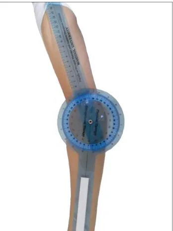
The carrying angle was measured using a clear plastic full-circle orthopedic goniometer (Fig 1) according to the method previously described by Amis and Miller.8 This method was used in several other studies9,10,11, and was reported to be accurate, with an error margin of ≤1 degree.9 The arm was abducted to 90 degrees over a straight table, with the forearm in full extension and supination. The hinge of the goniometer was positioned at the center of the cubital crease, and the arms of the goniometer arms were positioned parallel to the middle longitudinal axes of the arm and forearm. A minus sign (-) indicates varus angle, and a plus sign (+) indicates valgus angle. Both arms were measured, and the age, side, and gender of each child were recorded.
Statistical analysis
All data analyses were performed using SPSS Statistics version 18 (SPSS, Inc., Chicago, IL, USA). Mean normal
carrying angle reference values were calculated and statistically compared between and among age groups to identify differences relative to age, gender and elbow side. Data are presented as number, percentage, or mean ± standard deviation. Intraclass correlation coefficients were calculated and analyzed to measure interrater agreement. A p-value less than 0.05 was regarded as being statistically significant.
RESULTS
The 921 children that were included had both elbows measured for a total of 1,842 sides. There were 407 boys and 514 girls that were divided into 17 groups by year from 0 (at birth) to 16 years.
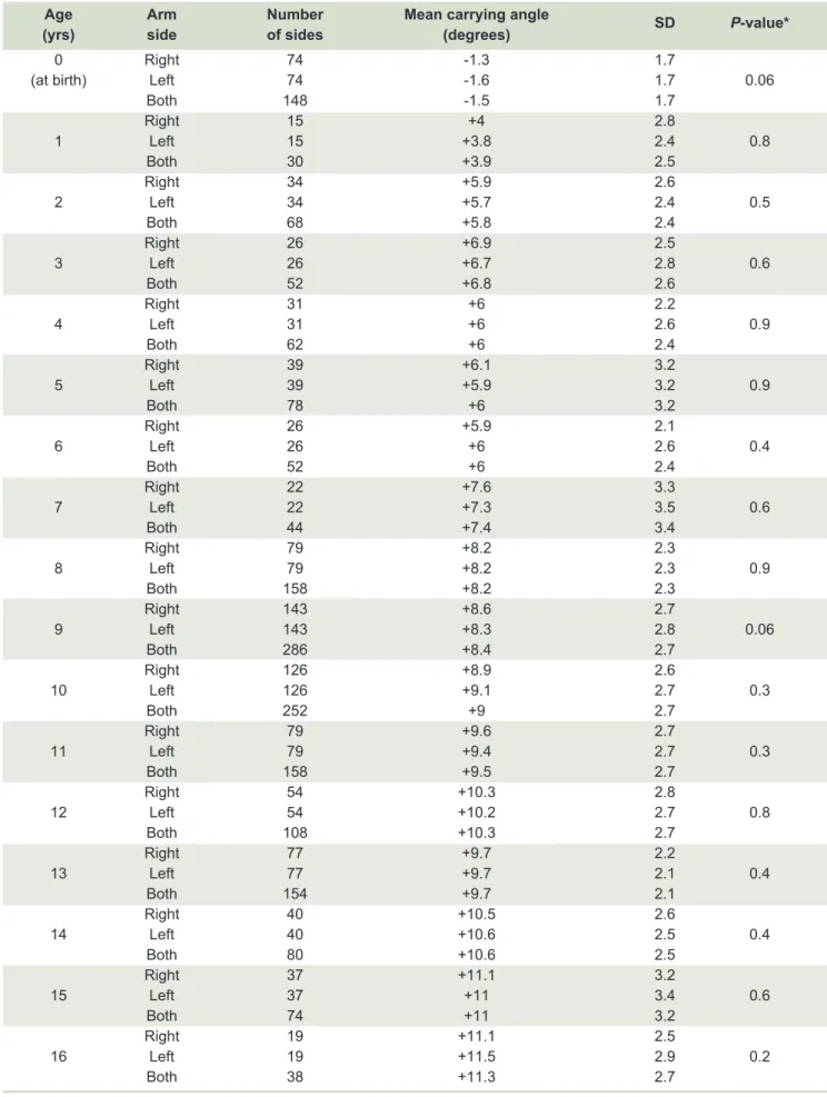
The mean normal carrying angle reference values for each age group by elbow side from birth to age 16 years are shown in Table 1. The mean carrying angle was lowest at birth (mean varus: 1.49 degrees) and highest in the 15- and 16-year age groups (mean valgus: 11
TABLE 1. Mean normal carrying angle reference values for each age group by elbow side from birth to age 16 years
Age Arm Number Mean carrying angle SD P-value*
(yrs) side of sides (degrees)
0 Right 74 -1.3 1.7
(at birth) Left 74 -1.6 1.7 0.06
Both 148 -1.5 1.7
Right 15 +4 2.8
1 Left 15 +3.8 2.4 0.8
Both 30 +3.9 2.5
Right 34 +5.9 2.6
2 Left 34 +5.7 2.4 0.5
Both 68 +5.8 2.4
Right 26 +6.9 2.5
3 Left 26 +6.7 2.8 0.6
Both 52 +6.8 2.6
Right 31 +6 2.2
4 Left 31 +6 2.6 0.9
Both 62 +6 2.4
Right 39 +6.1 3.2
5 Left 39 +5.9 3.2 0.9
Both 78 +6 3.2
Right 26 +5.9 2.1
6 Left 26 +6 2.6 0.4
Both 52 +6 2.4
Right 22 +7.6 3.3
7 Left 22 +7.3 3.5 0.6
Both 44 +7.4 3.4
Right 79 +8.2 2.3
8 Left 79 +8.2 2.3 0.9
Both 158 +8.2 2.3
Right 143 +8.6 2.7
9 Left 143 +8.3 2.8 0.06
Both 286 +8.4 2.7
Right 126 +8.9 2.6
10 Left 126 +9.1 2.7 0.3
Both 252 +9 2.7
Right 79 +9.6 2.7
11 Left 79 +9.4 2.7 0.3
Both 158 +9.5 2.7
Right 54 +10.3 2.8
12 Left 54 +10.2 2.7 0.8
Both 108 +10.3 2.7
Right 77 +9.7 2.2
13 Left 77 +9.7 2.1 0.4
Both 154 +9.7 2.1
Right 40 +10.5 2.6
14 Left 40 +10.6 2.5 0.4
Both 80 +10.6 2.5
Right 37 +11.1 3.2
15 Left 37 +11 3.4 0.6
Both 74 +11 3.2
Right 19 +11.1 2.5
16 Left 19 +11.5 2.9 0.2
Both 38 +11.3 2.7
A p-value<0.05 indicates statistical significance
degrees). Increase in the carrying angle was observed to progress to valgus 6 degrees at 6 years of age, and to valgus 11 degrees and stabilization at ≥15 years of age. There was no statistically significant difference between the mean carrying angle of the left and right side for any of the 17 evaluated age groups (p>0.05).
The mean carrying angle among all age groups, with the angle values of the left and right side added together, was valgus 7.8±4.1 degrees. In comparison of mean carrying angle between age group, the statistically significant difference was obtained between age group (p<0.001).
The mean carrying angle was valgus 6.9±3.8 degrees in boys, and valgus 8.5±4.1 degrees in girls. Girls demonstrated a significantly greater carrying angle than boys (p<0.001)
(Table 2).
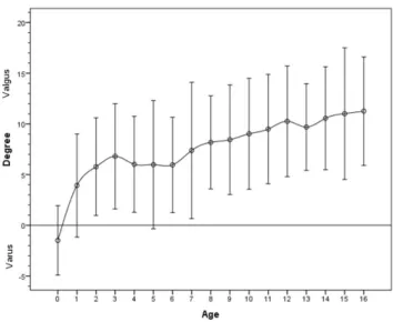
A graph describing the mean (±2 standard deviations) normal carrying angle reference value for each age group is shown in Fig 2.
The intraclass correlation coefficient (ICC) of interobserver variation between two observers in 537 cases was 0.848 (95% confidence interval: 0.818-0.872), which indicated minimal variation between observers.
TABLE 2. Comparison of carrying angle between boys and girls (N=921)
Gender Number Number Mean SD P-value
of children of sides Percent carrying angle
Boys 407 814 55.8% +6.9 3.8
Girls 514 1,028 44.2% +8.5 4.1
<0.001
A p-value<0.05 indicates statistical significance Abbreviation: SD, standard deviation
Fig 2. Normal carrying angle reference values (mean ± 2 standard deviations) from birth to 16 years. Degree values less than and greater than zero indicate varus of the elbow and valgus of the elbow, respectively.
DISCUSSION
This study provides new knowledge about normal carrying angle values from birth to adolescence, and about the development and progression of angle from slightly varus at birth to valgus and stable at 15-16 years of age (Table 1, Fig 2). The next study in carrying angle should focus on age group given the differences among age groups, especially in young children and adolescents. The present study and other studies found and reported age-dependent changes in the carrying angle.3,6,7 The findings of this study confirm the reported findings of other studies that the carrying angle increases with age as a result of skeletal growth and development.9,10,12
With the exception of the study by Beals5 who reported equality of carrying angle between genders, the present study and many others found a slightly greater carrying angle in girls than in boys.2-4,6,7,10-13 The difference in findings between Beals’ study and others may be due to differences in angle measurement method (measurement from radiographic imaging vs. measurement by goniometer), race of the study population, and sample size.
difficult to elicit from other age groups, we decided not to include this factor in our analysis.
The pediatric carrying angle data yielded by this study can be used as a comparative benchmark against clinical measurement in routine practice, or against radiographic measurement of the elbow.
CONCLUSION
This study established normal carrying angle reference values from birth to adolescence. Our results revealed that the elbow is slightly varus at birth, with increases in carrying angle until stabilization of skeletal growth and development at 15 years of age. The carrying angle is slightly greater in girls than in boys. This normal reference value data will benefit orthopedists that treat and follow pediatric patients with elbow-related disorders.
ACKNOWLEDGMENTS
The authors gratefully acknowledge Miss Siranart Kumpravat of the Research Division of the Department of Orthopaedic Surgery, Faculty of Medicine Siriraj Hospital, Mahidol University for assistance with data preparation and manuscript review.
Conflict of interest declaration: All authors declare no personal or professional conflicts of interest, and no financial support from the companies that produce and/ or distribute the drugs, devices, or materials described in this report.
Funding disclosure: This study was funded by a grant from the Faculty of Medicine Siriraj Hospital, Mahidol University, Bangkok, Thailand.
REFERENCES
1. Sinikumpu JJ, Victorzon S, Pokka T, Lindholm EL, Peljo T, Serlo W. The long-term outcome of childhood supracondylar humeral fractures: A population-based follow up study with a minimum follow up of ten years and normal matched comparisons. Bone Joint J. 2016;98B:1410-17.
2. Sudasna S, Seripantuwongsa V. The range of motion of the elbow,wrist and finger joints of the people of the northern part of Thailand. J Thai Orthop Assoc. 1985; 10: 93-122.
3. Baughman.FA, Higgins JV, Wadsworth TG, Denmaray M. The carrying angle in sex chromosome anomalies. JAMA. 1945;230:718-20.
4. Balasupramanian P, Madhuri V, Muliyil J. Carrying angle in children: a normative study. J Pediatr Orthop. 2006;15B:37-40.
5. Beals RK. The normal carrying angle of the elbow. A radiographic study of 422 patients. Clin Orthog Relat Res. 1976;119:94-6.
6. Sharma K, Mansur DI, Khanal K, Haque MK. Variation of carrying angle with age,sex,height and special reference to side. Kathmandu Univ Med J (KUMJ). 2013;11(44):315-8.
7. Dey S, Mandal L, Kundu B, Mondal M, Sett TK. Carrying angle of the elbow: it’s changes from childhood to adulthood: morphometric study in eastern India. Indian J Basic&Applied Med Res. 2013;2:823-30.
8. Amis AA, Miller JH. The elbow. Clin Rheum Dis. 1982;8:571-93.
9. Yilmaz E, Karakurt , Belhan O, Bulut M, Serin E, Avci M. Variation of carrying angle with age, sex, and special reference to side. Orthopedics. 2005;28:1360-3.
10. Allouh Mz, Khasawneh RR. Measurement of the carrying angle in Jordanians with respect to different body parameters. Jordan Med J. 2014;48: 93-101.
11. Zampagni ML, Casino D, Martelli S, Visani A, Marcacci M. A protocol for clinical evaluation of the carrying angle of the elbow by anatomic landmarks. J Shoulder Elbow Surg. 2008; 17(1):106-12.
12. Paraskevas G, Papadopoulos A, Papaziogas B, Spanidou S, Argiriadou H, Gigis J. Study of the carrying angle of the human elbow joint in full extension, a morphometric analysis. Surg Radiol Anat. 2004;26:19-23.