REGULATION OF ENDOTHELIAL PLASTICITY BY TGFβ IN TUMORS
Lin Xiao
A dissertation submitted to the faculty of the University of North Carolina at Chapel Hill in partial fulfillment of the requirements for the degree of Doctor of Philosophy
in the Department of Cell Biology and Physiology in the School of Medicine
Chapel Hill 2016
Approved by:
Andrew Dudley
Carol Otey
Vicki Bautch
Nancy Demore
© 2016 Lin Xiao
ABSTRACT
Lin Xiao: Regulation of Endothelial Plasticity by TGFβ in Tumors (Under the direction of Andrew Dudley)
Tumor endothelial cells (TEC) are abnormal in morphology, structure, and function. They
exhibit high cellular plasticity and may acquire stem-cell features in response to the aberrant
tumor microenvironment. Such ability of TEC is prominently manifested in a process termed
endothelial-mesenchymal transition, or EndMT. During EndMT, TEC lose their endothelial
characteristics and gain fibroblast gene expression, transdifferentiating into mesenchymal cells
in response to inflammatory factors, especially the chief inducer of EndMT, transforming growth
factor beta (TGFβ). Although EndMT has been described as a physiological process in cardiac development during embryogenesis, in tumors EndMT may contribute to vascular abnormalities,
generate tumor-promoting myofibroblasts, and lead to development of resistance to anti-cancer
therapies that target the tumor vasculature. This thesis aims to understand the mechanisms that
un derlie EndMT and identify regulatory factors that counteract this process in tumors.
During the project, we developed an efficient TEC isolation method that allowed us to
obtain highly pure TEC populations free of contaminating tumor or mesenchymal cells. We
found that the TEC isolated from mammary tumors possessed a unique gene signature that was
retained in culture. We also uncovered two distinct TEC populations that responded differently
Interestingly, TEC were found to possess an intrinsic protective mechanism whereby bFGF was
up-regulated and secreted to neutralize the effect of TGFβ during EndMT. Further elucidation of the molecular pathways that regulate TEC heterogeneity and plasticity may facilitate the
development of specific drugs to target myofibroblasts and to reverse myofibroblast
To Anthony, my amazing husband and best friend,
for a wonderful journey together in the pursuit of knowledge.
And for Grandpa, who will always be in my memory.
ACKNOWLEDGEMENTS
When I now look back on this journey that I had embarked on more than ten years ago
as I started my undergraduate studies in biology in 2005, I have so many wonderful memories
of mentors, friends, and family members who have encouraged and supported me. As a
registered nurse then who was inspired to become a scientist one day, I would not have been
able to reach this point without their kindness and help. In the course of my scientific training, I
have had the privilege to study at four universities in Australia, England, and the U.S. The
diverse academic environments that I have experienced and the many brilliant scientists whom I
have interacted with have been instrumental in shaping who I am today. For all those
opportunities, I am eternally grateful to God, and I thank the people who have helped me to
come to this point in my exciting journey in science.
I am especially thankful for my Ph.D. Advisor, Dr. Andrew Dudley, who has been a
wonderful mentor for the past five years, and has made my experience here at UNC truly
special. As a scientist, he is thoughtful, sharp, and critical; as an advisor, he is patient, kind,
encouraging, and good-natured – all great attributes for a perfect mentor. I feel privileged to be
in his lab, and appreciate very much his unconditional support throughout my research, giving
me unfettered opportunity to grow as a scientist. I would not have had such a wonderful
experience without my fantastic labmates either – they have helped to create a work
environment that is intellectually stimulating, and fun and enjoyable at the same time: in
particular, Jim Dunleavey, who has been a helper since day one when I joined the lab, and
Dr. Dae Joong Kim and Dr. Joan Chang, Sean Hicks, and previous lab members Clayton Davis,
Ailssa Vanderlinden, and Mimi Kim for their valuable assistance in my research.
A big thank you also to my committee members, Dr. Carol Otey, Dr. Vicki Bautch, Dr.
Nancy Demore, Dr. Natasha Snider, and Dr. Kay Lund, for their ongoing support and insightful
suggestions and comments on my projects, for their time and for being accommodating of my
requests for committee meetings, and for providing numerous reference letters, sometimes on
short notice. Both the Department of Cell Biology and Physiology and the McAllister Heart
Institute that I am affiliated with have provided a supportive and interactive learning
environment, and I have been fortunate to receive advice and help from many departmental
members as well as collaborators. The amazing team of past and current administrative staff,
including Vicki Morgan, Janice Warfford, and Adriana Tavernise in the Physiology Department,
and Rocky Riviella, Dean Riddick, Tracy Riley, Teresa Garner, and Sonia Colon at McAllister,
has helped me in grant applications in addition to many other matters, without which my
research would not have been able to move forward smoothly. My deep appreciation also goes
to Dr. Bob Bagnell, Victoria Madden, and Christine White at the Microscopy Services Laboratory
for their assistance with imaging;Kirk McNaughton and Ashley Ezzell at the Histology Research
Core Facility for processing my histology samples to perfection; and the staff at the MBRB
mouse facility for providing excellent animal service.
To my good friends in China, Australia, and the U.S, I am grateful for their
encouragement and for cheering me on. And last but not least, I would like to thank my parents
for being understanding and supportive all these years, my sister for her loyal friendship that I
always treasure, my grandma for her unconditional love since I was a little girl, and my husband
TABLE OF CONTENTS
ABSTRACT ... iii
ACKNOWLEDGEMENTS ... vi
TABLE OF CONTENTS ... viii
LIST OF FIGURES ... xi
LIST OF TABLES ... xiii
LIST OF MOVIES ... xiv
LIST OF ABBREVIATIONS ... xv
CHAPTER 1: Endothelial-Mesenchymal Transition and Its Regulators in Tumors ... 1
Overview ... 1
Endothelial heterogeneity and plasticity ... 1
Tumor endothelial cells (TEC) ... 5
Endothelial-mesenchymal transition (EndMT) ... 8
EndMT is critical in cardiac valve development ... 8
Pathological activation of EndMT in tumors and other diseases ... 9
Complex signaling networks regulate EndMT ... 16
Impact of pathological EndMT ... 23
Figures ... 26
Table ... 27
CHAPTER 2: Isolation and Long-Term Culture Expansion of Tumor-Specific
Endothelial Cells, ... 42
Overview ... 42
Introduction ... 43
Results ... 44
Discussion ... 46
Experimental Procedures ... 48
Figures ... 54
Tables ... 61
References ... 62
CHAPTER 3: Identification of a Stable Molecular Signature in Mammary Tumor Endothelial Cells that Persists In Vitro, ... 66
Overview ... 66
Introduction ... 67
Results ... 68
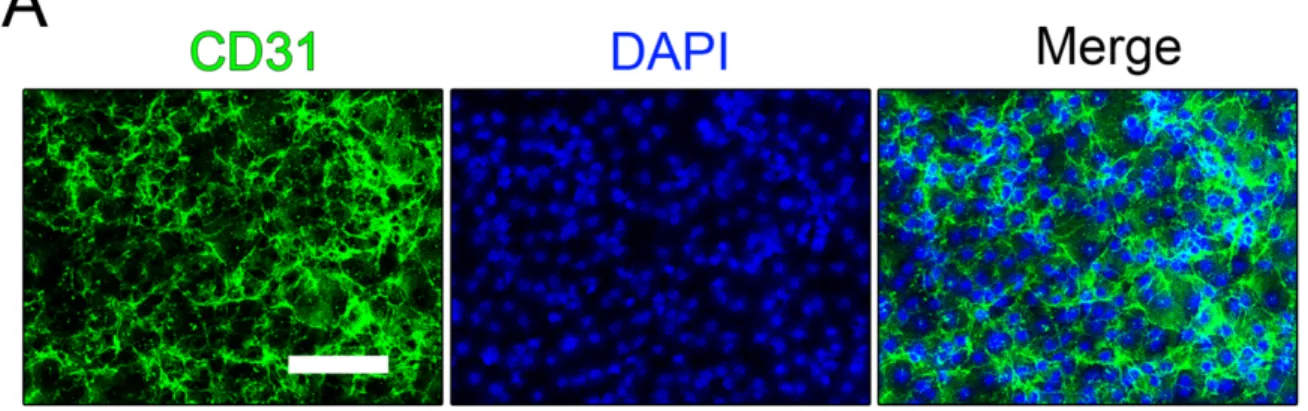
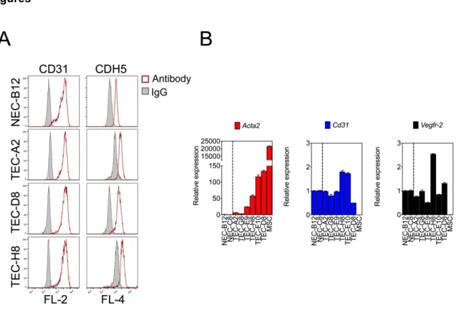
Isolated TEC maintain the expression of endothelial-selective genes and are free of mesenchymal cells and tumor cells ... 68
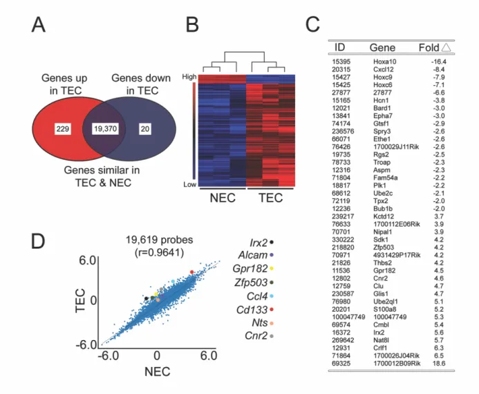
Genome-wide expression profiling reveals a distinct molecular signature in mammary TEC ... 69
Aberrant expression of TEC-selective genes persists in vitro ... 70
Discussion ... 70
Experimental Procedures ... 72
Figures ... 76
References ... 81
Endothelial-to-Mesenchymal Transition, ... 84
Overview ... 84
Introduction ... 85
Results and Discussion ... 86
Isolation of TEC with an intermediate EndMT phenotype ... 86
TGFβ induces diverse genetic signatures in different types of EC ... 88
Only a fraction of tumor vessels contain SMA+ endothelial cells in vivo ... 89
bFGF opposes the expression of some TGFβ target genes but augments the expression of others ... 90
EC challenged with TGFβ secrete their own bFGF which suppresses mesenchymal-like differentiation in secondary cultures ... 94
Experimental Procedures ... 96
Figures ... 102
Table ... 126
References ... 127
CHAPTER 5: Conclusions and Future Directions ... 131
Conclusions ... 131
Future Directions ... 132
1. What are the molecular mechanisms that dictate the differences between TGFβ responder EC and TGFβ non-responder EC? ... 132
2. How does bFGF counteract TGFβ-stimulated EndMT? ... 136
3. What is the role of endothelial TGFβ signaling in tumor progression? ... 139
Figures ... 141
LIST OF FIGURES
Figure 1-1. EndMT in development and disease. ... 26
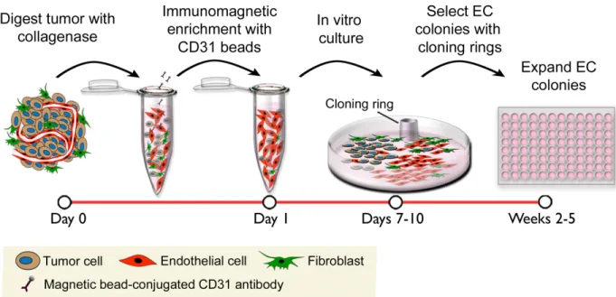
Figure 2-1. Schematic diagram of the EC isolation procedure. ... 54
Figure 2-2. Dil-Ac-LDL distinguishes EC from contaminating non-EC in live cultures. ... 55
Figure 2-3. Cloning rings and physical removal of non-EC produce EC cultures. ... 57
Figure 2-4. Expanded EC cultures retain endothelial marker CD31 and endothelial function in vitro. ... 58
Figure 2-5. EC isolated from Cdh5cre:ZsGreenl/s/l mice maintain brilliant ZsGreen fluorescence in culture. ... 60
Figure 3-1. Isolated TEC maintain the expression of endothelial-selective genes and are free of mesenchymal cells and tumor cells. ... 77
Figure 3-2. Genome-wide expression profiling reveals a distinct molecular signature in mammary TEC. ... 78
Figure 3-3. Aberrant expression of TEC-selective genes persists in vitro. ... 80
Figure 4-1. Characterization of NEC and TEC clones. ... 102
Figure 4-2. Subpopulations of EC undergo a spectrum of EndMT in response to TGFβ2 stimulation. ... 104
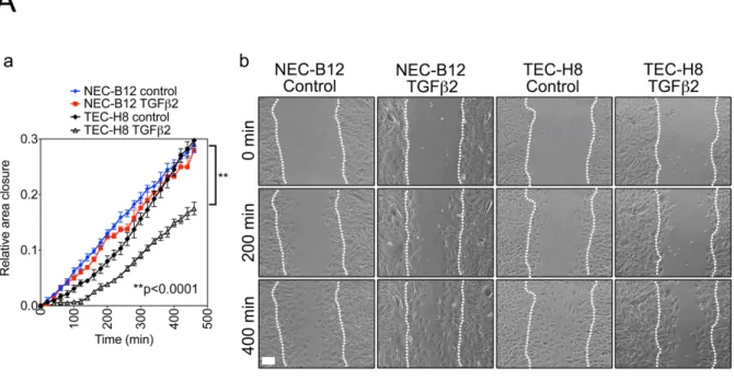
Figure 4-3. SMA+ TEC are functionally different from NEC. ... 105
Figure 4-4. Only a fraction of tumor vessels contain SMA+ TEC. ... 107
Figure 4-5. bFGF opposes the expression of some TGFβ target genes but augments the expression of others. ... 109
Figure 4-6. EC challenged with TGFβ secrete their own bFGF which suppresses mesenchymal-like differentiation in secondary cultures. ... 111
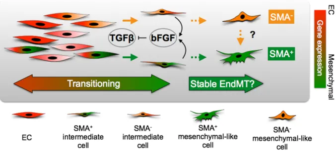
Figure 4-7. Schematic diagram of TGFβ and bFGF interactions during EndMT. ... 112
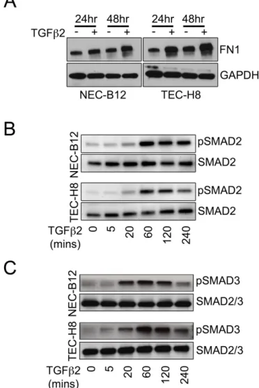
Figure 4-S1. Characterization of FN1 expression and SMAD2/SMAD3 phosphorylation in NEC versus TEC challenged with TGFβ2. ... 113
Figure 4-S3. Representative tile immunofluorescent images of mammary tumors
from Cdh5cre:ZsGreenl/s/l mice. ... 117
Figure 4-S4. bFGF reverses TGFβ2-induced morphological changes and target
gene expression in SMA+ TEC. ... 119
Figure 4-S5. bFGF suppresses TGFβ2-induced SMA expression in multiple EC clones. ... 120
Figure 4-S6. bFGF suppresses TGFβ2-induced mesenchymal gene expression in K-Ras lung TEC clones. ... 122
Figure 4-S7. bFGF weakly blocks TGFβ2-induced SMA expression through ERK/MAPK
signaling and inhibition of SMAD3 phosphorylation. ... 124
Figure 5-1. Inhibition of DNMTs by 5-Aza activates myofibroblast genes. ... 141
Figure 5-2. Depletion of DNMT1 by siRNA up-regulates myofibroblast genes in TGFβ non-responder EC clones. ... 142
Figure 5-3. Transient inhibition of DNMTs by 5-Aza induces sustained myofibroblast gene expression in TGFβ non-responders. ... 143
Figure 5-4. DNMT1 and SMA levels are inversely correlated in TGFβ responder and
non-responder clones. ... 144
Figure 5-5. Long-term TGFβ stimulation suppresses DNMT1 and induces persistent
SMA expression in non-responder EC. ... 145
Figure 5-6. TGFβ stimulates myofibroblast conversion from EC at two levels: suppression of DNMT1 and direct up-regulation of myofibroblast genes. ... 146
Figure 5-7. Inhibition of both MEK and ERK pathways blocks the antagonistic effect of
LIST OF TABLES
Table 1-1. Common endothelial markers expressed in mature blood and
lymphatic vasculature. ... 27
Table 2-1. PE-conjugated anti-CD31 antibody volumes required for different cell numbers. ... 61
Table 2-2. Anti-PE microbead volumes required for different cell numbers. ... 61
LIST OF MOVIES
Movie 4-S1. Three-dimensional projection of confocal z-stack immunofluorescent
LIST OF ABBREVIATIONS
aFGF acidic fibroblast growth factor
ALK anaplastic lymphoma kinase
BA blocking antibody
bFGF basic fibroblast growth factor
BMP bone morphogenetic protein
CAF cancer-associated fibroblast
CDH5 vascular endothelial cadherin (VE-cadherin)
CM conditioned media
CMM cerebral cavernous malformation
Cnn1 calponin 1
COL1 collagen 1
Co-SMAD common SMAD
Des desmin
DNMT DNA methyltransferase
EC endothelial cells
ECM extracellular matrix
EGF epithelial growth factor
Elk-1 ETS-domain containing protein-1
EMT epithelial-mesenchymal transition
End endoglin
EndMT endothelial-mesenchymal transition
FACS fluorescence-activated cell sorting
FBS fetal bovine serum
FN1 or Fn1 fibronectin 1
FSP-1 fibroblast-specific protein
FRS2 FGFR substrate 2
GEMM genetically engineered mouse model
HUVEC human umbilical vascular endothelial cells
I-SMAD inhibitory SMAD
KI kinase inhibitor
LAP latency-associated peptide
LG-DMEM low glucose (1 g/L D-glucose) DMEM
LTBP latent TGFβ binding proteins MEFs mouse embryonic fibroblasts
MEndT mesenchymal-endothelial transition
MI myocardiac infarction
NEC normal endothelial cells
PDGF platelet-derived growth factor
PDGFR platelet-derived growth factor receptor
PLCγ phospholipase Cγ1
qPCR quantitative real-time polymerase chain reaction
rpm rotations per minute
R-SMAD receptor-activated SMAD
siRNA small interfering RNA
SMA or Acta2 alpha smooth muscle actin
SMC smooth muscle cells
SRF serum response factor
Tagln of SM22 transgelin
TEC tumor-specific endothelial cells
TGFβR transforming growth factor beta receptor VEGF vascular endothelial growth factor
VEGFR vascular endothelial growth factor receptor
CHAPTER 1: Endothelial-Mesenchymal Transition and Its Regulators in Tumors
Overview
Endothelial cells (EC) form the innermost layer of the vascular system, lining the entire
blood and lymphatic vessels. This monolayer of cells, measuring an average of less than one
micron in thickness, is not merely part of passive conduits for nutrient and oxygen transport, but
is highly plastic and metabolically active. Studies of past decades have established that the
endothelium is integral to many physiological and pathological processes in virtually all tissues
of the body. In tumors, an aberrant microenvironment promotes dysregulated angiogenesis and
endothelial dysfunction. Tumor-specific EC (TEC) can acquire stem-cell futures and undergo
endothelial-mesenchymal transition (EndMT), where EC transdifferentiate into mesenchymal
cells, a process triggered mainly by a cytokine called transforming growth factor beta (TGFβ).
In the following chapter, a description of endothelial heterogeneity and plasticity of both
normal and tumor endothelium will be presented, along with summaries of studies on
developmental and pathological EndMT. Literature review of the regulatory factors of EndMT
will emphasize largely the pathways regulated by two opposing mediators of EndMT, namely,
TGFβ and bFGF, which are the main focus of this thesis. The final section of this introduction will discuss the implications of EndMT in tumors and other diseases, and will also include a
number of unanswered questions in the field for future studies.
Endothelial heterogeneity and plasticity
EC originate from a group of progenitor cells called angioblasts. Although how precisely
heterogeneous in their lineages. Studies of both avian species and mice have revealed that
some angioblasts and hematopoietic stem cells share the same common predecessors called
hemangioblasts, whereas other angioblasts derive independently from progressive restriction of
mesoderm in response to a series of growth signals, including Notch, vascular endothelial
growth factor (VEGF), and bone morphogenetic proteins (BMPs) 1,2. Angioblasts then develop
into the blood vascular system through two distinct processes: vasculogenesis where
angioblasts differentiate and assemble into a primitive vascular plexus of capillaries along with
some vessels such as presumptive dorsal aortae and cardinal veins, and angiogenesis where
new vessels are formed from the preexisting vasculature 1,3. Both processes can occur
concurrently once an initial vascular plexus is established, and the vascular network undergoes
extensive remodeling to establish a patent, functional system. Another parallel system, the
lymphatic vasculature, arises from the venous EC of the anterior cardinal vein. A subset of
these venous EC morph into lymphatic EC, which first form lymphatic sacs and then bud off the
cardinal vein, eventually ramifying into a separate network that returns the excessive
extravasated fluid to the blood circulation 4. Mature lymphatic EC are identified by a number of
molecular markers, including LYVE-1, PROX-1, and podoplanin, which are not observed in the
blood EC (Table 1-1). However, many of these markers are also expressed in other cell types
that are often derived from the same lineages (Table 1-1).
Although the endothelium is a single monolayer of cells that maintains the integrity of the
vasculature, endothelial heterogeneity is manifested from the early embryonic stage where
arterial and venous specification in the capillary plexus is determined by both intrinsic (e.g.,
epigenetic) and extrinsic (e.g., hemodynamic forces) factors 3,5. As the vascular network
integrates into the growing organs, EC specialize into subpopulations to meet the demands of
the local tissues, and start to display phenotypical, functional, and gene expression differences
The intracellular structural heterogeneity of EC was first revealed by electron microscopy
in the 1960s, an observation that suggests functional diversity of the endothelium 7. Distinct
endothelial morphologies were also observed in different vascular beds. For example, the brain
endothelium consists of tightly connected EC surrounded by a continuous basement membrane
(continuous endothelium), glomerulus EC in kidneys are penetrated with holes (fenestrated
endothelium), while liver EC are featured with ragged junctions and wrapped in a poorly formed
underlying basement membrane (discontinuous or sinusoidal endothelium) 8-10.
The advances of cell biology in the 1970s allowed successful culture expansion and
characterization of pure EC from human umbilical veins, acronymed HUVEC, which have since
been used as a standard tool for in vitro investigation of EC functions 11-14. Subsequent
immunohistochemical studies further discovered that subtypes of isolated EC are characterized
by different molecular markers 15-18. For instance, Weibel-Palade bodies are highly concentrated
in aortic EC compared with capillaries. Additionally, micro- and macro-vascular EC from various
tissues displayed not only tissue-specific responses to stimuli in vitro but also differential protein
expression, suggesting that the distinct molecular signatures and functions are retained in the
absence of their original microenvironments 19,20. More recently, Nolan and colleagues carried
out a comprehensive molecular profiling of capillary EC isolated from nine different organs 21.
Employing a method that involves “intravital labeling” of blood vascular EC by intravenous
injection of antibodies against endothelial markers, they isolated microvascular EC through flow
sorting and characterized these organ-specific EC subpopulations with microarray profiling.
Their findings demonstrate that EC express unique, organotypic clusters of transcription factors,
angiocrine factors, adhesion molecules, and chemokines that are consistent with their functions
in a specific organ. Furthermore, regeneration after tissue injury in different organs also elicits
divergent angiocrine responses in different vascular beds. For example, bone marrow and liver
and homeostasis compared to other EC subpopulations. Known EC markers such as Vegfr2
and Cdh5 levels are lowest in bone marrow-derived EC compared with other EC types, whereas
certain growth factors such as hepatocyte growth factor are markedly up-regulated only in liver
EC, not bone marrow EC, during injury-induced regeneration.
Because of the vastly diverse roles of EC in the growth, repair, and homeostasis of
virtually every organ in the body, it is not surprising that EC evolve to take on tissue-specific
characteristics. It is yet to be ascertained, however, what microenvironmental cues direct EC
specialization and how these tissue-specific traits are maintained and transmitted. To determine
the molecular mechanisms that control endothelial heterogeneity and to examine endothelial
functions pertaining to a specific tissue or organ, it is therefore important to isolate and culture
EC types derived from the tissue of interest. Although current EC isolation methods have been
improved significantly, researchers still encounter the same difficulties associated with the
isolation procedure as those early investigators did more than 40 years ago, primarily due to
tenacious contaminations of non-specific cell types. Part of this thesis (Chapter 1) will attempt to address this problem by outlining in detail a cloning-ring method that has consistently
produced highly pure EC cultures in our laboratory.
In addition to heterogeneity, EC also display remarkable adaptability and plasticity. In
intact post-developmental vessels, the endothelium generally remains quiescent. However,
under certain physiological or pathological conditions, such as pregnancy, inflammation, or
tumor growth, EC can be activated by angiogenic signals, such as bFGF and VEGF. They
proliferate and initiate sprouting and subsequent angiogenesis to repair or support new tissue
growth. They secrete an array of angiocrine factors that stimulate leukocyte infiltration and
instigate stem cell-mediated regeneration 22. Moreover, EC are able to transdifferentiate into
other cell types under both physiological and pathological conditions. During development,
endothelium through a novel cell transition 23. Time-lapse fluorescence confocal microscopy
reveals that individual aortic EC adopted a strong “bending” configuration and bulged out into
the sub-aortic space before transiting into hematopoietic cells 24. Endothelial-mesenchymal
transition, or EndMT, is another type of cellular process that EC undergo to generate stromal
cells during heart valve formation. EndMT can be re-activated in diseases like tumors and
fibrosis, where EC may revert to an embryonic-like stage and acquire stem cell-like features,
transdifferentiating into a variety of mesenchymal cell types. A more comprehensive review of
EndMT in both health and disease will be addressed in the later sections of this chapter.
Tumor endothelial cells (TEC)
TEC are essential in every step of tumor progression. Tumor cells cannot survive
beyond the size of 1 – 2 mm3 without blood supply, and many tumors remain dormant until an
angiogenic switch is triggered to recruit and generate blood vessels. Metastasis frequently
requires close EC-tumor cell interaction: metastatic cells first need to escape the primary sites
through intervasation and then extravasate at a distant site to establish metastases. In fact,
clusters of metastatic cells are often found in close proximity to the endothelium and TEC are
critical in promoting tumor stem cell growth and tumor progression 25. For instance, TEC in
lymphomas secrete an angiocrine factor Jag1, which activates the Notch signaling pathway and
downstream Hey1 transcription activities, promoting the expansion of aggressive and
chemoresistant tumor stem cell pools 26. In a melanoma model, tumor cells treated with
quiescent EC-conditioned media showed a slower growth curve and lower metastatic burden in
both tail-vein injection and xenograft tumor models, compared with the tumor cells stimulated
with media from activated EC (i.e., with an inflammatory phenotype), suggesting a pivotal role of
Observing these indispensable roles of the vasculature in tumor growth, Folkman in
1971 proposed a novel way to kill tumor cells by targeting tumor blood vessels, and later
pioneered the development of anti-angiogenic therapy 11. Since its inception, many
anti-angiogenic drugs primarily targeting VEGF-modulated anti-angiogenic signaling and TEC
proliferation have been developed, including a monoclonal antibody bevacizumab (Avastin)
against VEGFR2, a VEGF-trapping antibody, and tyrosine kinase inhibitors 28. However, clinical
benefit to prolong cancer patient survival has been limited due probably to either intrinsic
refractory or acquired resistance of the tumor vasculature 29. In fact, studies in recent years
have revealed that under the influence of an aberrant tumor microenvironment, tumor
vasculature deviates from its normal counterparts in morphology, function, gene expression,
and angiocrine signaling, which may contribute to the development of anti-angiogenic therapy
30. Tumor blood vessels are often distended, tortuous, leaky, and absent of the normal vascular
hierarchy 31. Tumor vasculature being constantly stimulated by angiogenic and inflammatory
factors display a hyper-angiogenic phenotype, up-regulating proangiogenic factors such as
VEGFRs, EPH receptors and ephrins, and endoglin 32-35.
Characterization of isolated TEC or tumor cell-conditioned EC further revealed that these
cells acquire inheritable genetic and epigenetic alterations. For example, Hida and colleagues
found that TEC isolated from melanoma and liposarcoma xenografts exhibited aneuploidy and
multiple centrosomes 36. TEC grew more rapidly and were more sensitive to growth factors like
bFGF and epidermal growth factor (EGF) than their normal counterparts 36. Comparison of
normal EC (NEC) and TEC isolated from human prostate tumors showed an epigenetic
signature of hypermethylation in the GSTP1 and RARβ2 promoters in TEC 37. Microarray profiling of tumor-conditioned EC identified 81 down-regulated genes in TEC, and further
examination of a subset of these gene promoters suggested that histone modification by histone
Chapter 3 that TEC isolated from mammary tumors express distinct gene signatures, which are persistent even after prolonged culturing and multiple passages, suggestive of aberrant
epigenetic modifications in these cells.
Another significant feature of TEC is that they acquire stem cell- or progenitor-like
plasticity. TEC attain a high proliferative potential that shortens their doubling time to 2.4 – 13
days, a rate that is 20 – 2000 times faster than quiescent NEC 40. Studies of TEC isolated from
high-metastatic melanomas (HM) showed that these TEC expressed higher levels of stem-cell
markers such as Sca-1 and CD90 compared to those from low-metastatic tumors 41. These
HM-TEC were highly proliferative and motile, and could differentiate into bone-like cells. Similar
observations were also made by Dudley and colleagues showing freshly isolated prostate TEC
not only had a mesenchymal-cell morphology, but also were able to dedifferentiate into cells
that resembled mesenchymal stem cells (MSC) 42. Like MSC, these prostate TEC when induced
by osteogenic media formed calcification and up-regulated bone markers such as osteopontin
and osteocalcin. Condrogenenic medium, on the other hand, stimulated the differentiation of
these TEC into cartilage-like cells which were stained positive for the cartilage dye Alcian blue,
and which also expressed cartilage-specific genes including Col2a1 and Sox9. Observation of
EndMT in tumors further supports the idea that the tumor endothelium can revert to its
developmental stage, gaining stemness and plasticity 42-44. Together, these data suggest that
TEC are highly adaptable to their environment. They proliferate to form new vessels, change
their secretion profiles to orchestrate tumor growth and metastasis, and even assume new
identities in response to environmental cues. Such abilities of TEC may give rise to a highly
adaptable tumor vasculature resembling “moving targets” that can consistently evade
Endothelial-mesenchymal transition (EndMT)
EndMT is critical in cardiac valve development
EndMT, a term coined to distinguish it from a closely related cellular event called
epithelial-mesenchymal transition (EMT), defines a process where EC lose endothelial identity
and gain mesenchymal characteristics 45. Individual transitioning EC may detach from the
endothelium, migrate into sub-endothelial interstitia, and transdifferentiate into
mesenchymal-like cell types. One of the most well characterized EndMT phenomena is the formation of
cardiac cushions and subsequent cardiac valves and septa during heart development (Figure
1-1). The heart tube formed in the early embryo (around embryonic day 9.5 in mice) consists of an
inner endothelial cell layer and several myocardial layers on the outside 46. The endocardium
initiates EndMT first by secreting an acellular substance called cardiac jelly into the interstitial
space to create swellings known as cardiac cushions. A subset of EC in the endocardium are
then activated and stimulated by TGFβ2 and TGFβ3 and other signaling pathways to undergo EndMT. These EC detach from the endocardium, migrate towards the underlying cardiac jelly,
transiently up-regulate the contractile protein alpha smooth muscle actin (SMA), and
transdifferentiate into mesenchymal cells to populate the cardiac septa and valve interstitium 47.
Using a Tie2cre:ROSA26R-LacZ lineage-tracing model, two groups have independently
demonstrated that cardiac valve interstitial cells derive from the EC of the endocardial cushions
48. Approximately 5-10% of fetal valve EC co-express both SMA and the endothelial marker
CD31, and 1% of the post-natal valve EC retain this feature, suggesting that a subpopulation of
mature valve EC continue to possess the ability to undergo EndMT by which the valve interstitial
cells may be replenished throughout adulthood 49. Another interesting observation is that these
valve EC are highly heterogeneous. EC clones isolated from adult cardiac valves exhibit
even after prolonged TGFβ exposure 50. While removal of serum and bFGF from the media can also trigger EndMT, VEGF addition was reported to counteract TGFβ-induced EndMT 50,51. A proportion of the clonal-derived mitral valve EC can be induced to undergo osteogenic and
chrondrogenic differentiation, but some other clones isolated from the same valves and
non-valve EC from carotid arteries are relatively less responsive to osteogenic signals 52. Taken
together, these data suggest that valve EC are heterogeneous in nature, and a subpopulation of
the EC in the mature valves retains stem cell features. This mesenchymal differentiation
potential can be reactivated under conducive environmental conditions. However, it remains
elusive why some EC have a higher propensity for EndMT than others, and what regulates the
differential responses in these EC subpopulations is not known.
Pathological activation of EndMT in tumors and other diseases
Tumors
EndMT in tumors was first reported by Zeisberg and colleagues in their landmark studies
of endothelial-derived fibroblasts in melanoma and pancreatic tumors. Using a lineage-tracing
model Tie2cre:ROSA26R-LacZ the group showed that EndMT-derived tumor fibroblasts
contributed to a mixture of heterogeneous stromal fibroblasts, which accounted for more than
40% of total cancer-associated fibroblasts, or CAF 44. Interestingly, the investigators discovered
that TEC differentiated into types of CAF: 30% of TEC-derived (i.e. β-gal+) tumor stromal fibroblasts expressed fibroblast-specific protein-1 (FSP-1), and another 12% were positive for
SMA, suggesting that different types of EndMT may exist in tumors. Confocal
immunofluorescence microscopy of human pancreatic tumors also revealed that endothelial
marker CD31+ co-localized with several mesenchymal markers including SMA, SM22α, N-cadherin, and FSP-1 53. Such transition to a mesenchymal phenotype in pancreatic tumors can
Our work, as discussed extensively in Chapter 4, has shown that clonal TEC isolated from the same tumor type were heterogeneous: some possessed mesenchymal features and
expressed high basal levels of myofibroblast marker SMA, whereas others resembled NEC and
expressed little to low levels of basal SMA. These different TEC subpopulations responded
differently to the EndMT inducer, TGFβ. SMA-high EC further up-regulated myofibroblast markers after TGFβ stimulation, but SMA-low EC were relatively resistant to TGFβ. To quantify EndMT in vivo, we employed a high-fidelity endothelial lineage-tracing model Cdh5Cre:
ZsGreenl/s/ which faithfully labels EC upon tamoxifen induction. However, we observed that in
mammary and lung tumors approximately 0-25% of microvessels contained TEC undergoing
EndMT per field examined. These SMA+ TEC in tumors are uncommon and often are isolated
individuals away from the tumor microvessels, as observed by us and others 55. We also found
that the frequency of EndMT varied in different tumor types and tumor models. For example,
K-RasG12D lung tumors and C3-TAg mammary tumors had much higher percentages of
transitioning TEC compared with the orthotopically implanted mammary tumors.
Although multiple groups have observed EndMT in tumors, to what extent EndMT
contributes to the CAF population is still a matter of debate. One concern is that the endothelial
lineage-tracing model Tie2cre:ROSA26R-LacZ used in earlier EndMT studies is not entirely
endothelial specific and labels a proportion of bone-marrow cells, which have been shown to
give rise to 20-40% SMA+ CAF in pancreatic tumor and gastric tumor models 56,57. Furthermore,
CAF markers used to identify transitioning TEC are often expressed by other stromal cells
juxtaposed with EC. For example, SMA is detected in perivascular cells including vascular
smooth muscle cells (SMC) and pericytes, and the latter are also positive for another CAF
protein, vimentin. This could greatly affect the accuracy of EndMT detection in vivo. Unbiased
fluorescence-activated cell sorting (FACS) using a high-fidelity, dual-reporter mouse model
better estimate EndMT frequency in different tumor types in future studies. Nevertheless, the
percentage of TEC that are undergoing EndMT could also be underestimated in in vivo studies,
as snapshots of tumors of a specific time point are often used to quantify EndMT in vivo. EndMT
may also be a dynamic process influenced by the tumor stage and microenvironment, and TEC
may assume an intermediate type vacillating between the endothelial and mesenchymal
phenotypes during tumor progression, which may produce varying numbers of transitioning EC
at specific stages of a tumor.
Despite the caveats of the in vivo models described above, EndMT has been
unequivocally confirmed in vitro from detection of co-expression of both mesenchymal and
endothelial markers in primary TEC or NEC challenged withTGFβ42,55,58-60. These in vitro studies have found that TEC acquire the plasticity and multipotency of stem cells, and can be
induced to differentiate into many cell types. Studies of tumor-endothelium interaction have
shown that EndMT promotes transmigration of melanoma cells through a TEC monolayer,
suggesting that EndMT may play a role in tumor metastasis 58. In fact, loss of endoglin, an
inhibitory regulator of TGFβ signaling, induces EndMT in a spontaneous pancreatic tumor model, Rip1-Tag2, and facilitates metastatic seeding in the liver due to a weakened endothelial
barrier 54. In addition, transitioning TEC could also serve as a source of growth factor such as
VEGF for stromal cell proliferation 55.
In our studies as outlined in Chapter 4, we identified two subpopulations of mammary TEC: one which transitions into myofibroblast-like cells by up-regulating SMA and other
myofibroblast markers after TGFβ challenge, and another which is relatively less responsive to TGFβ, suggesting that a proportion of TEC may be resistant to EndMT and this characteristic persists in culture (Figure 1-1). Other groups have also observed the same phenomenon in
different EC types. Ishisaki et al. reported a heterogeneous EC population that contained two
mesenchymal-like cells in a short time, and slow-differentiating cells that required more than
twice the length of TGFβ stimulation 60. Different endothelial TGFβ responsiveness is similarly confirmed in aortic valve EC 50. Some clonal valve EC respond to TGFβ treatment by up-regulating SMA, an effect that can be blocked by a soluble TGFβR2 antagonist. Other clonal populations do not respond to TGFβ, but increase SMA expression only after serum withdrawal, which is an effect independent of TGFβ pathway activation, as addition of TGFβR2 antagonist does not reverse the elevated SMA. These results together indicate that heterogeneous EC
subtypes may have distinct regulatory mechanisms of EndMT. Understanding the mechanisms
that control this difference between the two TEC subpopulations may help map out the
regulatory pathways of myofibroblast differentiation and design specific inhibitors that target
CAF in tumors.
Fibrotic diseases
Fibrosis featured by excessive scarring tissue is a common theme in chronic diseases of
many organs. Characteristics of fibrotic diseases include loss of normal epithelial function and
tissue architecture, increased collagen deposition, enhanced inflammatory reaction, and
persistent presence of SMA+ stromal cells, or myofibroblasts 61. Similar to CAF in tumors,
myofibroblasts perpetuate epithelial injury and aggravate fibrotic tissue formation by exerting
contraction force, secreting excessive extracellular matrix (ECM), and recruiting immune cells.
EndMT is believed to contribute to myofibroblast generation in a variety of fibrotic conditions
affecting the kidneys, lungs, and heart.
EndMT has been observed in different renal fibrosis models including obstructive
nephropathy, diabetic nephropathy, and the Alport (COL4A3 deletion) disease mouse models
62-64. Using co-staining of an endothelial marker CD31 with the mesenchymal markers FSP-1 or
FSP-1+/CD31+ cells accounted for 25-60% of all FSP-1+ fibroblasts, and SMA+/CD31+ 36-45% of all
SMA+ fibroblasts in kidney fibrosis. In another diabetic renal fibrosis study, 10-24% of SMA+
fibroblasts during the early stage of fibrosis development were of endothelial origin 65,
suggesting that EC are significant contributors of kidney fibrosis.
Endothelial-derived fibroblasts have also been identified in a bleomycin-induced lung
fibrosis mouse model that carries an endothelial lineage-tracing Tie2cre:LacZ reporter 66. EndMT
is reported to contribute to two subpopulations of fibroblasts, of which ~ 85% were
COL1-positive, and ~ 15% both COL1 and SMA COL1-positive, suggesting that EC may undergo different
types of EndMT in fibrosis. Furthermore, elevated Ras signaling potentiates TGFβ-stimulated mesenchymal gene expression and induces persistent EndMT phenotype upon TGFβ
withdrawal both in vitro and in vivo, implicating that a secondary change in the signaling network
augments TGFβ sensitivity of the endothelium and is required for irreversible fibroblast
differentiation. Inflammation can also sensitize the endothelium in intestinal fibrosis, where
pro-inflammatory factors, such as IL-1β and TNFα, work in synergy with TGFβ to drive EndMT 67.
In addition, EndMT has been described in myocardiac infarction (MI) and cardiac
fibrosis. Induction of Wnt signaling in subepicardial EC after an acute MI is associated with
mesenchymal transdifferentiation of these cells, a result supported by in vitro observations in
that induction of canonical Wnt pathway in cultured EC instigates EndMT 68. In a diabetic
cardiac fibrosis model described by Widyantoro et al., hyperglycemia induced EndMT through
up-regulation of endothelin, and EC-specific deletion of endothelin ameliorated cardiac fibrosis
and suppressed EndMT 69. Interestingly, endothelin augments TGFβ expression in diabetic hearts and in cultured EC treated with high glucose, thereby activating TGFβ signaling. TGFβ stimulation also significantly enhances endothelin mRNA production by almost three-fold,
or SMA are identified in pressure-overload cardiac fibrosis using Tie1cre:LacZ reporter mice, and
EndMT-derived cells contribute to 27 – 35% of the total fibroblast population 70. Activation of
EndMT in cardiac fibrosis may be mediated by various signaling and transcription regulators,
such as Toll-like receptor (TLR)5 and p53 71,72. Deletion of either of these factors can attenuate
interstitial fibroblast accumulation by lessening EndMT in pressure-overload cardiac fibrosis.
Generation of fibroblasts through EndMT in pressure overload-induced cardiac fibrosis,
however, was questioned by Moore-Morris and colleagues in a recent study where they utilized
multiple lineage-tracing reporter models to investigate the origins of fibroblasts in pressure
overload-induced cardiac fibrosis 73. The authors used a Wt1cre:Col1a1-GFP:ROSA-tdT double
reporter line to specifically label epicardial-derived fibroblasts, and a Tie2cre
:Col1a1-GFP:ROSA-tdT line to lineage trace endocardial-derived fibroblasts. They showed that these two lineages
contributed to the generation of a vast majority of resident cardiac fibroblasts (~95%) in an
uninjured heart during development. Transaortic constriction in adult mice induced pronounced
accumulation of cardiac fibroblasts both in ventricular myocardium and perivascular regions of
large coronary vessels. However, the percentages of the fibroblasts derived from the epicardium
or endocardium remained relatively constant in this pressure overload-induced cardiac fibrosis
model, demonstrating that resident fibroblasts, not other cellular sources, are responsible for
fibrosis formation. Furthermore, the investigators observed that the endothelial-lineage tracing
Tie1cre labeled non-specific immune cells which were also positive for FSP-1, which is a marker
used to identify cardiac fibroblasts in a previous study by Zeisberg et al., where the Tie1cre:LacZ
line was employed to show massive de novo generation of fibroblasts from mature EC through
EndMT 70.
Although these results by Moore-Morris et al. do not entirely exclude the occurrence of
EndMT in pressure overload cardiomyopathy, they suggest that the contribution of EndMT may
these studies could be due to differences in mouse genetic background (e.g., FVB/N or
C57BL/6 70 vs. Black Swiss 73), age of the animals (e.g., 12 – 24 weeks old 70 vs. 8 – 10 weeks
old 73), or length of the surgery, as all these factors can potentially influence cardiovascular
outcomes post-injury 74,75. Nevertheless, EndMT in many other fibrotic diseases has been
supported by a considerable amount of evidence, suggesting that the frequency of EndMT may
vary in different disease settings. These studies further demonstrated that fibroblasts are a
highly heterogeneous population that originates from many lineages. Multiple markers specific
for fibroblasts and myofibroblasts, such as COL1, SMA, and platelet-derived growth factor
(PDGF) α, and highly endothelial-specific inducible cre lines, such as Cdh5cre/ER,should
therefore be utilized in future studies to accurately determine the frequency of EndMT in fibrosis.
Vascular diseases
EndMT may contribute to arteriosclerotic plaque formation by promoting neointimal
hyperplasia, which in turn can drive the progression of numerous vascular conditions, including
pulmonary hypertension 76,77, vascular graft failure 78,79, and artherosclerosis 80. In addition,
EndMT may play an important role in the development of venous stenosis of the liver in
idiopathic portal hypertension 81. Increased serum TGFβ levels or perturbation of TGFβ
signaling in these vascular disorders probably triggers EndMT, whereas inflammation and
oscillatory shear stress during the disease progression can further boost EndMT and accelerate
atherogenesis 78,80. TGFβ signaling leads to downstream activation of SMAD2/3 pathway in the atherosclerotic endothelium, whereas suppression or deletion of SMAD2 and SMAD3 lessens
neointimal formation 79.
Cerebral cavernous malformation (CCM), which is mainly manifested as vascular
dysplasia in the brain, is a genetic condition caused by the loss-of-function mutations in one of
enlarged vascular lesions that can lead to severe cerebral hemorrhage. Recent evidence
strongly suggests that the pathological manifestations of both familial and sporadic CCM are a
direct consequence of EndMT 83. Loss of the Ccm1 gene in mice promotes EndMT through
decreasing EC markers and up-regulating a panel of mesenchymal genes, leading to cerebral
vascular leakage and dysplasia 84. Although the pathogenesis of CCM is not entirely clear, it is
believed that Ccm1 deficiency elevates BMP6 production, which acts synergistically with TGFβ to trigger SMAD-mediated EndMT 85.
Complex signaling networks regulate EndMT
TGFβ signaling
TGFβ is the single most important EndMT inducer. Three TGFβ isoforms (types 1, 2, and 3) that share a similar peptide structure have been identified. They belong to the TGFβ superfamily that comprises more than 30 ligands categorized into six subgroups, including
TGFβs and other members consisting of activins/inhibins, nodals, BMPs, growth differentiation factors, and Mullerian inhibiting substance 86. TGFβ is synthesized as a homodimeric proprotein that contains an N-terminal pro-peptide region, termed as the latency-associated peptide (LAP),
and a C-terminal polypeptide region. Intracellular proteolytic cleavage of LAP from the TGFβ proprotein produces the mature TGFβ peptide, which forms an inactive complex with LAP. This inactive TGFβ complex is sequestered by latent TGFβ binding proteins (LTBP) and deposited into the ECM upon secretion 87. Subsequent activation of TGFβ requires mobilization of the
TGFβ peptide from the ECM by proteases or thrombospondin regulated by complex signals in response to ECM damage or other extracellular perturbations 88. All three TGFβs bind to the same receptors and activate them through similar mechanisms; however, local ligand
availability and receptor affinity may determine their differential biological effects during
Seven type I receptors and five type II receptors for the TGFβ superfamily ligands are found in humans, and ligand binding induces formation of a receptor complex comprising of two
type I and two type II components, all of which are serine/threonine kinases. Once released
from ECM, TGFβ peptides bind to the type II receptor TGFBR2, which then recruit and activate the type I receptor TGFBR1 (or anaplastic lymphoma kinase 5 [ALK5]) to induce downstream
signaling 90. The signal strength depends on differential ligand affinity for TGFBR1 and
mediation of co-receptors including endoglin and betaglycan (TGFBR3) 91,92.
TGFβ signaling is intricately complex and highly cellular-context dependent. Its outcome is influenced by cell type, microenvironment, interaction with other signaling pathways and
transcription factors, epigenetic landscape, and innate genetic variations 86,93. Many different
downstream signaling factors can be activated by TGFβ, including the canonical transcription factors, SMADs, and other non-SMAD mediators. In vertebrates, there are eight SMADs: six
receptor-activated SMADs (R-SMADs), a common SMAD (Co-SMAD) named SMAD4, and two
inhibitory SMADs (I-SMADs) which include SMAD6 and SMAD7 94. R-SMADs 2 and 3 are
predominantly activated by TGFβ binding of TGFBR1, whereas R-SMADs 1, 5, and 8 are phosphorylated mainly by BMP-regulated activation of other receptors such as ALK1, ALK3,
and ALK6. Activated R-SMAD dimerizes and binds to SMAD4 before nuclear translocation.
I-SMADs, on the other hand, can bind to R-SMADs to block their transcription activation ability.
TGFβ also regulates gene expression by SMAD-independent pathways, including Ras-mediated ERK, p38 MAPK, PI3K/Akt, and Rho-like GTPase signaling 95.
TGFβ-stimulated EndMT was first observed in cultured bovine aortic EC in the early 1990’s 96. The induction of EndMT by TGFβ was later confirmed in vivo by Brown and colleagues, who reported that neutralizing TGFβ signaling with a TGFβR2 blocking antibody blocks the activation of EndMT during cardiac cushion development. 97. In pathological
by activating both SMAD-dependent and SMAD-independent pathways 27,98,99, but contribution
of each of these pathways in EndMT may be context-dependent. In this thesis project, we have
found that TGFβ mediated EndMT in mammary tumor EC through both SMAD2/3 and PI3K signaling, and blocking of these pathways attenuated TGFβ activity (Chapter 4). Activation of SMAD and non-SMAD signaling can in turn lead to transcriptional up-regulation of EndMT
master transcription factors such as Snail, Slug ZEB-1, SIP-1, Twist, and LEF-1, which then
allow the induction of mesenchymal genes and suppression of endothelial genes 99,100.
Other regulatory pathways can also interact with TGFβ signaling to either augment or inhibit EndMT. For instance, TGFβ coordinates with several BMPs and Notch signaling to synergistically stimulate mesenchymal gene expression in valve EC during cardiac valve
development46. Induction of EndMT in cultured HUVEC has been shown to require synergistic
action of IL1β and TGFβ, although only TGFβ is necessary for sustaining mesenchymal gene expression 101. Depletion of an inhibitory co-receptor endoglin sensitizes EC to TGFβ stimulation and promotes EndMT in tumors, while a TGFβ superfamily member BMP7 antagonizes TGFβ -induced EndMT both in vitro and in vivo 54,70,102. In addition to BMP7, tyrosine kinase receptor
ligands such VEGF and bFGF have been shown to counteract TGFβ signaling and reverse EndMT in normal valve EC and the arteriosclerotic endothelium respectively 51,78. This thesis
work has also found that bFGF, but not VEGF, acts as a potent inhibitor of EndMT, completely
blocking TGFβ-stimulated myofibroblast gene induction in EC isolated from mammary tumors. (More on bFGF and TGFβ interaction will be discussed in the following section.) Together, these studies suggest that TGFβ mediates EndMT in a context of complex signaling network, and its effect is likely controlled by an array of diverse factors including EC heterogeneity,
bFGF
FGFs are a pleiotropic group of ligands that signal through four highly conserved
receptor tyrosine kinases (RTKs), FGF receptors (FGFRs) 1 – 4. They regulate a plethora of
major cellular processes during development, ranging from early embryonic mesoderm
patterning to the formation of multiple organ systems 32-35,103. In EC, FGFR1 and FGFR2 are the
most predominant isoforms, and upon ligand binding they dimerize and autophosphorylate to
activate downstream signaling cascades. A total of 18 distinct mammalian FGFs have been
discovered, among which the prototype FGFs, acidic fibroblast growth factor (aFGF) and bFGF,
are particularly critical in early endothelial specification and vascular development 104.
aFGF and bFGF share 55% sequence homology and they exert similar biological effects
on many different cell types, although their signal strength depends on the expression of
subtypes and splice variants of FGFRs and on the ligand-receptor affinity 105. In particular, bFGF
is well known for its potent pro-proliferative and angiogenic effect on EC. Cultured mesoderm
cells cannot differentiate into endothelial progenitors in the absence of bFGF 106. bFGF is also
pivotal in the maintenance of endothelial VEGFR2 expression that is indispensable in
VEGF-stimulated angiogenesis 107. In mature vasculature post development, endothelial-specific FGF
signaling is essential in injury response, as endothelial deletion of both FGFR1 and FGFR2
significantly delays wound healing of the skin and impairs hypoxia-induced angiogenic response
in the retina 108.
In addition to regulating EC functions, emerging evidence shows that bFGF signaling
intersects with TGFβ pathways to influence downstream cellular events during EndMT. High-throughput screenings of 60 different combinations of ECM proteins singled out bFGF as the
directly blocks TGFβ-induced EndMT in arteriosclerotic lesions, whereas suppression of bFGF signaling through inflammation-mediated FGFR1 down-regulation renders EC prone to EndMT
78. In lymphatic vessels, bFGF inhibits EndMT and maintains lymphatic EC identity by opposing
TGFβ-activated SMAD2 through Ras/ERK MAPK signaling 111. bFGF also decreases TGFβ -stimulated SMA and mesenchymal gene expression in pericytes and airway SMC 112,113. The
close bFGF homologue, aFGF, acts similarly to bFGF to counteract TGFβ signaling. Deprivation of aFGF or TGFβ stimulation, for example, induces mesenchymal genes SM22 and calponin in HUVEC, while treatment with aFGF blocks TGFβ-induced gene expression and decreases the acquired SMC-like contractility 60. This antagonistic effect of aFGF on TGFβ, transduced through the MAPK/ERK pathway, is also observed in fibroblasts and in epithelial cells during
EMT 114-116.
The antifibrotic effect of bFGF is additionally demonstrated by studies of hypertrophic
scars and keloid tissue formation during wound healing, where bFGF effectively deceased
collagen production, ECM proteins, and SMA expression in fibroblasts through suppressing
SMAD-dependent pathways 117. More remarkably, administration of bFGF evidently reduced
scar tissue and inhibited the profibrogenic effect of TGFβ in in vivo wound-healing models and in clinical studies, indicating that the therapeutic benefits of bFGF are likely due to inhibition of
myofibroblast generation 118-121.
An intriguing reciprocal interaction between TGFβ and bFGF is also discovered by this thesis work in that TGFβ treatment of EC can induce bFGF production and secretion, which inhibits TGFβ signaling in these same cells (Chapter 4). Conditioned media harvested from TGFβ-stimulated EC contained high levels of bFGF, which suppressed mesenchymal gene induction and preserved endothelial markers in a fresh batch of EC induced to undergo EndMT.
enhanced by TGFβ in human lung fibroblasts, and this secreted bFGF can in turn activate ERK pathway through autocrine activation of its own receptors 122.
Surprisingly, bFGF is also implicated in the promotion of EMT, and in some instances,
EndMT. Studies of avian heart development suggested that both TGFβ and bFGF are inducers of EMT of the epicardium during coronary vasculogenesis 123. Isolated epicardial cells in
response to the combination treatment of bFGF and TGFβ increased motility and proliferation, and penetrated the underlying matrix, which is a characteristic of EMT. In a different study
where the epicardial-lineage cells were generated from human pluripotent stem cells, addition of
bFGF augmented TGFβ-stimulated expression of mesenchymal markers including SMA and vimentin, although bFGF-induced proliferation and expression of EMT transcription factor SNAI1
were counteracted by TGFβ124. Similarly, bFGF is shown to induce EndMT in corneal EC 125,126.
These conflicting results may be due to a number of reasons. First, bFGF signaling, like
TGFβ pathways, is highly complex and cellular-context dependent. Intrinsic factors such as cell heterogeneity and extrinsic factors such as culture conditions may influence cellular responses
to bFGF. Distinctive cell types and tissue may also respond to bFGF differently. The second
compounding issue is that the hallmarks and markers of EndMT and EMT are not precisely
defined, and studies on EndMT and EMT often use different readouts (gene expression, cell
shape, motility). Such diverse characterization methods may lead to different interpretations and
conclusions in different studies.
In addition, it is possible that bFGF acts synergistically with TGFβ as a fibroblast mitogen, promoting myofibroblast proliferation after mesenchymal transition is triggered by
showed that a two-step mechanism likely occurred during myogenesis: pulse exposure of TGFβ was required to initiate myogenesis at the early stage of myogenesis, whereas bFGF, albeit not
necessary for myogenic commitment, was indispensible in maintaining long-term cell survival
and proliferation. These data suggest that time-sensitive regulation of bFGF may be important in
myofibroblast generation. It is therefore conceivable that bFGF may have a biphasic effect on
EMT or EndMT: counteracting the pro-differentiation effect of TGFβ signaling at the initiation stage, but promoting myofibroblast growth once the mesenchymal differentiation pathway is
committed.
Other signaling pathways
Various other pathways have also been implicated in EndMT. BMP7 inhibits EndMT 70,81,
whereas BMP6 acts synergistically with TGFβ to activate EndMT through SMAD1/3-dependent pathways in CCM 84,85. Studies by different groups have also reported the importance of Wnt-β -catenin signaling in EndMT. Mice with endothelial-specific deletion of β-catenin fail to develop heart cushion due to impaired EndMT during development 128, but in adult cardiac EC
injury-triggered canonical Wnt signaling activation leads to the initiation of EndMT in myocardiac
infarction 68. In a CCM mouse model, loss of Ccm3 gene triggers Wnt-independent β-catenin,
which initiates EndMT in the cerebral vasculature 129, indicating that β-catenin can dynamically regulate EndMT depending on the cell context. Depletion of Tie1 can also induce EndMT by
up-regulating the EndMT transcription factor Slug through ERK1/2, ERK5, and Akt cascades 53.
Moreover, hypoxia-stimulated HIF1 pathway, which is a major regulator of EMT, has been
shown to contribute to EndMT activation in many fibrotic diseases 130-135.
MicroRNAs (miRNAs) in EndMT
miRNAs have recently emerged as important new players in EndMT, and they regulate
several target genes. Global perturbation of miRNA levels has been observed during
pathological EndMT 136. Based on their roles, EndMT-associated miRNAs can be categorized
into two groups: pro-EndMT and anti-EndMT miRNAs. Pro-EndMT miRNAs antagonize
inhibitory regulators, thereby up-regulating EndMT signaling. Examples include miRNA-21,
which promotes EndMT by activating Akt pathway through silencing of a phosphatase and
tensin homolog in EC 137. miR-342-5p is another EndMT inducer which initiates EndMT probably
by down-regulating endoglin, a TGFβ signaling inhibitor 138. On the other hand, anti-EndMT miRNAs frequently target positive regulators to curb profibrotic signaling and mesenchymal
gene expression. An aforementioned miRNA regulated by bFGF, let-7, for example, maintains
endothelial identity by counteracting TGFβ signaling, whereas reduced let-7 expression by disruption of bFGF signaling activates EndMT 78. Another miRNA, miR-125b, can suppress
profibrotic signals in cardiac EC by down-regulation of p53 136. Decreased miR-200b levels have
been observed during EndMT in diabetic hearts, and over-expression of miR-200b reverses
high glucose-activated EndMT by suppressing multiple TGFβ downstream targets in cardiac EC 139.
Impact of pathological EndMT
The significance of EndMT in tumors and other diseases is multifold. First, EndMT
serves as a cellular source for CAF in tumors or myofibroblast in fibrotic diseases. CAF are
activated fibroblasts in tumor stroma identified by the expression of the prototypical marker
SMA, and they promote tumor progression by supporting primary tumor growth and stimulating
tumor metastasis 140. Similarly, myofibroblasts in fibrotic diseases can exacerbate fibrotic
conditions through multiple mechanisms, including disrupting normal ECM and sustaining
inflammatory response, which lead to loss of organ functions 61. Secondly, vascular function and
integrity may be affected by transitioning EC detaching and migrating away from the
abnormal vascular morphology of the cerebral vasculature in CCM 69,84, while in tumors
increased metastasis may result from EndMT 54,58. Alteration of endothelial functions may also
contribute to parenchymal hypoxia, leading to detrimental consequences in hypoxia-sensitive
organs such as the kidney 141. Thirdly, aberrant endothelial signaling as a result of EndMT may
promote tissue inflammation, and cause dysregulated angiogenesis and abnormal angiocrine
profiles. Finally, loss of endothelial markers, especially VEGFR2 which is an anti-angiogenic
target in the treatment of late-stage tumors, during EndMT could potentially facilitate the evasion
of anti-angiogenic therapies.
Another aspect of EndMT shown collectively by us and others using different disease
and lineage tracing models is that EC are heterogeneous and respond to TGFβ differently. Subsets of EC can transition into distinct fibroblast subpopulations with distinctive molecular
markers, for example, COL1+ or SMA+ fibroblasts. Our results showed that only a proportion of
mammary TEC transdifferentiate into SMA+ myofibroblast-like cells in response to TGFβ, whereas other TEC do not. It has been recently proposed that in tumors CAF are a polarized
cell type consisting of tumor-restricting (Type I) and tumor-promoting (Type II) populations 140.
While Type I CAF are SMA- and inhibit tumor growth, Type II CAF are activated fibroblasts that
up-regulate an array of mesenchymal markers including FSP-1, vimentin, PDGF receptor
(PDGFR) β, and the prototypical myofibroblast marker SMA. In fact, acquisition of SMA
expression, or myofibroblast contractility, determines cell fate and function during mesenchymal
stem cell differentiation 142. However, it is unclear what regulates the heterogeneity of CAF and
their precursors, and why some precursor cells are resistant to TGFβ challenge and remain SMA-. Furthermore, mechanisms that reverse or inactivate myofibroblast gene expression
during EndMT are not well defined. It also remains elusive if different subpopulations of CAF
Future studies that delineate the molecular mechanisms that regulate these processes will
Figures
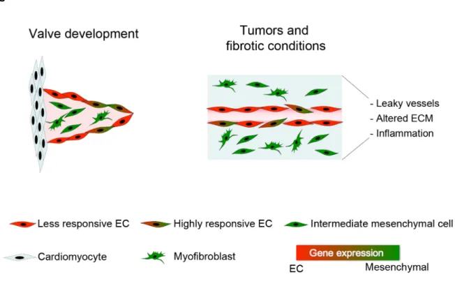
Figure 1-1. EndMT in development and disease.
EC undergo EndMT during cardiac valve formation in development. In certain pathological conditions such as fibrosis and tumors, EC can respond to TGFβ and other inflammatory factors to transdifferentiate into myofibroblasts. The endothelium consists of a heterogeneous
Table
Table 1-1. Common endothelial markers expressed in mature blood and lymphatic vasculature.
Markers Blood EC Lymphatic EC Other cell types/tissues CD31 (PECAM-1) 143 + + Megakaryocytes, subsets of B and T lymphocyte, monocytes, neutrophils
CD34 73,144 Heterogeneous + Fibroblasts; hematopoietic cells CD144 (VE-Cadherin
or Cadherin 5 [CDH5]) 145
+ + Popliteal lymph node sinus
macrophages
Claudin 5 146-148 +; more
concentrated in the blood-brain barrier and endoneurial blood-nerve barrier
+ Pancreatic acinar cells, alveolar lung cells, intestinal epithelium
E-selectin 149-151 Proliferating or inflammatory endothelia
Activated lymphatic EC
EPH receptor B4 152 Venous EC - Ephrin B2 152 Arterial EC -
Factor VIII 153 Heterogeneous - Platelets, megakaryocytes Friend leukemia
integration 1 transcription
factor (FLI1 or ERGB) 144
+ - Lymphocytes
LYVE-1 153 - + Macrophage marker Mac-1-positive cells
Podoplanin 153 - + Nerve sheaths, lingual gland myoepithelial cells
PROX1 154,155 - + Renal papilla Scavenger receptor
type 1 (DiI-Ac-LDL uptake)154
+; higher in capillary EC
+ Smooth muscle cells,
macrophages
Stem-cell antigen 1 (Sca-1) 6,156
Heterogeneous, possibly endothelial progenitor cells
- Hematopoietic stem cells, stem/progenitor cells in other organs, perivascular cells TIE-1 157,158 +; stronger in
arterial EC
+ Hematopoietic stem cells
TIE-2 159 + + Monocytes and macrophages VCAM 160 Inflammatory
endothelia
-
VEGFR1 15,161 + - Monocytes, vascular SMC, hematopoietic cells, retina VEGFR2 161-166 + + Retina, neural stem/progenitor
cells VEGFR3 167 Fenestrated blood
endothelia
+
von Willebrand factor
REFERENCES
1. Chappell, J. C. & Bautch, V. L. Vascular development: genetic mechanisms and links to vascular disease. Current Topics in Developmental Biology 90, 43–72,
doi:10.1016/S0070-2153(10)90002-1 (2010).
2. Udan, R. S., Culver, J. C. & Dickinson, M. E. Understanding vascular development. Wiley Interdisciplinary Reviews: Developmental Biology 2 (3), 327–346,
doi:10.1002/wdev.91 (2012).
3. Aird, W. C. Endothelial Cell Heterogeneity. Cold Spring Harbor Perspectives in Medicine 2 (1), a006429–a006429, doi:10.1101/cshperspect.a006429 (2012).
4. Hong, Y.-K. & Detmar, M. Prox1, master regulator of the lymphatic vasculature
phenotype. Cell and Tissue Research 314 (1), 85–92, doi:10.1007/s00441-003-0747-8 (2003).
5. Carmeliet, P. Angiogenesis in life, disease and medicine. Nature 438 (7070), 932–936, doi:10.1038/nature04478 (2005).
6. Aird, W. C. Phenotypic heterogeneity of the endothelium: I. Structure, function, and mechanisms. Circulation Research 100 (2), 158–173, doi:10.1161/01.
RES.0000255691.76142.4a (2007).
7. Florey The endothelial cell. British Medical Journal 2 (5512), 487–490, doi:10.1136/bmj.2.5512.487 (1966).
8. Wisse, E. An electron microscopic study of the fenestrated endothelial lining of rat liver sinusoids. Journal of Ultrastructure Research 31 (1), 125–150 (1970).
9. Karnovsky, M. J. The ultrastructural basis of transcapillary exchanges. The Journal of general physiology 52 (1), 64Suppl–95s (1968).
10. Farquhar, M. G., Wissig, S. L. & Palade, G. E. Glomerular permeability I. Ferritin transfer across the normal glomerular capillary wall. 1961. Journal of the American Society of Nephrology : JASN 10 (12), 2645–2662 (1999).
11. Folkman, J. Tumor angiogenesis: therapeutic implications. The New England Journal of Medicine 285 (21), 1182–1186, doi:10.1056/NEJM197111182852108 (1971).
12. Lewis, L. J., Hoak, J. C., Maca, R. D. & Fry, G. L. Replication of human endothelial cells in culture. Science 181 (4098), 453–454 (1973).
13. Jaffe, E. A., Nachman, R. L., Becker, C. G. & Minick, C. R. Culture of human endothelial cells derived from umbilical veins. Identification by morphologic and immunologic