Synthesizing a New DNA Strand
• Enzymes called DNA polymerases catalyze the elongation of new DNA at a replication fork
• Most DNA polymerases require a primer and a DNA template strand
• The rate of elongation is about 500 nucleotides per second in bacteria and 50 per second in
human cells
• Each nucleotide that is added to a growing DNA strand is a nucleoside triphosphate
• dATP supplies adenine to DNA and is similar to the ATP of energy metabolism
• The difference is in their sugars: dATP has deoxyribose while ATP has ribose
Figure 16.14
New strand Template strand
Sugar Phosphate Base Nucleoside triphosphate DNA polymerase Pyrophosphate 5 5 5 5 3 3 3 3 OH OH OH
P P i
2 P i
Antiparallel Elongation
• The antiparallel structure of the double helix affects replication
• Along one template strand of DNA, the DNA polymerase synthesizes a leading strand
continuously, moving toward the replication fork
Leading strand
Lagging strand
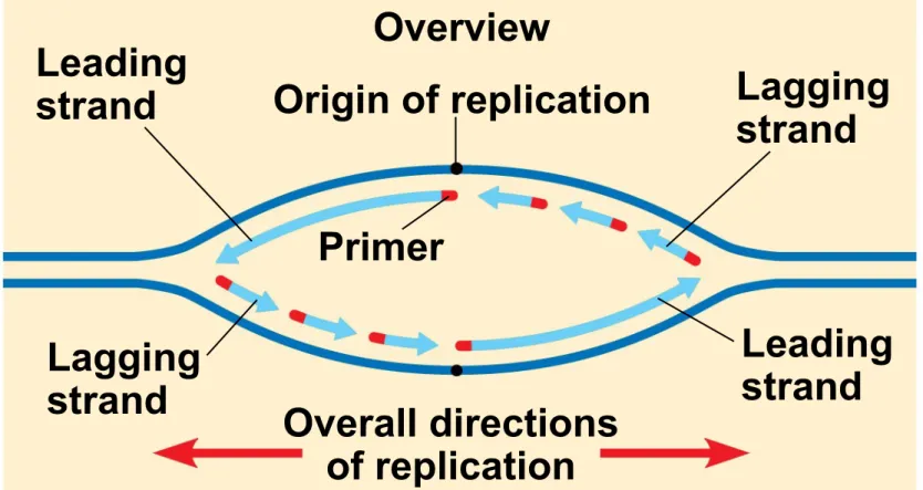
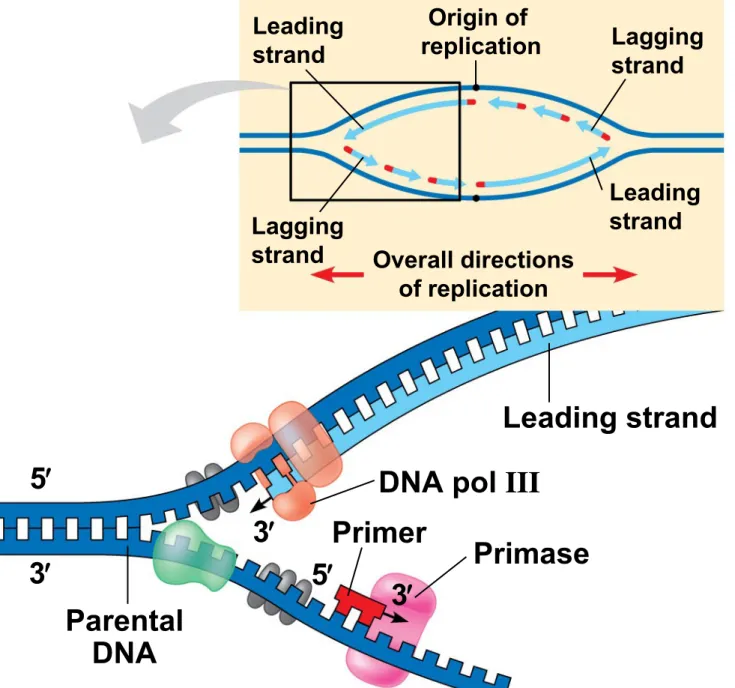
Overview
Origin of replication Lagging strand Leading strand Primer Overall directions of replication Origin of replication RNA primer Sliding clamp
Figure 16.15a
Leading strand
Lagging strand
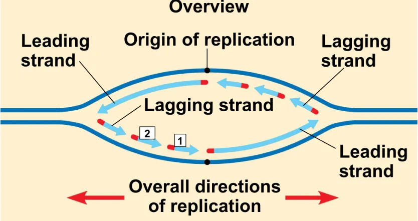
Overview
Origin of replication Lagging strand
Leading strand Primer
Origin of replication
RNA primer Sliding clamp
DNA pol III
Parental DNA
3
5
5
3
3
5
3
5
3
5
3
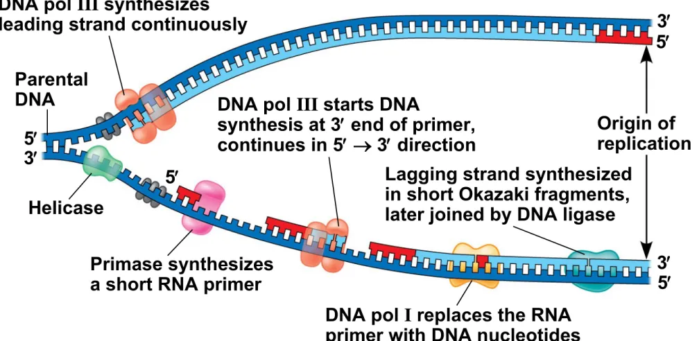
• To elongate the other new strand, called the
lagging strand, DNA polymerase must work in the
direction away from the replication fork
• The lagging strand is synthesized as a series of segments called Okazaki fragments, which are joined together by DNA ligase
Origin of replication Overview Leading strand Leading strand Lagging strand Lagging strand Overall directions of replication Template strand RNA primer for fragment 1
Okazaki fragment 1
RNA primer for fragment 2
Figure 16.16a
Origin of replication Overview
Leading strand
Leading strand Lagging strand
Lagging strand
Overall directions of replication
Template strand
3
3
5
Figure 16.16b-2
Template strand
RNA primer for fragment 1 3
3
3
3
5
5
5
5
Template strand
RNA primer for fragment 1
Okazaki fragment 1 3
3
3
3
3
3
5
5
5
5
5
Figure 16.16b-4
Template strand
RNA primer for fragment 1
Okazaki fragment 1 RNA primer
Template strand
RNA primer for fragment 1
Okazaki fragment 1 RNA primer
Figure 16.16b-6
Template strand
RNA primer for fragment 1
Okazaki fragment 1 RNA primer
for fragment 2 Okazaki fragment 2
Overview Leading strand Origin of replication Lagging strand Leading strand Lagging
strand Overall directions of replication
Leading strand
DNA pol III
DNA pol III Lagging strand
DNA pol I DNA ligase Primer Primase Parental DNA 5 5 5 5 3 3 3 3 3
3 2 1
Figure 16.17a Overview Leading strand Origin of replication Lagging strand Leading strand Lagging
strand Overall directions of replication
Leading strand
DNA pol III
Overview Leading strand Origin of replication Lagging strand Leading strand Lagging
strand Overall directions of replication
Leading strand
Primer
DNA pol III
DNA pol I
Lagging strand DNA ligase 5 5 5 3 3
3 3
4
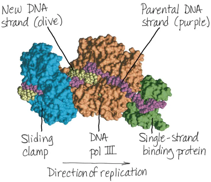
The DNA Replication Complex
• The proteins that participate in DNA replication
form a large complex, a “DNA replication machine”
• The DNA replication machine may be stationary during the replication process
• Recent studies support a model in which DNA
polymerase molecules “reel in” parental DNA and “extrude” newly made daughter DNA molecules
Parental DNA
DNA pol III
Leading strand
Connecting
protein Helicase
Lagging strand DNA
pol III
Lagging strand template 5 5 5 5 5 5
3 3
3 3
3
Proofreading and Repairing DNA
• DNA polymerases proofread newly made DNA, replacing any incorrect nucleotides
• In mismatch repair of DNA, repair enzymes
correct errors in base pairing
• DNA can be damaged by exposure to harmful chemical or physical agents such as cigarette smoke and X-rays; it can also undergo
spontaneous changes
• In nucleotide excision repair, a nuclease cuts
out and replaces damaged stretches of DNA
Evolutionary Significance of Altered DNA
Nucleotides
• Error rate after proofreading repair is low but not zero
• Sequence changes may become permanent and
can be passed on to the next generation
• These changes (mutations) are the source of the genetic variation upon which natural selection
operates
Replicating the Ends of DNA Molecules
• Limitations of DNA polymerase create problems for the linear DNA of eukaryotic chromosomes
• The usual replication machinery provides no way to complete the 5 ends, so repeated rounds of
replication produce shorter DNA molecules with uneven ends
Figure 16.20
Ends of parental DNA strands
Leading strand Lagging strand
Last fragment Next-to-last fragment Lagging strand RNA primer
Parental strand Removal of primers and replacement with DNA where a 3 end is available
Second round of replication
Further rounds of replication New leading strand
New lagging strand
Ends of parental DNA strands
Leading strand Lagging strand
Last fragment Next-to-last fragment
Lagging strand RNA primer
Parental strand Removal of primers and
replacement with DNA
where a 3 end is available
3
3
3
5 5
Figure 16.20b
Second round of replication
Further rounds of replication New leading strand
New lagging strand
Shorter and shorter daughter molecules 3
3
3
• Eukaryotic chromosomal DNA molecules have special nucleotide sequences at their ends called
telomeres
• Telomeres do not prevent the shortening of DNA molecules, but they do postpone the erosion of genes near the ends of DNA molecules
Figure 16.21
• If chromosomes of germ cells became shorter in every cell cycle, essential genes would eventually be missing from the gametes they produce
• The shortening of telomeres might protect cells from cancerous growth by limiting the number of cell divisions
• There is evidence of telomerase activity in cancer cells, which may allow cancer cells to persist
Concept 16.3 A chromosome consists of a
DNA molecule packed together with proteins
• The bacterial chromosome is a double-stranded, circular DNA molecule associated with a small amount of protein
• Eukaryotic chromosomes have linear DNA molecules associated with a large amount of protein
•
Chromatin
,
a complex of DNA and protein,
is found in the nucleus of eukaryotic cells
•
Chromosomes fit into the nucleus through
an elaborate, multilevel system of packing
DNA double helix (2 nm in diameter)
DNA, the double helix
Nucleosome
(10 nm in diameter)
Histones
Histones
Histone tail
H1
Figure 16.22b
30-nm fiber
30-nm fiber
Loops Scaffold
300-nm fiber
Chromatid (700 nm)
Replicated chromosome (1,400 nm) Looped domains
(300-nm fiber) Metaphase
Figure 16.22d
Figure 16.22f
• Chromatin undergoes changes in packing during the cell cycle
• At interphase, some chromatin is organized into a 10-nm fiber, but much is compacted into a 30-nm fiber, through folding and looping
• Though interphase chromosomes are not highly condensed, they still occupy specific restricted regions in the nucleus
5
Figure 16.23c
5
• Most chromatin is loosely packed in the nucleus during interphase and condenses prior to mitosis
• Loosely packed chromatin is called euchromatin
• During interphase a few regions of chromatin (centromeres and telomeres) are highly
condensed into heterochromatin
• Histones can undergo chemical modifications that result in changes in chromatin organization
Sugar-phosphate backbone
Nitrogenous bases
Hydrogen bond
G
G G
G
C
C C
C
A
A
A
T T
Figure 16.UN03
DNA pol III synthesizes
leading strand continuously
Parental
DNA DNA pol III starts DNA
synthesis at 3 end of primer, continues in 5 3 direction
Origin of replication
Helicase
Primase synthesizes a short RNA primer
DNA pol I replaces the RNA primer with DNA nucleotides 3 3 3 5 5 5 5